Diverse roles of Phosphatidylinositol transfer protein PITPβ in eukaryotic cells
Kristina Elizabeth Ile
A dissertation submitted to the faculty of the University of North Carolina at Chapel Hill in partial fulfillment of the requirements for the degree of Doctor of Philosophy in the
School of Medicine (Cell and Developmental Biology)
Chapel Hill 2009
Abstract
Kristina Ile: Diverse roles of phosphatidylinositol transfer protein PITPβ in eukaryotic cells
(Under the direction of Vytas A. Bankaitis)
Phosphatidylinositol transfer proteins (PITPs) provide a dynamic interface between membrane dynamics and lipid signaling. PITPα and PITPβ encode for two soluble class I, metazoan specific PITPs. However, despite the fact that PITPα and PITPβ share77 and 95% primary sequence identity and similarity (respectively), PITPα and PITPβare functionally distinct and play nonredundantfunctions in cells. Whereas PITPα knockout mice have been created, and these mice display neuronal, intestinal, and liver pathologies, PITPβ functionality remains largely unknown.
In this work, I addressed the distinguishing properties of PITPβ and its role in mammalian and zebrafish cells. We identified regions of PITPβ that contributed to its
unique properties: Golgi localization and sphingomyelin (SM) binding. Two single point mutations are sufficient to ablate the SM binding properties of PITPβ, and the Golgi
localization was attributed to the combination of two regions of the protein. Next, siRNA and morpholino analyses, in mammalian cells and zebrafish respectively, were developed to understand the physiological role of PITPβ in cells. In mammalian cells, PITPβ was essential for cell survival. Particularly, PITPβ-depleted cells displayed disorganized Golgi networks and anterograde trafficking defects. Surprisingly, PITPβ was not
outer segment of the double cone cells of the eye – a cell type required for detection of red and green light. The lack of a housekeeping role for PITPβ led us to examine the roles of other PITPs in zebrafish. A novel third soluble PITP in zebrafish, PITPγ, was
identified, though its function is not yet determined because we have no means of
detecting the protein levels to ascertain knockdown. PITPα, however, had an unexpected role in early development: reduction of PITPα led to an arrest at an early stage of
Acknowledgments
I would like to thank my advisor, Vytas Bankaitis, for his excellent mentoring. He has provided me both freedom to learn and explore on my own, and helpful advice when I needed it. I will always be grateful for all that I have learned from him.
I would also like to thank the members of the Bankaitis lab, from whom I’ve learned a lot, and who have made the lab a fun place to work. Thanks especially to my bench neighbors Carl, Jim, Malika, Sweety and Shriya, and to fellow graduate students Emily and Lydia, all of whom have been great colleagues and friends.
Thank you to my parents, sister, and brother-in-law, who have always supported me in graduate school and in everything else.
Table of Contents
List of Tables ... .vii
List of Figures ... viii
Abbreviations... xi
Chapter 1 (Introduction) ...1
Part 1a ...1
1a Figures...20
1a References ...26
Part 1b ...35
1b References...43
Chapter 2 -Targeting of PITPβ to the trans-Golgi network...47
Introduction...48
Materials and Methods...50
Results...55
Discussion...71
Figures/Tables...77
References...101
Chapter 3 –siRNA mediated knockdown of PITPβ...105
Introduction...106
Results...112
Discussion...119
Figures...122
References...128
Chapter 4 –Function of PITPs in zebrafish...131
Introduction...132
Materials and Methods...135
Results...143
Discussion...160
Figures...167
References...191
Chapter 5 – Discussion ...196
Appendix: Part A: Mouse models of PITPβ...208
Part I: Gene trap mouse...208
Part II: Transgenic PITPα/β mouse ...212
Part III: Floxed PITPβ mouse ...217
Tables/Figures...222
References...227
Part B: Record of plasmids ...229
pRE plasmids ...229
pCTY plasmids ...237
List of Tables
Table 2.1: Summary of effect of missense substitutions on PITP localization in COS-7 cells ...100 Table 3.1 (Supplementary): Primers used to amplify zebrafish PITP
List of Figures
Figure 1.1: A lipid metabolic nanoreactor ...20
Figure 1.2: Phospholipid transfer reaction...21
Figure 1.3: PITP crystal structures...22
Figure 1.4: The proposed Sec14p nanoreactor. ...23
Figure 1.5: The Sec14-nodulin PITP family in A. thaliana. ...24
Figure 1.6: The metazoan PITP family in humans. ...25
Figure 2.1:Endogenous PITP localization profiles ...77
Figure 2.2: PITPβ localizes specifically to TGN membranes ...79
Figure 2.3: C-terminal PITPβ localization elements necessary for TGN association ..81
Figure 2.4: C-terminal PITPβ localization elements sufficient for redirecting PITPα to TGN membranes. ...83
Figure 2.5: PITPβ localization to TGN membranes is independent of phospholipid loading...86
Figure 2.6: General PITP elements required for PITPβ localization to TGN membranes...88
Figure 2.7: Properties of PITPβW202W203 interaction with membranes. ...89
Figure 2.8: W202W203 and C-terminal BOX motifs in TGN targeting ...91
Figure 2.9: PITPβ localization and protein kinases C. ...92
Figure 2.10: C-terminal PITPβ localization elements necessary for TGN association...94
Figure 2.11: C-terminal PITPβ localization elements sufficient for redirecting PITPα to TGN membranes. ...95
Figure 2.12: Comparison of PITPβ and PITPβQGQR localization in MEFs...96
Figure 3.1: Several PITPβ siRNAs are capable of reducing PITPβ levels in mammalian
cells. ...122
Figure 3.2: Reduced PITPβ levels lead to changes in pan-Golgi morphology...123
Figure 3.3: Trafficking through the trans-Golgi is delayed in PITPβ-depleted cells. ...125
Figure 3.4: The localization of lipid binding proteins is unaffected in PITPβ siRNA-treated cells ...127
Figure 4.1: DrPITPβ and DrPITPγ isoforms. ...167
Figure 4.2: PI synthesis in yeast ...169
Figure 4.3: Temporal and spatial expression of DrPITPβ and DrPITPγ...171
Figure 4.4: DrPITPβ immunolocalization in the adult zebrafish retina...173
Figure 4.5: The DrPITPβ and double cone cell-specific zpr-1 antigen co-localize in adult retina wholemounts...175
Figure 4.6: Reduced DrPITPβ expression results in a loss of zpr-1 staining...176
Figure 4.7: DrPITPβ morphants present double cone cell structural defects...178
Figure 4.8: Identification of zpr1 antigen as arrestin-3-like. ...179
Figure 4.9:DrPITPα morphants fail at an early stage of development. ...181
Figure 4.10: DrPITPβ function in double cone cell outer segment biogenesis and maintenance ...183
Figure 4.11 (Supplemental Figure 1): All PITP isoforms are expressed in the adult zebrafish eye ...185
Figure 4.12 (Supplemental Figure 2): Blue opsin and rhodopsin expression are unaffected in DrPITPβ morphants ...186
Figure 4.13 (Supplemental Figure 3): At 5dpf, DrPITPβ morphants display no defects in electroretinograms (ERGs) measuring their response to light ...187
Figure 4.14 (Supplemental Figure 4): MAb zpr1 only detects Arr3L, and not other retinal and ubiquitously expressed arrestins. ...188
Figure A.2- A BAC/PAC was modified by recombination techniques to create a
transgenic PITPα/PITPβ mouse...225
Figure A.3 – A targeting vector to create a conditional PITPβ knockout mouse is
List of abbreviations
BFA: Brefeldin ABSA: bovine serum albumin DAG: diacylglycerol
FBS: fetal bovine serum GFP: green fluorescent protein MEF: mouse embryo fibroblasts
PITP: phosphatidylinositol transfer protein PKD: protein kinase D
Chapter 1 - Introduction
Chapter 1a
Chapter 1a reprinted by permission from Macmillan Publishers Ltd: Nature Chemical
Biology.
Reference: Ile KE, Schaaf G, Bankaitis VA. Phosphatidylinositol transfer proteins and
cellular nanoreactors for lipid signaling. Nat Chem Biol. 2006 Nov;2(11):576-83.
Copyright 2006
Abstract
Membrane lipids function as structural molecules, reservoirs for second messengers,
membrane platforms that scaffold protein assembly and regulators of enzymes and ion
channels. Such diverse lipid functions contribute substantially to cellular mechanisms for
fine-tuning membrane-signaling events. Meaningful coordination of these events requires
exquisite spatial and temporal control of lipid metabolism and organization, and reliable
mechanisms for specifically coupling these parameters to dedicated physiological
processes. Recent studies suggest such integration is linked to the action of
phosphatidylinositol transfer proteins that operate at the interface of the metabolism,
trafficking and organization of specific lipids.
Intro
An important feature of eukaryotic membranes is their lipid heterogeneity (van
criteria of lipid headgroup and backbone composition alone. When one also considers
acyl-chain heterogeneity (that is, heterogeneity in the chemical nature of the fatty-acyl
groups that are incorporated into lipid molecules), the number of lipid molecular species
rises to over 1,000. Why such diversity if the role of membranes is simply to act as
barriers that define the boundary of the cell and physically segregate biochemically
distinct internal compartments from one another? We now know that membrane surfaces
are sites of robust interfacial chemical reactions that are necessary for proper
maintenance of organellar and cellular homeostasis. Thus, membranes serve as platforms
for intricate orchestration of the complex biochemistry of signal transduction. Because
signaling systems are intrinsic properties of eukaryotic cells, the microanatomy of the
membrane surface must also be sufficiently complex to satisfy the intricacies in the
organization of such systems.
Recent progress in many laboratories is leading to an increasing appreciation for
the complexity of the interface between lipids and proteins, even when one considers
only the cytoplasmic leaflet of cellular membranes. As one simple example,
phosphatidylinositol-4,5-bisphosphate (PtdIns(4,5)P2) constitutes only ~1 mole percent
of total lipid in mammalian plasma membranes (van Meer, 2005). Yet PtdIns(4,5)P2 has
many roles in production of second messengers(Berridge and Irvine, 1989; Majerus,
1992; Nishizuka, 1995; Rhee, 2001). PtdIns(4,5)P2 also regulates membrane-protein
interactions (Shaw, 1996; Hurley and Meyer, 2001; Lemmon, 2003), enzyme and
ion-channel activity (Suh and Hille, 2005), membrane trafficking to and from the plasma
membrane (Martin, 2001; Simonsen et al., 2001; Wenk and De Camilli, 2004), activity of
2004) and, indirectly, transcription and mRNA transport (Kronke, 1999; Odom et al.,
2000). The picture becomes even more complicated when other phosphorylated forms of
phosphatidylinositol (PtdIns) are considered. There are seven distinct species of
phosphoinositides (PIPs) in higher eukaryotic cell membranes, of which PtdIns(4,5)P2 is
one. There is growing evidence that each of these individual PIP isomers also has unique
biological activity. This chemical diversity offers impressive prospects for intricate and
combinatorial fine-tuning of PIP-dependent regulation of complex cell functions. In this
regard, PIPs are uniquely eukaryotic phospholipids.
Other lipids, previously relegated to 'housekeeping' status, are also more
functionally versatile than first believed. Enzyme activation by ceramide-based lipids is a
highly active area of investigation (Pettus et al., 2002; Spiegel and Milstien, 2003; Pettus
et al., 2004). Phosphatidylserine (PtdSer) activates specific protein kinase C isozymes
(Stahelin et al., 2003), phosphatidylethanolamine (PtdEtN) organization imparts spatial
organization to the actin cytoskeleton (Iwamoto et al., 2004) and phosphatidylcholine
(PtdCho) is a substrate for various phospholipases that release hydrolytic products (for
example, phosphatidic acid, lysophosphatidic acid and arachidonic acid) having diverse
signaling capabilities (Exton, 1990; Moolenaar, 1995; Singer et al., 1997). The cellular
lipid signaling circuitry that connects these regulatory pathways is very complex. For
example, PtdCho metabolism intersects at several points with PIP metabolism, which
indicates a cross-talk between these two signaling systems that permits their integration
(Liscovitch and Cantley, 1995; Sciorra et al., 2002). It is now clear that lipids are not
homogeneously distributed along the surface of any membrane system. Rather, they are
regulation on interfacial protein binding and/or enzymatic reactions. Moreover, it is
becoming increasingly apparent that populations of lipid molecules are functionally
segregated from others of the same species with respect to their involvements in specific
intracellular activities. Such functionally distinct populations of specific lipids are
operationally referred to as 'pools' (Cleves et al., 1991a; Hinchliffe et al., 1998). A
precise definition of what 'pools' means in a physical sense is often unclear because there
are many physical explanations for how a functional lipid pool can be created and
maintained. Some of these concepts are developed further below.
How are lipid pools segregated in the face of powerful subversive forces, such as
lateral lipid diffusion and membrane trafficking, that one might expect to erase higher
order blueprints for lipid organization? Unrestrained lipids are mobile in the plane of a
membrane bilayer (with diffusion coefficients in the range of 0.1–1.0 μm2 s-1) after all (Schwille et al., 1999). Given that yeast cells have a diameter of 5 μm, whereas
mammalian cells are roughly 10–20 μm in each dimension, this is a considerable rate of
movement. Vesicle trafficking is also a highly active process that would be expected to
confound mechanisms for maintaining nonrandom distribution of lipids. The entire
picture becomes even more confused when the robust contributions of 'household' lipid
metabolism (both biosynthesis and turnover) are superimposed, particularly when one
considers that lipids with signaling power are continuously generated and consumed by
such constitutive metabolic pathways (Schwille et al., 1999). Obviously, the coupling of
lipid pools to privileged biological functions demands the timely metabolism and/or
organization of specific lipids. One mechanism for doing so is to interface lipid
metabolic steps themselves, or the organization of the products of such metabolism. This
concept implies the existence of very small machines, or nanoreactors, in which
metabolic and signaling reactions take place and their products are subsequently
organized.
Herein, we explore the notion that the biological functions of an understudied
class of eukaryotic proteins, the phosphatidylinositol transfer proteins (PITPs), adhere to
some of the key principles implicit in a nanoreactor concept. We also discuss the
emerging evidence that PITPs have key roles in defining the functional identities of
individual lipid pools. These individual pools are, in turn, dedicated to the regulation of
specific biological functions. For the purpose of this review, we define nanoreactor to
mean a functional network connecting a phospholipid-bound PITP, at least one
phospholipid metabolic enzyme, and a specific lipid-binding component in addition to the
PITP (or PITP domain in the case of multidomain proteins; Fig. 1). This basic notion is
inspired by work done some 15 years ago by Konrad Sandhoff and colleagues, who
demonstrated the existence of activator proteins required for efficient turnover of
sphingolipids in lysosomes. Although these sphingolipid activator proteins resemble
PITPs in their behavior as lipid transfer proteins in vitro (see below), they actually
function as substrate-presentation subunits that mark a bound phospholipid for
degradation (Furst and Sandhoff, 1992). The metabolic channeling, by a subunit with
lipid-transfer activity, of a privileged lipid molecule to a specific enzyme is an example
of what we define as a lipid metabolic nanoreactor.
To place this discussion in historical context, it has long been appreciated that
cells express lipid transfer proteins; that is, provisionally cytosolic proteins that catalyze
the desorption of lipid monomers from bilayers in vitro. In some (but not all) cases, these
transfer proteins have lipid-binding specificity and are stable as lipid–protein complexes
in solution. The sphingolipid activator proteins introduced above belong to this general
class of lipid transfer proteins and have binding specificity for sphingolipids (Furst and
Sandhoff, 1992). PITPs are uniquely eukaryotic proteins that also have both the property
of lipid-binding specificity and the ability to form stable protein–phospholipid complexes
in solution. PITPs catalyze exchange of either PtdIns or PtdCho between membranes in
vitro (Wirtz, 1997; Hsuan and Cockcroft, 2001; Phillips et al., 2006b). A cycle for such a
transfer reaction is illustrated in Figure 2. PITPs contain one phospholipid binding site
per monomer, with a decided preference for PtdIns over PtdCho. PITPs exchange a
bound phospholipid for a PtdIns or PtdCho monomer that resides in a bona fide
membrane bilayer. This is done very efficiently and without ATP or other cofactors.
PITPs abstract a phospholipid molecule from a monolayer, which indicates that bilayer
organization is not an obligatory requirement for action. As a result, it has been taken
somewhat as an article of faith that PITPs do not access, or otherwise disturb, the luminal
leaflet of intracellular membranes. Whether this is actually true remains to be rigorously
shown. How PITPs couple conformational changes to phospholipid recognition, and eject
a bound phospholipid in favor of binding and desorbtion of another phospholipid
molecule, remains unknown.
PITPs fall into two families on the basis of primary sequence relationships and
Sec14-like and metazoan PITPs have nearly identical in vitro activities but share no
primary sequence similarity and, as shown in Figure 3, have unrelated three-dimensional
folds. The Sec14 fold defined a novel fold when a crystal structure for the yeast Sec14p
PITP was solved (Sha et al., 1998; Phillips et al., 1999). By contrast, the metazoan PITP
fold belongs to the steroidogenic acute response protein–related START-domain family
(Yoder et al., 2001; Schouten et al., 2002; Tilley et al., 2004). In that respect, it groups
with other lipid transfer proteins (for example, ceramide transfer protein (Hanada et al.,
2003), PtdCho transfer protein (Roderick et al., 2002) and the putative Kes1p (also called
Osh4p) sterol transfer protein (Li et al., 2002; Im et al., 2005)) that either share primary
sequence similarity with START domains or are otherwise known to adopt similar
structural shapes.
If the extent of gene expansion is a fair measure of functional importance, then
the Sec14 family is an important one indeed. Sec14-like proteins make up a protein
superfamily for which >500 member proteins can be discerned in available databases,
and the expansion of this superfamily is clear in even the simplest unicellular eukaryotes.
The yeast Saccharomyces cerevisiae expresses six Sec14-like proteins, whereas higher
eukaryotes (humans, mice, Drosophila melanogaster, Caenorhabditis elegans,
Arabidopsis thaliana) each harbor in excess of 20. The founding member of this
superfamily is the yeast Sec14p, and it is arguably the best studied of the group (see
below). It is almost certain that not all Sec14p-like proteins are PITPs. Some Sec14-like
proteins (or proteins with Sec14 domains) have specific PIP-binding activities, and these
comprise an interesting class in their own right. Some examples include the
protein tyrosine phosphatase (Gu et al., 1992; Huynh et al., 2003), Rho nucleotide
exchange factors such as Dbl and Ost/Dbs (Whitehead et al., 1997; Cheng et al., 2002;
Rossman et al., 2002), the rasGAP neurofibromin (D'Angelo et al., 2006) and plant
PIP-binding proteins implicated in osmoregulation (Kearns et al., 1998; Monks et al., 2001).
The sheer number of Sec14-like proteins in eukaryotic cells argues for extensive
diversification of function, but it remains uncertain whether PtdIns and PIP binding is a
common feature of all Sec14p-like proteins. Some bind other hydrophobic ligands (such
as vitamin E and retinaldehyde) (Stocker et al., 2002; Min et al., 2003; Panagabko et al.,
2003). However, functional studies already suggest that this evolutionary expansion
cannot be explained by diversification of substrate binding specificity alone. We
anticipate that many mammalian Sec14-like proteins retain PITP activity, and that the
PITP complement in metazoans is underestimated.
By contrast, the metazoan PITP family is not highly expanded, and no members
of this family have yet been recognized in the genomes of even the higher plants. An
enigmatic case is presented by the slime mold Dictyostelium discoideum, which
expresses four Sec14-like and four metazoan PITP–like proteins. It is notable that the two
classes of PITPs are so similar with respect to in vitro activity yet so different in
structural construction and evolutionary expansion. Is this a case for convergent
evolution, or does it reflect some functional distinction that is invisible in in vitro
systems? In our view, the evidence suggests the latter. The difficulties in interpreting the
in vitro lipid-transfer assays are known (Rogers and Bankaitis, 2000). Even in cell-free
assays that are more directed, metazoan PITPs and Sec14p are functionally
Jones et al., 1998; Simon et al., 1998). This equivalence is not recapitulated in in vivo
studies, however. Those studies consistently report a high degree of functional distinction
between individual PITPs. In this review, we will primarily discuss the lipid nanoreactor
concept in the context of the Sec14-like PITP family. We will close with a discussion of
how this concept may also apply to the metazoan PITPs.
Yeast Sec14p and Sec14-like proteins
We now appreciate that a primary function for PITPs in cells is to coordinate lipid
metabolism and signaling with membrane trafficking. The first indications to this effect
came from studies of the main yeast PITP (Sec14p), which is essential for protein
transport from the yeast trans-Golgi network (TGN) and for yeast cell viability (Bankaitis
et al., 1990; Cleves et al., 1991b). Sec14p uses both its PtdIns- and PtdCho-bound forms
to regulate many aspects of lipid metabolism. The net purpose of this is to generate a
membrane-lipid environment conducive to optimal activity of core components of the
TGN vesicle-budding machinery (Fig. 4). Both PtdCho metabolism and conversion of
PtdIns to PtdIns(4)phosphate are subject to regulation by Sec14p. That is, Sec14p
regulates the metabolic fate of specific pools of PtdCho and PtdIns that are specifically
interfaced with the activity of a yeast Golgi subcompartment (Bankaitis et al., 1990;
Cleves et al., 1991b; Guo et al., 1999; Hama et al., 1999; Rivas et al., 1999). The
localized interplay among Sec14p, lipid metabolism, and proteins of the Arf GTPase
cycle define at least one, and perhaps two, TGN lipid nanoreactors (that is,
Sec14p-PtdCho-based and Sec14p-PtdIns-based nanoreactors; see below).
Why argue the nanoreactor concept for Sec14p, when it seems equally plausible
example, PtdIns) or vice versa (for example, PtdCho)? The reasons are several. First,
unlike the case for mammalian cells, PtdCho and PtdIns are the most abundant
phospholipids in yeast cell membranes. Elevating PtdIns levels to 40 mole percent of
total bulk phospholipid in yeast membranes does not circumvent the Sec14p requirement
for protein transport from the TGN (Jones et al., 1998). Second, loss of activity of one
specific pathway for PtdCho biosynthesis is one of the mechanisms that permits cell
viability and TGN functionality in the absence of Sec14p (ref. (Cleves et al., 1991b)).
Third, a mutant Sec14p that retains PtdCho-transfer activity but is ablated for
PtdIns-transfer activity retains substantial function in vivo (Phillips et al., 1999). Presumably,
this mutant Sec14p is incapable of driving vectorial movement of any phospholipid
species—a seemingly necessary condition for meaningful lipid transfer between
membranes in cells. It is difficult to conceive how these various data are consistent with
lipid transfer mechanisms, but the data are readily consistent with precisely tuned
metabolic control mechanisms for Sec14p function.
Though the details of this Sec14p-dependent combinatorial regulation of TGN
vesicle trafficking await resolution, the ability of a peripheral membrane protein to
execute function at the TGN provides a clear example of a dedicated coupling of a PITP
to a specific physiological function. In the case of PtdCho metabolism, Sec14p is
proposed to act as a lipid sensor that modulates the antagonistic effects of household lipid
metabolism on an essential TGN process regulated by multiple lipids (for example,
PtdCho, diacylglycerol and PIPs). This set of reactions is proposed to couple to activity
of the Arf GTPase cycle (Fig. 4) and defines a Sec14p-PtdCho nanoreactor in the TGN
the Pik1p PtdIns-4-OH kinase of the yeast TGN (Pik1p and Stt4p are the two
demonstrated PtdIns-4-OH kinases). Which proteins couple to this particular metabolic
reaction remains unknown, but one candidate is the sterol-binding protein Kes1p/Osh4p
(Fig. 4). This Sec14p-PtdIns/Pik1p circuit defines a Sec14p-PtdIns nanoreactor in the
TGN (see also Fig. 1a).
Other studies in yeast are also inconsistent with the notion that PITPs execute the
types of 'one size fits all' activities that are reported by in vitro reconstitutions. Sec14p is
only one of six yeast Sec14-like proteins, and several of the other Sec14 homologs
(SFHs) are biochemically related to Sec14p on the basis of phenotypic rescue or
determined phospholipid-transfer activities (Li et al., 2000; Schnabl et al., 2003; Routt et
al., 2005). Testimony to the diversification of biological function of Sec14-domain
proteins is apparent from analysis of these SFH proteins. First, SFH proteins are required
for optimal activation of the yeast PtdCho-hydrolyzing enzyme phospholipase D (PLD).
This is in contrast to the case for Sec14p, whose deficiency activates PLD (Sreenivas et
al., 1998; Xie et al., 1998; Xie et al., 2001). Second, the Sec14-like Sfh4p is uniquely
required for coordinating the localization and assembly of an enzymatic machinery
dedicated to nonmitochondrial decarboxylation of PtdSer to PtdEtN (Wu et al., 2000; Wu
and Voelker, 2004; Routt et al., 2005). This machinery is posited to involve formation of
intermembrane contact sites between the ER and a TGN or endosomal compartment.
Such contact sites serve as portals for nonvesicular trafficking of lipids between distinct
compartments, and lipid trafficking via this mechanism is likely to represent a
quantitatively significant and underappreciated pathway for intracellular lipid transport
contact site requires an intricate organization of acidic phospholipids that help orchestrate
the activity of proteins that chaperone formation of this class of intermembrane contact
site. In other words, this particular intermembrane contact machine is coupled to an
Sfh4p-dependent metabolic nanoreactor. Third, Sfh5p has the unique ability to promote
expansion of what seems to be a plasma-membrane pool of PtdIns(4,5)P2 (ref. (Routt et
al., 2005). This pool optimizes the activity of specific t-SNARES (components of the
plasma-membrane machinery for vesicle docking and fusion). We propose that the
Sfh5p-dependent nanoreactor consists of Sfh5p, the Stt4p PtdIns-4-OH kinase, the Mss4p
PtdIns(4)phosphate-5-OH kinase and specific t-SNAREs (Routt et al., 2005). Finally,
yeast PITPs control actin organization in yeast. But, whereas Sec14p has a constitutive
role in this process, SFH isoforms are involved in evoked pathways for actin
reorganization (Routt et al., 2005).
The conclusions derived from study of the SFH proteins are instructive on several
counts with regard to the nanoreactor concept. The budding yeast PITPs execute
functions unique to individual members of this family, and one can imagine that the
coupling of any single PITP to a particular PtdIns kinase may underly functional
specification. Yet these proteins, including Sec14p, act at least in part through the same
Stt4p PtdIns-4-OH kinase isoform (Routt et al., 2005). Thus, specific PIP pools are
defined not only by the lipid kinases that generate them, but also by the individual PITP
that couples to a particular lipid kinase. This interface demonstrates a considerable
plasticity. Coupling of both the PtdIns kinase isoform and the PITP is subject to
Stt4p in vegetative cells, but it is dependent on Sec14p and Pik1p in sporulating cells
(Engebrecht, 2003; Rudge et al., 2004).
Multidomain Sec14-like PITPs and nanoreactors
Potentially the most notable example of a PITP-dependent nanoreactor comes
from studies of two-domain Sec14-nodulin proteins of higher plants (Fig. 5a). Unlike
Sec14p, these are membrane-associated proteins, at least on the basis of the one case that
has been analyzed (Kapranov et al., 2001; Vincent et al., 2005). The Sec14 domains are
PITP modules that stimulate PIP synthesis, whereas the C-terminal nodulin domains
represent membrane targeting units. Root-hair PtdIns(4,5)P2 homeostasis and
organization is dependent on AtSfh1p function, and there are strong similarities between
the nonrandom distributions of the AtSfh1p Sec14-nodulin and PtdIns(4,5)P2 in A.
thaliana root hairs (Vincent et al., 2005). This is a prime example in which the
distribution of a PITP sets the distribution of a PIP, and Sec14-nodulin proteins may do
so via PIP binding. There are examples of multidomain Sec14-like proteins that bind
PIPs, such as the Sec14-GOLD proteins that are conserved across the higher Eukaryota
(including mammals) (Anantharaman and Aravind, 2002; McLaughlin and Murray, 2005;
Phillips et al., 2006b). Multidomain Sec14 proteins present interesting cases for the PITP
nanoreactor concept.
The chemical properties of nodulin domains suggest the way in which PIP
synthesis and organization may be coordinated by a multidomain PITP. Nodulin domains
have highly basic C-terminal tails with properties reminiscent of PIP-binding motifs (Fig.
5b). Given the ability of unstructured and highly basic protein domains to sequester
2005), Sec14-nodulin proteins theoretically have the capability to regulate the spatial
organization of the very PIP molecules whose synthesis they promote. Available
evidence, though admittedly scant, does suggest that Sec14-nodulin proteins can
functionally discriminate PIP pools in the context of polarized membrane-trafficking
pathways. As PIPs are known regulators of membrane trafficking and of actin
cytoskeleton dynamics, we posit that it is through the functional specification of PIP
pools that Sec14-nodulin proteins evoke their specific biological effects.
The concept of PIP pools in the context of Sec14-nodulin function suggests other
interesting possibilities. One attractive conjecture is that nodulin domains sequester PIPs
via high-affinity, but regulated, electrostatic interactions. This sequestration could
potentially be controlled so that these PIPs are rendered accessible to the proper effectors
(or enzymes) on cue. One hypothetical mechanism for such regulated 'release' involves
calmodulin. Basic unstructured polypeptides are excellent binding substrates for
Ca2+-calmodulin (Yamniuk and Vogel, 2004; McLaughlin and Murray, 2005), which suggests
the possibility that calmodulin activation could loosen the 'nodulin clamp' and release a
spatially restricted PIP pool in response to a temporal program responsive to cytosolic
Ca2+ flux. The fact that A. thaliana encodes 13 distinct Sec14-nodulin proteins (Vincent
et al., 2005) provides an indication of the potential diversity such a strategy may hold for
the coupling of distinct Sec14-nodulin–dependent PIP metabolic nanoreactors to
developmental pathways for membrane morphogenesis.
Metazoan PITPs: few but potent
As indicated above, the metazoan PITP family is quantitatively much more sparse
genes, two of which encode membrane-bound isoforms (Fig. 6). Yet some of the same
functional principles discussed above for Sec14p-like PITPs are likely to apply to these
proteins. For example, the ability of these PITPs to stimulate PIP synthesis is a common
denominator in their involvements in numerous exocytic reactions (for which they act as
reconstituted stimulators) and in plasma-membrane receptor and G protein–coupled
hydrolysis of PIPs by phospholipase C (Ohashi et al., 1995; Cunningham et al., 1996;
Jones et al., 1998; Simon et al., 1998). This is in contrast to the results of in vivo studies
that highlight the importance and functional specificities of individual metazoan PITPs.
Genetic insufficiencies for the soluble PITPα lead to a perinatal morbidity in mice
characterized by chylomicron retention disease (CRD), hypoglycemia and
spinocerebellar neurodegeneration (Weimar et al., 1982; Hamilton et al., 1997; Alb et al.,
2003). With regard to the neurological component of this disease, recent results indicate
that efficient recognition of axonal guidance cues via the netrin receptor requires PITPα
(Xie et al., 2005). Soluble-PITP defects in D. melanogaster lead to defective oogenesis
that results from dysfunction of the actin cytoskeleton (Gatt and Glover, 2006; Giansanti
et al., 2006).
Membrane-associated isoforms of metazoan PITPs also exist (but why, if lipid
transfer is the operative function?), and these isoforms may yet prove to be analogs of the
higher plant Sec14-nodulin proteins discussed above. An inherited and light-enhanced
retinal degeneration in flies results from loss of function of the membrane-associated
RdgB protein. RdgB expression is highly tissue-specific, and the 280-residue PITP
(Milligan et al., 1997). One mammalian RdgB ortholog is Nir2, and mice lacking this
protein die before embryonic day 7.5 (ref. (Chang et al., 1997)).
Why do metazoans invest in development of a small PITP family that is unrelated
to Sec14 PITPs when trivial expansion of the already large Sec14 protein superfamily
would presumably generate sufficient numbers of functionally unique PITPs to satisfy the
same requirements? This puzzle is deepened by indications that mammals apply the same
sets of functional-specificity rules to the small cohort of PITPs that they apply to the
Sec14-like proteins. The PITPα and PITPβ isoforms are very similar, but they are
functionally nonredundant (Alb et al., 2003). In part, this is likely the result of differential
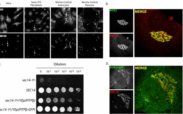
distribution of these isoforms within cells. PITPβ is a TGN-associated protein (Phillips et
al., 2006a), whereas PITPα is localized to the cytosol and the nuclear matrix (De Vries et
al., 1996; Phillips et al., 2006a). However, the cases of PITPβ and the
membrane-associated Nir2 cannot be so easily dismissed. Both Nir2 and PITPβ localize to TGN
membranes (Litvak et al., 2005; Phillips et al., 2006a), yet these two proteins define a
functionally nonredundant pair of PITPs.
The building evidence suggests that a lipid metabolic nanoreactor concept applies
to metazoan PITPs as well as the Sec14-like PITPs. What is perhaps more notable, given
the structural dissimilarity between these classes of proteins, is that the lipid metabolic
nanoreactors under metazoan PITP control resemble those described for Sec14-like
PITPs. For example, the Nir2 PITP regulates a TGN lipid metabolic nanoreactor that
coordinates diacylglycerol and PtdCho metabolism with protein trafficking in a fashion
very similar to that seen in yeast for Sec14p (Litvak et al., 2005). In this instance, the
kinase D family (Liljedahl et al., 2001; Baron and Malhotra, 2002; Litvak et al., 2005).
The function of PITPβ in the TGN is unknown, but speculations have been made about a
PITP-PLD interface on mammalian Golgi membranes (Liscovitch and Cantley, 1995).
Such a PITP-PLD interface offers the prospect of a lipid metabolic nanoreactor that
couples with the Arf small GTPase cycle in regulating vesicle trafficking. A role for
PITPβ in regulating the Arf GTPase cycle and Golgi vesicle trafficking would also be
directly analogous to models for Sec14p function in yeast (Phillips et al., 2006b).
Returning to comparisons of the functional scenarios for Nir2 and PITPβ, the data further
emphasize that the functional specification of PITPs can be achieved via differential
coupling of distinct lipid metabolic nanoreactors to sets of distinct effectors.
Along those same lines, mouse PITPα nullizygosity evokes some phenotypes that
resemble human CRD (Alb et al., 2003), a disorder caused by ER-localized Sar1 GTPase
insuffiency (Jones et al., 2003). Sar1 is a distant member of the Arf GTPase family. Thus,
physiological coupling of the soluble metazoan PITPs to the action of small GTPase
catalytic cycles may prove a common theme. Why a specialized trafficking cargo
transport pathway from the ER (the primary site of lipid synthesis in the cell) requires
PITPα for efficient operation is an intriguing question—one that again suggests that lipid
delivery is not the final word in terms of metazoan PITP function.
Future directions and future challenges
The idea that PITPs functionally define specific lipid metabolic pools, and do so
via the action of dedicated lipid metabolic nanoreactors, is supported by a growing body
of data. Much remains to be discovered, however. As a first step, we need to decipher the
metabolism and ultimately to biological readout. The physical nature of PITP-dependent
nanoreactors also begs analysis. Certainly intermembrane contact sites provide one
appealing (but not well-supported) notion (Holthuis and Levine, 2005). Functional
analyses are obviously required for assessing the nature of PITP-dependent lipid
metabolic nanoreactors, but these will likely prove tricky. For instance, the physiological
fidelity of PITP activity is sensitive to protein dosage (Skinner et al., 1993; Li et al.,
2000). New ideas for how to approach these questions are needed.
From a more general perspective, lipid metabolism cannot be viewed as an
averaged activity in any single membrane system—much less in the cell. Unfortunately,
we are ill equipped to analyze the mosaic nature of lipid metabolism in vivo.
Development of new technologies that report, with spatial and temporal resolution, the
activities of specific lipid metabolic nanoreactors in living cells would represent a
tremendous advance. New and powerful vital-imaging approaches are required, and
possibilities for exploration include methods based on fluorescence resonance energy
transfer (FRET) that score activated conformations of protein components of specific
nanoreactors. Such status-sensitive reporters are available for Cdc42 GTPases (Nalbant et
al., 2004). PITPs provide at least a conceptual starting point for thinking about how one
would design such vital 'activity' sensors.
Finally, the concepts discussed herein will extend beyond the PITPs. There are
other lipid transfer proteins (for example, ceramide transfer protein; (Hanada et al.,
2003), the putative sterol transporter Kes1p/Osh4p and other members of the large
oxysterol-binding protein family (Li et al., 2002; Im et al., 2005; Raychaudhuri et al.,
interfaced with signaling events. Finally, a highly diverse system of metabolic
nanoreactors clearly demands multiple levels of higher order integration of these
machines. The physical basis for regulation of cellular lipid metabolism and signaling in
such microenvironments, and how the action of lipid metabolic nanoreactors is integrated
at higher levels, are two important questions for the next decade.
Acknowledgments
This work was supported by grants from the US National Institutes of Health to V.A.B.
K.E.I. and G.S. were also supported by a Predoctoral Training Grant from the US
National Institutes of Health and a Postdoctoral Fellowship from the Deutsche
Figures
Figure 1.1 A lipid metabolic nanoreactor.
Two scenarios for Sec14p-dependent nanoreactors relevant to this discussion are shown.
(a) Sec14p binds substrate PtdIns and channels the bound PtdIns to a metabolic enzyme
(for example, PtdIns kinases), and the ultimate metabolic product (in this illustration,
PtdIns(4,5)P2) then binds another protein (or protein domain), thereby leading to a
nonrandom organization of the product. (b) Sec14p binds PtdCho and is thereby
programmed to regulate a specific phospholipid metabolic pathway (in this case the
cytidine diphosphocholine pathway for PtdCho biosynthesis that involves the sequential
activity of a choline kinase (CKIase), a choline-phosphate cytidylyltransferase (CCTase)
and a cholinephosphotransferase (CPTase)). The lipid compositional consequences of the
Sec14p-dependent regulation in that nanoreactor are transmitted to the action of
Figure 1.2: Phospholipid transfer reaction.
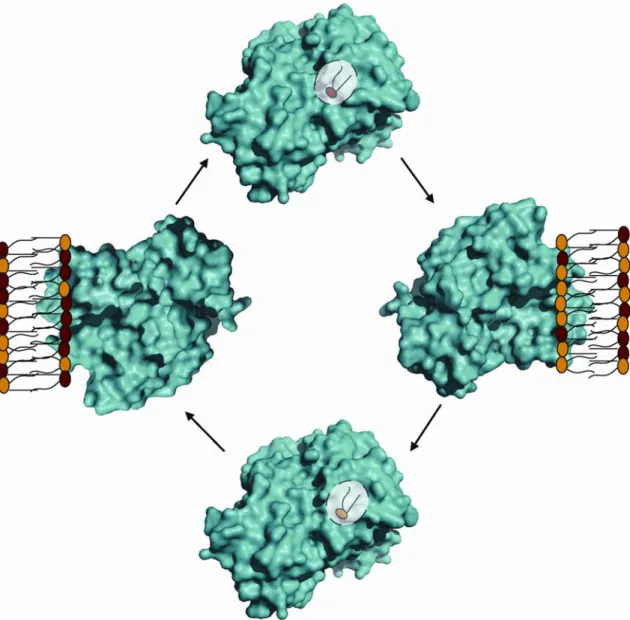
A cycle for PITP-mediated phospholipid exchange is shown. This reaction proceeds
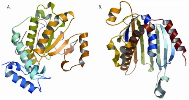
Figure 1.3: PITP crystal structures.
(a,b) Three-dimensional structures of apo forms of yeast Sec14p (a) and mammalian
PITPα (b). The apo-Sec14p fold consists of twelve α-helices, six β-strands and eight 310
-helices (bound detergent not shown). PITPα is a START-domain protein, and the fold
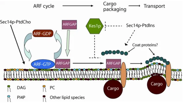
Figure 1.4: The proposed Sec14p nanoreactor.
Sec14p bound to PtdCho downregulates PtdCho biosynthesis to generate a DAG-rich,
PtdCho-poor membrane domain that is conducive to the recruitment and activation of
specific ArfGAPs. Genetic and biochemical data indicate that the relevant ArfGAPs are
activated by DAG and inhibited by PtdCho (Yanagisawa et al., 2002). These ArfGAPs
potentiate later steps in secretory cargo packaging and vesicle biogenesis on TGN
membranes (Lewis et al., 2004; Lee et al., 2005; Robinson et al., 2006). The
PtdIns-bound form of Sec14p independently potentiates a distinct step in the vesicle biogenesis
pathway by promoting synthesis of a PtdIns(4)phosphate (PI4P) pool that regulates
downstream effector proteins (perhaps the PIP and sterol-binding protein Kes1p/Osh4p,
Figure 1.5: The Sec14-nodulin PITP family in A. thaliana.
(a) Schematic of the two general domain arrangements of the 13 A. thaliana
Sec14-nodulin proteins. The Sec14p (blue), Sec14-nodulin (green) and hydrophobic (red) domains are
shown. Members of the family that conform to each arrangement are identified at left. (b)
Shown is an alignment of the C-terminal half of each of 13 A. thaliana nodulin domains
of Sec14-nodulin proteins. These nodulin domains are grouped into three classes on the
basis of sequence similarity. Conserved residues are highlighted in red and basic residues
in the extreme C termini of these proteins are highlighted in blue. The bar at the top
highlights sequences predicted to reside in coiled coils. For purposes of comparison,
known high-affinity PtdIns(4,5)P2 binding sites for myristoylated alanine-rich C-kinase
substrate (MARCKS) protein and the NMDA (N-methyl-D-aspartate) receptor are shown
Figure 1.6: The metazoan PITP family in humans.
Schematic alignment of the five human PITPs of the metazoan PITP family. The PITP
domain (blue), FFAT (green; sequence that binds an ER-localized receptor of the
VAP/Scs2 family; (Loewen et al., 2003) and DDHD (brown; a putative metal-binding
domain) are shown. The primary sequence identities shared by each PITP domain to
References
Alb, J.G., Jr., Cortese, J.D., Phillips, S.E., Albin, R.L., Nagy, T.R., Hamilton, B.A., and Bankaitis, V.A. (2003). Mice lacking phosphatidylinositol transfer protein-alpha exhibit spinocerebellar degeneration, intestinal and hepatic steatosis, and hypoglycemia. J Biol Chem 278, 33501-33518.
Anantharaman, V., and Aravind, L. (2002). The GOLD domain, a novel protein module involved in Golgi function and secretion. Genome Biol 3, research0023.
Bankaitis, V.A., Aitken, J.R., Cleves, A.E., and Dowhan, W. (1990). An essential role for a phospholipid transfer protein in yeast Golgi function. Nature 347, 561-562.
Baron, C.L., and Malhotra, V. (2002). Role of diacylglycerol in PKD recruitment to the TGN and protein transport to the plasma membrane. Science 295, 325-328.
Berridge, M.J., and Irvine, R.F. (1989). Inositol phosphates and cell signalling. Nature
341, 197-205.
Chang, J.T., Milligan, S., Li, Y., Chew, C.E., Wiggs, J., Copeland, N.G., Jenkins, N.A., Campochiaro, P.A., Hyde, D.R., and Zack, D.J. (1997). Mammalian homolog of
Drosophila retinal degeneration B rescues the mutant fly phenotype. J Neurosci 17,
5881-5890.
Cheng, L., Rossman, K.L., Mahon, G.M., Worthylake, D.K., Korus, M., Sondek, J., and Whitehead, I.P. (2002). RhoGEF specificity mutants implicate RhoA as a target for Dbs transforming activity. Mol Cell Biol 22, 6895-6905.
Cleves, A., McGee, T., and Bankaitis, V. (1991a). Phospholipid transfer proteins: a biological debut. Trends Cell Biol 1, 30-34.
Cleves, A.E., McGee, T.P., Whitters, E.A., Champion, K.M., Aitken, J.R., Dowhan, W., Goebl, M., and Bankaitis, V.A. (1991b). Mutations in the CDP-choline pathway for phospholipid biosynthesis bypass the requirement for an essential phospholipid transfer protein. Cell 64, 789-800.
Cunningham, E., Tan, S.K., Swigart, P., Hsuan, J., Bankaitis, V., and Cockcroft, S. (1996). The yeast and mammalian isoforms of phosphatidylinositol transfer protein can all restore phospholipase C-mediated inositol lipid signaling in cytosol-depleted RBL-2H3 and HL-60 cells. Proc Natl Acad Sci U S A 93, 6589-6593.
D'Angelo, I., Welti, S., Bonneau, F., and Scheffzek, K. (2006). A novel bipartite phospholipid-binding module in the neurofibromatosis type 1 protein. EMBO Rep 7,
De Vries, K.J., Westerman, J., Bastiaens, P.I., Jovin, T.M., Wirtz, K.W., and Snoek, G.T. (1996). Fluorescently labeled phosphatidylinositol transfer protein isoforms (alpha and beta), microinjected into fetal bovine heart endothelial cells, are targeted to distinct intracellular sites. Exp Cell Res 227, 33-39.
Defacque, H., Bos, E., Garvalov, B., Barret, C., Roy, C., Mangeat, P., Shin, H.W., Rybin, V., and Griffiths, G. (2002). Phosphoinositides regulate membrane-dependent actin assembly by latex bead phagosomes. Mol Biol Cell 13, 1190-1202.
Engebrecht, J. (2003). Cell signaling in yeast sporulation. Biochem Biophys Res Commun 306, 325-328.
Exton, J.H. (1990). Signaling through phosphatidylcholine breakdown. J Biol Chem 265,
1-4.
Furst, W., and Sandhoff, K. (1992). Activator proteins and topology of lysosomal sphingolipid catabolism. Biochim Biophys Acta 1126, 1-16.
Gatt, M.K., and Glover, D.M. (2006). The Drosophila phosphatidylinositol transfer protein encoded by vibrator is essential to maintain cleavage-furrow ingression in cytokinesis. J Cell Sci 119, 2225-2235.
Giansanti, M.G., Bonaccorsi, S., Kurek, R., Farkas, R.M., Dimitri, P., Fuller, M.T., and Gatti, M. (2006). The class I PITP giotto is required for Drosophila cytokinesis. Curr Biol
16, 195-201.
Gu, M., Warshawsky, I., and Majerus, P.W. (1992). Cloning and expression of a cytosolic megakaryocyte protein-tyrosine-phosphatase with sequence homology to retinaldehyde-binding protein and yeast SEC14p. Proc Natl Acad Sci U S A 89,
2980-2984.
Guo, S., Stolz, L.E., Lemrow, S.M., and York, J.D. (1999). SAC1-like domains of yeast SAC1, INP52, and INP53 and of human synaptojanin encode polyphosphoinositide phosphatases. J Biol Chem 274, 12990-12995.
Hama, H., Schnieders, E.A., Thorner, J., Takemoto, J.Y., and DeWald, D.B. (1999). Direct involvement of phosphatidylinositol 4-phosphate in secretion in the yeast Saccharomyces cerevisiae. J Biol Chem 274, 34294-34300.
Hanada, K., Kumagai, K., Yasuda, S., Miura, Y., Kawano, M., Fukasawa, M., and Nishijima, M. (2003). Molecular machinery for non-vesicular trafficking of ceramide. Nature 426, 803-809.
Hay, J.C., and Martin, T.F. (1993). Phosphatidylinositol transfer protein required for ATP-dependent priming of Ca(2+)-activated secretion. Nature 366, 572-575.
Hinchliffe, K.A., Ciruela, A., and Irvine, R.F. (1998). PIPkins1, their substrates and their products: new functions for old enzymes. Biochim Biophys Acta 1436, 87-104.
Holthuis, J.C., and Levine, T.P. (2005). Lipid traffic: floppy drives and a superhighway. Nat Rev Mol Cell Biol 6, 209-220.
Hsuan, J., and Cockcroft, S. (2001). The PITP family of phosphatidylinositol transfer proteins. Genome Biol 2, REVIEWS3011.
Hurley, J.H., and Meyer, T. (2001). Subcellular targeting by membrane lipids. Curr Opin Cell Biol 13, 146-152.
Huynh, H., Wang, X., Li, W., Bottini, N., Williams, S., Nika, K., Ishihara, H., Godzik, A., and Mustelin, T. (2003). Homotypic secretory vesicle fusion induced by the protein tyrosine phosphatase MEG2 depends on polyphosphoinositides in T cells. J Immunol
171, 6661-6671.
Im, Y.J., Raychaudhuri, S., Prinz, W.A., and Hurley, J.H. (2005). Structural mechanism for sterol sensing and transport by OSBP-related proteins. Nature 437, 154-158.
Iwamoto, K., Kobayashi, S., Fukuda, R., Umeda, M., Kobayashi, T., and Ohta, A. (2004). Local exposure of phosphatidylethanolamine on the yeast plasma membrane is implicated in cell polarity. Genes Cells 9, 891-903.
Janmey, P.A., and Lindberg, U. (2004). Cytoskeletal regulation: rich in lipids. Nat Rev Mol Cell Biol 5, 658-666.
Jones, B., Jones, E.L., Bonney, S.A., Patel, H.N., Mensenkamp, A.R., Eichenbaum-Voline, S., Rudling, M., Myrdal, U., Annesi, G., Naik, S., Meadows, N., Quattrone, A., Islam, S.A., Naoumova, R.P., Angelin, B., Infante, R., Levy, E., Roy, C.C., Freemont, P.S., Scott, J., and Shoulders, C.C. (2003). Mutations in a Sar1 GTPase of COPII vesicles are associated with lipid absorption disorders. Nat Genet 34, 29-31.
Jones, S.M., Alb, J.G., Jr., Phillips, S.E., Bankaitis, V.A., and Howell, K.E. (1998). A phosphatidylinositol 3-kinase and phosphatidylinositol transfer protein act synergistically in formation of constitutive transport vesicles from the trans-Golgi network. J Biol Chem
Kapranov, P., Routt, S.M., Bankaitis, V.A., de Bruijn, F.J., and Szczyglowski, K. (2001). Nodule-specific regulation of phosphatidylinositol transfer protein expression in Lotus japonicus. Plant Cell 13, 1369-1382.
Kearns, M.A., Monks, D.E., Fang, M., Rivas, M.P., Courtney, P.D., Chen, J., Prestwich, G.D., Theibert, A.B., Dewey, R.E., and Bankaitis, V.A. (1998). Novel developmentally regulated phosphoinositide binding proteins from soybean whose expression bypasses the requirement for an essential phosphatidylinositol transfer protein in yeast. Embo J 17,
4004-4017.
Kostenko, E.V., Mahon, G.M., Cheng, L., and Whitehead, I.P. (2005). The Sec14
homology domain regulates the cellular distribution and transforming activity of the Rho-specific guanine nucleotide exchange factor Dbs. J Biol Chem 280, 2807-2817.
Kronke, M. (1999). Biophysics of ceramide signaling: interaction with proteins and phase transition of membranes. Chem Phys Lipids 101, 109-121.
Lee, S.Y., Yang, J.S., Hong, W., Premont, R.T., and Hsu, V.W. (2005). ARFGAP1 plays a central role in coupling COPI cargo sorting with vesicle formation. J Cell Biol 168,
281-290.
Lemmon, M.A. (2003). Phosphoinositide recognition domains. Traffic 4, 201-213.
Lewis, S.M., Poon, P.P., Singer, R.A., Johnston, G.C., and Spang, A. (2004). The ArfGAP Glo3 is required for the generation of COPI vesicles. Mol Biol Cell 15,
4064-4072.
Li, X., Rivas, M.P., Fang, M., Marchena, J., Mehrotra, B., Chaudhary, A., Feng, L., Prestwich, G.D., and Bankaitis, V.A. (2002). Analysis of oxysterol binding protein homologue Kes1p function in regulation of Sec14p-dependent protein transport from the yeast Golgi complex. J Cell Biol 157, 63-77.
Li, X., Routt, S.M., Xie, Z., Cui, X., Fang, M., Kearns, M.A., Bard, M., Kirsch, D.R., and Bankaitis, V.A. (2000). Identification of a novel family of nonclassic yeast
phosphatidylinositol transfer proteins whose function modulates phospholipase D activity and Sec14p-independent cell growth. Mol Biol Cell 11, 1989-2005.
Liljedahl, M., Maeda, Y., Colanzi, A., Ayala, I., Van Lint, J., and Malhotra, V. (2001). Protein kinase D regulates the fission of cell surface destined transport carriers from the trans-Golgi network. Cell 104, 409-420.
Liscovitch, M., and Cantley, L.C. (1995). Signal transduction and membrane traffic: the PITP/phosphoinositide connection. Cell 81, 659-662.
Loewen, C.J., Roy, A., and Levine, T.P. (2003). A conserved ER targeting motif in three families of lipid binding proteins and in Opi1p binds VAP. Embo J 22, 2025-2035.
Majerus, P.W. (1992). Inositol phosphate biochemistry. Annu Rev Biochem 61, 225-250.
Martin, T.F. (2001). PI(4,5)P(2) regulation of surface membrane traffic. Curr Opin Cell Biol 13, 493-499.
McLaughlin, S., and Murray, D. (2005). Plasma membrane phosphoinositide organization by protein electrostatics. Nature 438, 605-611.
Milligan, S.C., Alb, J.G., Jr., Elagina, R.B., Bankaitis, V.A., and Hyde, D.R. (1997). The phosphatidylinositol transfer protein domain of Drosophila retinal degeneration B protein is essential for photoreceptor cell survival and recovery from light stimulation. J Cell Biol
139, 351-363.
Min, K.C., Kovall, R.A., and Hendrickson, W.A. (2003). Crystal structure of human alpha-tocopherol transfer protein bound to its ligand: implications for ataxia with vitamin E deficiency. Proc Natl Acad Sci U S A 100, 14713-14718.
Monks, D.E., Aghoram, K., Courtney, P.D., DeWald, D.B., and Dewey, R.E. (2001). Hyperosmotic stress induces the rapid phosphorylation of a soybean phosphatidylinositol transfer protein homolog through activation of the protein kinases SPK1 and SPK2. Plant Cell 13, 1205-1219.
Moolenaar, W.H. (1995). Lysophosphatidic acid signalling. Curr Opin Cell Biol 7,
203-210.
Nalbant, P., Hodgson, L., Kraynov, V., Toutchkine, A., and Hahn, K.M. (2004). Activation of endogenous Cdc42 visualized in living cells. Science 305, 1615-1619.
Nishizuka, Y. (1995). Protein kinase C and lipid signaling for sustained cellular responses. Faseb J 9, 484-496.
Odom, A.R., Stahlberg, A., Wente, S.R., and York, J.D. (2000). A role for nuclear inositol 1,4,5-trisphosphate kinase in transcriptional control. Science 287, 2026-2029.
Ohashi, M., Jan de Vries, K., Frank, R., Snoek, G., Bankaitis, V., Wirtz, K., and Huttner, W.B. (1995). A role for phosphatidylinositol transfer protein in secretory vesicle
formation. Nature 377, 544-547.
Panagabko, C., Morley, S., Hernandez, M., Cassolato, P., Gordon, H., Parsons, R., Manor, D., and Atkinson, J. (2003). Ligand specificity in the CRAL-TRIO protein family. Biochemistry 42, 6467-6474.
Pettus, B.J., Chalfant, C.E., and Hannun, Y.A. (2004). Sphingolipids in inflammation: roles and implications. Curr Mol Med 4, 405-418.
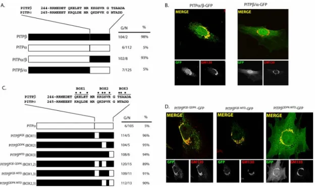
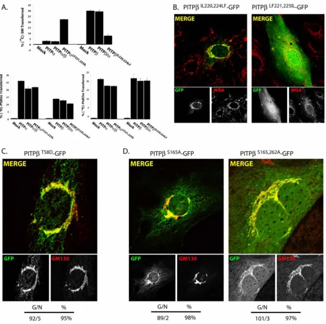
Phillips, S.E., Ile, K.E., Boukhelifa, M., Huijbregts, R.P., and Bankaitis, V.A. (2006a). Specific and nonspecific membrane-binding determinants cooperate in targeting
phosphatidylinositol transfer protein beta-isoform to the mammalian trans-Golgi network. Mol Biol Cell 17, 2498-2512.
Phillips, S.E., Sha, B., Topalof, L., Xie, Z., Alb, J.G., Klenchin, V.A., Swigart, P., Cockcroft, S., Martin, T.F., Luo, M., and Bankaitis, V.A. (1999). Yeast Sec14p deficient in phosphatidylinositol transfer activity is functional in vivo. Mol Cell 4, 187-197.
Phillips, S.E., Vincent, P., Rizzieri, K.E., Schaaf, G., Bankaitis, V.A., and Gaucher, E.A. (2006b). The diverse biological functions of phosphatidylinositol transfer proteins in eukaryotes. Crit Rev Biochem Mol Biol 41, 21-49.
Raychaudhuri, S., Im, Y.J., Hurley, J.H., and Prinz, W.A. (2006). Nonvesicular sterol movement from plasma membrane to ER requires oxysterol-binding protein-related proteins and phosphoinositides. J Cell Biol 173, 107-119.
Rhee, S.G. (2001). Regulation of phosphoinositide-specific phospholipase C. Annu Rev Biochem 70, 281-312.
Rivas, M.P., Kearns, B.G., Xie, Z., Guo, S., Sekar, M.C., Hosaka, K., Kagiwada, S., York, J.D., and Bankaitis, V.A. (1999). Pleiotropic alterations in lipid metabolism in yeast sac1 mutants: relationship to "bypass Sec14p" and inositol auxotrophy. Mol Biol Cell 10, 2235-2250.
Robinson, M., Poon, P.P., Schindler, C., Murray, L.E., Kama, R., Gabriely, G., Singer, R.A., Spang, A., Johnston, G.C., and Gerst, J.E. (2006). The Gcs1 Arf-GAP mediates Snc1,2 v-SNARE retrieval to the Golgi in yeast. Mol Biol Cell 17, 1845-1858.
Roderick, S.L., Chan, W.W., Agate, D.S., Olsen, L.R., Vetting, M.W., Rajashankar, K.R., and Cohen, D.E. (2002). Structure of human phosphatidylcholine transfer protein in complex with its ligand. Nat Struct Biol 9, 507-511.
Rogers, D.P., and Bankaitis, V.A. (2000). Phospholipid transfer proteins and physiological functions. Int Rev Cytol 197, 35-81.
Rossman, K.L., Worthylake, D.K., Snyder, J.T., Siderovski, D.P., Campbell, S.L., and Sondek, J. (2002). A crystallographic view of interactions between Dbs and Cdc42: PH domain-assisted guanine nucleotide exchange. Embo J 21, 1315-1326.
Stt4p PtdIns-4-kinase and modulate function of late stages of exocytosis in vegetative yeast. Traffic 6, 1157-1172.
Rudge, S.A., Sciorra, V.A., Iwamoto, M., Zhou, C., Strahl, T., Morris, A.J., Thorner, J., and Engebrecht, J. (2004). Roles of phosphoinositides and of Spo14p (phospholipase D)-generated phosphatidic acid during yeast sporulation. Mol Biol Cell 15, 207-218.
Schnabl, M., Oskolkova, O.V., Holic, R., Brezna, B., Pichler, H., Zagorsek, M.,
Kohlwein, S.D., Paltauf, F., Daum, G., and Griac, P. (2003). Subcellular localization of yeast Sec14 homologues and their involvement in regulation of phospholipid turnover. Eur J Biochem 270, 3133-3145.
Schouten, A., Agianian, B., Westerman, J., Kroon, J., Wirtz, K.W., and Gros, P. (2002). Structure of apo-phosphatidylinositol transfer protein alpha provides insight into
membrane association. Embo J 21, 2117-2121.
Schwille, P., Haupts, U., Maiti, S., and Webb, W.W. (1999). Molecular dynamics in living cells observed by fluorescence correlation spectroscopy with one- and two-photon excitation. Biophys J 77, 2251-2265.
Sciorra, V.A., Rudge, S.A., Wang, J., McLaughlin, S., Engebrecht, J., and Morris, A.J. (2002). Dual role for phosphoinositides in regulation of yeast and mammalian
phospholipase D enzymes. J Cell Biol 159, 1039-1049.
Sha, B., Phillips, S.E., Bankaitis, V.A., and Luo, M. (1998). Crystal structure of the Saccharomyces cerevisiae phosphatidylinositol-transfer protein. Nature 391, 506-510.
Shaw, G. (1996). The pleckstrin homology domain: an intriguing multifunctional protein module. Bioessays 18, 35-46.
Simon, J.P., Morimoto, T., Bankaitis, V.A., Gottlieb, T.A., Ivanov, I.E., Adesnik, M., and Sabatini, D.D. (1998). An essential role for the phosphatidylinositol transfer protein in the scission of coatomer-coated vesicles from the trans-Golgi network. Proc Natl Acad Sci U S A 95, 11181-11186.
Simonsen, A., Wurmser, A.E., Emr, S.D., and Stenmark, H. (2001). The role of phosphoinositides in membrane transport. Curr Opin Cell Biol 13, 485-492.
Singer, W.D., Brown, H.A., and Sternweis, P.C. (1997). Regulation of eukaryotic
phosphatidylinositol-specific phospholipase C and phospholipase D. Annu Rev Biochem
66, 475-509.
Skinner, H.B., Alb, J.G., Jr., Whitters, E.A., Helmkamp, G.M., Jr., and Bankaitis, V.A. (1993). Phospholipid transfer activity is relevant to but not sufficient for the essential function of the yeast SEC14 gene product. Embo J 12, 4775-4784.
Spiegel, S., and Milstien, S. (2003). Sphingosine-1-phosphate: an enigmatic signalling lipid. Nat Rev Mol Cell Biol 4, 397-407.
Sreenivas, A., Patton-Vogt, J.L., Bruno, V., Griac, P., and Henry, S.A. (1998). A role for phospholipase D (Pld1p) in growth, secretion, and regulation of membrane lipid synthesis in yeast. J Biol Chem 273, 16635-16638.
Stahelin, R.V., Rafter, J.D., Das, S., and Cho, W. (2003). The molecular basis of differential subcellular localization of C2 domains of protein kinase C-alpha and group IVa cytosolic phospholipase A2. J Biol Chem 278, 12452-12460.
Stocker, A., Tomizaki, T., Schulze-Briese, C., and Baumann, U. (2002). Crystal structure of the human supernatant protein factor. Structure 10, 1533-1540.
Suh, B.C., and Hille, B. (2005). Regulation of ion channels by phosphatidylinositol 4,5-bisphosphate. Curr Opin Neurobiol 15, 370-378.
Tilley, S.J., Skippen, A., Murray-Rust, J., Swigart, P.M., Stewart, A., Morgan, C.P., Cockcroft, S., and McDonald, N.Q. (2004). Structure-function analysis of human [corrected] phosphatidylinositol transfer protein alpha bound to phosphatidylinositol. Structure 12, 317-326.
van Meer, G. (2005). Cellular lipidomics. Embo J 24, 3159-3165.
Vincent, P., Chua, M., Nogue, F., Fairbrother, A., Mekeel, H., Xu, Y., Allen, N., Bibikova, T.N., Gilroy, S., and Bankaitis, V.A. (2005). A Sec14p-nodulin domain phosphatidylinositol transfer protein polarizes membrane growth of Arabidopsis thaliana root hairs. J Cell Biol 168, 801-812.
Weimar, W.R., Lane, P.W., and Sidman, R.L. (1982). Vibrator (vb): a spinocerebellar system degeneration with autosomal recessive inheritance in mice. Brain Res 251,
357-364.
Wenk, M.R., and De Camilli, P. (2004). Protein-lipid interactions and phosphoinositide metabolism in membrane traffic: insights from vesicle recycling in nerve terminals. Proc Natl Acad Sci U S A 101, 8262-8269.
Whitehead, I.P., Campbell, S., Rossman, K.L., and Der, C.J. (1997). Dbl family proteins. Biochim Biophys Acta 1332, F1-23.
Wirtz, K.W. (1997). Phospholipid transfer proteins revisited. Biochem J 324 ( Pt 2),
Wu, W.I., Routt, S., Bankaitis, V.A., and Voelker, D.R. (2000). A new gene involved in the transport-dependent metabolism of phosphatidylserine, PSTB2/PDR17, shares sequence similarity with the gene encoding the phosphatidylinositol/phosphatidylcholine transfer protein, SEC14. J Biol Chem 275, 14446-14456.
Wu, W.I., and Voelker, D.R. (2002). Biochemistry and genetics of interorganelle aminoglycerophospholipid transport. Semin Cell Dev Biol 13, 185-195.
Wu, W.I., and Voelker, D.R. (2004). Reconstitution of phosphatidylserine transport from chemically defined donor membranes to phosphatidylserine decarboxylase 2 implicates specific lipid domains in the process. J Biol Chem 279, 6635-6642.
Xie, Y., Ding, Y.Q., Hong, Y., Feng, Z., Navarre, S., Xi, C.X., Zhu, X.J., Wang, C.L., Ackerman, S.L., Kozlowski, D., Mei, L., and Xiong, W.C. (2005). Phosphatidylinositol transfer protein-alpha in netrin-1-induced PLC signalling and neurite outgrowth. Nat Cell Biol 7, 1124-1132.
Xie, Z., Fang, M., and Bankaitis, V.A. (2001). Evidence for an intrinsic toxicity of phosphatidylcholine to Sec14p-dependent protein transport from the yeast Golgi complex. Mol Biol Cell 12, 1117-1129.
Xie, Z., Fang, M., Rivas, M.P., Faulkner, A.J., Sternweis, P.C., Engebrecht, J.A., and Bankaitis, V.A. (1998). Phospholipase D activity is required for suppression of yeast phosphatidylinositol transfer protein defects. Proc Natl Acad Sci U S A 95, 12346-12351.
Yamniuk, A.P., and Vogel, H.J. (2004). Calmodulin's flexibility allows for promiscuity in its interactions with target proteins and peptides. Mol Biotechnol 27, 33-57.
Yanagisawa, L.L., Marchena, J., Xie, Z., Li, X., Poon, P.P., Singer, R.A., Johnston, G.C., Randazzo, P.A., and Bankaitis, V.A. (2002). Activity of specific lipid-regulated ADP ribosylation factor-GTPase-activating proteins is required for Sec14p-dependent Golgi secretory function in yeast. Mol Biol Cell 13, 2193-2206.
Yoder, M.D., Thomas, L.M., Tremblay, J.M., Oliver, R.L., Yarbrough, L.R., and Helmkamp, G.M., Jr. (2001). Structure of a multifunctional protein. Mammalian
phosphatidylinositol transfer protein complexed with phosphatidylcholine. J Biol Chem
276, 9246-9252.
York, J.D., Odom, A.R., Murphy, R., Ives, E.B., and Wente, S.R. (1999). A
Chapter 1b
The PITP proteins
The PITP superfamily of proteins is defined by the ability of the proteins to
transfer lipids between membranes in vitro. Within the superfamily, there are two
subfamilies that share the same biochemical activity, but do not have any sequence or
structural similarity. These families are called the Sec14 PITP family, named for the
founding yeast PITP, and the Class I or Metazoan PITPs. In many cases, there are more
Sec14 family proteins in a given organism than PITP members. The most striking case is
Drosophila, where there are 15 Sec14 family members and just three metazoan PITPs
(Phillips et al, 2006). The reason for the many family members in the Sec14 family may
be related to the ability of these proteins to bind to different lipids (such as the α
-tocopherol transfer proteins), and the fact that Sec14 domains are often found in
association with other domains such as the GOLD or nodulin domains. The PITP family,
which is the focus of my thesis, and thus this review, is a smaller family with members
that are often essential for survival.
Sec14
While Sec14 is not related to the PITPs in structure or sequence, the functional
interchangability of these molecules suggests that Sec14 is still a relevant model for PITP
function.
The first clues to Sec14 function came from finding mutations in yeast genes that
allowed yeast to grow despite a non-functional Sec14 molecule (known as bypass