Research Article
Biofunctional Activities of
Equisetum
ramosissimum
Extract: Protective Effects against
Oxidation, Melanoma, and Melanogenesis
Pin-Hui Li,
1Yu-Pin Chiu,
2Chieh-Chih Shih,
3Zhi-Hong Wen,
3Laura Kaodichi Ibeto,
4,5Shu-Hung Huang,
6,7,8,9Chien Chih Chiu,
10Dik-Lung Ma,
11Chung-Hang Leung,
12Yaw-Nan Chang,
2and Hui-Min David Wang
1,3,7,131Department of Fragrance and Cosmetic Science, Kaohsiung Medical University, 100 Shih-Chuan 1st Road,
San Ming District, Kaohsiung 807, Taiwan
2Department of Biotechnology, National Formosa University, 64 Wenhua Road, Huwei Township, Yunlin County 632, Taiwan
3Department of Marine Biotechnology and Resources, National Sun Yat-Sen University, 70 Lianhai Road, Gushan District,
Kaohsiung 804, Taiwan
4Department of Biological Science, University of North Carolina at Chapel Hill, Coker Hall, CB No. 3280, 120 South Road,
Chapel Hill, NC 27599-3280, USA
5Department of Biomedical Science, Barry University, 11300 Northeast 2nd Avenue, Miami, FL 33161, USA
6Graduate Institute of Medicine, College of Medicine, Kaohsiung Medical University, 100 Shih-Chuan 1st Road, San Ming District,
Kaohsiung 807, Taiwan
7Center for Stem Cell Research, Kaohsiung Medical University, 100 Shih-Chuan 1st Road, San Ming District, Kaohsiung 807, Taiwan
8Division of Plastic Surgery, Department of Surgery, Kaohsiung Medical University Hospital, Kaohsiung Medical University,
Kaohsiung 807, Taiwan
9Department of Surgery, Faculty of Medicine, College of Medicine, Kaohsiung Medical University, Kaohsiung 807, Taiwan
10Department of Biotechnology, Kaohsiung Medical University, Kaohsiung 807, Taiwan
11Department of Chemistry, Hong Kong Baptist University, Kowloon Tong, Hong Kong
12State Key Laboratory of Quality Research in Chinese Medicine, Institute of Chinese Medical Sciences, University of Macau, Macau
13Graduate Institute of Natural Products, Kaohsiung Medical University, Kaohsiung 807, Taiwan
Correspondence should be addressed to Yaw-Nan Chang; [email protected] and Hui-Min David Wang; [email protected]
Received 26 February 2016; Revised 28 April 2016; Accepted 8 May 2016
Academic Editor: Pavel Pospisil
Copyright © 2016 Pin-Hui Li et al. This is an open access article distributed under the Creative Commons Attribution License, which permits unrestricted use, distribution, and reproduction in any medium, provided the original work is properly cited.
Equisetum ramosissimum, a genus of Equisetaceae, is a medicinal plant that can be separated into ethyl acetate (EA),
dichloromethane (DM),n-hexane (Hex), methanol (MeOH), and water extracts. EA extract was known to have potent antioxidative properties, reducing power, DPPH scavenging activity, and metal ion chelating activity. This study compared these five extracts in terms of their inhibiting effects on three human malignant melanomas: A375, A375.S2, and A2058. MTT assay presented the notion that both EA and DM extracts inhibited melanoma growth but did not affect the viabilities of normal dermal keratinocytes (HaCaT) or fibroblasts. Western blot analyses showed that both EA and DM extracts induced overexpression of caspase proteins in all three melanomas. To determine their roles in melanogenesis, this study analyzed theirin vitrosuppressive effects on mushroom tyrosinase. All extracts except for water revealed moderate suppressive effects. None of the extracts affected B16-F10 cells proliferation. EA extract inhibited cellular melanin production whereas DM extract unexpectedly enhanced cellular pigmentation in B16-F10 cells. Data for modulations of microphthalmia-associated transcription factor, tyrosinase, tyrosinase-related protein 1, and tyrosinase-related protein 2 showed that EA extract inhibited protein expression mentioned above whereas DM extract had the opposite effect. Overall, the experiments indicated that the biofunctional activities of EA extract contained in food and cosmetics protect against oxidation, melanoma, and melanin production.
1. Introduction
Mitochondria, chloroplasts, and peroxisomes produce reac-tive oxygen species (ROS) through respiration and photo-synthesis [1, 2]. Studies suggested that the changes in cellular homeostasis caused by high ROS levels can result in oxidative damage [3]. To avoid ROS oxidative injuries, the defense radical scavenging systems used by the human being were separated into enzymatic and nonenzymatic mechanisms. Antioxidant enzymes and substances could reduce oxidative damage by decreasing production of ROS and radicals [2]. These agents include glutathione and catalase, glutathione reductase, superoxide dismutase, and glutathione peroxidase. Others include 𝛼-lipoic acid, carotenoids, coenzyme Q10, flavonoids, antioxidative minerals (copper, zinc, manganese, and selenium), and cofactors (folic acid and vitamins A, B1, B2, B6, B12, C, and E). Generally, the above antioxidative materials are applied in synergic ways with each other against various free radical types [1–3].
Malignant melanoma is among the most incursive and life-threatening malignant tumors [4]. Skin cancer can result from exposure to ultraviolet (UV) radiation emitted by the sun and by halogen lamps. Experimental studies of metastatic melanoma are very challenging because systemic treatments are often ineffective and the rapid spread of melanoma cells to retain an intensive property of the cellular spreading which happens later is pathologically confusing [5]. Although melanoma is not a major cause of tumorous symptoms, melanoma is a major cause of death in patients with skin cancer. Treating melanoma is difficult due to its resistance to conventional chemoradiotherapy [6]. No effective therapies for metastatic melanoma are currently available, and effective drugs are urgently needed.
Melanocytes are located in the basal epidermal layer and in the hair follicles. Generally, UV radiation produces pig-ment by diametric stimulation of melanocytes [7]. Melanin pigmentation has been recognized by many factors; the permeation of sunlight is a well recognized source of melanin pigmentation; specifically, UV radiation causes darkening of the skin and/or sunburn. UV is the most common reason of changes in the visible countenance of human skin. Unusual melanogenesis is a characteristic of many human skin disorders, including abnormal pigmentation, nevi, and melanoma [8].
Many recent studies have investigated the biological functions of natural extracts and their potential applications as health foods, as active ingredients in cosmetics, and as leading compounds in new medicines [9, 10]. Equisetum ramosissimum,a genus of Equisetaceae, is a medicinal plant administered to treat hemorrhage, urethritis, jaundice, and hepatitis [11]. Although the antioxidant activities ofE. ramo-sissimum were identified [12], its biological activities have not been examined. Therefore, this study elucidated the potential protective effects ofE. ramosissimumextract against oxidation, melanoma, and melanogenesis.
2. Materials and Methods
2.1. Chemicals and Reagents. Ascorbic acid (vitamin C), 3-(4,5-dimethylthiazol-2-yl)-2,5-diphenyltetrazolium bromide
(MTT), L-3,4-dihydroxyphenylalanine (L-DOPA), dimethyl sulfoxide (DMSO), 1,1-diphenyl-2-picrylhydrazyl (DPPH), ethanol, ethylenediaminetetraacetic acid (EDTA), ferrous chloride (FeCl2⋅4H2O), ferric chloride (FeCl3), kojic acid, methanol, potassium ferricyanide (K3Fe(CN)6), 3-tert-butyl-4-hydroxyanisole (BHA), and L-tyrosine were purchased from Sigma-Aldrich Company (St. Louis, MO, USA). Dul-becco’s modified Eagle’s medium (DMEM) and fetal bovine serum (FBS) were obtained from Gibco BRL (Gaithersburg, MD, USA). Other chemical buffers and reagents were pur-chased at the highest available purity and quality.
2.2. Plant Material Extraction and Isolation. Two authors of this study (Dr. Chieh-Chih Shih and Professor Zhi-Hong Wen) prepared the extracts as follows. First, methanol (2.2 L) was requested in two extraction procedures ofE. ramosissi-mumfrom a powder consisting of groundE. ramosissimum plant, and the extract was then refluxed for 30 minutes. After a filtering procedure, the extract was concentrated to obtain methanol (MeOH) crude extract, 9.52 g. The crude extract was added to distilled water (200 mL) and then partitioned withn-hexane (Hex, 1.95 g), dichloromethane (DM, 0.67 g), and ethyl acetate (EA, 0.26 g), which left an aqueous layer (H2O, 6.44 g). All fractions were concentrated, freeze-dried, dissolved in DMSO to obtain a stock solution (500 mg/mL), and then diluted with DMEM to the required concentrations.
2.3. Assays of Antioxidant Effects
2.3.1. Reducing Power Assay. Assays of the reducing power of the crude extracts were performed as described in the literature [13]. Briefly, dissimilar concentrations of each extract were blended with 85𝜇L of 67 mM sodium phosphate buffer (pH 6.8) and 2.5𝜇L of 20% K3Fe(CN)6. The admixture was kept at 50∘C for 20 min. After addition of 160𝜇L 10% TCA, the admixture was centrifuged for 10 min at 3,000 g. The supernatant (75𝜇L) was mixed with 2% FeCl3 (25𝜇L), and absorbance was read with a spectrophotometer (BioTek Co., Winooski, VT, USA) at 700 nm with a BHA solution as a positive control. High absorbance was interpreted as a high capacity for metal ion reduction.
2.3.2. DPPH∙ Radical Scavenging Ability Assay. DPPH is a stable free radical with a violet color. Reaction of DPPH∙ with an antioxidant provides hydrogen, which results in decreased absorbance at 517 nm. DPPH assay was performed as described previously, with some minor modification [13]. Various concentrations of E. ramosissimum extracts were added to 100𝜇L of aqueous stable DPPH∙ (60𝜇M) solution and allowed to stand at room temperature for 60 min. Vitamin C was used as a positive control. Low absorbance was interpreted as a high DPPH scavenging ability. The calculation for free radical scavenging activity (%) is done as follows:
Scavenging activity(%) = 𝐴control− 𝐴sample
𝐴control × 100%. (1)
previously [14]. Briefly, various concentrations of extract were dissolved in DMSO and added to a 10𝜇L solution of FeCl2⋅4H2O (2 mM). Next, 20𝜇L ferrozine (5 mM) was added, and the admixture was shaken vigorously for 10 minutes. The absorbance was 562 nm. EDTA was used as a positive control, and the formula employed to calculate metal chelating activity is as follows:
Metal chelating activity(%)
= 𝐴control𝐴 − 𝐴sample
control × 100%.
(2)
2.4. Cell Line Cultures. Human and rat melanoma cell lines were obtained from Bioresource Collection and Research Center (Taiwan): A375 (BCRC number 60039), A375.S2 (BCRC number 60263), A2058 (BCRC number 60240), and B16-F10 (BCRC number 60031). Human fibroblasts were separated from the foreskin primary culture (Institutional Review Board, KMUH-IRB-990269). Cells were cultured in DMEM supplemented with 10% FBS and 1% antibiotics. Human skin keratinocytes, HaCaT cells, were cultured in Keratinocyte-SFM (Gibco, USA) supplemented with bovine pituitary extract and human recombinant epidermal growth factor. All cell lines were cultured in 5% CO2at 37∘C. 2.5. MTT Assay of Cell Viability. The influences of extracts on cell development were estimated with MTT assay [15]. Cells were seeded at8 × 103cells/well in 96-well plates and incubated for 24 h before addition of extracts. After 24 h, MTT solution was dispensed into each well. After 2 h, the culture medium was discarded, and DMSO was added to each well. The absorbance of the formazan salt was 595 nm, and the cell viability was computed as follows:
Cell viability(%) = 𝐴control− 𝐴sample
𝐴control × 100%. (3)
2.6. Western Blot Analysis. This analysis was performed as described previously with some minor modifications [16].1× 106cells were treated with extracts or with the vehicle control
for 24 h, and the cells were then harvested and lysed with RIPA lysis buffer. Equal amounts of protein were separated by sodium dodecyl sulfate-polyacrylamide gel electrophoresis and next transferred into a polyvinylidene fluoride mem-brane. The membranes were incubated with corresponding primary antibodies and afterwards incubated with secondary antibodies corresponding with the primary antibodies. The signals were visualized with a chemiluminescence detection kit (Amersham, Piscataway, NJ, USA).
2.7. Mushroom Tyrosinase Measurement. Mushroom tyrosi-nase activity was evaluated as described previously with some minor modifications [17]. Samples were incubated with mushroom tyrosinase (25 U/mL), and L-tyrosine (2 mM) in phosphate buffer (pH 6.8) was added. The mixtures were then kept at 37∘C for 30 minutes. Kojic acid was used as a positive
control. Tyrosinase inhibitory activity was determined by the following equation:
Mushroom tyrosinase inhibition(%)
= [(𝐴 − 𝐵) − (𝐶 − 𝐷)]
(𝐴 − 𝐵) × 100%,
(4)
where𝐴is the optical density (OD490) without testing extract; 𝐵is OD490with tyrosinase and without testing extract;𝐶is OD490 with testing extract; and𝐷is OD490 with tyrosinase and without testing extract.
2.8. Melanin Quantification Assessment. This analysis was demonstrated as described in the literature with some minor modifications [17]. Cell pellets were liquefied with 1.0 N NaOH, warmed to 80∘C for 1 hour, and centrifuged at 10,000 g for 10 minutes. The quantity of melanin was determined by extracting the supernatant and running it through the spectrophotometer, which gave a result of 475 nm.
2.9. Statistical Analysis. Biofunctional assays of theE. ramo-sissimumextracts in each platform were performed in trip-licate. Results were expressed as means ±SD. Analysis of variance was used for data analysis. A𝑝value less than 0.05 was considered statistically significant.
3. Results and Discussion
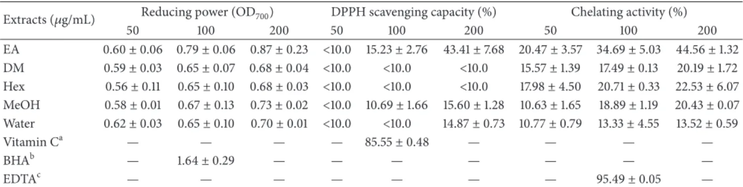
3.1. Antioxidant Activity of E. ramosissimum. Free radicals have roles in signal travel and in physiological, metabolic, and immune reactions. Even though free radicals are needed for normal healthy biochemical processes in the body, they have severe negative health effects [18, 19]. One of the study aims was to test antioxidative properties, which were examined by ferric reducing powers, DPPH radical scavenging capacities, and metal chelating power activities.
First, a simple, rapid, and reliable test was used to measure Fe(III)-ferricyanide complex synthesis. In this test, the extracts reducing properties of E. ramosissimum were indicated by changes in the color of the solution (from light yellow to different shades of green and blue). Table 1 presented the notion that the reducing power of EA extract resulted in stronger dose-dependent suppressive effects com-pared to the other four extracts and showed the highest scavenging of0.87 ± 0.23at 200𝜇g/mL.
The second oxidation inhibitory assay was DPPH radical scavenging test. As antioxidants stabilize DPPH radicals, the color of DPPH solution changes from violet to yellow as diphenylpicrylhydrazine is formed. Table 1 illustrated the results for the five extracts, and the comparisons presented the notion that the EA extract had the strongest radical scavenging effects in a dose-dependent manner.
Table 1: Antioxidant activities ofE. ramosissimumextracts, including reducing power, DPPH free radical scavenging activity, and ferrous ion chelating power.
Extracts (𝜇g/mL) Reducing power (OD700) DPPH scavenging capacity (%) Chelating activity (%)
50 100 200 50 100 200 50 100 200
EA 0.60±0.06 0.79±0.06 0.87±0.23 <10.0 15.23±2.76 43.41±7.68 20.47±3.57 34.69±5.03 44.56±1.32 DM 0.59±0.03 0.65±0.07 0.68±0.04 <10.0 <10.0 <10.0 15.57±1.39 17.49±0.13 20.19±1.72 Hex 0.56±0.11 0.65±0.10 0.68±0.03 <10.0 <10.0 <10.0 17.98±4.50 20.71±0.33 22.53±6.07 MeOH 0.58±0.01 0.67±0.13 0.73±0.02 <10.0 10.69±1.66 15.60±1.28 10.63±1.65 18.89±1.19 20.43±0.07 Water 0.62±0.03 0.65±0.10 0.70±0.01 <10.0 <10.0 14.87±0.73 10.77±0.79 13.33±4.55 13.52±0.59
Vitamin Ca — — — — 85.55±0.48 — — — —
BHAb — 1.64±0.29 — — — — — — —
EDTAc — — — — — — — 95.49±0.05 —
All statistics are presented as average values±SD;𝑛 = 3.aVitamin C (100 mM) was utilized as a positive control for DPPH assay;bEDTA (100 mM) was used as a positive control for analysis of metal chelating ability;cBHA (100 mM) was applied as a positive control for analysis of reducing power. Assays not performed in this study were indicated by dashes.
Water MeOH
DM
EA Hex
0 50 100
H
aC
aT cell via
b
ili
ty (%)
Concentration (𝜇g/mL)
∗ ∗ ∗ ∗ ∗ ∗ ∗
∗
5 10 25 50 100 5 10 25 50 100 5 10 25 50 100 5 10 25 50 100 5 10 25 50 100
Co
n
tr
o
l
(a)
0 50 100
Fib
rob
last cell via
b
ili
ty (%)
∗ ∗ ∗
∗
Water MeOH
DM
EA Hex
Concentration (𝜇g/mL)
5 10 25 50 100 5 10 25 50 100 5 10 25 50 100 5 10 25 50 100 5 10 25 50 100
Co
n
tr
o
l
(b)
Figure 1: Effects ofE. ramosissimumextracts on viability of normal human cells according to MTT assay. Suppression of cell viability was measured in (a) HaCaT and (b) fibroblast cells cultured with 5, 10, 25, 50, and 100𝜇g/mL EA, DM, Hex, MeOH, and water extracts. The results for the control group cultured without extracts were shown on the left (gray line). All experimental data were presented as average values± SD;𝑛 = 3;∗𝑝 < 0.05.
of antioxidative properties. The extracts showed low-to-moderate Fe2+scavenging activities at concentrations of 50– 200𝜇g/mL, and EA extract possessed the highest value of 44.56 ± 1.32at 200𝜇g/mL.
3.2. Cytotoxicity of E. ramosissimum on Human Melanoma Cells and Normal Cells. Melanoma is a malignant tumor that starts in a certain type of skin cell and is activated when the abnormal cells in the affected part of the body begin to proliferate in an uncontrolled manner [20]. Metastatic malig-nant melanomas are highly resistant to existing therapies and have a very poor prognosis, and thus new treatment strategies are urgently needed. In early stages of the development of new chemoprotective substances, the main considerations are normal cell allergic responses, sensitivity and potential reactions, and toxic side effects [9]. MTT method was applied to evaluate the cytotoxic effectivenesses ofE. ramosissimum extracts on normal human skin cells, including epidermal keratinocytes (HaCaT) and dermal fibroblasts in Figure 1. These two cells were treated with various concentrations (0
to 100𝜇g/mL) to compare dose-dependent impacts. In both cells, high doses (100𝜇g/mL) of the five E. ramosissimum extracts had minor effects, and all cellular viabilities exceeded 65% after a 24-hour treatment. That is, theE. ramosissimum extracts had no severe discernible toxic effects on human normal cells.
∗
∗ ∗ ∗
∗ ∗
∗
∗ ∗ ∗
∗ ∗
Water MeOH
DM
EA Hex
Concentration (𝜇g/mL)
5 10 25 50 100 5 10 25 50 100 5 10 25 50 100 5 10 25 50 100 5 10 25 50 100
Co
n
tr
o
l
0 50 100
A375 cell via
b
ili
ty (%)
(a)
∗
∗ ∗
∗ ∗
∗ ∗
∗
∗ ∗
∗ ∗ ∗
∗
Water MeOH
DM
EA Hex
Concentration (𝜇g/mL)
5 10 25 50 100 5 10 25 50 100 5 10 25 50 100 5 10 25 50 100 5 10 25 50 100
Co
n
tr
o
l
0 50 100
A375.S2 cell via
b
ili
ty (%)
(b)
∗ ∗ ∗
∗
∗ ∗ ∗ ∗
∗
Water MeOH
DM
EA Hex
Concentration (𝜇g/mL)
5 10 25 50 100 5 10 25 50 100 5 10 25 50 100 5 10 25 50 100 5 10 25 50 100
Co
n
tr
o
l
0 50 100
A2058 cell via
b
ili
ty (%)
(c)
Figure 2: Effects ofE. ramosissimumextracts on viability of human melanoma cells according to MTT assay. Suppression of cell viability was measured in (a) A375, (b) A375.S2, and (c) A2058 cells cultured with 5, 10, 25, 50, and 100𝜇g/mL EA, DM, Hex, MeOH, and water. The results for the control group cultured without extract were demonstrated on the left (gray line). All experimental data were presented as average values±SD;𝑛 = 3;∗𝑝 < 0.05.
decreased to 50%. MeOH extract had no effect on the A2058 cells, and the water extract had no apparent cytotoxic effect on the three melanoma cell types. According to our statistical data, theE. ramosissimumextracts have little harmful effects on normal skin cells, and these extracts, particularly EA, DM, and Hex extracts, actually inhibit melanoma cellular proliferation.
3.3. Effects of EA and DM Extracts on Caspase Proteins in Melanoma Cells. Caspase is a family of cysteine-aspartic proteases that mediates type I programmed cell death (apop-tosis), and more than 10 family members have been identified so far [22]. The activation of caspase-associated proteins is essential for apoptosis induced by various apoptotic stimuli. These changes include blebbing, cell shrinkage, nuclear frag-mentation, chromatin condensation, and chromosomal DNA fragmentation; however, the failure of cancer cell apoptosis is a major contributor to tumor development and autoimmune disease [23].
Caspase-9 initiates an apoptotic cascade by cleaving and activates 3 [24]. However, the maturation of caspase-9 requires autocatalytic cleavage by apoptosomes released by damaged mitochondria [25]. The cleavage of caspase-3
activates caspase-6 and caspase-7; the protein itself is pro-cessed and activated by 8, 9, and caspase-10. The activation of caspase-3 induces cellular apoptosis and proteolysis in specific substrates [26]. Figure 3 showed how caspase affected the apoptotic process induced by EA and DM extracts ofE. ramosissimumin the three human melanoma cells. Remarkable alterations caused by molecular proteins associated with apoptosis included increased proteolysis induced by caspase-3 and caspase-9. A low concentration (10𝜇g/mL) of EA extract induced stimulated enzymes in A375, A375.S2, and A2058 cells. Although DM extract also triggered caspase-3 and caspase-9, its effect was smaller compared to a similar dose of EA extract. Notably, 50𝜇g/mL DM extract was needed to trigger caspase-3 and caspase-9 in A2058 cells. The cellular proteins 3 and caspase-9 have important roles in the regulation of nuclear DNA damage caused by apoptosis and in the decomposition of organelles. Our experiments demonstrated that EA induced caspase protein changes, and similar results were also shown from DM extract, but lower.
Con. 10 50 100 Con. 10 50 100
Cleaved caspase-3
Cleaved caspase-9
GAPDH
EA extract (𝜇g/mL) DM extract (𝜇g/mL)
(a) A375
GAPDH Cleaved caspase-3
Cleaved caspase-9
Con. 10 50 100 Con. 10 50 100
EA extract (𝜇g/mL) DM extract (𝜇g/mL)
(b) A375.S2
GAPDH Cleaved caspase-3
Cleaved caspase-9
Con. 10 50 100 Con. 10 50 100
EA extract (𝜇g/mL) DM extract (𝜇g/mL)
(c) A2058
Figure 3: Expressions of caspase proteins in (a) A375, (b) A375.S2, and (c) A2058 cells after treatment with 10, 50, and 100𝜇g/mL EA and DM extracts for 24 h. Band darkness indicated relative protein expressions in comparison with GAPDH.
pigment biosynthesis reactions. Other enzymes only adjust to differences in synthesis of eumelanin and pheomelanin [10, 27, 28]. To determine whether the extracts inhibited melanin synthesis by suppressing tyrosinase,in vitro tyrosi-nase activity was tested in mushroom type. In Table 2, it was shown that all extracts except for water had dose-dependent (5–100𝜇g/mL) inhibiting effects on the mushroom tyrosinase system. At a concentration of 100𝜇g/mL, the repressive effect of EA extract was slightly lower than that of kojic acid. At this concentration, EA had the strongest inhibiting effect (approximately 40%).
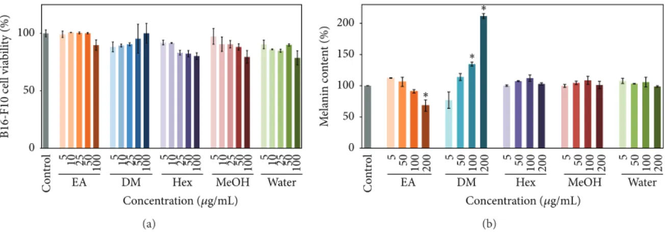
3.5. Melanin Content of B16-F10 Cells. Melanin is the source of skin color and can protect the skin from UV radiation damage which induces DNA mutations. Despite its protective functions, superabundance of melanin negatively affects the skin, which then causes social problems [29, 30]. To demon-strate the skin whitening effects ofE. ramosissimumand its
Table 2: Mushroom tyrosinase inhibition by different concentra-tions ofE. ramosissimumextracts.
Concentrations (𝜇g/mL)
Mushroom tyrosinase inhibition (%)
5 50 100
EA 22.42±0.25 24.40±4.90 38.93±3.09 DM 20.69±4.53 21.30±1.10 23.79±3.84 Hex 20.54±0.64 22.33±0.69 23.82±4.25 MeOH 18.54±2.70 20.45±0.36 23.27±1.67 Water 17.88±3.46 19.37±0.12 23.82±1.24 Kojic acida — — 30.23±5.68
All statistics are presented as average values±SD;𝑛 = 3.aKojic acid (100𝜇g/mL) was applied as a positive control. Assays not performed in this study were indicated by dashes.
Water MeOH DM
EA Hex
Concentration (𝜇g/mL)
5 10 25 50 100 5 10 25 50 100 5 10 25 50 100 5 10 25 50 100 5 10 25 50 100
Co
n
tr
o
l
0 50 100
B16-F10 cell via
b
ili
ty (%)
(a)
∗ ∗
∗
Water MeOH
DM
EA Hex
Concentration (𝜇g/mL)
5 50 100 200 5 50 100 200 5 50 100 200 5 50 100 200 5 50 100 200
Co
n
tr
o
l
0 50 100 150 200
M
ela
nin co
n
ten
t (%)
(b)
Figure 4: (a) Effects ofE. ramosissimumextracts on viability of B16-F10 cells according to MTT assay and (b) effects ofE. ramosissimum
extracts on melanin content quantification of all extracts were processed with 5, 50, 100, and 200𝜇g/mL, respectively. The results for the control group cultured without extracts were shown on the left (gray line). All experimental results were presented as average values±SD;
𝑛 = 3;∗𝑝 < 0.05.
MITF
TRP-2
GAPDH TRP-1 Tyrosinase
Con. 10 50 100
EA extract (𝜇g/mL)
(a)
MITF
TRP-2
GAPDH TRP-1 Tyrosinase
Con. 10 50 100
DM extract (𝜇g/mL)
(b)
Figure 5: Expressions of tyrosinase, TRP-1, TRP-2, and MITF after treatment withE. ramosissimumextracts. B16-F10 cells were treated with EA and DM extracts at concentrations of 10, 50, and 100𝜇g/mL for 24 h. Protein expressions were shown in comparison with GAPDH.
did not substantially harm B16 cell viability. In Figure 4(b), we presented the notion that the EA extract had the ability to decrease melanin production by about 32% at 200𝜇g/mL in a dose-dependent trend from concentrations of 5 to 200𝜇g/mL. In contrast, 200𝜇g/mL DM extract substantially augmented melanin production (112%), and the effects of DM extract were dose-dependent. Other extracts, including Hex, MeOH, and water, did not substantially change melanin production, even at the maximum experimental dose of 200𝜇g/mL. These experimental results suggested that the EA extract had potential applications as a whitening agent in cosmetic products whereas the DM extract could be used as a skin darkening agent.
3.6. Expression of Melanogenesis-Related Proteins in B16-F10 Cells. A well known role of microphthalmia-associated
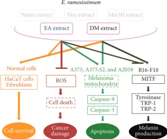
E. ramosissimum
DM extract
MeOH extract Hex extract
Water extract
HaCaT cells Fibroblasts
Cell survival
ROS
Cell death
Cancer damage
Melanoma mitochondria
Caspase-9
Caspase-3
MITF
Tyrosinase TRP-1 TRP-2
Melanin production Apoptosis
EA extract
Normal cells A375, A375.S2, and A2058 B16-F10
Figure 6: Schematic diagram of biofunctions ofE. ramosissimum
extracts in human skin cells, including normal cell survival, apop-totic pathways of melanoma, and melanogenesis.
4. Conclusion
In summary, the experiments in this study demonstrated that the most beneficial of the five fraction extracts of E. ramosissimumwas EA because of its multiple biofunctional properties (Figure 6). The experimental outcomes showed that, by acting as an antioxidant ingredient and electron donor, EA extract discontinued or terminated free radical chain reactions. In human melanoma, EA and DM extracts affected the viabilities of melanoma cells and showed low toxicity in both normal human cells, HaCaT cells and fibroblasts. To understand the mechanisms of cell death, we performed western blot analyses of protein expressions in melanoma cells, which pointed out that both extracts induced caspase-3 and caspase-9, both of which have vital roles in apoptosis. Research evaluations of the potential use of EA extract as a whitening agent illustrated that it inhibited mushroom tyrosinase activity and the synthesis of melanin; in contrast, DM extract increased the quantity of melanin. Western blot analyses showed that EA and DM extracts decreased and increased melanin content, respectively, by regulating MITF, tyrosinase, Trp-1, and Trp-2. Whereas this study established the biological functions ofE. ramosissimum, our future studies will further investigate the components and mechanisms of these compounds.
Competing Interests
The authors have no competing interests regarding the publication of this study.
Authors’ Contributions
Pin-Hui Li and Yu-Pin Chiu equally contributed to this study.
Acknowledgments
This work was supported by grants from the Ministry of Science and Technology, Taiwan (MOST104-2622-E-037-001; MOST104-2622-E-037-003-CC2; MOST104-2221-E-037-005-MY2; and MOST104-2628-E-037-001-MY3), from the Center for Stem Cell Research, Kaohsiung Medical University, Kaohsiung, Taiwan (KMU-TP104G00 and KMUTP104G02-05), from Kaohsiung Medical University, Taiwan (KMU-DK105005), from Kaohsiung Medical University, NSYSU-KMU Joint Research Project, Taiwan (NSYSUKMU105-P 007), and from the Center for Infectious Disease and Cancer Research, Kaohsiung Medical University, Taiwan (KMU-TP104E18).
References
[1] K. Apel and H. Hirt, “Reactive oxygen species: metabolism, oxidative stress, and signal transduction,” Annual Review of
Plant Biology, vol. 55, pp. 373–399, 2004.
[2] ´A. Guti´errez-Uzquiza, M. Arechederra, P. Bragado, J. A. Aguirre-Ghiso, and A. Porras, “p38𝛼mediates cell survival in response to oxidative stress via induction of antioxidant genes: effect on the p70S6K pathway,”Journal of Biological Chemistry, vol. 287, no. 4, pp. 2632–2642, 2012.
[3] C. Richardson, S. Yan, and C. G. Vestal, “Oxidative stress, bone marrow failure, and genome instability in hematopoietic stem cells,”International Journal of Molecular Sciences, vol. 16, no. 2, pp. 2366–2385, 2015.
[4] K.-N. Kim, G. Ahn, S.-J. Heo et al., “Inhibition of tumor growth in vitro and in vivo by fucoxanthin against melanoma B16F10 cells,”Environmental Toxicology and Pharmacology, vol. 35, no. 1, pp. 39–46, 2013.
[5] A. Ascenso, T. Pedrosa, S. Pinho et al., “The effect of lycopene preexposure on UV-B-irradiated human keratinocytes,”
Oxida-tive Medicine and Cellular Longevity, vol. 2016, Article ID
8214631, 15 pages, 2016.
[6] C.-C. Lee, L.-Y. Chiou, J.-Y. Wang et al., “Functional ginger extracts from supercritical fluid carbon dioxide extraction via in vitro and in vivo assays: antioxidation, antimicroorganism, and mice xenografts models,”The Scientific World Journal, vol. 2013, Article ID 210845, 8 pages, 2013.
[7] R. Ghosh, D. Guha, S. Bhowmik, and S. Karmakar, “Antioxidant enzymes and the mechanism of the bystander effect induced by ultraviolet C irradiation of A375 human melanoma cells,”
Mutation Research, vol. 757, no. 1, pp. 83–90, 2013.
[8] W.-S. Liu, Y.-D. Kuan, K.-H. Chiu et al., “The extract of
Rhodobacter sphaeroides inhibits melanogenesis through the
MEK/ERK signaling pathway,”Marine Drugs, vol. 11, no. 6, pp. 1899–1908, 2013.
[9] C.-C. Lee, Y.-T. Chen, C.-C. Chiu, W.-T. Liao, Y.-C. Liu, and H.-M. David Wang, “Polygonum cuspidatum extracts as bioactive antioxidaion, anti-tyrosinase, immune stimulation and anticancer agents,”Journal of Bioscience and Bioengineering, vol. 119, no. 4, pp. 464–469, 2015.
[10] M.-G. Lee, S.-Y. Kuo, S.-Y. Yen et al., “Evaluation of
Cinnamo-mum osmophloeumKanehira extracts on tyrosinase suppressor,
wound repair promoter, and antioxidant,”The Scientific World
Journal, vol. 2015, Article ID 303415, 7 pages, 2015.
and flavonoids isolated from Equisetum arvense,” Journal of
Ethnopharmacology, vol. 95, no. 2-3, pp. 421–424, 2004.
[12] S. Paulsamy, D. Moorthy, K. Nandakumar et al., “Evaluation of
in vitroantioxidant potential of methanolic extracts of the ferns,
Actiniopteris radiata(Sw) Link andEquisetum ramosissimum
Desf,” International Journal of Research and Development in
Pharmacy & Life Sciences, vol. 2, no. 3, pp. 451–455, 2013.
[13] H.-M. Wang, Y.-T. Chou, Z.-L. Hong et al., “Bioconstituents from stems of Synsepalum dulcificum Daniell (Sapotaceae) inhibit human melanoma proliferation, reduce mushroom tyrosinase activity and have antioxidant properties,”Journal of
the Taiwan Institute of Chemical Engineers, vol. 42, no. 2, pp.
204–211, 2011.
[14] H.-M. Wang, J.-L. Pan, C.-Y. Chen et al., “Identification of anti-lung cancer extract fromChlorella vulgarisC-C by antioxidant property using supercritical carbon dioxide extraction,”Process
Biochemistry, vol. 45, no. 12, pp. 1865–1872, 2010.
[15] H.-M. Wang, C.-Y. Chen, and Z.-H. Wen, “Identifying melano-genesis inhibitors from Cinnamomum subavenium with in vitro and in vivo screening systems by targeting the human tyrosinase,”Experimental Dermatology, vol. 20, no. 3, pp. 242– 248, 2011.
[16] C.-L. Lin, R.-F. Chen, J. Y.-F. Chen et al., “Protective effect of caffeic acid on paclitaxel induced anti-proliferation and apop-tosis of lung cancer cells involves NF-𝜅b pathway,”International
Journal of Molecular Sciences, vol. 13, no. 5, pp. 6236–6245, 2012.
[17] X. Chen, B. Zhang, X. Yuan et al., “Isoliquiritigenin-induced differentiation in mouse melanoma B16F0 cell line,”Oxidative
Medicine and Cellular Longevity, vol. 2012, Article ID 534934, 11
pages, 2012.
[18] X. H. Pan, X. L. Zhang, H. L. Sun, J. Zhang, M. Yan, and H. Zhang, “Autophagy inhibition promotes 5-fluorouraci-induced apoptosis by stimulating ROS formation in human non-small cell lung cancer A549 cells,”PLoS ONE, vol. 8, no. 2, Article ID e56679, 2013.
[19] A. Godic, B. Poljˇsak, M. Adamic, and R. Dahmane, “The role of antioxidants in skin cancer prevention and treatment,”
Oxidative Medicine and Cellular Longevity, vol. 2014, Article ID
860479, 6 pages, 2014.
[20] D.-L. Ma, L.-J. Liu, K.-H. Leung et al., “Antagonizing STAT3 dimerization with a rhodium(III) complex,” Angewandte
Chemie—International Edition, vol. 53, no. 35, pp. 9178–9182,
2014.
[21] M. Liu and S. Douthwaite, “Resistance to the macrolide antibi-otic tylosin is conferred by single methylations at 23S rRNA nucleotides G748 and A2058 acting in synergy,”Proceedings of
the National Academy of Sciences of the United States of America,
vol. 99, no. 23, pp. 14658–14663, 2002.
[22] T. Nakagawa, H. Zhu, N. Morishima et al., “Caspase-12 mediates endoplasmic-reticulum-specific apoptosis and cytotoxicity by amyloid-𝛽,”Nature, vol. 403, no. 6765, pp. 98–103, 2000. [23] H. A. Harrington, K. L. Ho, S. Ghosh, and K. C. Tung,
“Con-struction and analysis of a modular model of caspase activation in apoptosis,”Theoretical Biology and Medical Modelling, vol. 5, article 26, 2008.
[24] P.-F. Wu, C.-C. Chiu, C.-Y. Chen, and H.-M. D. Wang, “7-Hydroxydehydronuciferine induces human melanoma death via triggering autophagy and apoptosis,”Experimental
Derma-tology, vol. 24, no. 12, pp. 930–935, 2015.
[25] Y. Pang, X. C. Bai, C. Yan et al., “Structure of the apoptosome: mechanistic insights into activation of an initiator caspase from
Drosophila,”Genes & Development, vol. 29, no. 3, pp. 277–287,
2015.
[26] S.-C. Lo, Y. Wang, M. Weber, J. L. Larson, K. Scearce-Levie, and M. Sheng, “Caspase-3 deficiency results in disrupted synaptic homeostasis and impaired attention control,”The Journal of
Neuroscience, vol. 35, no. 5, pp. 2118–2132, 2015.
[27] J. V. Gruber and R. Holtz, “Examining the impact of skin lighteners in vitro,”Oxidative Medicine and Cellular Longevity, vol. 2013, Article ID 702120, 7 pages, 2013.
[28] E. Neagu, G. Paun, C. Albu, and G.-L. Radu, “Assessment of acetylcholinesterase and tyrosinase inhibitory and antioxidant activity ofAlchemilla vulgarisandFilipendula ulmariaextracts,”
Journal of the Taiwan Institute of Chemical Engineers, vol. 52, pp.
1–6, 2015.
[29] M. Brenner and V. J. Hearing, “The protective role of melanin against UV damage in human skin,”Photochemistry and
Photo-biology, vol. 84, no. 3, pp. 539–549, 2008.
[30] C.-Y. Chen, L.-C. Lin, W.-F. Yang, J. Bordon, and H.-M. D. Wang, “An updated organic classification of tyrosinase inhibitors on melanin biosynthesis,”Current Organic
Chem-istry, vol. 19, no. 1, pp. 4–18, 2015.
[31] K. S. Hoek, N. C. Schlegel, O. M. Eichhoff et al., “Novel MITF targets identified using a two-step DNA microarray strategy,”
Pigment Cell and Melanoma Research, vol. 21, no. 6, pp. 665–
676, 2008.
[32] J.-Y. Lee, “Down-regulation of MITF, TRP-1, TRP-2, and tyrosi-nase expressions by compounds isolated from Pruni persicae Flos in murine B16F10 melanoma,”Journal of the Korean Society