The Non-Operative Management of Symptomatic Knee Osteoarthritis
By
Katlyn M. Joyner, PA-S, MSEd, LAT, ATC
A Capstone Paper submitted to the faculty of the University of North Carolina at Chapel Hill
in partial fulfillment of the requirements for the degree of Master of Health Sciences
in the Physician Assistant Program
BACKGROUND
The knee is the largest synovial joint in the body and is a common cause of lower-limb disability in patients over the age of 60.1 One of the most common ailments affecting the knee is osteoarthritis, an inflammatory process that leads to pain and altered joint function.2,3
Osteoarthritis (OA) of the knee affects a large number of Americans and is a common cause of disability in older adults.1,4 The prevalence of arthritis in the U.S. is high with 29.3% of persons aged 45-64 and 49.6% of persons over 65 reporting a doctor-diagnosed arthritis.5 It is estimated that by the year 2030, the number of people over 65 with osteoarthritis will reach more than 70 billion.6 This high prevalence combined with the potential of knee osteoarthritis to lead to
permanent disability places a large burden on the healthcare system.
Osteoarthritis was once thought of as a “wear and tear” process but over the years, medical research has shown that the pathogenesis of OA is multifactorial and includes
previous trauma, congenital malformations, or even metabolic (ex: Rickets, chondrocalcinosis, etc.) or endocrine (ex: hyperparathyroidism, acromegaly, etc.) disorders.2
Modern advances in healthcare are allowing patients to live longer and as a result, the number of patients suffering from osteoarthritis will only continue to increase.1,2 As the number
or patients suffering from OA increases, so will the burden on physicians and advanced practice providers (APPs) to care for these patients. Therefore, physicians and APPs should make themselves aware of the various modalities that are available to patients to help them manage their symptoms. While one approach or modality might work for one patient, it may not work for the next and so it is vital that we offer each patient an individualized treatment plan. Treatment plans should focus on each patient’s current symptoms, their normal activity level, and the outcomes they hope to achieve. Patient-centered outcomes include decreased pain, fewer limitations in their daily life, and less morning stiffness. The definitive treatment of knee OA is total knee arthroplasty (TKA) or total joint replacement. TKA is indicated only in patients with advanced OA when conservative treatment has been tried without success.2 The purpose of this
paper is to discuss the non-surgical management of knee OA and to compare stem cell injections vs. platelet-rich plasma injections to corticosteroid or hyaluronic acid injections in decreasing pain and improving function in patients over the age of 60 with knee osteoarthritis.
GENERAL APPROACH TO THE PATIENT WITH KNEE OSTEOARTHRITIS
History
The European League Against Rheumatism (EULAR) published a set of
recommend that the initial assessment should include “the person’s physical status, activities of daily living, participation, mood and health education needs, health beliefs, and motivation to self-manage”.1 Patients with knee osteoarthritis will typically present to their primary care physician with complaints of pain, joint swelling, joint stiffness, a decrease in range of motion, and/or stiffness upon first awakening in the morning.7 Patients will often complain of pain with the initiation of movement and an increased sensitivity to cold and/or damp weather.2
Mechanical symptoms such as swelling, grinding, catching, or locking are suggestive of
damaged cartilage or bony fragments that may produce severe limitations on range of motion.6 As the disease progresses, patients begin to lose active range of motion, their pain becomes constant, and they have significant impairment in their ability to walk.2,3,4 These symptoms often lead to disability and limit a patient’s participation in their activities of daily living.1 A personal history of high-impact activities such as sports, genetic factors, gender, and one’s occupational history factor in to an individual’s risk of developing osteoarthritis.2,5,8 Additional risk factors for the development of knee OA include race/ethnicity, obesity, history of prior injury/surgery, physical activity, and mechanical factors.9,10
Physical Exam
a cause of a painful and acutely inflamed knee. McMurray and Appley Compression tests are helpful in evaluating for meniscus tear and should also be included in the initial assessment.2
Laboratory and Radiographic Findings
Arthrocentesis of the knee joint is necessary to rule out infection in patients presenting with an acute effusion and painful knee. The synovial fluid should be sent for analysis to rule out other potential causes of arthritis of the knee (rheumatoid arthritis, gout, Pseudogout,
infectious arthritis, etc.).6 Radiographs of the knee, both anterior-posterior and lateral views are the initial test of choice in diagnosing knee OA. Common radiographic findings include
narrowing of the joint space, osteophyte formation, and subchondral sclerosis and cysts.11
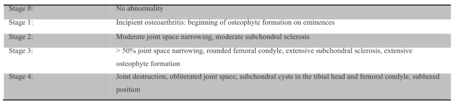
Radiographs can also be used to stage the disease and follow the progression of disease over time. A patient’s symptoms combined with their physical exam findings and radiographic evidence can be used to stage the disease process and attempt to correlate the disease to a typical course.2 Staging of knee osteoarthritis is based on the staging system of Kellgren and Lawrence where stage 0 is no abnormality and stage 4 is a combination of joint destruction, obliterated joint space, subchondral cysts, and a subluxed position (Table 1).2 Figures 1-2 are examples of a patient with stage 3 osteoarthritis of the right knee and stage 4 osteoarthritis on the left knee from multiple views.
Table 1: The staging of osteoarthritis of the knee after Kellgren and Lawrence
Stage 0: No abnormality
Stage 1: Incipient osteoarthritis: beginning of osteophyte formation on eminences
Stage 2: Moderate joint space narrowing, moderate subchondral sclerosis
Stage 3: > 50% joint space narrowing, rounded femoral condyle, extensive subchondral sclerosis, extensive osteophyte formation
Stage 4: Joint destruction, obliterated joint space, subchondral cysts in the tibial head and femoral condyle, subluxed position
MANAGEMENT
Treatment options for knee osteoarthritis are extensive but most guidelines recommend starting with a non-pharmacological approach such as: 1) education and self-management; 2) exercise and weight loss; 3) assistive devices; and 4) complimentary integrative medicine (CIM) approaches.12,13 Common CIM approaches include massage and acupuncture. Once patients have exhausted non-pharmacological approaches, their treatment can be augmented with pharmacologic management. First-line pharmacologic management of OA is acetaminophen followed by topical NSAIDs, oral NSAIDS, and other agents (capsaicin and tramadol).12 Other pharmacologic agents include intra-articular corticosteroids, hyaluronic acid, platelet-rich plasma, and stem cell injections.12,13 This section will focus on the various non-pharmacologic, pharmacologic and intra-articular injections (corticosteroids and hyaluronic acid) as well as the evolving role of regenerative medicine injections (platelet-rich plasma and mesenchymal stem cells) in the treatment of knee osteoarthritis.
Figure 1: Bilateral AP view of a 74 y.o. female seen in clinic with stage III knee OA on the right and stage IV knee OA on the left. Note the extreme narrowing of the joint space, osteophyte formation and subchondral sclerosis.
Non-pharmacologic Treatment Options
Conservative management is the first step in treating patients with knee OA and includes patient education, lifestyle modifications, weight loss in certain patient populations, and self-management of symptoms. Patient education is crucial in helping patients understand the disease process associated with knee OA and allows them to decrease or eliminate potentially damaging activities that could speed up the disease process.2,12–15 According to the 2014 Osteoarthritis Research Society International (OARSI) guidelines for the non-surgical management of knee OA, patients suffering from chronic musculoskeletal pain showed moderate benefit in decreasing pain and disability by participating in self-management programs.13 Interventions aimed at
promoting weight loss are among the initial recommendations in obese patients for the treatment of osteoarthritis.10 According to a systematic review conducted in 2014 by Nelson, Allen,
Golightly, Goode, and Jordan seven guidelines regarding weight loss in patients with hip or knee OA were evaluated. Of those 7 guidelines, 5 gave strong recommendations in favor of weight loss for patients with knee OA.12 A systematic review and meta-analysis from 2007 showed that
a ten percent reduction in body weight in obese individuals led to reduction in pain and disability as well as the reduction of multiple risk factors for knee OA.16
Manual therapy is another important component in the non-pharmacologic management of knee OA. Varying manual therapy techniques are used to decrease pain and improve
function.17 A randomized controlled trial from 2008 looked at the effect of manual therapy on
pain associated with knee OA.15 43 patients ages 45-70 were randomly allocated to either the intervention or control group. The intervention group received treatments consisting of
for the mobilization and manipulations. After completing the two weeks of treatment, the intervention group reported an improvement in pain compared to the control group, which reported no change in symptoms.15 The manual therapy techniques allow for stretching of the patellofemoral joint capsule, which decreases restriction allowing for greater joint mobility and a decrease in pain. Other studies have also looked at the effect of manual therapy on OA
symptoms and found that manual therapy works to improve patient’s symptoms. By decreasing the amount of pain the patient is in allows the patient to participate more readily in rehabilitation programs and daily activities.18,19
Aerobic exercise and strength training play a pivotal role in the treatment of knee OA. Multiple systematic reviews 7,12 and a critical narrative review from 2016 20 all make strong recommendations in favor of including both water- and land-based exercise and strength training in the management of OA of the knee. The purpose of rehabilitation for treating knee
osteoarthritis is to improve range of motion, strengthen the muscles around the joint, improve proprioception and increase aerobic capacity.20,21 Aerobic exercise can be either water- or
of the person and local availability”.1 As for the frequency, duration, and intensity of the exercise programs, these should also be individualized to the patient but allow for gradual progression over time.1 A major benefit of exercise in the management of knee OA is that many forms of exercise are inexpensive, making it available to all patients regardless of insurance benefits and socioeconomic status.
Studies have also looked at the efficacy of bracing and other assistive devices for patients with knee OA. Devices such as orthotics, cushioned heels, or wedges are thought to work by unloading the mechanical stress of the knee and therefore relieve pain and improve joint
function.2 Knee braces such as either valgus or varus unloader braces for medial and lateral knee
OA respectively are recommended by the OARSI guidelines based on data from a 2011 systematic review.12,13,23 The systematic review found that knee braces were effective in decreasing pain, stiffness, and the number of doses of NSAIDS patients were taking. Patients also showed improvement in proprioception and physical function as well as the amount of condylar separation achieved by the brace.23 The American Academy of Orthopaedic Surgeons
(AAOS) were unable to recommend for or against the use of valgus directing force braces (medial unloader brace) due to a lack of compelling evidence whether in favor of or against unloader braces.24 In regards to medial and lateral heel wedges, the OARSI guidelines recommended in favor of their use if directed by an appropriate specialist.13 This
However, the AAOS recommended against their use in patients with medial knee OA based on the results of five studies; all of which showed no significant improvement in pain or physical function.24 The OARSI guidelines also recommend the use of a cane or walking stick in patients with knee OA as it could help decrease pain and possibly improve quality of life. However, they strongly encourage further research in this area to confirm this recommendation.13
Pharmacologic Treatment Options
Pharmacologic modalities continue to play a significant role in the treatment of
osteoarthritis of the knee and are mainly aimed at decreasing pain and inflammation in order to improve function and allow patients to return to their normal daily activities. Acetaminophen continues to be considered first-line in the treatment of knee OA; it is inexpensive and an effective analgesic with a more favorable side-effect profile compared to NSAIDs.12,27,28 Acetaminophen works to inhibit the production of prostaglandins in the brain and spinal cord. Common side effects include renal and hepatic toxicity, elevated alkaline phosphatase, elevated bilirubin, and hypersensitivity reactions. Doses greater than 4 g/day or concurrent alcohol use further increase a patient’s risk for hepatotoxicity.27 Acetaminophen is initially administered on an as needed basis but as the severity of OA worsens patients may require scheduled doses in order to reach adequate pain relief. Recommended doses of acetaminophen are 325 mg every 4 to 6 hours or 1000 mg (1 g) every 6 to 8 hours with a max dose of 4000 mg per day.27 Patients with inadequate relief from acetaminophen may not be taking a sufficient dose for the
appropriate amount of time. Patients are recommended to take up to 4 g per day (in divided doses) for 4 to 6 weeks in order to achieve a sufficient trial.27 The OARSI guidelines
not recommended as a first-line analgesic. They were unable to recommend for or against the use of acetaminophen based on inconclusive evidence24. The American College of
Rheumatology (ACR) makes a conditional recommendation in favor of the use of acetaminophen initially for the management of knee OA.21 Before patients are started on an acetaminophen
regimen, a careful history and review of current medications is essential to reduce the risk of adverse effects and unintentional acetaminophen overdose.27 Patients with chronic liver disease can still use acetaminophen but require more frequent monitoring.
For patients who fail to experience relief in symptoms after an adequate trial of acetaminophen, non-steroidal anti-inflammatory drugs (NSAIDs) can be used.21 Topical
NSAIDs minimize systemic exposure seen with oral agents but are only appropriate in patients with superficial joint OA (hand, knee).12,13,21,27 In the United States, diclofenac is the only topical NSAID available. While topical NSAIDs have a decreased risk for GI side effects they pose an increased risk of dermatitis, pruritis, and phototoxicity.27 According to the ACR
recommendations from 2012, topical NSAIDs are preferred over oral agents in patients 75 years of age or older.21 For patients who fail to respond to topical NSAIDs or suffer from OA of deeper joints such as the hip, oral NSAIDs should be used.21,27 NSAIDS are also a reasonable first-line therapy for patients with moderate-to-severe OA.27 NSAIDs work by inhibiting both they cyclooxygenase enzymes (COX-1 and COX-2) that are responsible for synthesizing prostaglandins.27 They are frequently associated with gastrointestinal (GI) side effects such as
Other common side effects associated with NSAID use include cardiovascular (CV) side effects such as stroke or myocardial infarction and renal impairment. Patients with existing glomerular disease, renal insufficiency, hypercalcemia, heart failure, or cirrhosis or patients with true volume depletion are at increased risk for acute renal failure with the use of NSAIDs.29
Since the use of NSAIDs can lead to acute kidney injury (AKI), acute interstitial nephritis (AIN), nephrotic syndrome, and papillary necrosis, their use should be limited in the aforementioned patient populations and in patients with decreased glomerular filtration rate (GFR).30 NSAIDs should be avoided completely in patients with a GFR less than 60 mL/min/1.73 m2.21,27,30 Selective NSAIDs (ex. celecoxib) have a lower GI side effect profile but are associated with an increased risk for CV side effects. NSAIDS can interfere with the anti-platelet effects of aspirin, and in patients with known cardiovascular disease they are associated with an increased risk of adverse cardiovascular events such as death, myocardial infarction (MI), heart failure, and stroke.20,27,31
Patients respond differently to the various types of oral NSAIDs and therefore may have to try multiple agents before finding the NSAID that works best for them. Dosing also varies widely between the different classes of NSAIDs and clinicians should consult current guidelines for appropriate dosing recommendations.27 Clinicians should take into account the patient’s preference, previous response to NSAIDs, frequency or dosing, and history of comorbid conditions when deciding in favor of or against any particular agent.27 Patients should also be
considered to have the lowest risk for adverse cardiovascular events and is should therefore be the first-line agent in patients with a history of cardiovascular disease.
The AAOS strongly recommends the use of either topical or oral NSAIDs for patients with knee OA.24 According to the OARSI guidelines, non-selective and selective NSAIDs are
recommended in patients without comorbid conditions.8 The concomitant use of a PPI is also recommended in patients with moderate to high risk of adverse GI effects or the presence of comorbid conditions.13 Topical NSAIDs are recommended in patients with knee-only OA. They are not recommended in patients with multiple joint OA.13 The ACR recommends that topical or oral NSAIDs be used if a patient fails to have a satisfactory response to
acetaminophen. They too advocate for the addition of a PPI for patients who require chronic NSAID use.21
If patients continue to remain refractory to treatment after adequate trials of
acetaminophen and NSAIDs, tramadol is a reasonable next step. Although tramadol is a weak opiod, it is still considered a controlled substance.4 It works by inhibiting the reuptake of
serotonin and norepinephrine to achieve its analgesic effects. It has no effect however on the inflammatory process that contributes to the pathogenesis of OA.27 Tramadol can be used as a single agent or it can be used to augment a patient’s current regimen of acetaminophen or NSAIDs. Common side effects associated with tramadol include dizziness, vertigo, nausea, vomiting, constipation, and lethargy.32 Concomitant use of tramadol and antidepressants has
recommends the use of tramadol for patients 75 years and older in who the use of NSAIDs is not recommended.20 The OARSI guidelines do not separate low-potency opioids from traditional opioids and therefore conclude that the use of opioids in the long-term treatment of OA has limited usefulness.13
Potent opioids such as oxycodone, hydromorphone, morphine, and transdermal fentanyl patches are only recommended in patients with comorbid conditions that prevent the use of NSAIDs or in patients with grade 4 OA in whom total joint replacement is not an option.27,32 When initiating opioids, clinicians should start at the lowest possible dose and in combination with other non-opioid therapies (non-pharmacological therapies, acetaminophen, or NSAIDs).27
Potent opioids have a similar side effect profile as tramadol with the addition of sedation and respiratory depression. A systematic analytic review from 2016 compared the effects of orals NSAIDs to opioids in the treatment of knee OA. They found that opioids provide similar analgesic effects as NSAIDs and should only be used when all other treatments have failed.4 Before prescribing opioids, clinicians should conduct a thorough history and physical exam as well as ensure that previous non-opioid therapy has failed.27 The ACR had no recommendation regarding the use of opioids in the treatment of OA21 and the AAOS could neither recommend for or against their use due to inconclusive evidence.24
Intra-articular Injections
Intra-articular (IA) corticosteroid injections have been a mainstay in the treatment of knee osteoarthritis for many years despite inconclusive data to support their use.12 IA corticosteroids work to provide pain relief and decrease inflammation by inhibiting the synthesis of
corticosteroids are considered to have multiple advantages over systemic corticosteroids including increased bioavailability, decreased systemic exposure, and less adverse effects.34 These injections can more directly target the pathophysiology associated with OA while maximizing efficacy and decreasing drug cost.34,35 Triamcinolone preparations are the most
commonly used and are approved by the United States Food and Drug Administration (US FDA).35 Although IA corticosteroid injections are considered relatively safe, they do carry a risk of pain, swelling, or infection at the site of injection as well as systemic hyperglycemia.10,35 Patients typically experience pain relief and improvement in other associated symptoms for up to 4 weeks after the inection.34 There is concern among physicians and APPs that prolonged use of
IA corticosteroids can have adverse effects on articular cartilage and potentially accelerate the progression of the disease. Therefore most physicians and APPs recommend limiting the use of IA corticosteroids to 3-4 injections in any given joint per year.34,35
Studies over the years that have looked at the efficacy of using intra-articular
improvement in physical function. Unfortunately the authors found the quality of evidence to be low and the results inconclusive.37 The OARSI guidelines recommend the use of intra-articular corticosteroid injections for the treatment of knee OA based on two systematic reviews (SRs) that showed a short-term clinically significant decrease in pain.13 The AAOS were unable to
recommend for or against the use of intra-articular corticosteroids “due to a lack of compelling evidence that has resulted in unclear balance between benefits and potential harm”.24 The ACR conditionally recommends the use of intra-articular corticosteroid injections in the initial management of patients with symptomatic knee OA.21
Hyaluronic acid is a naturally occurring glycosaminoglycan in normal, healthy joint fluid and provides shock absorption, joint lubrication, and energy dissipation.27,38–40 IA administration of hyaluronic acid, also known as viscosupplementation has been a common modality in the treatment of knee OA since its approval by the US FDA in 2001.35 The use of IA hyaluronic acid has been reported to increase chondrocyte proliferation and decrease chondrocyte apoptosis which is thought to slow the progression of disease.34,35 Similarly to IA corticosteroids,
viscosupplementation is commonly used in patients who have not had satisfactory relief from acetaminophen or NSAIDs.34 There are currently seven different formulations approved by the US FDA and administration varies from 1 to 5 weekly injections.34 Although they are widely used, there remains a division of opinion regarding their efficacy and role in the treatment of knee OA.
A systematic review of overlapping meta-analyses from 2015 found that IA hyaluronic injections are a safe and viable treatment option for the patients with knee OA and their effects are long-term, lasting up to 26 weeks.38 Their inspection of 14 different meta-analyses
function.38 A 2009 systematic review and meta-analysis found that IA corticosteroids are more effective in the short-term (up to 4 weeks) while IA hyaluronic acid is a more effective long-term (8 weeks or more) modality.40 An article from July 2017 compared the effectiveness of IA hyaluronic acid and corticosteroid injections in delaying the time to surgery in patients with knee OA. The authors found that there was no significant difference in the risk for surgery between the two groups and their results could not support the use of IA hyaluronic acid in the treatment of knee OA.39 A Cochrane review from 2010 found viscosupplementation to be an effective treatment for knee OA and that its effects on pain and function were superior to placebos.41 The same review found viscosupplementation to have more prolonged effects than IA corticosteroids and based on their results supported the use of IA hyaluronic acid in the treatment of knee OA.41 The OARSI guidelines were uncertain in their recommendation on the use of IA hyaluronic acid in the treatment of knee OA based on inconsistent conclusions and conflicting results.13 The ACR had no recommendation regarding the use of IA hyaluronic injections and the AAOS could not recommend the use of IA hyaluronic acid.21,24 The AAOS based their recommendation on
the lack of efficacy from the results of 14 studies that assessed the use of IA hyaluronic acid.24
Regenerative Medicine Intra-articular Injections
As evidenced by the information above, the focus of current treatment modalities,
Platelet-rich plasma (PRP) is an autologous (from self) blood product derived from whole blood that has been centrifuged to produce a heavily concentrated number of platelets above an individual’s baseline.42 PRP has been used for several years in certain dental procedures and has also been found to play a role in the treatment of soft tissue lesions such as chronic ulcers, tendonopathies, and fasciitis.43,44 The growth factors that are released from the platelets have been shown to promote chondrogenesis and mesenchymal stem cell proliferation, increase anti-inflammatory mediators, and decrease proanti-inflammatory mediators through various pathways.45 Although PRP injections are widely used in the treatment of knee osteoarthritis there remains a lack of concrete evidence in the literature to support its role in the treatment of knee OA.
The AAOS were unable to recommend in favor of the use of PRP injections in the treatment of knee OA base on inconclusive evidence and recommend that practitioners use clinical judgment in deciding whether or not to use PRP injections in the treatment of patients with symptomatic knee OA that has failed other treatment modalities. They also recommend that practitioners continue to seek out new research that could help clarify the role of PRP injections in the treatment of knee OA.24 The ACR and OARSI do not consider PRP injections in knee OA treatment in their current guidelines.35
Mesenchymal stem cells (MSCs) are immunosuppressive cells that are found in abundance in various types of adult tissue such as bone marrow, adipose, umbilical cord tissue/blood, synovium, and periosteum.48,49 MSCs were first characterized by Dr. Alexander Friedenstein over forty years ago and are suggested to have a fundamental role in repairing and regenerating tissue.50 Over the past decade, they have continued to attract attention as a
promising therapeutic modality for knee OA.49 The ability of MSCs to differentiate into many cell types such as bone and/or cartilage allows them to have the potential to revolutionize the treatment of cartilage defects associated with osteoarthritis.51,52 In addition to the ability to differentiate into a variety of cell types, MSCs also play a significant role in the 1) control and modulation of inflammation; 2) inhibition of programed cell death (apoptosis); 3) stimulation of the endogenous proliferation and repair of cells; and 4) expansion of blood flow to joints.49,53 The overall goal of MSCs in the treatment of knee OA is to aid in the self-healing process of the cartilage, ultimately resulting in symptom relief.53
stem cells would be considered less likely to be rejected by the host’s immune system, the process of harvesting the cells from the patient and allowing time for cell expansion prior to injection or implantation makes the procedure both expensive and time-consuming.54 A
randomized controlled trial from 2015 looked at the efficacy of treating knee OA with allogeneic bone marrow MSCs.54 Thirty patients (17 females and 13 males) with chronic knee OA that had been unsuccessfully treated with conventional methods for at least 6 months were enrolled in the study. The control group received injections of hyaluronic acid while the experimental group received allogeneic bone marrow MSC injections. The authors’ results found that the use of allogeneic MSCs for the treatment of knee OA are safe, feasible, and effective. The group receiving allogeneic MSCs reported a 38-42% improvement in their pain while the hyaluronic acid group (control group) reported only a 10-14% improvement in pain.54
Other areas of debate in current research include harvest sites, mechanism of delivery, whether or not to use a scaffold such as platelet-rich plasma, and the number of cells needed per treatment to reach therapeutic effects. Adult MSCs can be harvested from either adipose tissue, bone marrow, or umbilical cord tissue or blood but the best source remains unclear.48–52 Bone marrow has traditionally been the preferred harvest site but recent research has discovered that the number of MSCs obtained from bone marrow aspirates is insufficient.50 The use of adipose tissue as a harvest site is becoming increasingly more common due to the ease of the lipoaspirate procedure and the abundance of cells obtained.50,55 A randomized controlled pilot study is
injections will lead to better cartilage formation and in the long-term improve the efficacy in the prevention of secondary knee OA.55
The most effective delivery method of the MSCs also continues to be under evaluation. MSCs can either be injected into the joint or they can be implanted using a technique similar to autologous chondrocyte transplantation (ACT).50 Results from current preclinical trials that are evaluating the efficacy of the injection method have shown that intra-articular injections of MSCs are improving function. However, these same trials have shown inconsistent results when evaluating whether or not intra-articular MSC injections have any effect on cartilage
restoration.50 A comparative matched-pair analysis from 2015 evaluated the effectiveness of the
injection of MSCs compared to the implantation of MSCs for knee OA. The study enrolled 14 men and 26 women and divided them into two separate groups. One group received arthroscopic MSC injection and the other group underwent arthroscopic MSC implantation. The major finding of the study showed that the patients receiving MSC implantation showed greater improvement (p value = .041) in cartilage regeneration at second-look arthroscopic surgery compared to those receiving MSC injection.53 The authors concluded that “a single simple injection of MSCs is insufficient for the repair of damaged articular cartilage…because of limited cell retention and survival at the target site”.53
HA combined with MSCs enhances the migration of synovial cells and chondrocytes to the location of the cartilage defect. HA may also work to enhance the adherence of MSCs to the cartilage defect itself.50 Several articles make mention of the uncertainty of the number of MSCs to reach a therapeutic dose but the authors don't attempt to address this in their current
research.49,53 According to Wolfsstadt et al., “the optimal dose, frequency, timing, and number of injections remains unclear”.49
CONCLUSION
Osteoarthritis (OA) is a progressive condition that leads to the degeneration of cartilage and severely inhibits the patient’s level of function. OA is not caused by a single, isolated event, but instead is the cumulative result of aging and trauma. Symptomatic knee OA affects as many as 10% of men and 13% of women over the age of 60 and will only increase as the population ages. Symptoms associated with knee OA lead to substantial disability and increasing medical costs.9 The high prevalence combined with the fact that patients are living longer means that the
burden on the health care system to care for patients with OA will only continue to grow. It is imperative that clinicians educate themselves on the various treatment modalities available in order to develop an individualized treatment plan for their patients that is both efficacious and low cost.
suffering from knee OA.1,12,13 Although studies have looked extensively into the efficacy of rehabilitation exercises, controversy remains over the type of exercise, effective dosage and/or frequency, duration, and intensity. More extensive studies are warranted to further evaluate the most effective modality, intensity, duration, and frequency. Dr. Maura Iverson recommends that exercise programs be viewed similarly to medications; that each modality has its own associated risks and benefits.7 The overall consensus of the various guidelines is that physical exercise and rehabilitation are effective in decreasing pain and improving function. Exercise programs should be initiated on a individualized basis based on the patient’s exercise tolerance and previous level of function.
Pharmacologic modalities include acetaminophen, NSAIDs, weak opioids, and intra-articular injections such as corticosteroids and hyaluronic acid. The guidelines have a strong consensus that pharmacologic modalities be initiated in a stepwise approach with the initial drug of choice being acetaminophen. If patients fail to obtain relief from their current pharmacologic modality, the guidelines recommend confirming that the patient has received an adequate trial (therapeutic dose for the recommended period of time) before escalating therapy. Although popular in the treatment of knee OA, the efficacy of intra-articular (IA) injections such as
corticosteroids and hyaluronic acid continue to remain under debate. The guidelines vary in their recommendations for IA corticosteroid injections with some organizations recommending in favor of IA corticosteroids and some recommending neither in favor nor against their use.13,21,24
There appears to be consensus among the guidelines for the use of hyaluronic acid (HA)
injections with the majority of organizations being unable to recommend for or against its use in the treatment of knee OA.13,21,24 These organizations base their recommendations on
that total joint arthroplasty should only be performed when conservative treatment has failed.2 Regenerative medicine injections such as PRP and MSCs have recently been suggested to potentially reverse or correct the underling disease process associated with knee osteoarthritis. The thought behind these injections is that they potentially allow for remodeling of the joint, which allows for healing. This leads to improvement in pain and function; ultimately allowing the patient to delay or even avoid surgery altogether.10 PRP injections for the treatment of knee OA have been gaining popularity in recent years. Recent RCTs have found that PRP injections are effective in decreasing the amount of pain associated with knee OA but the evidence for supporting these findings comes with a high risk of bias.46 Another RCT from 2015 showed no
benefit in using PRP injections for the treatment of knee OA when compared to HA injections.47 The collective results from recent studies regarding the role of PRP injections in the treatment of knee OA is inconclusive. PRP injections could very well be a viable treatment option for
Another promising modality in the treatment of knee OA is the use of mesenchymal stem cell (MSC) injections. Unfortunately, the majority of current research in this area has been completed using animal subjects and design methods of current studies remains highly variable. Similarly to PRP injections, the lack of a standardized treatment protocol in the use of MSC injections hinders researchers ability to fully evaluate the potential role of MSCs in the treatment of knee OA. Areas to address in such protocols include 1) autologous versus allogeneic MSCs; 2) harvest site (bone marrow, adipose, or umbilical cord); 3) mechanism of delivery (injected vs. implanted); 4) use of a scaffold; and 5) the number of MSCs per treatment. Perhaps one of the greatest areas of discord in current research is the mechanism of delivery of MSCs to the area of the chondral defect. Whether it be injection or implantation, the delivery of MSCs to the site of the defect must be an extremely efficient one in order for MSCs to be a viable therapeutic option. Some researchers even argue that efficient delivery of MSCs to the site of the chondral defect is more important than the number of MSCs delivered to the site of the defect.53 Therefore, more studies on human subjects are needed to evaluate the efficacy of MSC intra-articular injections versus implantation.
APPENDIX 1: RESEARCH METHODS
A PubMed search was completed using the key words stem cell injections, corticosteroid injections, platelet-rich plasma injections, and knee osteoarthritis. Additional search terms included non-pharmacological treatment of osteoarthritis and treatment guidelines for knee osteoarthritis. An UpToDate search was also completed using the key words knee osteoarthritis. In order to obtain additional articles for this clinical review a reference search of the systematic reviews and treatment guidelines identified during the primary PubMed and UpToDate searches was used to identify additional pertinent articles.
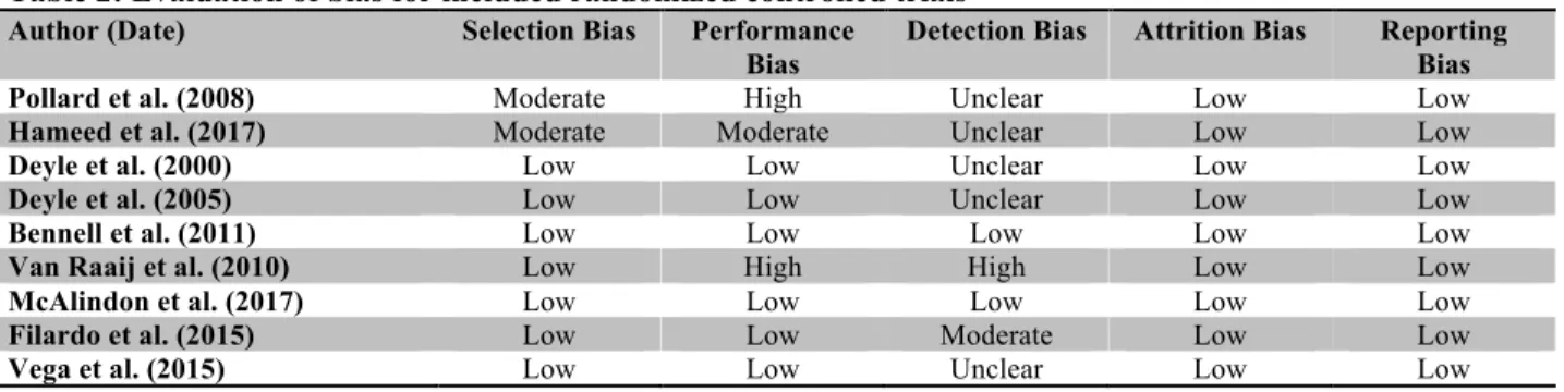
APPENDIX 2: RISK OF BIAS EVALUATION FOR INCLUDED RANDOMIZED CONTROLLED TRIASLS
Table 2: Evaluation of bias for included randomized controlled trials
Author (Date) Selection Bias Performance
Bias
Detection Bias Attrition Bias Reporting
Bias
Pollard et al. (2008) Moderate High Unclear Low Low
Hameed et al. (2017) Moderate Moderate Unclear Low Low
Deyle et al. (2000) Low Low Unclear Low Low
Deyle et al. (2005) Low Low Unclear Low Low
Bennell et al. (2011) Low Low Low Low Low
Van Raaij et al. (2010) Low High High Low Low
McAlindon et al. (2017) Low Low Low Low Low
Filardo et al. (2015) Low Low Moderate Low Low
Vega et al. (2015) Low Low Unclear Low Low
APPENDIX 3: QUALITY OF EVIDENCE FOR INCLUDED COCHRANE REVIEWS
Table 3: Quality of evidence for included Cochrane reviews
Author (Date) Summary of Quality of Evidence
Hari et al. (2015) Using GRADE analysis the authors graded the quality of evidence as low for all of their
findings; meaning that they have little confidence in these results. The authors attributed this to being due to the results being, in general, highly discordant across studies and mainly based on small studies of low quality.
Bellamy et al. (2010) Trials included in the systematic review were assessed separately by two reviewers for
APPENDIX 4: ANALYSIS OF SYSTEMATIC REVIEWS
Table 4: Level of evidence among included systematic reviews
Author (Date) Summary Statement Level of Evidence*
Smith et al. (2016)4 In the evaluation of pain reduction in patients with OA treated with
NSAIDs, less potent opioids, and potent opioids the mean reduction in WOMAC pain was comparable between the three classes.
Ia
Pas et al. (2017)8 Based upon the high risk of bias in all of the studies included in this
review, there is no high level of evidence for stem cell therapy in the treatment of patients with knee OA. The authors do not recommend the use of stem cell therapy for patients with knee OA.
Ia
Nelson et al. (2014)12 An essential agreement on many recommendations for OA
management was found in guidelines from various societies. The authors concluded that the deficit is not in the lack of quality guidelines but in getting those guidelines disseminated and
implemented in the primary care settings and other specialty settings. IV
Larmer et al. (2014)14 Authors found that that within the guidelines for the management of
knee OA, exercise and education were the most strongly recommended. There is strong evidence to support the use of exercise, electrical-based therapy, equipment, education, diet and weight loss, manual therapy, and self-management.
IV
Christensen et al. (2007)16 Authors determined that physical disability could be improved by
weight loss. Overweight individuals should initiate a 10% reduction in body weight.
Ia
Jansen et al. (2011)22 Exercise therapy with manual joint mobilization helps decrease pain
(effect size 0.69) compared to strength training (effect size 0.38) or exercise therapy (effect size (0.38) when used alone.
Ia
Raja et al. (2011)23 Despite heterogeneity in current studies and a lack of clinical trials,
knee braces and foot orthoses have been shown to help decrease the level of stress transmitted though the medial compartment of the knee.
Ib
Campbell et al. (2015)38 Intra-articular viscosupplementation is a safe and viable treatment
option for knee IA with effects that can last up to 26 weeks. Ia
Bannuru et al. (2009)40 Intra-articular corticosteroids is more effective in the short term (up
to 4 weeks) and intra-articular hyaluronic acid is more effective in the long term (4-26 weeks)
Ia
Khoshbin et al. (2013)42 When compared to hyaluronic acid or normal saline injections,
multiple intra-articular PRP injections, when given in a sequence, may have beneficial effects in patients with mild to moderate knee OA beginning at approximately 6 months post-injection.
Ia
Meheux et al. (2016)45 Intra-articular PRP injections results in significant clinical
improvement up to 12 weeks post-injection in patients with symptomatic knee OA.
Ib
Laudy et al. (2015)46 Intra-articular PRP injections are more effective in decreasing pain
and improving function in patients with knee OA when compared to placebo and hyaluronic acid injections.
Ia
Xia et al. (2015)51 There is potential for MSC injections to have a significant effect on
pain and potential benefit on physical function in patients with knee OA at 3, 12, and 24 months post-injection but the evidence is inconclusive.
Ia
*Level of Evidence: Ia = meta-analyses of randomized controlled trials (RCT); Ib = at least 1 RCT; IIa = at least 1 controlled trial (CT) without randomization); IIb = at least 1 type of quasi-experimental study; III = descriptive studies (comparative, correlation, or case-control studies); IV = expert committee reports or opinions and or clinical experience of respected authorities.
BIBLIOGRAPHY
1. Fernandes L, Hagen KB, Bijlsma JWJ, et al. EULAR recommendations for the non-pharmacological core management of hip and knee osteoarthritis. Ann Rheum Dis. 2013;72(7):1125-1135. doi:10.1136/annrheumdis-2012-202745.
2. Michael JW, Schluter-Brust KU, Eysel P. The epidemiology, etiology, diagnosis, and treatment of osteoarthritis of the knee. Dtsch Arztebl Int. 2010;107(9):152-162. doi:10.3238/arztebl.2010.0152.
3. Loeser R. Pathogenesis of osteoarthrtis. In: Post TW, ed. UpToDate. Waltham, NA: UpToDate; 2017. uptodate.com.
4. Smith SR, Deshpande BR, Collins JE, Katz JN, Losina E. Comparative pain reduction of oral non-steroidal anti-inflammatory drugs and opioids for knee osteoarthritis: Systematic analytic review. Osteoarthr Cartil. 2016;24(6):962-972. doi:10.1016/j.joca.2016.01.135. 5. Arthritis-related statistics. Centers for Disease Control and Prevention.
6. Hellman DB, Imboden JB. Degenerative Joint Disease (Osteoarthritis). In: Papdakis MA, McPhee SJ, Rabow MW, eds. 2016 Current Medical Diagnosis and Treatmet. 55th ed. New York, NY: McGraw Hill Education; 2016:812-815.
7. Iversen MD. Rehabilitation Interventions for Pain and Disability in Osteoarthritis. AJN,
Am J Nurs. 2012;112(3):S32-S37. doi:10.1097/01.NAJ.0000412649.02926.35.
8. Pas HI, Winters M, Haisma HJ, Koenis MJ, Tol JL, Moen MH. Stem cell injections in knee osteoarthritis: a systematic review of the literature. Br J Sports Med. 2017:bjsports-2016-096793. doi:10.1136/bjsports-2017:bjsports-2016-096793.
9. Zhang Y, Jordan JM. Epidemiology of Osteoarthritis. Clin Geriatr Med. 2010;26(3):355-369.
10. Richards MM, Maxwell JS, Weng L, Angelos MG, Golzarian J. Intra-articular treatment of knee osteoarthritis: from anti-inflammatories to products of regenerative medicine.
Phys Sportsmed. 2016;44(2):101-108. doi:10.1080/00913847.2016.1168272.
11. Herring W. Primary Osteoarthrits (Also known as Primary Degenerative Arthritis, Degenerative Joint Disease-DJD). In: Learning Radiology Recognizing the Basics. ; 2016:256-258.
12. Nelson AE, Allen KD, Golightly YM, Goode AP, Jordan JM. A systematic review of recommendations and guidelines for the management of osteoarthritis: The Chronic Osteoarthritis Management Initiative of the U.S. Bone and Joint Initiative. Semin Arthritis
Rheum. 2014;43(6):701-712. doi:10.1016/j.semarthrit.2013.11.012.
13. McAlindon TE, Bannuru RR, Sullivan MC, et al. OARSI guidelines for the non-surgical management of knee osteoarthritis. Osteoarthr Cartil. 2014;22(3):363-388.
doi:10.1016/j.joca.2014.01.003.
14. Larmer PJ, Reay ND, Aubert ER, Kersten P. Systematic review of guidelines for the physical management of osteoarthritis. Arch Phys Med Rehabil. 2014;95(2):375-389. doi:10.1016/j.apmr.2013.10.011.
15. Pollard H, Ward G, Hoskins W, Hardy K. The effect of a manual therapy knee protocol on osteoarthritic knee pain: a randomised controlled trial. J Can Chiropr Assoc.
2008;52(4):229-242.
16. Christensen R, Bartels EM, Astrup A, Bliddal H. Effect of weight reduction in obese patients diagnosed with knee osteoarthritis: a systematic review and meta-analysis. Ann
17. Hameed R, Waqas M, Akhtar F, Joseph R, Niazi A. Effect of Manual Therapy on Knee Osteoarthritis ( OA ) Pain , A Randomized Control Trial. 2017;2(4):19-22.
18. Deyle G, Henderson N, Matekel R, Ryder M, Garber M, Allison S. Effectiveness of Manual Physical Therapy and Exercise in Osteoarthritis of the Knee: A Randomized, Controlled Trial. Ann Intern Med. 2000;132(3).
19. Deyle GD, Allison SC, Matekel RL, et al. Physical Therapy Treatment Effectiveness for Osteoarthritis of the Knee: A Randomized Comparison of Supervised Clinical Exercise and Manual Therapy Procedures Versus a Home Exercise Program. Phys Ther.
2005;85(12):1301-1317.
20. Nguyen C, Lef??vre-Colau MM, Poiraudeau S, Rannou F. Rehabilitation (exercise and strength training) and osteoarthritis: A critical narrative review. Ann Phys Rehabil Med. 2016;59(3):190-195. doi:10.1016/j.rehab.2016.02.010.
21. Hochberg MC, Altman RD, April KT, et al. American College of Rheumatology 2012 recommendations for the use of nonpharmacologic and pharmacologic therapies in osteoarthritis of the hand, hip, and knee. Arthritis Care Res. 2012;64(4):465-474. doi:10.1002/acr.21596.
22. Jansen MJ, Viechtbauer W, Lenssen AF, Hendriks EJM, de Bie RA. Strength training alone, exercise therapy alone, and exercise therapy with passive manual mobilisation each reduce pain and disability in people with knee osteoarthritis: a systematic review. J
Physiother (Australian Physiother Assoc. 2011;57(1):11-20.
http://search.ebscohost.com/login.aspx?direct=true&db=cin20&AN=104570589&site=eh ost-live.
23. Raja K, Dewan N. Efficacy of Knee Braces and Foot Orthoses in Conservative Management of Knee Osteoarthritis. Am J Phys Med Rehabil. 2011;90(3):247-262. doi:10.1097/PHM.0b013e318206386b.
24. American Academy of Orthopaedic Surgeons Academy A, AAOS. Treatment of Osteoarthritis of the Knee: Evidence-Based Guideline 2nd Edition Adopted by the American American Academy of Orthopaedic Surgeons Board of Directors. Am Acad
Orthop Surg Board Dir. 2013:973. doi:10.5435/JAAOS-21-09-577.
25. Bennell KL, Bowles K-A, Payne C, et al. Lateral wedge insoles for medial knee osteoarthritis: 12 month randomised controlled trial. Bmj. 2011;342(may18 3):d2912-d2912. doi:10.1136/bmj.3):d2912-d2912.
26. Van Raaij TM, Reijman M, Brouwer RW, Bierma-Zeinstra SMA, Verhaar JAN. Medial knee osteoarthritis treated by insoles or braces a randomized trial. Clin Orthop Relat Res. 2010;468(7):1926-1932. doi:10.1007/s11999-010-1274-z.
27. Smith SM, Dietrich E, Gums JG. Osteoarthritis. In: Chisholmm-Burns MA, Wells BG, Schwinghammer TL, Malone PM, Kolesar JM, DiPiro JT, eds. Pharmacotherapy
Principles and Practice. 3rd ed. New York, NY: McGraw Hill Medical; 2013:1041-1053.
28. Zhang W. Does paracetamol (acetaminophen) reduce the pain of osteoarthritis?: a meta-analysis of randomised controlled trials. Ann Rheum Dis. 2004;63(8):901-907.
doi:10.1136/ard.2003.018531.
29. Luciano R, Perazella MA. NSAIDs: Acute kidney injury (acute renal failure). In: Post TW, ed. UpToDate. Waltham, MA: UpToDate; 2017. www.uptodate.com.
30. Solomon DH. Nonselective NSAIDs: Overview of adverse effects. In: Post TW, ed.
31. Solomon DH. Nonselective NSAIDs: Adverse cardiovascular effects. In: Post TW, ed.
UpToDate. Waltham, MA: UpToDate; 2017.
32. Deveza LA, Bennell K. Management of knee osteoarthritis. In: Post TW, ed. UpToDate2. Waltham, MA: UpToDate; 2017.
33. Yavuz U, Sökücü S, Albayrak A, Ö ztürk K. Efficacy comparisons of the intraarticular steroidal agents in the patients with knee osteoarthritis. Rheumatol Int. 2012;32(11):3391-3396. doi:10.1007/s00296-011-2188-0.
34. Evans CH, Kraus VB, Setton LA. Progress in intra-articular therapy. Nat Rev Rheumatol. 2013;10(1):11-22. doi:10.1038/nrrheum.2013.159.
35. Wehling P, Evans C, Wehling J, Maixner W. Effectiveness of intra-articular therapies in osteoarthritis: a literature review. Ther Adv Musculoskelet Dis. 2017;9(8):183-196. 36. McAlindon TE, LaValley MP, Harvey WF, et al. Effect of Intra-articular Triamcinolone
vs Saline on Knee Cartilage Volume and Pain in Patients With Knee Osteoarthritis. Jama. 2017;317(19):1967. doi:10.1001/jama.2017.5283.
37. Jüni P, Hari R, Aws R, et al. Intra-articular corticosteroid for knee osteoarthritis ( Review ) SUMMARY OF FINDINGS FOR THE MAIN COMPARISON. 2015;(10).
doi:10.1002/14651858.CD005328.pub3.www.cochranelibrary.com.
38. Campbell KA, Erickson BJ, Saltzman BM, et al. Is Local Viscosupplementation Injection Clinically Superior to Other Therapies in the Treatment of??Osteoarthritis of the Knee: A Systematic Review of??Overlapping Meta-analyses. Arthroscopy. 2015;31(10):2036-2045. doi:10.1016/j.arthro.2015.03.030.
39. Shewale AR, Barnes CL, Fischbach LA, et al. Comparative Effectiveness of Intra-Articular Hyaluronic Acid and Corticosteroid Injections on the Time to Surgical Knee Procedures. 2017.
40. Bannuru RR, Natov NS, Obadan IE, Price LL, Schmid CH, McAlindon TE. Therapeutic trajectory of hyaluronic acid versus corticosteroids in the treatment of knee osteoarthritis: A systematic review and meta-analysis. Arthritis Rheum. 2009;61(12):1704-1711.
doi:10.1002/art.24925.
41. Bellamy N, Campbell J, Robinson V, Gee T, Bourne R, Wells G. Viscosupplementation for the treatment of osteoarthritis of the knee. Cochrane Database Syst Rev.
2006;19(2):CD005321.
doi:10.1002/14651858.CD005321.pub2.www.cochranelibrary.com.
42. Khoshbin A, Leroux T, Wasserstein D, et al. The Efficacy of Platelet-Rich Plasma in the Treatment of Symptomatic Knee Osteoarthritis: A Systematic Review With Quantitative Synthesis. Arthrosc J Arthrosc Relat Surg. 2013;29(12):2037-2048.
doi:10.1016/j.arthro.2013.09.006.
43. Knop E, De Paula LE, Fuller R. Platelet-rich plasma for osteoarthritis treatment. Rev Bras
Reumatol. 2016;56(2):152-164. doi:10.1016/j.rbre.2015.07.002.
44. Ornetti P, Nourissat G, Berenbaum F, Sellam J, Richette P, Chevalier X. Does platelet-rich plasma have a role in the treatment of osteoarthritis? Joint Bone Spine.
2016;83(1):31-36. doi:10.1016/j.jbspin.2015.05.002.
45. Meheux CJ, McCulloch PC, Lintner DM, Varner KE, Harris JD. Efficacy of Intra-articular Platelet-Rich Plasma Injections in Knee Osteoarthritis: A Systematic Review.
46. Laudy ABM, Bakker EWP, Rekers M, Moen MH. Efficacy of platelet-rich plasma
injections in osteoarthritis of the knee: a systematic review and meta-analysis. Br J Sports Med. 2015;49(10):657-672. doi:10.1136/bjsports-2014-094036.
47. Filardo G, Di Matteo B, Di Martino A, et al. Platelet-Rich Plasma Intra-articular Knee Injections Show No Superiority Versus Viscosupplementation. Am J Sports Med. 2015;43(7):1575-1582. doi:10.1177/0363546515582027.
48. Pers YM, Ruiz M, Noël D, Jorgensen C. Mesenchymal stem cells for the management of inflammation in osteoarthritis: State of the art and perspectives. Osteoarthr Cartil. 2015;23(11):2027-2035. doi:10.1016/j.joca.2015.07.004.
49. Wolfstadt JI, Cole BJ, Ogilvie-Harris DJ, Viswanathan S, Chahal J. Current Concepts.
Sports Health. 2015;7(1):38-44. doi:10.1177/1941738114529727.
50. Freitag J, Bates D, Boyd R, et al. Mesenchymal stem cell therapy in the treatment of osteoarthritis: reparative pathways, safety and efficacy – a review. BMC Musculoskelet
Disord. 2016;17(1):230. doi:10.1186/s12891-016-1085-9.
51. Xia P, Wang X, Lin Q, Li X. Efficacy of mesenchymal stem cells injection for the management of knee osteoarthritis: a systematic review and meta-analysis. Int Orthop. 2015;39(12):2363-2372. doi:10.1007/s00264-015-2785-8.
52. Richardson SM, Kalamegam G, Pushparaj PN, et al. Mesenchymal stem cells in
regenerative medicine: Focus on articular cartilage and intervertebral disc regeneration.
Methods. 2016;99:69-80. doi:10.1016/j.ymeth.2015.09.015.
53. Kim YS, Kwon OR, Choi YJ, Suh DS, Heo DB, Koh YG. Comparative Matched-Pair Analysis of the Injection Versus Implantation of Mesenchymal Stem Cells for Knee Osteoarthritis. Am J Sports Med. 2015;43(11):2738-2746.
doi:10.1177/0363546515599632.
54. Vega A, Martín-Ferrero MA, Del Canto F, et al. Treatment of Knee Osteoarthritis With Allogeneic Bone Marrow Mesenchymal Stem Cells. Transplantation. 2015;99(8):1681-1690. doi:10.1097/TP.0000000000000678.