Intestinal Regeneration After Irradiation: Stem Cells and Sox9
Brooks Scull
A thesis submitted to the faculty of the University of North Carolina at
Chapel Hill in partial fulfillment of the requirements for the degree Master of
Science in the Department of Cell and Molecular Physiology.
Chapel Hill
2009
ii
Abstract
Brooks Scull: Intestinal Regeneration After Irradiation: Stem Cells and
Sox9
(Under the direction of P. Kay Lund)
Whole body irradiation is a useful challenge to study the response of intestinal epithelial stem cells (IESC) to genetic damage. High dose irradiation causes complete loss of the proliferative zone within intestinal crypts excluding a few stem/multipotent cells that can regenerate the intestinal epithelial lining. Whole body irradiation models using high dose irradiation are not ideal for studying the regenerative process due to poor animal survival. Little is known about direct effects of irradiation on IESC due to lack of biomarkers. This study developed a high dose abdominal irradiation model with increased survival allowing for analysis of regeneration to day 9 after irradiation and potentially longer. The expression pattern of the putative IESC marker, Sox9, was evaluated and compared with proliferation markers. We provide evidence that
iii
Acknowledgments
First and foremost, I would like to thank my advisor, Dr. Kay Lund who has been an incredible and irreplaceable mentor. Her scientific knowledge and integity are an inspiration. She started my science career which she continues to nurture and guide for which I am forever in debt.
I would also like to thank the members of my committee, Dr. Michael Helmrath and Dr. Scott Magness for their scientific guidance and advise. They have also been inspirational and integral to my development as a scientist.
I would like to thank my many colleagues and friends in the Cell and Molecular Physiology department. Most notabley, the past and present members of the Lund Lab, especially Jim Simmons, Randy Fuller, Kate Hamilton, Vicki Newton and Nicole
Ramocki for their friendship and support.
iv
Table of Contents
List of Tables………...………..….v
List of Figures………..……….vi
Chapters I. Introduction………..1
II. Methods………..………12
III. Results………19
IV.Diescussion………..……..……45
Appendice 1……….………55
Appendice 2……….………56
Appendice 3……….…………58
v
List of Tables
Tablevi
List of Figures
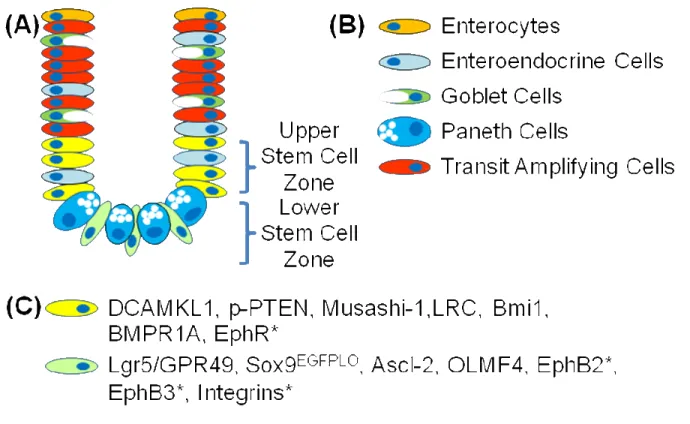
Figure1. Intestinal cell lineages and intestinal stem cell (IESC) markers……….10 2. Mouse placement in the X-RAD 320 from Precision X-Ray………17 3. Body weights and survival rates survival rates for CD1
and C57BL/6 mice………..27 4. H&E staining of non-irradiated small intestinal photographed
at 5X magnification………..….………..28 5. H&E staining of small intestinal at day 3 after irradiation photographed
at 5X magnification………..……….………..29 6. H&E staining of small intestinal at day 5 after irradiation photographed
at 5X magnification………..………..……….30 7. H&E staining of small intestinal at day 7 after irradiation photographed
at 5X magnification………….………..………..31 8. H&E staining of small intestinal at day 9 after irradiation photographed
at 5X magnification…….………..………..…32 9. H&E staining of proximal and distal colon after irradiation photographed
at 4X and 10X magnification….………33 10. Colocalization of SOX9 protein and Ki67 in microcolonies and hyper-
regenerative areas after irradiation……….………..……….34-35 11. Colocalization of SOX9 protein and Ki67 in crypts of proximal colon….…...36 12. Colocalization of SOX9 protein and Ki67 in crypts of distal colon….…...37-38 13. SOX9 protein colocalizes with a subset of EdU positive cells in sub-
epithelial areas and at the base of hyper-regenerative crypts……….…..39-40 14. Sox9EGFPLO cells are lost at day 3 and then show vast expansion in
vii
16. Sox9EGFPHI cells colocalize with most Substance P expressing cells after irradiation……….……43 17. Lysozyme positive Paneth cells did not colocalize with Sox9EGFP cells
Chapter 1
Introduction
Intestinal Physiology
The intestine is a tubular structure consisting of several layers including an outer serosa, longitudinal and circular smooth muscle, the submucosa and an inner mucosa. The mucosa comprises a single layer of epithelial cells facing the lumen, the underlying lamina propria which contains fibroblasts, myofibroblasts, blood vessels and resident immune cells, and the muscularis mucosa. The epithelial lining functions in digestion and absorption of ingested nutrients and as a barrier protecting the underlying tissue from toxins, pathogens and commensal microbiota in the lumen. The epithelial lining of the small intestine is organized into a crypt-villus axis with crypts representing
2
crypt axis and onto the villi or surface epithelium creating a spatially organized hierarchy of cells along the crypt-villus axis . The intestine contains four differentiated cell
lineages (Potten & Loeffler, 1990). In the small intestine the lineages include
3
Intestinal Epithelial Stem Cells
4
These studies and characteristics of IESC have led to a concept that there may be two populations of IESC, the ‘+4’ IECS lying above Paneth cells and the CBC lying between Paneth cells, and exemplifies the fact that there is still a need for conclusive evidence to confirm the precise location and identity of IESC. For example, LRC are not solely localized at +4, but are distributed at positions 2-7 from the crypt base, which can include the CBC population. Punitive IESC have also been identified that distinctly mark cells in the +4 IESC or CBC zones. Thus one proposal by Dr. Susan Henning at UNC suggests that upper stem cell zone (USZ) and lower stem cell zone (LSZ) may be better terminology. Many aspects of IESC including their location will remain unverified until valid IESC markers in the USZ and LSZ are defined. It is also important to show multipotency of these IESC in vitro as well as in vivo for verification that they are true IESC.
IESC Markers
Recently several IESC markers have been proposed which are preferentially expressed in the USZ or LSZ (Figure 1). Indirect evidence that bone morphogenic protein receptor 1a (BMPR1A) and phosphorylated-phosphatase and tensin homolog deleted on chromosome 10 (p-PTEN) are markers of IESC in the USZ has been demonstrated using label retaining assays and mouse knockout models. The phenotype of BMPR1A and PTEN knockouts showed that deregulation of these
pathways led to expansion of the IESC zone and an increase in proliferation leading to intestinal polyposis (He et al., 2004), (He et al., 2007). Evidence using microarray analysis of small intestinal crypts and short-term label retaining assays indicated that
5
potential IESC marker in the USZ (Giannakis et al., 2006). The microarray from theses
same studies identified that Eph receptors could make good candidates for cell surface
markers of IESC. This was further strengthened by findings in EphB2 and EphB3 gene
deletion models which demonstrated a disruption of cell migration and through
characterization of the expression pattern of Eph receptors (Batlle et al., 2002). Some
integrins including α2β1 also make good candidates as surface markers of IESC
(Beaulieu, 1992). Musashi-1 shows potential as an IESC marker, since Musashi-1
positive cells colocalize with label retaining cells and the proliferation marker Ki67 in the
base of crypts (He et al., 2007; Potten et al., 2003). However Musashi-1 staining can
often localize to CBC, +4 cells and also cells at positions higher in the crypt then
thought to represent IESC. Strong experimental evidence suggests that Lgr5 is a true
IESC marker. Both lineage tracing models and in vitro pluipotency have demonstrated
that Lgr5 positive cells are CBC cells that give rise to all intestinal cell lineages (Barker,
van Es, Kuipers, Kujala, van den Born, Cozijnsen, Haegebarth, Korving, Begthel,
Peters, & Clevers, 2007a; Sato et al., 2009). Genetic lineage tracing has also been
used to show that cells expressing Sox9 give rise to all differentiated cell types in the
embryonic intestine {{155 Akiyama,H. 2005; }}. Mice that have a Sox9 gene deletion do
not develop Paneth cells suggest that Sox9 is required for Paneth cell differentiation
however is not required for the development of the other cell lineages {{156
Mori-Akiyama,Y. 2007; }}. Redundancy in function across the Sox protein family is a
common theme and therefore compensatory effects of other SOX proteins could
account for differentiation of the other cell lineages in the absence of SOX9 expression
6
the CBC and to cells within the USZ or transit amplifying zones {{157 Blache,P. 2004; }}.
Recently, Sox9 transcriptional activity was visualized in the intestine using a Sox9EGFP
reporter mouse. Differential expression of Sox9EGFP was observed within the USZ and
LSZ. Cells expressing high levels of Sox9EGFP (Sox9EGFPHI) where localized to the USZ
and to cells on the villus. Further evaluation of these cells revealed that they were
post-mitotic or non-proliferating cells with an enteroendocrine phenotype {{116
Formeister,E.J. 2009; }}. Cells expressing low levels of Sox9EGFP (Sox9EGFPLO) localized
to the LSZ/CBC and these cells were shown to be enriched for Lgr5 and other stem cell
markers {{116 Formeister,E.J. 2009; }}. Thus Sox9EGFP may mark terminally
differentiated enteroendocrine cells in the USZ, as well as LSC/CBC. A key question is
whether the non-dividing enteroendocrine cells expressing high levels of Sox9 in the
USZ may in some instances adopt an IESC phenotype. Lineage tracing and reporter
mouse models have shown great potential in identifying IESC, but to date the
expression pattern of IESC markers has not been fully characterized after injury such as
irradiation which creates an ideal model to study IESC and their role in regeneration
after complete crypt loss {{12 Potten,C.S. 2004; }}.
Irradiation as a model to study IESC
7
epithelium {{12 Potten,C.S. 2004; }}. A wide range of doses have been tested and have shown that cells within the USZ or +4 zone are highly susceptible to radiation–induced apoptosis which peaks at radiation doses as low as 1Gy and is maximal as early as 4 hours after irradiation. The highest rate of apoptosis is around position 4 above the crypts, but is induced highly throughout the entire crypt base {{144 Potten,C.S. 1998; }}. A recent study compared radiosensitivity of USZ or +4 cells and LSZ/CBC and
suggested that LSZ/CBC cells were more resistant requiring a high dose of 10Gy for peak apoptosis (Barker, van Es, Kuipers, Kujala, van den Born, Cozijnsen, Haegebarth, Korving, Begthel, Peters, & Clevers, 2007a). The colon appears to be less sensitive to radiation-induced apoptosis at early time points after irradiation and does not show any region specific apoptosis along the length of the crypt (Cai, Roberts, Bowley, Hendry, & Potten, 1997). Indeed the identity of colon IESC is less well defined than small intestine IESC. Radiation has also been used to show that IESC in the small intestine have a unique ability to protect their genome from mutation {{129 Potten,C.S. 2002; }}. This, along with the high rate of apoptosis in the IESC zone after injury, may present a mechanism by which the small intestine prevents proliferation of genetically damaged cells and maintains an extremely low rate of cancer formation after genotoxic injury. The intestine also seems to be able to effectively recognize the degree of injury
8
been considered putative IESC, but irradiation may be altering their phenotype and how they compare to IESC before injury has not been established. Combining a high dose irradiation model that shows the greatest increase in clonogenic cells and regeneration, with lineage tracing or reporter mouse models could effectively determine any
differences between IESC before and after irradiation.
Mediators of regeneration after irradiation
It is noteworthy that relatively little is known about the local, paracrine or autocrine mediators of regeneration after irradiation. Some studies suggest that
endothelial cells may be required for appropriate regeneration after irradiation although this is controversial (Schuller et al., 2007; Wang, Boerma, Fu, & Hauer-Jensen, 2007). Another recent study suggests that the protein, p53 upregulated modulator of apoptosis (PUMA), regulates the radiosensitivity of IESC. PUMA, which works in a p53
dependent manner, was seen to be highly upregulated in the intestine after irradiation and gene deletion of PUMA led to increased regeneration after irradiation. Interestingly, IESC in the USZ appeared to show greater apoptosis then cells in the LSZ (Qiu et al., 2008). However, detection and quantification of cells in the LSZ was limited due to the difficulty in recognizing these slender cells between more prominent Paneth cells.
The Sox9EGFP reporter mouse would provide an effective model to visualize these cells in the LSZ, as well as to quantify the expression of PUMA on the USZ and LSZ
and determine the underlying mechanisms regulating PUMA through the isolation of
Sox9EGFP cells for molecular analysis. It would be interesting if the increased apoptosis
9
identify the importance of these cells in the maintenance of the IESC niche. This is
consistent with a previously hypothesized enteroendocrine cell-based quiescent intestinal stem cell niche, which has not been confirmed experimentally (Radford & Lobachevsky, 2006).
Therapies for regeneration
Furthermore, the suppression of PUMA could provide an effective therapy to
protect IESC from radiation damage. Insulin-like growth factor-I (IGF-I) suppresses
PUMA independently of p53 and could be an effective therapy (Han et al., 2001). IGF-I
has also been seen to promote SOX9 expression and survival of SOX9 positive cells
after irradiation (Ramocki et al., 2008). In other tissues, IGF-I plays a major role in the
survival, expansion and regeneration of stem cells (Arsenijevic, 2005; Ye & D'Ercole,
2006). IGF-I seems to mediate the actions of growth hormone (GH) and GLP2, the only
approved or under trial trophic therapies for human intestinal disease, respectively
(Dube, Forse, Bahrami, & Brubaker, 2006; Krysiak, Gdula-Dymek,
Bednarska-Czerwinska, & Okopien, 2007). However, one complication with using growth factors as
therapies for cancer patients undergoing radiation therapy is that they are desensitizing
therapies which may protect not only the normal tissue, but also the tumor tissue
rendering them ineffective. Interestingly, GH administered before radiation exposure
has been seen to protect normal tissue but not implanted adenocarcinoma from
10
Crypt microcolony assay
Irradiation has also been widely used to test therapies that can protect IESC or increase regeneration {{15 Potten,C.S. 2003; }}. Many of these studies utilize the crypt microcolony assay, which compares the number of regenerating crypts, as defined by a given number of dividing cells, after irradiation in animals with and without treatment (Khan, Shui, Ning, & Knox, 1997)(Booth, Booth, Williamson, Demchyshyn, & Potten, 2004)(Ishizuka et al., 2003). The crypt microcolony assay is an indirect measurement of IESC activity and is often dependent on using a high dose of irradiation that does not allow for analysis of the complete regenerative process due to limited animal survival. Many of the irradiation models that have been used are limited by either using a low dose of irradiation that only eliminates a portion of the IESC population or use of a high dose of irradiation that is limited by poor survival of animals.
Hypothesis
This study focuses on development of a high dose abdominal irradiation model that allows for survival of only IESC that can regenerate the intestine. It is hypothesized that this model will increasing the animal survival rate so that the regenerative response of IESC can be further characterized. This abdominal irradiation model will also used to characterize the expression pattern of the putative IESC marker Sox9. It is further hypothesized that Sox9 will provide a useful marker of IESC during the regenerative
11
Figure 1: Intestinal cell lineages and intestinal stem cell (IESC) markers. (A)
Chapter 2
Methods
Experimental Mice
The mice used in these experiments included wild-type C57BL/6 mice and Sox9EGFP mice. The Sox9EGFP mice were provided by Dr. Scott Magness (University of North Carolina at Chapel Hill). Sox9EGFP transgenics were originally generated for the Gene Expression Nervous System Atlas (GENSAT) BAC Transgenic Project. Frozen embryos were obtained from the Mutant Mouse Regional Resource Center and reconstituted in foster mice as previously described (Gong et al., 2003) within the Mouse Mutant Resource Core at UNC-Chapel Hill. These mice are on a CD-1
background and were genotyped by visualization of enhanced green fluorescent protein (EGFP) expression in tail snips (Formeister et al., 2009).
Abdominal irradiation model
13
isolation cubicle in room 1131 of the Neuroscience Research Building. This is
necessary because mice cannot be returned to SPF housing after being removed to another facility.
Tissue Collection
The mice were killed on days 3, 4, 5, 7 and 9 after irradiation. Mice were injected with 5-ethynyl-2’-deoxyuridine (EdU, Invitrogen A10044, Carlsbad, CA) at a
concentration of 4µg/g body weight 90 minutes before they were killed. The entire intestine was removed and flushed with PBS. Samples were taken from the duodenum, jejunum, mid-gut, ileum, and proximal and distal colon for histology or biochemical analyses. Samples for protein or RNA were snap frozen in liquid nitrogen and stored at -80ºC. Samples for histology or immunostaining were fixed for 24 hours in 4%
paraformaldehyde (PFA) freshly made in PBS. These samples were then cryo-protected in 10% sucrose for 24 hours followed by 30% sucrose for 24 hours. The samples were then embedded in Optimal Cutting Temperature (OCT) medium, frozen on dry ice and stored at -80ºC. Tissue sections were cut at 6-10µm thickness and placed on positively charged slides for histology or immunostaining.
Histology and detection of proliferating cells
14
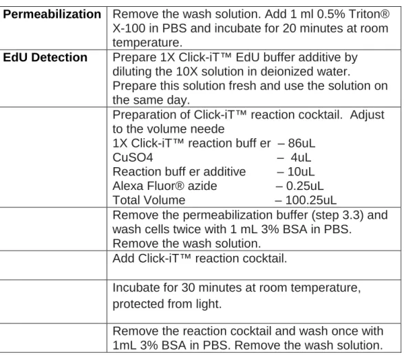
Proliferating cells were detected based on incorporation of EdU into newly
synthesized DNA or immunostaining for Ki67. EdU assays use a two-step click reaction that is based on detection of incorporated EdU with an Alexa Fluor® azide dye (Alexa Fluor® 488) which gives green fluorescence, or Alexa Fluor® 594 which gives red fluorescence (Click-iT™ EdU Alexa Fluor® 488 and 594 Imaging Kits, Invitrogen, Carlsbad, CA). EdU contains a small alkyne group that is detected by an azide containing fluorescent dye. EdU offers advantages over commonly used BrdU based methods. Because the Alexa Fluor® dye is small, only mild permeabilization is required to detect incorporated nucleotide. No DNA denaturation or antibody-based detection is needed which makes the system faster and more compatible with dual labeling for other proteins. EdU was detected following manufacturers instructions through steps 3.3 – 4.6 (see Table 1, pg 18). Ki67 was detected by immunofluorescence as outlined below.
Immunohistochemistry
If sections were first stained for EdU, primary antibodies were added directly after removal of Alexa fluor dye and the final wach of the Click-iT™ procedure. If EdU
15
retrieval after the initial wash steps by incubating the sections at 125ºC for 30 seconds followed by 90ºC for 10 seconds in Reveal Decloaker RTU (no. RV1000MMRTU; Biocare Medical, Concord, CA). These samples were then moved into PBS for 20 minutes at room temperature. After primary antibody incubation, sections were washed 3 times and secondary antibodies were added at the following concentrations, anti-Rabbit- Alexafluor 488 (1:500, no. Z-25302, Molecular Probes, Eugene,OR), anti-Rabbit-594 (1:250; no. 711-505-152 Jackson Immunoresearch Laboratories, West Grove, PA) and anti-Rat-594 (1:250; no. 712-505-150 Jackson Immunoresearch Laboratories, West Grove, PA). Ki67 required amplification using anit-Mouse IgG-Biotinylated(1:200; Jackson Immunoresearch Laboratories, West Grove, PA)followed by Alexafluor-555-labeled streptavidin (1:250, no.S32355, Molecular Probes). Sections were then washed 3 times and nuclei were stained with Bisbenzamide (1:20000) and Draq5 (1:1000 no. BOS-889-001; Biostatus, San Diego, CA) in Tris for 10 min. This was followed by a 5 minute wash and then the slides were cover slipped using Hydromount (National Diagnost, no. HS-106, Atlanta, GA).
A major goal of these studies was to assess if Sox9 marked proliferating cells in microcolonies and regenerating crypts between days 3 – 9 after irradiation. Thus the localization of immunoreactive Sox9 was compared with Ki67 or EdU on the same sections.
Quantification of Sox9EGFP
16
(Formeister 2009) indicate two populations of Sox9EGFP cells defined by high
expression, Sox9EGFPHI, and low expression, Sox9EGFPLO. In normal, uninjured small intestine, Sox9EGFPHI cells in crypt and villus appear to represent post-mitotic cells
17
Figure 2: Mouse placement in the X-RAD 320 from Precision X-Ray. Each mouse
is kept anesthetized with its head placed in an open syringe connected to a continuous tube supplying 2% isoflurane. The lower half of each mouse is placed within the
18
Permeabilization Remove the wash solution. Add 1 ml 0.5% Triton® X-100 in PBS and incubate for 20 minutes at room temperature.
EdU Detection Prepare 1X Click-iT™ EdU buffer additive by diluting the 10X solution in deionized water.
Prepare this solution fresh and use the solution on the same day.
Preparation of Click-iT™ reaction cocktail. Adjust to the volume neede
1X Click-iT™ reaction buff er – 86uL CuSO4 – 4uL Reaction buff er additive – 10uL Alexa Fluor® azide – 0.25uL Total Volume – 100.25uL
Remove the permeabilization buffer (step 3.3) and wash cells twice with 1 mL 3% BSA in PBS.
Remove the wash solution. Add Click-iT™ reaction cocktail.
Incubate for 30 minutes at room temperature, protected from light.
Remove the reaction cocktail and wash once with 1mL 3% BSA in PBS. Remove the wash solution.
Chapter 3
Results
Body weight and survival
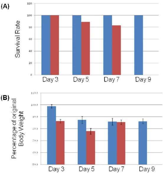
Figures 3A and 3B (pg 27) show body weights and survival rates for CD1 and C57BL/6 mice used in this study. CD1 mice showed 100% survival out to 9 days, the latest time point tested to date. Survival of C57BL/6 mice was slightly less although 80% of animals survived up to day 7. To date we have not extensively tested C57BL/6 mice through the 9 day time point. Both CD1 and C57BL/6 mice lost weight after irradiation. Peak loss for C57BL/6 mice occurred at day 5 and animals began to regain weight by day 7. After initial weight loss at day 5, CD1 mice maintained body weight through day 9. The high survival rates and increased or maintained body weights of C57BL/6 and CD1 mice through days 7 – 9 indicate that the abdominal irradiation model will be valuable for assessing crypt regeneration and effects of irradiation on stem cells through and beyond day 9.
Regional effects of abdominal radiation on crypt damage and regeneration
20
and mice at 3 (Figure 5, pg 30-31), 5 (Figure 6, pg 32-33), 7 (Figure 7, pg 34-35), and 9 (Figure 8, pg 36-37) days after irradiation. The duodenum showed the least tissue damage and had a regenerative response as early as day 3 after irradiation (Figures 4A and 5A, pg 30-33). This was followed by an increasing hyperplastic response
characterized by deep, densely staining hyper-regenerative crypts from days 5 through 9 (Figures 6A, 7A and 8A, pg 32-37). The duodenum then showed almost complete recovery to a normal physiological state, although with some remaining areas of regeneration and crypt fission, by day 9 (Figure 8A, pg 36-37). In the jejunum, there was almost complete crypt loss with the appearance of a few small regenerative crypts and sub-epithelial microcolonies at day 3 after irradiation (Figure 4B and 5B, pg 28-31). There were increasing numbers of hyper-regenerative crypts that continued to expand from days 5 through 9 (Figure 6B, 7B and 8B, pg 32-37). At day 9, there was recovery of the villi, but the crypts continued to show a hyperplastic response demonstrated by an increase in cell number and size (Figure 8B, pg 36-37). The ileum showed the greatest tissue damage with fewer, shortened villi and almost no crypts at day 3 (Figures 4C and 5C, pg 28-31). A regenerative response demonstrated by a recovery in the number of villi and hyperplastic crypts was seen at day 5 (Figure 6C, pg 32-33). This was followed by the appearance of hyper-regenerative crypts and varying recovery of villi at days 7 and 9 after irradiation (Figure 7C and 8C, pg 34-37).
The subsequent analysis of intestinal stem cells (IESC) in small intestine was focused on the jejunum due to the ideal injury response to irradiation characterized by crypt loss, proliferating IESC and microcolony formation by day 3, followed by
21
studies. This process is a useful injury and regeneration model for the study of stem cells, because the intestinal crypts have been lost due to massive apoptosis and
regeneration is believed to be mediated by a few multipotent clonogenic stem cells that go on to regenerate the intestinal crypts and epithelium (Potten, 2004).
Figure 9 (pg 38) shows the response of the proximal and distal colon to 14Gy abdominal irradiation. Neither proximal nor distal colon showed complete crypt loss. Crypt damage was evident at days 3 and 5, being more apparent in the distal colon. In both proximal and distal colon there was the appearance of large hyper-regenerative crypts by day 7 with adjacent regions showing continued evidence of crypt loss.
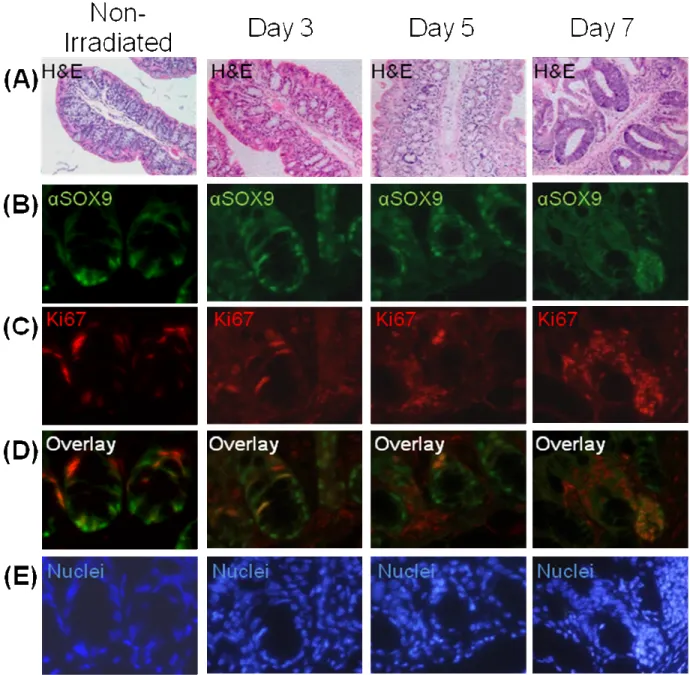
A subset of proliferating cells express SOX9 protein during crypt regeneration after irradiation
22
consistent with previous studies and the hypothesis of increased stem/multipotent cell recruitment after high dose irradiation (Hendry et al., 1992). SOX9 and Ki67 were colocalized to a subset of regenerating cells in hyper-regenerating crypts. There were also SOX9 and Ki67 positive cells that did not co-label. This could be reflective of the observation that SOX9 marks non-cycling enteroendocrine cells that would not be positive for Ki67 and Ki67 marks transit amplifying cells that are not positive for SOX9.
Figures 11 and 12 (pg 41-43) show SOX9 and Ki67 localization in non-irradiated colon and colon through days 3 – 7 after irradiation. In proximal colon of non-irradiated mice SOX9 positive cells were located at the base of the crypts. Most Ki67 cells did not overlap with SOX9 although weak Ki67 staining appeared to co-localize with SOX9 in some cells at the crypt base. In hyper-regenerative crypts at days 5 and 7 there was clear overlap between some strongly labeled Ki67 cells and SOX9. At day 3, SOX9 positive cells at the crypt base did not label with Ki67 while at day 7 hyper-regenerative crypts showed clear co-staining for Ki67 and SOX9, although there was not complete overlap. Figure 10 (pg 39-40) shows data for distal colon. In non-irradiated colon, SOX9 was localized to cells at the crypt base and there was clear overlap between a subset of these cells and Ki67 staining. On day 3, Ki67 staining was less obvious. However, in hyper-regenerative crypts at days 5 and 7 there was expansion of the SOX9 positive cells at the base (day 5) and throughout hyper-regenerative crypts (day 7) and virtually all of these co-labeled with Ki67.
23
microcolonies and sub-epithelial areas. At later time points, SOX9 colocalized with EdU in regenerative areas and hyper-regeneratative crypts (Figure13, pg 44-45). SOX9 was observed to colocalize with only a subset of EdU positive cells. The subset of
co-staining cells was limited to small microcolonies at day 3 and at the base of the hyper-regenerating crypts at later time points. This provides indirect evidence that SOX9 may mark early multipotent regenerating stem cells rather than proliferating cells at higher locations in the crypt.
Sox9EGFP expression after irradiation
The Sox9EGFP reporter mouse was used to further analyze the expression pattern of Sox9 after radiation-induced injury and during regeneration. The Sox9EGFP reporter mice breed normally and the EGFP transgene does not cause any negative phenotype as described by Formeister (Formeister, 2009). Consistent with findings of Formeister, non-irradiated jejunum showed Sox9EGFPLO cells at the crypt base and Sox9EGFPHI cells at higher levels in the crypts and on the villi (Figure 14, pg 46). At day 3 after irradiation, Sox9EGFP was expressed at the very base of villi and in some sub-epithelial groups of cells that may represent microcolonies. High expressing Sox9EGFP (Sox9EGFPHI) cells were identified at day 3, while low expressing Sox9EGFP (Sox9EGFPLO) cells were not detectable (Figure 14, pg 46). Throughout hyper-regenerative crypts at days 5 and 7 after irradiation, there was a robust expansion of Sox9EGFPLO cells with few Sox9EGFPHI cells being detected in most crypts (Figure 14, pg 46).
24
number of Sox9EGFP cells in normal crypts compared to regenerative and
hyper-regenerative crypts (Figure 15, pg 47). Sox9EGFP could not be counted at day 7 due to technical reasons. There were no crypt structures identified at day 3 after irradiation and, therefore, the number of Sox9EGFP cells per crypt could not be quantified at that time. Additionally, Sox9EGFPHI cells were present in sub-epithelial areas that could not be identified as crypts at day 3. In regenerative crypts at days 5 and 9 after irradiation, no change occurred in the mean number of Sox9EGFPHI cells compared to crypts in non-irradiated mice. Sox9EGFPLO cells could not be found at day 3 after irradiation. At day 5 after irradiation, the number of Sox9EGFPLO cells increased almost three-fold in hyper-regenerative crypts. The number of Sox9EGFPLO cells returned to just above non-irradiated numbers in crypts at day 9 (Figure 15, pg 47). The expansion of Sox9EGFPLO cells provides indirect evidence that they play an important role in the regenerative process.
It should be noted that while there was not an expansion in the mean number of Sox9EGFPHI cells per crypt at days 5 and 9, there was the appearance of occasional large crypts with greater numbers of Sox9EGFPHI cells (Figures 16A, pg 48). This suggests that local factors within individual crypts during regeneration may dictate the regeneration of Sox9EGFPHI cells.
The cell counting data of Sox9EGFPHI and Sox9EGFPLO should be considered as a preliminary qualitative-semi-quantitative test of the patterns of regeneration of
25
the quantitative data need to be captured from tissue sections prepared soon after tissue collection and with samples mounted on the same slide. This is because there was variation in the intensity of Sox9EGFP across slides. Additionally, given the dramatic expansion in Sox9EGFP cells between days 3 and 4, future studies should include
evaluation of day 4. It should be noted however that preliminary FACS analysis for GFP provides confirmatory evidence that Sox9EGFPLO cells are expanded at day 5 after
irradiation (Van Landeghem and Magness personal communication).
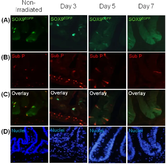
Colocalization of Sox9EGFPHI cells and Substance P
Sox9EGFPHI cells have previously been shown to be mature/post-mitotic enteroendocrine cells, because these Sox9EGFPHI cells colocalized with the
enteroendocrine cell markers Substance P and Chromogranin-A but not with Ki67 or the secretory/enteroendocrine progenitor marker, Neurogenin3 (Formeister et al., 2009). Substance P immunostaining was used to determine if Sox9EGFPHI cells exhibit
enteroendocrine phenotype after irradiation. At day 3, colocalization of Substance P was detected in a majority of Sox9EGFPHI cells (Figure 16, pg 48). Thus, even in hyper-regenerative crypts, Sox9EGFPHI cells were immunopositive for an enteroendocrine marker. It will be of interest to compare EdU and Substance P staining in the same crypts to assess if these cells are post-mitotic. Note that Figure 16A shows an example of a hyper-regenerative crypt with multiple Sox9EGFPHI and Substance P labeled cells at day 5 contrasting with a crypt composed almost solely of Sox9EGFPLO cells at day 7.
26
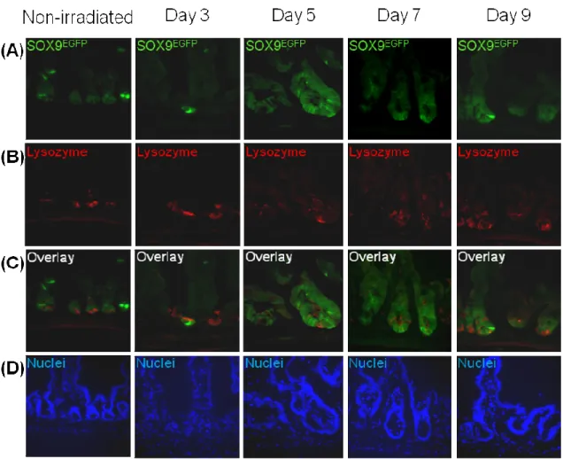
Previously, two populations of Lysozyme positive Paneth cells have been observed that exist at a 1:1 ratio, one with and one without SOX9 protein; however, SOX9EGFP is not associated with Lysozyme positive Paneth cells which appear to be CBCs between Paneth cells (Formeister et al., 2009). SOX9EGFP was evaluated after Lysozyme immunofluorescence staining to determine if this remained true after irradiation. At day 3 after irradiation, Lysozyme positive cells had lost their normal granulated appearance and the staining seemed more diffuse throughout the cytoplasm (Figure 17B, pg 49). This may be because the cells were releasing their lysozyme-containing granules or because they are apoptotic, which can be further evaluated with Caspase-3 staining (Marshman, Ottewell, Potten, & Watson, 2001). The Lysozyme positive cells at day 3 did not colocalize with SOX9EGFP (Figure 17C, pg 49). At days 5 and 7 after irradiation, Lysozyme immunostaining returned to a more granulated
27
Figure 3: Body weights and survival rates for CD1 and C57BL/6 mice. (A) Survival
rates of CD1 (blue bars) and C57BL/6 (red bars) after irradiation. N=7, 5, 3 and 2 for CD1 mice and N=11, 9 and 6 for C57BL/6 mice for each day after irradiation
29
Figure 4: Normal Small Intestinal Physiology. H&E staining of non-irradiated small intestinal photographed at 5X magnification. (A) Duodenum (B) Jejunum (C)
31
Figure 6: Small Intestinal Damage at day 3 after irradiation. H&E staining
photographed at 5X magnification. (A) Duodenum with arrow showing regenerative
33
Figure 6: Regenerative response in the small intestine at day 5 after irradiation. H&E staining photographed at 5X magnification. Arrows point to regenerative crypts (A) Duodenum (B) Jejunum with recovery of crypts (C) Ileum showing recovery
35
Figure 7: Hyper regenerative response at day 7 after irradiation. H&E staining photographed at 5X magnification. Arrows show examples of hyper-regenerative crypts that have an increase in size and cell density (A) Duodenum with an increase
37
Figure 8: Increase in regeneration and crypt fission at day 9 after irradiation. H&E staining photographed at 5X magnification. Arrows show areas of crypt fission in the duodenum and jejunum as well as regeneration in the ileum. (A) Duodenum (B)
38
Figure 9: Colon has shorted crypts followed by areas of regeneration after irradiation. H&E staining photographed at 4X and 10X magnification.. Arrows show areas of regeneration. (A) Proximal colon at 4x. (B) Proximal colon at 10x. (C)
40
Figure 10: SOX9 protein and Ki67 colocalize in microcolonies and
hyper-regenerative areas after irradiation. (A) H&E staining at higher magnification in areas
41
Figure 11: SOX9 protein and Ki67 colocalize in crypts of proximal colon. (A) H&E
staining at higher magnification in areas representative of immunofluorescence staining.
(B) SOX9 protein (SOX9) in green. (C) Ki67 staining in red. (B) SOX9 protein (SOX9)
43
Figure 12: SOX9 protein and Ki67 colocalization in crypts of distal colon. (A) H&E
staining at higher magnification in areas representative of immunofluorescence staining.
(B) SOX9 protein (SOX9) in green. (C) Ki67 staining in red. (B) SOX9 protein (SOX9)
45
Figure 13: SOX9 protein colocalizes with a subset of EdU positive cells in sub-epithelial areas and at the base of hyper-regenerative crypts. (A) SOX9 protein
46
Figure 14: Sox9EGFPLO cells are lost at day 3 and then show vast expansion in hyper-regenerative crypts at days 5 and 7 after irradiation. (A) H&E staining of
47
Figure 15: There is no change in the number of Sox9EGFPHI cells after irradiation, while Sox9EGFPLO cells increase almost 3 fold. Quantification of Sox9EGFPHI ( ) and Sox9EGFPLO cells ( ). (A) Sox9EGFPexpression. (B) Corresponding nuclear staing. (C) Sox9EGFP cells were counted in 15 crypts from 1 animal at each time point. There is no change in the number of Sox9EGFPHI cells at any time point, while
Sox9EGFPLO cells showed a 2.93 fold change (6.47±0.413 vs. 19±1.919) in
48
Figure 16: Sox9EGFPHI cells colocalize with most Substance P expressing cells after irradiation. (A) Distribution of Sox9EGFPHI and Sox9EGFPLO cells after irradiation.
49
Figure 17: Lysozyme positive Paneth cells did not colocalize with Sox9EGFP cells and did not show an increase at any time point after irradiation. (A) Sox9EGFP
Chapter 4
Discussion
Whole body irradiation provides a useful experimental model, but has the potential disadvantage that a majority of WT animals die between 5 and 7 days after irradiation precluding analyses of complete crypt and villus regeneration (Qui
2008)(Potten, 2004). With this in mind, we adapted an abdominal irradiation model for our studies with the goal of improve animal survival beyond 5 – 7 days. In addition, abdominal irradiation may better mimic radiation-induced damage that occurs clinically due to regional radiation for abdominal malignancy (Kountouras, Zavos 2008). Our study using abdominal irradiation revealed that a majority of animals survived up to 7 – 9 days and began to gain or maintain weight. This suggests that the model will prove valuable for evaluating later time points in future studies. This model was then used to evaluate the expression pattern of the putative IESC marker, Sox9.
51
studies have focused on days 3 through 5 after irradiation when colonies of proliferating cells or expanded crypts with proliferating cells are believed to reflect clonal expansion of intestinal stem cells surviving the injury (George 2009, Qiu 2008). However,
relatively few published studies have evaluated crypt loss and regeneration after abdominal radiation along the entire length of small and large intestine in the same animals after irradiation (Freeman 2001). Our studies revealed clear regional
differences in crypt loss after abdominal radiation. In the small intestine, the duodenum was most resistant to radiation-induced crypt loss with crypts remaining at all times studied, but regenerative responses still occurring by days 7 and 9. Jejunum and ileum exhibited complete crypt loss by days 3 and 5 with ileum showing significantly greater damage. The differential response to irradiation defined here by increasing injury from the proximal to the distal end of the small intestine may be reflective of differences in stem cell properties within each region. In the duodenum, there may be more IESC, these IESC may be less sensitive or have greater regenerative capacity in response to irradiation. In this regard it is noteworthy that one recently defined stem cell marker Bmi1 appears to be selectively expressed in proximal small intestine suggesting differences in stem cell populations throughout the intestine (Sangiorgi & Capecchi, 2008). Future studies to better understand the mechanisms that allow duodenal stem cells to survive and regenerate will be relevant to strategies that may increase
regeneration in other areas of the intestine.
52
intestine. Neither proximal nor distal colon showed complete crypt loss at days 3 and 5. Furthermore, it has been observed that germ-free mice are less susceptible to radiation induced crypt damage (Crawford & Gordon, 2005). Despite this, by day 7 there was a marked increase in regenerating crypts, occurring with the appearance of inflammatory infiltrates. While more needs to be done to verify inflammation, such as staining for immune cells, this observation may reflect that damage in the colon after irradiation may lead to barrier defects that expose the underlying lamina propria to high levels of luminal microbiota that initiate an inflammatory response. This inflammatory response may be aiding regeneration after irradiation. We also speculate that the greater damage to the ileum compared to more proximal regions of the small intestine could also reflective of a difference in the inflammatory response and microbiota. Comparisons of crypt loss and regeneration across the small intestine and colon may be useful in understanding the role of inflammation during regeneration after irradiation in a setting of different degrees of damage-induced inflammation.
53
The expression pattern and molecular properties of Sox9 positive cells was further evaluated using a Sox9EGFP reporter and it has been reported that Sox9EGFPLO cells are crypt base columnar (CBC) with stem cell like properties during a normal physiological state (Formeister et al., 2009). In this study we used a model of high dose irradiation to test if Sox9EGFP expression is useful for identifying IESC after injury.
Results show that Sox9EGFPLO cells appear to be eliminated at day 3 after high dose irradiation when clonogenic IESC are present. The loss or reduction of Sox9EGFPLO cells, that are hypothesized to be IESC, emphasized the dynamic nature of IESC after injury and the regenerative process.
One interesting observation was that Sox9EGFPHI cells are still visible at day 3 after high dose irradiation, many of which colocalize with Substance P. Further
characterization of these cells with other markers such as Neuorgenin 3 is needed, but it remains a possibility that these Sox9EGFPHI cells revert back to a stem cell like
54
It is also possible that upon injury, Substance P expressing Sox9EGFPHI cells enhance the survival of surrounding quiescent IESC that are not initially identified by Sox9EGFP expression and have an ability to protect their original genome (Potten, Owen, & Booth, 2002). The close proximity of quiescent stem cells and Sox9EGFPHI cells, both located in the upper stem cell zone, makes it plausible that there may be interactions between these cells. However, the location of quiescent IESC at position 4 above the base of the crypt is the cellular site of greatest frequency of apoptosis after irradiation (Wilkins et al., 2002). Therefore, quiescent IESC may not be the best candidate for regenerative IESC. In this case, Sox9EGFPHI cells may influence surrounding cells to adapt a stem cell-like phenotype. There is evidence that Substance P increases
regeneration after irradiation (Kang, Kim, Yi, & Son, 2009). The release of Substance P from Sox9EGFPHI cells could conceivably provide a paracrine mechanism driving the increase in proliferation and regeneration of adjacent IESC. At present this possibility is speculative, but could be tested by Substance P neutralization or administration. The stimulation by Substance P of surrounding cells could lead them to a phenotype more like CBC that could then be identified as Sox9EGFPLO cells at later time points after irradiation. This would account for the vast increase in Sox9EGFPLO cells in hyper-regenerative crypts at later time points after irradiation.
The increase in Sox9EGFPLO cells in hyper-regenerative crypts after irradiation is not accompanied by an increase in Paneth cells, which have been seen to decrease after irradiation (Brennan, Carr, Seed, & McCullough, 1998). The increase in
55
or stem cells. An interaction between Sox9EGFPHI and Sox9EGFPLO cells is supported by the observation that some hyper-regenerative crypts marked with an extensive number of Sox9EGFPLO cells also have Sox9EGFPHI cells often times at almost regular intervals between the Sox9EGFPLO cells. This interaction may be unique to regeneration and the presence of Sox9EGFPHI cells in the upper stem cell region of only some crypts during a normal physiological state could be a protective mechanism for situations when an increase in regeneration is needed, such as after injury. Further understanding of the unique expression pattern and molecular characteristic of Sox9EGFP cells after injury could help us to define the IESC phenotype and niche driving the regenerative process.
This study would be greatly enhanced with further evaluation of current samples and more animals using confocal microscopy. In this regard, I recently completed additional experiments to increase the sample size to at least four animals per time point at 3 – 9 days after irradiation. Ongoing analyses of these samples and detailed characterization of EdU colocalization with IESC biomarkers or markers of differentiated cells will provide important information to optimally design future growth factor
interventions. We also have begun fluorescent-activated cell sorting (FACS) analysis to quantitatively assess the relative proportions of Sox9EGFPHI and Sox9EGFPLO cells at different times after irradiation. This could confirm the absence of Sox9EGFPLO cells at day 3 after irradiation as the regenerative process may only require a few surviving stem cells and the identification of Sox9EGFPLO cells at day 3 after irradiation by
56
FACS sorting can also be used to isolate Sox9EGFP cells for microarray analysis. Comparing the molecular phenotype of Sox9EGFP cells from non-irradiated mice with cells from mice at different time points after irradiated could identify genes that are important in stimulating regeneration. These genes will be relevant to developing
therapies to promote IESC regeneration in patients exposed to irradiation. It would also determine the molecular differences between steady-state IESC and IESC
post-irradiation. Understanding these differences is important, because as this study exemplifies there are major dramatic differences in Sox9 cells during steady-state regeneration and during the regenerative process stimulated by injury.
Sox9EGFP cells isolated by FACS can also be used for in vitro studies. One in
vitro study that would test the dedifferentiation of Sox9EGFPHI cells into IESC could be done by isolating Sox9EGFPHI cells from normal intestinal tissue, exposing them to high-dose irradiation and maintaining them in culture to determine if they dedifferentiate. Sox9EGFPLO cells could also be isolated and treated with Substance P to determine if Substance P increases proliferation.
This study focused on proliferation during the regenerative process after high dose irradiation, but this abdominal irradiation model in conjunction with stem cell
57
is reached at 10Gy (Barker, van Es, Kuipers, Kujala, van den Born, Cozijnsen, Haegebarth, Korving, Begthel, Peters, & Clevers, 2007b). Therefore the expression pattern of Sox9EGFP should be evaluated at 4 hours after irradiation at both high and low doses of irradiation to determine the relative radiosensitivity of Sox9EGFPHI cells in the USZ and Sox9EGFPLO cells in the LSZ.
This irradiation model can also be used for preclinical testing of potential therapies for patients undergoing abdominal radiation therapy. It is estimated that 60-80% of cancer patients that undergo radiation therapy show signs of acute bowel toxicity which include diarrhea, abdominal pain and nausea. Delayed bowel toxicity is characterized by malabsorption and bowel dysmotility (Hauer-Jensen, Wang, Boerma, Fu, & Denham, 2007). Multiple growth factors have been investigated for their
therapeutical properties, but this has been based on their ability to increase the number of microcolonies at early time points after in high-dose irradiation (Potten et al., 1995). The use of this abdominal irradiation model with Sox9EGFP reporter mice, other reporter mice or lineage tracing models can directly test the expansion and differentiation of stem cells throughout the regenerative process.
Insulin-like growth factor I (IGF-I) has potential as a therapy for abdominal irradiation patients. IGF-I transgenic mice have enhanced intestinal growth and show increase proliferation in crypts (Ohneda, Ulshen, Fuller, D'Ercole, & Lund, 1997). It has also been shown that IGF-I decreases apoptosis in crypts at 4 hours after 5Gy of
58
radiosensitivity of cells in the IESC zone after irradiation (Han et al., 2001; Qiu et al., 2008). The model of abdominal irradiation developed in this study can be used to
determine if IGF-I treatment protects Sox9EGFP cells from apoptosis. It will be interesting to diescover if IGF-I protects Sox9EGFPHI cells at low doses of irradiation, while protecting Sox9EGFPLO cells at high doses of irradiation, and to observe any difference in the
regenerative process.
The abdominal irradiation model developed in this study provides a useful model for studying IESC and regeneration. This model resulted in increased survival
compared to previously models used in other studies. This will allow for better analysis of regeneration after irradiation and the role of IESC during this process. It was
observed in this study that Sox9 marked proliferating cells in microcolonies and
59
60
Appendix 1
Biochomoendoscopy and Ileo-cecal resection of CONV-IL-10 null mice
The focus of my independent research project on the irradiation model is
presented in the main thesis. I also participated in two other studies aimed at improved visualization of gastrointestinal tumors using molecular probes and development of an ileo-cecal resection model in IL-10 null mice, as a model to analyze stem cells during post-surgical adaptive growth of small intestine in a setting of inflammation.
Co-authorship on two publications resulted from my participation in these projects as listed below:
Zhang H, Morgan D, Cecil G, Burkholder A, Ramocki N, Scull B, Lund PK.
Biochromoendoscopy: molecular imaging with capsule endoscopy for detection of polypoid lesions in the GI tract. Gastrointestinal Endoscopy 2008; 68(3):520-7. PMID: 18499106.
Rigby RJ, Hunt MR, Scull BP, Helmrath MA, Lund PK. A new animal model of post-surgical inflammatory bowel disease and fibrosis: the effect of commensal microflora. Gut 2009. PMID: 19398439. In Press.
61
Appendix 2
Biochromoendoscopy: molecular imaging with capsule endoscopy for
detection of polypoid lesions in the GI tract.
BACKGROUND: Current capsule endoscopy (CE) provides minimally invasive technology for GI imaging but has limited ability to diescriminate different types of polyps. Near infrared fluorescent (NIRF) probes activated by biomarkers upregulated in adenomas (eg, cathepsin B) are potentially powerful tools to distinguish premalignant or malignant lesions from benign or inflammatory lesions. OBJECTIVES: To examine whether CE can be integrated with NIRF probes to detect adenomas and whether cathepsin B-activated NIRF probes are activated by benign or inflammatory lesions. DESIGN: Mouse models of adenomas, hyperplactic/lymphoid polyps, and acute or chronic intestinal inflammation were injected intravenously with a cathepsin B-activated probe (Prosense 680). Dissected intestine was imaged with CE under white or NIRF light. For NIRF excitation (680 nm), dichroic and emission (700 nm) filters were
62
used. CONCLUSIONS: We demonstrate proof of the principle that
63
Appendix 3
A new animal model of post-surgical inflammatory bowel disease and
fibrosis: the effect of commensal microflora.
OBJECTIVE: Ileo-cecal resection (ICR) is common in Crohn's disease (CD). Inflammation and fibrosis frequently recur at the site of anastomosis or in the small intestine (SI). No animal models of post-surgical inflammation and fibrosis exist. We developed a model of ICR in IL-10 null and wild-type (WT) mice to test the hypothesis that that ICR promotes post-surgical inflammation and fibrosis in SI or anastomosis of genetically susceptible IL-10 null, but not WT or germ free (GF)-IL-10 null mice. DESIGN: GF-IL-10 null mice were conventionalized (CONV) and 3 weeks later
randomized to ICR, transection (T) or no treatment (NoTx). Age-matched conventionally raised (CONV) WT and GF-IL-10 null mice received ICR, T or NoTx. Animals were killed 28 days later. MAIN OUTCOME MEASURES: Histological scoring, real-time PCR for TNFalpha and collagen, and immunostaining for CD3+ T cells, assessed
inflammation and fibrosis. RESULTS: After ICR, CONV-IL-10 null, but not CONV-WT mice, developed significant inflammation and fibrosis in SI and inflammation in
64
65 References
Arsenijevic, Y. (2005). Future perspectives: From stem cells and IGF biology to the clinic. Advances in Experimental Medicine and Biology, 567, 385-412.
Barker, N., van Es, J. H., Kuipers, J., Kujala, P., van den Born, M., Cozijnsen, M., et al. (2007a). Identification of stem cells in small intestine and colon by marker gene Lgr5. Nature, 449(7165), 1003-1007.
Barker, N., van Es, J. H., Kuipers, J., Kujala, P., van den Born, M., Cozijnsen, M., et al. (2007b). Identification of stem cells in small intestine and colon by marker gene Lgr5. Nature, 449(7165), 1003-1007.
Batlle, E., Henderson, J. T., Beghtel, H., van den Born, M. M., Sancho, E., Huls, G., et al. (2002). Beta-catenin and TCF mediate cell positioning in the intestinal epithelium by controlling the expression of EphB/ephrinB. Cell, 111(2), 251-263.
Beaulieu, J. F. (1992). Differential expression of the VLA family of integrins along the crypt-villus axis in the human small intestine. Journal of Cell Science, 102 ( Pt 3)(Pt 3), 427-436.
Bjerknes, M., & Cheng, H. (2002). Multipotential stem cells in adult mouse gastric epithelium. American Journal of Physiology.Gastrointestinal and Liver Physiology,
283(3), G767-77.
Bjerknes, M., & Cheng, H. (2006). Intestinal epithelial stem cells and progenitors.
Methods in Enzymology, 419, 337-383.
Booth, C., Booth, D., Williamson, S., Demchyshyn, L. L., & Potten, C. S. (2004). Teduglutide ([Gly2]GLP-2) protects small intestinal stem cells from radiation damage. Cell Proliferation, 37(6), 385-400.
Brennan, P. C., Carr, K. E., Seed, T., & McCullough, J. S. (1998). Acute and protracted radiation effects on small intestinal morphological parameters. International Journal
of Radiation Biology, 73(6), 691-698.
Cai, W. B., Roberts, S. A., Bowley, E., Hendry, J. H., & Potten, C. S. (1997). Differential survival of murine small and large intestinal crypts following ionizing radiation.
International Journal of Radiation Biology, 71(2), 145-155.
66
Crawford, P. A., & Gordon, J. I. (2005). Microbial regulation of intestinal radiosensitivity.
Proceedings of the National Academy of Sciences of the United States of America, 102(37), 13254-13259.
de Santa Barbara, P., van den Brink, G. R., & Roberts, D. J. (2003). Development and differentiation of the intestinal epithelium. Cellular and Molecular Life Sciences :
CMLS, 60(7), 1322-1332.
Dube, P. E., Forse, C. L., Bahrami, J., & Brubaker, P. L. (2006). The essential role of insulin-like growth factor-1 in the intestinal tropic effects of glucagon-like peptide-2 in mice. Gastroenterology, 131(2), 589-605.
Formeister, E. J., Sionas, A. L., Lorance, D. K., Barkley, C. L., Lee, G. H., & Magness, S. T. (2009). Distinct SOX9 levels differentially mark Stem/Progenitor populations and enteroendocrine cells of the small intestine epithelium. American Journal of
Physiology.Gastrointestinal and Liver Physiology,
Giannakis, M., Stappenbeck, T. S., Mills, J. C., Leip, D. G., Lovett, M., Clifton, S. W., et al. (2006). Molecular properties of adult mouse gastric and intestinal epithelial progenitors in their niches. The Journal of Biological Chemistry, 281(16), 11292-11300.
Gibson, P. R. (2004). Apoptosis or necrosis--colonic epithelial cell survival. Novartis
Foundation Symposium, 263, 133-45; diescussion 145-50, 211-8.
Gong, S., Zheng, C., Doughty, M. L., Losos, K., Didkovsky, N., Schambra, U. B., et al. (2003). A gene expression atlas of the central nervous system based on bacterial artificial chromosomes. Nature, 425(6961), 917-925.
Han, J., Flemington, C., Houghton, A. B., Gu, Z., Zambetti, G. P., Lutz, R. J., et al. (2001). Expression of bbc3, a pro-apoptotic BH3-only gene, is regulated by diverse cell death and survival signals. Proceedings of the National Academy of Sciences
of the United States of America, 98(20), 11318-11323.
Harrisingh, M. C., Perez-Nadales, E., Parkinson, D. B., Malcolm, D. S., Mudge, A. W., & Lloyd, A. C. (2004). The Ras/Raf/ERK signalling pathway drives schwann cell dedifferentiation. The EMBO Journal, 23(15), 3061-3071.
Hauer-Jensen, M., Wang, J., Boerma, M., Fu, Q., & Denham, J. W. (2007). Radiation damage to the gastrointestinal tract: Mechanisms, diagnosis, and management.
Current Opinion in Supportive and Palliative Care, 1(1), 23-29.
67
He, X. C., Zhang, J., Tong, W. G., Tawfik, O., Ross, J., Scoville, D. H., et al. (2004). BMP signaling inhibits intestinal stem cell self-renewal through suppression of wnt-beta-catenin signaling. Nature Genetics, 36(10), 1117-1121.
Hendry, J. H., Roberts, S. A., & Potten, C. S. (1992). The clonogen content of murine intestinal crypts: Dependence on radiation dose used in its determination. Radiation
Research, 132(1), 115-119.
Ishizuka, S., Martin, K., Booth, C., Potten, C. S., de Murcia, G., Burkle, A., et al. (2003). Poly(ADP-ribose) polymerase-1 is a survival factor for radiation-exposed intestinal epithelial stem cells in vivo. Nucleic Acids Research, 31(21), 6198-6205.
Kang, M. H., Kim, D. Y., Yi, J. Y., & Son, Y. (2009). Substance P accelerates intestinal tissue regeneration after gamma-irradiation-induced damage. Wound Repair and
Regeneration : Official Publication of the Wound Healing Society [and] the European Tissue Repair Society, 17(2), 216-223.
Khan, W. B., Shui, C., Ning, S., & Knox, S. J. (1997). Enhancement of murine intestinal stem cell survival after irradiation by keratinocyte growth factor. Radiation
Research, 148(3), 248-253.
Krysiak, R., Gdula-Dymek, A., Bednarska-Czerwinska, A., & Okopien, B. (2007). Growth hormone therapy in children and adults. Pharmacological Reports : PR,
59(5), 500-516.
Marshman, E., Ottewell, P. D., Potten, C. S., & Watson, A. J. (2001). Caspase activation during spontaneous and radiation-induced apoptosis in the murine intestine. The Journal of Pathology, 195(3), 285-292.
Morante, J., Vallejo-Cremades, M. T., Gomez-Garcia, L., Vazquez, I., Gomez-de-Segura, I. A., Sanchez, M., et al. (2003). Differential action of growth hormone in irradiated tumoral and nontumoral intestinal tissue. Digestive Diseases and
Sciences, 48(11), 2159-2166.
Ohneda, K., Ulshen, M. H., Fuller, C. R., D'Ercole, A. J., & Lund, P. K. (1997).
Enhanced growth of small bowel in transgenic mice expressing human insulin-like growth factor I. Gastroenterology, 112(2), 444-454.
Potten, C. S. (1992). The significance of spontaneous and induced apoptosis in the gastrointestinal tract of mice. Cancer Metastasis Reviews, 11(2), 179-195.
68
Potten, C. S., & Booth, C. (1997). The role of radiation-induced and spontaneous apoptosis in the homeostasis of the gastrointestinal epithelium: A brief review.
Comparative Biochemistry and Physiology.Part B, Biochemistry & Molecular Biology, 118(3), 473-478.
Potten, C. S., Booth, C., Tudor, G. L., Booth, D., Brady, G., Hurley, P., et al. (2003). Identification of a putative intestinal stem cell and early lineage marker; musashi-1.
Differentiation; Research in Biological Diversity, 71(1), 28-41.
Potten, C. S., & Grant, H. K. (1998). The relationship between ionizing radiation-induced apoptosis and stem cells in the small and large intestine. British Journal of Cancer,
78(8), 993-1003.
Potten, C. S., & Loeffler, M. (1990). Stem cells: Attributes, cycles, spirals, pitfalls and uncertainties. lessons for and from the crypt. Development (Cambridge, England),
110(4), 1001-1020.
Potten, C. S., Merritt, A., Hickman, J., Hall, P., & Faranda, A. (1994). Characterization of radiation-induced apoptosis in the small intestine and its biological implications.
International Journal of Radiation Biology, 65(1), 71-78.
Potten, C. S., Owen, G., & Booth, D. (2002). Intestinal stem cells protect their genome by selective segregation of template DNA strands. Journal of Cell Science, 115(Pt 11), 2381-2388.
Potten, C. S., Owen, G., Hewitt, D., Chadwick, C. A., Hendry, H., Lord, B. I., et al. (1995). Stimulation and inhibition of proliferation in the small intestinal crypts of the mouse after in vivo administration of growth factors. Gut, 36(6), 864-873.
Qiu, W., Carson-Walter, E. B., Liu, H., Epperly, M., Greenberger, J. S., Zambetti, G. P., et al. (2008). PUMA regulates intestinal progenitor cell radiosensitivity and
gastrointestinal syndrome. Cell Stem Cell, 2(6), 576-583.
Radford, I. R., & Lobachevsky, P. N. (2006). An enteroendocrine cell-based model for a quiescent intestinal stem cell niche. Cell Proliferation, 39(5), 403-414.
Ramocki, N. M., Wilkins, H. R., Magness, S. T., Simmons, J. G., Scull, B. P., Lee, G. H., et al. (2008). Insulin receptor substrate-1 deficiency promotes apoptosis in the putative intestinal crypt stem cell region, limits apcmin/+ tumors, and regulates Sox9. Endocrinology, 149(1), 261-267.
Rindi, G., Leiter, A. B., Kopin, A. S., Bordi, C., & Solcia, E. (2004). The "normal" endocrine cell of the gut: Changing concepts and new evidences. Annals of the
69
Sancho, E., Batlle, E., & Clevers, H. (2004). Signaling pathways in intestinal
development and cancer. Annual Review of Cell and Developmental Biology, 20, 695-723.
Sangiorgi, E., & Capecchi, M. R. (2008). Bmi1 is expressed in vivo in intestinal stem cells. Nature Genetics, 40(7), 915-920.
Sato, T., Vries, R. G., Snippert, H. J., van de Wetering, M., Barker, N., Stange, D. E., et al. (2009). Single Lgr5 stem cells build crypt-villus structures in vitro without a mesenchymal niche. Nature, 459(7244), 262-265.
Schuller, B. W., Rogers, A. B., Cormier, K. S., Riley, K. J., Binns, P. J., Julius, R., et al. (2007). No significant endothelial apoptosis in the radiation-induced gastrointestinal syndrome. International Journal of Radiation Oncology, Biology, Physics, 68(1), 205-210.
Wang, J., Boerma, M., Fu, Q., & Hauer-Jensen, M. (2007). Significance of endothelial dysfunction in the pathogenesis of early and delayed radiation enteropathy. World
Journal of Gastroenterology : WJG, 13(22), 3047-3055.
Wilkins, H. R., Ohneda, K., Keku, T. O., D'Ercole, A. J., Fuller, C. R., Williams, K. L., et al. (2002). Reduction of spontaneous and irradiation-induced apoptosis in small intestine of IGF-I transgenic mice. American Journal of Physiology.Gastrointestinal
and Liver Physiology, 283(2), G457-64.
Winton, D. J., & Brooks, R. A. (1998). Analysis of DNA damage and repair
accompanying differentiation in the intestinal crypt. Philosophical Transactions of
the Royal Society of London.Series B, Biological Sciences, 353(1370), 895-902.
Ye, P., & D'Ercole, A. J. (2006). Insulin-like growth factor actions during development of neural stem cells and progenitors in the central nervous system. Journal of
Neuroscience Research, 83(1), 1-6.