Mutations
Protease*
Printed in U.S.A.
That Alter the Activity of the Rous Sarcoma Virus
(Received for publication, November 4,1991)
Bjorn GrindeS, Craig E. Cameron$, and Jonathan LeisV
From Case Western Reserve University, School of Medicine, Cleveland, Ohio 44106
Irene T. Weber
)I
and Alexander WlodawerFrom the National Cancer Znstitute-Frederick Cancer Research and Development Center, ABL-Basic Research Program, Frederick, Maryland 21 702
Haim Burstein, Diane Bizub, and Anna Marie Skalka
From the Fox Chase Cancer Center, Institute for Cancer Research, Philadelphia, Pennsylvania 191 1 1
Mutations designed by analysis of the Rous sarcoma virus (RSV) and human immunodeficiency virus (H1V)- 1 protease (PR) crystal structures were introduced into 1) the substrate binding pocket, 2) the substrate en- closing “flaps,” and 3) surface loops of RSV PR. Each mutant PR was expressed in Escherichia coli. Changes in activity were detected by following cleavage of a truncated (NC-PR) precursor polypeptide in E. coli and cleavage of synthetic peptide substrates representing RSV and HIV-1 PR cleavage sites in vitro. Mutations in the substrate binding pocket exchanged amino acid residues located close to the substrate in the HIV-1 PR for structurally equivalent residues in the RSV PR. Changing histidine 65 to glycine (H65C) gave an in- active enzyme, while a double mutant R105P,G106V, as well as the triple mutant, H65G,R105P,G106V, produced enzymes which showed significant activity toward a substrate that represented a HIV-1 cleavage site. Mutating the catalytic aspartate (D37S) or an adjacent conserved alanine to threonine (A40T), pro- duced inactive enzymes. In contrast, the substitution A40S was active, but showed a reduced rate of catal-
ysis. Mutations in the flaps of conserved glycines (G69L, G70L) produced inactive PRs. Two extended RSV PR surface loops were shortened to the size found in HIV- 1 PR and resulted in drastically reduced activ- ity. These results have confirmed some of the basic predictions made from structural models but have also revealed unexpected roles and interactions in the pro- tein.
* This work was supported in part by United States Public Health Service Grants CA38046 (to J. L.), CA47486, CA06927, and RR05539, an appropriation from the Commonwealth of Pennsylvania (to A. M. S.), and National Cancer Institute, Department of Health and Human Services Contract NO1 CO1 74101 with ABL (to A. W.). The costs of publication of this article were defrayed in part by the payment of page charges. This article must therefore be hereby marked “advertisement” in accordance with 18 U.S.C. Section 1734 solely to indicate this fact.
$ Present address: Dept. of Virology, National Institute of Public Health, Oslo, Norway. Recipient of a fellowship from the American Cancer Society, Ohio Division, and support from the International Union Against Cancer and the Norwegian National Research Council.
Recipient of Predoctoral Fellowship GM13628 from the National Institutes of Health.
II To whom all correspondence and reprint requests should be addressed.
11 Present address: Jefferson Cancer Institute, Thomas Jefferson University, Philadelphia, PA 19107.
Recent interest in the retroviral aspartic protease stems from its essential role in retroviral replication and its potential as a therapeutic target to block HIV’ infection (for reviews see Refs. 1 and 2). The PR is responsible in uiuo for processing viral gag and gug-pol precursor polypeptides, whose products are required for the production of mature infectious virus particles (3-5). To do so, the PR must specifically cleave 7-9 unique sequences, depending upon the virus. At present, it is not clear how the PR recognizes such a diverse group of substrates. The minimum sequence required for cleavage, defined by structural and biochemical studies, is 7 amino acids (P4-P3’); inspection of these various sequences (6) does not reveal a consensus. The only obvious recurrent feature is that amino acids at or near the cleavage sites tend to be hydrophobic.
The RSV and HIV-1 PRs recognize different substrates and exhibit very different catalytic rates (7-9); the HIV-1 PR is approximately 10-fold more active than the RSV enzyme. To begin to understand the relationship between PR structure and function, we have compared the high resolution crystal- lographic structures of the RSV (10, 11) and HIV-1 (12-19) PRs for similarities and differences that might be related to specificity. This information has been used to select targets for site-directed mutational analysis, which for this report includes the surface loops, the substrate-enclosing flap region, and the active site of the RSV PR. Some mutational analyses of the RSV and HIV PRs have been reported previously (20- 23). Substitution of one or both of the catalytic aspartate residue(s) has always resulted in an inactive enzyme. In addition, Loeb et ul. (20) have identified three regions of the HIV-1 PR that are very sensitive to amino acid substitution. These regions include the amino acids surrounding the cata- lytic triad, the flaps, and a region in HIV-1 PR corresponding to T74 to R87. All of these regions are expected to include amino acids directly involved in substrate binding.
Despite differences in molecular size and sequence, the three-dimensional structures of the two PRs resemble each other, as well as cellular aspartic proteases such as pepsin (24), rhizopuspepsin (25), and endothiapepsin (26). The retro- viral PRs are unique, however, since they are only half as
The abbreviations used are: HIV, human immunodeficiency virus; RSV, Rous sarcoma virus; AMV, avian myeloblastosis virus; PR, retroviral protease; NC, nucleocapsid; SDS, sodium dodecyl sulfate; HEPES, 4-(2-hydroxyethyl)-l-piperazineethanesulfonic acid. All amino acids and numbers placed in italics refer to the sequence of the HIV-1 PR.
9482
Mutational
Analysis
ofa Retroviral Protease
large as the cellular enzymes and must dimerize to form acatalytically active enzyme. The structural alignment of the
HIV and RSV enzymes (shown in Fig.
lA)
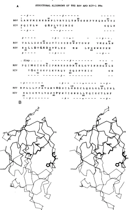
reveals 31 identical residues and 9 additional conservative amino acid substitu- tions. Since the HIV-1 PR has 99 residues, compared to 124for the RSV PR, parts of the latter structure do not correspond to the former. Most of these differences are localized to two surface loops and the flaps which are smaller in the HIV-1 PR (Fig. 1B).
The structural similarity between the two PRs is especially evident around the active site where 44 C , backbone atoms superimpose within a 0.5-A root mean square deviation (11).
This similarity allowed us to use structural models for HIV-1 PR complexed to substrate-based peptide inhibitors (14-19)
to predict amino acid residues in the RSV PR substrate binding pocket. Thirteen residues were observed to be in close proximity to a substrate-based inhibitor in HIV-1 PR and are indicated by bold letters in Fig. lA. Nine of these residues were identical in the RSV PR. Three of these residues were sufficiently different to be candidates that may discriminate between substrates of the two PRs. To test this hypothesis, amino acids found in the HIV PR were introduced into comparable structural positions in the RSV enzyme (Fig. 1B).
Analysis of the activity of these altered RSV PRs on peptide substrates representing known RSV and HIV-1 cleavage sites verified that the candidate residues do influence selection of substrate by PR.
EXPERIMENTAL PROCEDURES
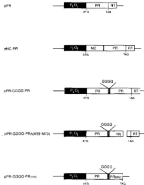
Bacterial Cells and DNA Constructs-The expression vectors used for the mutagenesis, pPR and pNC-PR (Fig. 2), were as previously described (21). They contain, respectively, the RSV protease and the nucleocapsid-protease fusion protein (precursor p23). The viral genes are under the control of a temperature-sensitive X cI repressor. Two strains of Escherichia coli, AR68 and MC1061, were used for cloning and expression. In MC1061 cells, the repressor is supplied in trans from the plasmid pRK24&Its, while in AR68 cells, the repressor is supplied by an integrated copy of the X genome containing cI857. The construction of plasmid pPR-GGGG-PR and details for purification of linked PR dimers are as previously described (28). The plasmid pPR-GGGG-PRA(A59-M73) was created by oligodeoxynucleotide- directed mutagenesis of the parent vector pPR-GGGG-PR. The plas- mid pPR-GGGG-PR(H1V) was constructed by substituting the distal DNA subunit of PR from pPR-GGGG-PR with a DNA fragment encoding the HIV-1 PR from the vector pPROT/RT (a gift from Stuart LeGrice, Case Western Reserve University).
Bacterial Cell Growth and Induction-The bacteria were grown at 30 "C in M9 medium containing 0.5% glucose and 0.5% Casamino acids or in YT media. Expression from the PL promoter was induced by shifting the culture to 42 "C at midlog phase. Cells were collected 3 h postinduction, or as indicated, and protein was analyzed by polyacrylamide gel electrophoresis (27) and immunoblotting (21, 28)
using antisera directed against NC or PR.
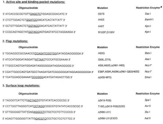
Mutagenesis-Site-directed oligodeoxynucleotide mutagenesis was
used as previously described (21) to introduce amino acid changes (substitutions and/or deletions) into the protease coding region of pPR and pNC-PR. Oligodeoxynucleotides used for mutagenesis are
pPR and pNC-PR (PuuI) and gapped DNA from the expression shown in Fig. 3. The oligodeoxynucleotides were mixed with linearized
vectors ( p P R BglII and BssHII; pNC-PR BssHII and BarnHI). To construct pNC-PR (H65G,R105P,G106V), the mutant plasmid pNC- PR (H65G) was digested with EcoRI and BglII, and the fragment containing the H65G substitution was subcloned into EcoRI/BgZII- digested and purified pNC-PR (R105P,G106V). The same strategy was used to construct pPR (H65G,R105P,G106V) from pPR (H65G) and pPR (R105P,G106V).
Preparation of Retrouiral Proteases-Inclusion bodies were pre- pared from E. coli induced for expression of the RSV PR as previously described (21). The cells were disrupted with EDTA and lysozyme followed by treatment with Triton X-100 or sonication. The pellet was collected and washed by sequential suspension and centrifugation (6-8 times), first with 25% sucrose, 10 mM 2-mercaptoethanol and 25 mM sodium phosphate, pH 7.5, then with 1-2 M urea. The inclusion
bodies were solubilized in 8 M urea (with 150 mM 2-mercaptoethanol), and the protease was refolded by dialysis (0.5-1 mg of protein/ml) a t 23 "C against a decreasing concentration of urea (4,2, and 1 M for 2 h each). The final dialysis (against 25 mM sodium phosphate, pH 7.5, 150 mM NaCl, and 10 mM 2-mercaptoethanol) was overnight. The clarified soluble fraction containing active PR was stored at 4 "C. The preparations were at least 95% pure as determined by SDS- polyacrylamide gel electrophoresis with Coomassie staining (28). Pro- tein concentration was determined using the Bio-Rad Protein Assay Kit (Bio-Rad Laboratories) and bovine serum albumin as a standard. In some experiments, wild type and mutant PR subunits were mixed in 8 M urea and renatured together.
Purified HIV-1 PR was a generous gift from Dr. C. Z. Giam (Dept. of Medicine, Case Western Reserve University). Purified HIV-2 PR was generously provided by S. LeGrice (Dept. of Medicine, Case Western Reserve University). AMV PR, prepared from viral particles by the procedure of Alexander et al. (29), was obtained from Molecular Genetic Resources (Tampa, FL). The RSV and AMV PRs, which differ in primary sequence by two amino acids, are enzymatically indistinguishable and have been used interchangeably in these stud- ies.
The efficiency of refolding of the bacterial derived RSV PR varied from preparation to preparation. Therefore, the specific activity of all refolded enzymes was assessed by the method of Tomasselli et al.
(30), which involves titrating PR concentrations against a nanomolar
or stoichiometric inhibitor and extrapolating to the inhibitor concen-
tration that yields complete loss in activity. The inhibitor used was PPCV(phe-statine)AMTM (31). The RSV recombinant PRs refolded with an efficiency that varied between 20 and 40%, while the HIV PR refolded with close to 100% efficiency.
Peptides-Peptides were synthesized and purified as previously described (28). Lyophilized preparations were dissolved in 1 mM 2- mercaptoethanol. Concentrations were determined by amino acid composition analysis. In addition to the amino acids assumed to interact with the protease, all peptides contained an N-terminal Pro (to prevent reaction with fluorescamine), and 2 C-terminal arginines (to improve solubility). The peptides used in the present study are listed in Table I.
Proteolytic Assay-The assay was based on the reaction of fluores- camine (Pierce) with primary amines produced at the P1' position after cleavage (21). The assay mixture (25 pl) contained 2.4 M NaCl
(1 M for the HIV PRs), 0.1 M sodium phosphate (pH 5.9), 0.2 mM 2- mercaptoethanol, 50-300 nM protease (active dimers according to titration), and 10-400 p~ peptide. The reaction was started by adding the protease and stopped by adding 175 pl of 0.5 M sodium borate (pH 8.5). The incubation time (37 "C) varied from 1 to 45 min. With HIV-1 PR, incubation times longer than 8 min were avoided, as this enzyme had a half-life of approximately 20 min when incubated in the absence of substrate (presumably due to autocatalysis).
The amount of cleaved peptide was determined by adding 20 pl of
0.05% (w/v) fluorescamine, mixing vigorously, and measuring the net
increase in fluorescence intensity a t excitation and emission wave- lengths of 386 and 477 nm, respectively, using a Perkin-Elmer LS- 5B Spectrofluorometer with both excitation and emission slits set at 10 nm. Fluorescence was converted to nanomoles of product formed using a standard curve obtained by reacting fluorescamine with the peptide FQAY (CH,N)PLR. This peptide was used as a standard since
TABLE I
Peptides used as PR substrates
Virus
cleavage site Location of
Peptide sequence"
RSV gagMA-p2 PTSCY-HCGTRR
g a g CA
-
NCb PLIM-AWNRRg a g NC
-
PR PPAVS-
LAMTFdRRp o l PR-RT PATVL
-
TVALRRp o l IN-p4 PLFA-GISDRR
HIV
-
1 g a g CA-NCa PARVL -AEAMRRg a g CA-NCb PATIM-MQRERR
g a g NC
-
p6a PGNF-LQSRRg a g NC
-
p6b PRQAN-FLGKRRp o l RT- IN PRKIL-FLLXRR
Mutational Analysis
ofa
Retroviral Protease
it was found that the reaction of fluorescamine with short peptides was somewhat different from the reaction with free amino acids or proteins. The presence of high concentrations of peptides quenched the fluorescence readings. Measurements made in the presence of 200
p~ peptide or above were therefore corrected for quenching: 7.5% for 200 phi and proportionally more at higher peptide concentrations. The extent of quenching was found to be approximately the same with all substrates used in this study. All assays were performed in triplicate using several independent PR preparations. The standard deviation was less than 10%. Correct cleavage of selected substrates was verified by HPLC separation of products and amino acid com- position analysis. Two of the HIV-1 peptides (Table I) contain lysine residues with amino groups. The pK. values of these groups are approximately 10.5 so that at pH 8.5 they are mostly protonated and therefore do not react efficiently with the fluorescamine. However, an increase in the background fluorescence of these substrates of 2- to 3-fold over peptides without lysine residues was noted. Nonethe- less, activity measurements reported here were at least 10-fold above background fluorescence in each case.
Kinetic Anulysis-For each substrate peptide, 5-7 different concen- trations were tested and the reaction was stopped before 20% of the substrate was cleaved. Within that limit, the reaction was shown to
be linear with time, implying that the turnover could be used as a measure of initial velocity. Kinetic parameters were determined by fitting the velocity versus substrate concentration data to the Mi- chaelis-Menten equation using the program NFIT (Island Products, Galveston, TX). The R2 values were greater than 0.99. Standard deviations for the K,,, values were less than 20% and for the kc, values less than 10%.
Zmmunoblotting-Proteins from uninduced E. coli extracts or from
E. coli induced to express NC-PR fusion protein were separated on SDS-18% polyacrylamide gels (28), visualized, and transferred to membranes as previously described (28). The membranes were incu- bated with precleared rabbit anti-RSV PR serum (diluted 1/5000), washed, and incubated with biotinylated goat anti-rabbit antibody followed by the Vectastain Elite reagent, and the peroxidase reaction was visualized using Indophane blue. Where indicated, the mem- branes were incubated with precleared rabbit anti-RSV NC or PR serum in phosphate-buffered saline containing 0.1% Tween 20 and 1% bovine serum albumin for 1 h at 37 "C. After washing twice with phosphate-buffered saline containing 0.1% Tween 20, the membranes were incubated with 20-50 pCi of lZ6I-labeled protein G (Amersham) in phosphate-buffered saline containing 0.1% Tween 20 and 1% bovine serum albumin for 1 h at room temperature. The membranes were then washed three times as above, air-dried, and exposed to Kodak X-OMAT AR-5 x-ray film at -70 "C with an intensifying screen for 10-20 h.
Molecular Modeling-The crystal structures of the native RSV PR (11) and HIV-1 PR, as well as of the complexes of the latter with inhibitorb) (14-19), were examined on an Evans and Sutherland PS390 computer graphics system using the program FRODO (32). The residues forming the subsites were determined directly for the HIV-1 PR complexed with inhibitors, and the corresponding residues in RSV PR were obtained from the structural and sequence align- ments. The structural superimposition of the C, atoms from the subunits of RSV and HIV-1 PRs (33, 34) was used to deduce the residues to be deleted in the extended surface loops of RSV PR and in the flaps.
RESULTS
Rationale for PR Mutations
The superimposed crystal structures of HIV-1 and RSV PRs were compared in order to locate regions that would be expected t o influence substrate binding and catalysis or to have other interactions with the polyprotein precursors or
other mature viral proteins. The active site residues of the two PRs are found in virtually identical conformations. Much of their subunit structure is very similar as well, despite the fact that the two enzymes differ in specificity and rates of catalysis. Their role in regulation of the viral life cycle may also differ since the RSV P R is present in both the gag and gag-pol precursors and thus may have other functions in addition to proteolytic cleavage of precursors. Based upon this structural analysis, a series of mutations in (a) the active
site, ( b ) the substrate binding pocket, and (c) on the protein surface have been designed to explore structure/function re- lationships of the PR and its role in viral replication. Fig. 1B
shows the location of the altered amino acids and structures. It should be noted that the retroviral PR is a homodimer. Thus, when mutations are introduced into the monomeric subunit, they are de facto double mutations in the native PR. This is true except for the mutations placed into a P R in which the subunits are covalently linked.
Actiue Site Mutants-Several types of active site mutations have been constructed. These include substituting serines for the catalytic aspartic acid residues (D37) to prove that these residues are essential for catalysis. Serine was chosen since it was not expected to drastically alter the structure of the catalytic site. Serine and threonine were also substituted for alanine 40. This alanine residue is highly conserved in all retroviral PRs. In the closely related nonviral aspartic pro- teases, serine and threonine are conserved in the correspond- ing positions. It has been suggested that this difference may account for the lower pH optimum of the cellular enzymes (35, 36).
An important feature of the active site is the flap structure, a n extended surface loop that lies over the substrate binding region (Fig. 1B). It is present in all aspartic proteases and may affect substrate binding and/or catalysis. In retroviral PRs there are two flaps, one per subunit in the dimer, in contrast to the single flap of nonviral aspartic proteases. However, the flap sizes are not conserved. For instance, the RSV P R flaps contain three more amino acids than the HIV-
1 P R flaps. RSV PR mutants were constructed which con- tained either a single flap or which substituted the smaller HIV-1 flaps for those found in the RSV PR. The flaps are also characterized by a series of conserved glycines, which are presumed to provide flexibility to the loop conformation. These residues may also have a steric role in binding sub- strate. In order to test the importance of these residues, glycine 69 and 70 were mutated to bulkier leucine residues.
RSV Specificity Mutants-The substrate binding region of the retroviral protease was initially defined by modeling a peptide substrate based on the position of the inhibitor in the co-crystal structure of rhizopuspepsin with a reduced peptide inhibitor (25, 33). This was later substantiated by examina- tion of the crystal structures of HIV-1 P R with inhibitors (14-19). This analysis indicated that the peptide backbones
9484
Mutational
Analysis ofa Retroviral Protease
A STRUCTURAL ALIGNMENT OF THE RSV AND HIV-1 PRs
"
8 "
1 10
" _
- 8 " " -" _ "
RSV L A M T M E H K D R P L V R V I L T N T G S H P V K Q R S V Y I
HIV P Q I T L W Q R P F o V T I R I G G Q L K
20 3 0
1
"
8 " " - 8 " " " "
8 " " - 8 - " e - - "
8 " .
RSV T A L L D S G A D I T I I S E E D % P T D W P V M E A A N
HIV E A L L D T G A D D T V L E E M N L P G K W K P K M
40 60
R " " " 8 " - " 8 "
20 30 40
.
f l a p. . . .
" " " 8 " " " -" _
-
RSV P Q I H G I G G G I P M R K S R D M I E L G V I N R D G S L E
HIV I G G I G G F I K V R Q Y D Q I P V E I C G H
70 BO 80
50
" " " " "
60
p - " " " " "
- 8 " " " - 8 "
" - ~ " "
"1M)
R -
RSV R P L L L F P A V A M V R G S I L G R D C L Q G L G L R L T N L
HIV K A I G T V L V G P T P V N I I G R N L L T Q I G C T L N F
110 120
70
"
8 0
8 " " " - 8 -
" _
90 ol"" "B
R -
"
,..?
V
r
..(
. .
....?
FIG. 1. Structure and sequence comparison of the RSV and HIV-1 PRs. A , structural alignment of the
RSV and HIV-1 PRs. The numbers of amino acids for the RSV and HIV-1 PRS are indicated. Secondary structure elements (CY helices and p structure) are indicated above and below the sequences for the RSV and HIV-1 PRs,
respectively. Thirteen amino acid residues of HIV-1 PR with side chains in close proximity to a substrate-based inhibitor are shown in bold letters. Of these, four are different in RSV (also shown in bold letters). B , tracing of the
C, backbone of the RSV PR showing sites of mutations. The two subunits of the RSV PR are drawn in the medium
line (top) and thin line (bottom). Residues belonging to the flap were modeled in the top subunit only. This modeling was for illustrative purposes only and may not represent the true structure. Mutations and deletions discussed in this paper are identified in the top subunit only. The deletions are shown in dashed lines. Amino acid substitution mutations next to the deletions are not shown separately. Side chains of the mutated residues, or main chain atoms for glycines, are shown in thick lines. More detail about the mutations is provided in Fig. 3.
ing R105P and G106V, and one containing all three substi- tutions.
Surface Loops-The other obvious structural difference be- tween the two enzymes is that the RSV PR has two extended surface loops from residues 19 to 28 (loop A) and from 86 to 90 (loop B), compared to shorter loops present in HIV PR. Many other retroviral PRs show the shorter loops character- istic of HIV-1 PR at these two regions (37). Exceptions are bovine leukemia virus and human T-cell leukemia virus,
dues were removed and substitutions were made to include glycine at the predicted turn regions as occurs in HIV-1 PR.
Preparation of Mutant PR and PR-containing Precursor Fragments
Fig. 2 shows a map of the region in RSV DNA that contains the PR coding sequences. The indicated fragments were cloned in an E. coli plasmid vector downstream from the bacteriophage X PL promoter and an ATG initiator codon (21). The pPR clone expresses a protein that is predicted to be identical with the viral PR with the exception of the initiator methionine (which is often removed in bacterial cells). The pNC-PR expresses a protein that contains PR upstream from the NC sequences as normally occurs in the viral gag precursor protein. The pPR-GGGG-PR clone ex- presses a PR dimer linked by 4 glycine residues. The plasmid expression clones were subjected to site-directed mutagenesis, as described under “Experimental Procedures,” to introduce the mutations listed in Fig. 3.
Expression and Analysis of Mutant PRs in Bacterial Extracts
We have shown previously that the NC-PR protein pro- duced by the clone pNC-PR described in Fig. 2 processes itself in E. coli to form mature NC and PR products (38). Fig. 4 shows a comparison of products observed in extracts of bac- teria that express NC-PR precursor fragments with different catalytic site mutations. The viral proteins were detected by immunoblot analysis using an anti-PR serum. Extracts of cells that express the wild type precursor fragment contained both NC-PR and PR, as expected. The same two proteins were detected in the extracts from cells expressing the NC- PR(A40S) mutant, although in this case there appeared to be
PPR
*TO TAB
PNC PR NC I PR I RT
A T 0 T I 0
pPR-GGGG~PR
A m TAG
GGGG . pPR-GGGG-PRb(A59 M731
e
P R ‘ L I Q+A T 0 ,A0
GGGG
pPR-GGGG~PR:w PR IPR+
A T 0 TAB
FIG. 2. Molecular clones of PR and NC-PR. pPR and pNC- PR are E. coli expression clones describedpreviously (21) that contain the viral PR or nucleocapsid and protease (NC-PR) genes, respec- tively. The numbers correspond to the Rous sarcoma virus sequence.
PLOL is the leftward promoter derived from X phage DNA. pPR-
GGGG-PR is an E. coli expression clone described previously (28) that contains a covalently linked PR dimer. GGGG (hatched box) indicates a 4-glycine linker that fuses the two PR subunits. pPR-GGGG- PRAfA59-M73) is an expression plasmid that contains a covalently linked PR with a truncated flap in the indicated subunit. pPR-GGGG- PR(HZV) is an expression plasmid containing a covalently linked PR dimer with one RSV and one HIV subunit. The initiation codon provided by the expression vector is indicated (ATG). The genetically engineered amber stop codon (TAG) is shown at the end of the PR domain.
less of the PR product made. Free PR was not detected in cells expressing the other mutant proteins, including A40T and D37S. In these cases, there was some antibody-reactive protein in intermediate positions that probably reflects diges- tion by bacterial proteases. We also note that the mutant NC- PR proteins migrate slightly differently from the wild type protein in this gel system.
Plusticity of the PR Substrate Binding Pocket
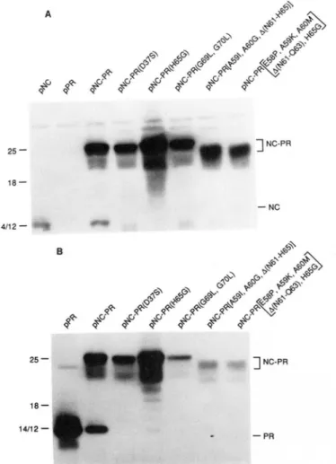
A similar analysis was carried out with mutant PRs con- taining substitutions and/or deletions in the flap region of the RSV PR. This region appears to be very sensitive to mutation as indicated by the results from bacterial lysates shown in Fig. 5. Substitutions at positions 65, 69, and 70, exchange of the HIV-1 PR flaps for those in RSV, or removal of one of the RSV flaps (pPR-GGGG-PRA(A59-M73) in Fig. 2) produced inactive PRs. Also, a mosaic PR (pPR-GGGG- PR(H1V) in Fig. 2) containing one HIV-1 and one RSV subunit was inactive (data not shown).
The enzymatic studies of the RSV PR(H65G) mutant were of particular interest. When substituted singly, glycine at position 65 produces an RSV enzyme which was inactive on the RSV NC-PR precursor. However, if this mutation was combined with two other RSV to HIV-1 PR amino acid exchanges, R105P and Gl06V, activity on this substrate was restored (Fig. 6). The RSV PR mutant containing only R105P,G106V was active. These results indicate that unfa- vorable conformational changes in the flaps introduced by H65G can be compensated by additional changes in the sub- strate binding pocket.
Purification of Mutant PRs
While analysis of cleavage of the NC-PR precursor in bacterial extracts provides a rapid screen for mutations that change PR activity, accurate quantitation of these effects is not possible. To facilitate such studies, selected PR mutants were expressed as the free enzyme and purified from bacterial extracts. With the exception of HIV-2 PR, all of the proteins were recovered from the inclusion body fraction. The PRs were solubilized by suspension in urea and renatured by sequential dialysis. A RSV wild type PR prepared by this procedure had the same substrate preference as the PR puri- fied from AMV. However, the efficiency of refolding varied from preparation to preparation. In order to correct for dif- ferences in estimates of the specific activities of the different PR preparations, renatured PRs were titrated with a sub- strate-based inhibitor, and the activities were related to the number of active dimers as described under “Experimental Procedures.” The titration experiments indicated that with the better bacterial-derived preparations 30-40% of the pro- tein was folded correctly.
Activity of PR Mutants
9486
Mutational Analysis
of
a Retroviral Protease
1. Active site and bindina DOCket mutations;
Ollgonucleotlde
5’ ATCACCGCGCTGTTC&CKTGGAGCGGACATC 3’
5’ CTGTTGGACTCTGGATCCGACATCACTATTATT 3’
5’ GCTGTTGGACTCTGGTACCGACATCACTATTATT 3’
SCCGCAGTAGCTATmAGTGAGTATCCTAGGAAGA3’
2. FlaD mutations:
Ollgonucleotlde
STGGAGGCCGCGAACCCCCAGATCGGTGGGATAGGAGGGGGA 3’
5’ ATCCATGGGATAGGATT-CCCATGCGAAAA 3’
STTGGCCAGTGATGGAGATAGGGGGGATAGGAGGGGGY
5’ CGATTGGCCAGTGATGCCTAAGATGATCGGGGGGATAGGAGGGGGGA 3’
5’ TGATGGAGCGAAAATCCCGGGACATGATAGAGTTGG 3’
3. Surface loor, mutations:
Ollgonucleollde
5’ TAGGGTCATTCTGASZGIGTGTATATCACCGCGC 3’
5’ CCTTGGTTAGGGTCATCCTAGGCGGCGTGTATATCACCGCGC 3’
5’GTTGGGGGTTATTGAGAGG.‘XICTGCTCCTCTTCCCCG3’ 5’AGAGTTGGGGGTTATTG.G?&GECCCTGCTCCTCTTCC 3’
Mutallon Restrlctlon Enzyme x
D37s Sac I
A40S BamH I
A40T Kpn I
R105P.Gl06V Kpn I
Mutallon Restrlctlon Enzyme
H65G Bstx I
G69L.G70L Ase I
A591,A60G.A(N61-H65) Nco 1.
E%tP,A59K,A60Mb(N61-063)H65G NCO I.
A(A59M73) Sma I
Mutation Reslrlctlon Enzyme
A(N19.RZS) spel
TlSG,A(N19-R2S)S29G Avrll
A(NS6.L91) stu I
A(NS6-L91)E92G Aat I1
ti A new restriction site was introduced in each oligonucleotide (see undelined nucleotides) to allow srceening of mutant PR-containing plasmids. Loss of the original Nco I restriction site.
FIG. 3. RSV PR mutations. Oligodeoxynucleotides used to place mutations into the pPR and pNC-PR plasmids by site-directed mutagenesis are as indicated. New restriction enzyme sites (underlined) were introduced to facilitate identification of mutated clones. Mutations are listed by the amino acid and position number of the RSV PR followed by the substituted amino acid. Deletion mutations are indicated by A followed by the position number of the first and last deleted amino acids.
18.4
-
14.3-
3 NC-PR
FIG. 4. Effect of active site mutations o n PR activity. Im- munoblotting of cell lysates from induced bacterial cultures express-
ing NC-PR and NC-PR mutant proteins containing NC-PR, NC-
PR(D37S), and NC-PR(A40S or A40T). E. coli were grown a t 30 “C in minimal medium to an ODm of 0.8 and then a t 42 “C for 2 or 4 h to induce expression of the cloned DNAs. Samples of bacteria from uninduced and induced cultures were fractionated on an 18% poly-
acrylamide-SDS gel. AMV PR was included as a marker. After
electrophoresis, proteins were transferred to an Immobilon membrane and PR was detected using rabbit anti-PR serum and peroxidase staining as described under “Experimental Procedures.” The numbers
above the lanes indicate time (h) of incubation a t the induction temperature. * denotes position of PR where applicable.
peared to be slightly more active than wild type a t pH values below 5.5 (Fig. 7).
No activity was detected with the purified D37S, A40T, H65G, and G69L, G70L PRs (Table 11). When the denatured D37S, A40T and G69L, G70L proteins were mixed with equimolar or twice equimolar amounts of denatured wild type AMV PR, respectively, and refolded as described under “EX- perimental Procedures,” the amount of active wild type PR
activity recovered was as predicted for a binomial distribution of the various dimer forms (Table 111). This indicates that the mutant subunits were able to form stoichiometric complexes with those of wild type protein. Therefore, we conclude that their lack of activity is not due to incorrect folding.
Activity of PR Mutants with Modified Surface Loops
Mutant PRs with one or two of the surface loops shortened were also purified. If one of the loops was shortened, then the mutant exhibited only 2 to 3% of the activity associated with the wild type RSV PR (Table 11). If both of the loops were shortened, no activity was detected. When introduced into an infectious virus DNA clone, the amount of residual activity found in these “loopless” PRs was insufficient to support normal processing of gag precursor polypeptides in uiuo, and infectious progeny virions were thus not obtained (data not shown).
Mutational Analysis
a Retroviral Protease
9487
TABLE I1
25
-
18
-
14/12
-
25
-
10
-
1412-
] NC-PR
-
NCB
9.“
] NC-PR
-
PRFIG. 5. Effect of flap mutations on PR activity. Immunoblot analysis of NC-PR proteins containing flap mutations in the PR and expressed in E. coli. Cell lysates were fractionated on a SDS-15% polyacrylamide gel, and the virus-related proteins were detected with rabbit antiserum directed against the NC (panel A ) or the PR (panel B ) with ’251-labeled protein G as described under “Experimental Procedures.” Molecular size markers in kilodaltons are indicated on the left. The positions of NC-PR and PR are indicated on the right.
~ ”.
43-
25
-
+ NC-PR18-
14112-
6-
FIG. 6. Effect of substrate binding pocket mutations on PR activity. Immunoblot analysis of NC-PR proteins containing sub- strate binding pocket mutations and expressed in E. coli. Cell lysates were analyzed as described in the legend to Fig. 5 except that only anti-PR serum was used to detect proteins.
Activities of RSVproteases with active site or structural mutations
PR mutation Activity”
% recombinant wild type
<0.1 23 <0.1 <0.1
<o.
1 D37SA40S A40T G69L,G70L H65G
ALoop A 3
AT.nnn R 2
‘Activity was determined as described under “Experimental Pro- cedures” using mutated RSV PRs as indicated and the RSV NC-PR peptide substrate. Activity of the wild type PR is 575 nmol of peptide cleaved/min/mg of enzyme.
100
-
80
-
2
6 0 --
-
:
% 4 0 -
a
20
-
I
0 . , . , . , . , . , . , . I
2 3 4 5 6 7 8 9
J
=e;
pH
FIG. 7. The pH dependence of AMV and A40S RSV PR.
AMV PR (46 ng) or RSV PR(A40S) (150 ng) was incubated with the NC-PR peptide substrate using 100 mM citrate for pH 3-7 and 100 mM HEPES for pH 7.9 instead of sodium phosphate buffer. The rate of cleavage was determined as described under “Experimental Pro-
cedures.” 100% activity for the AMV and RSV A40S PRs is defined
as 584 and 81 pmol of peptide cleaved, respectively, in 30 min.
El,
AMV PR,
.,
RSV PR(A40S).TABLE 111
Renaturation mixing experiments with RSV and mutant PR subunits
Ratio of wild
subunits
PR added” type to mutant PR activityb
Wild type (WT) PR(A40T) PR(G69L,G70L) PR(D37S)
WT
+
PR(A40T)WT
+
PR(A40T)W T
+
PR(G69L,G70L)W T
+
PR(G69L,G70L)W T
+
PR(D37S)WT
+
PR(D37S)nmoll90 min
2.1
0.0 0.0
0.0
1:l 0.42
1:2 0.27
1:l 0.77
1:2 0.29
1:l 0.41
1:2 0.21
a Wild-type RSV and mutant PR subunits were solubilized from
the inclusion body fraction with 8 M urea and renatured either alone or in mixtures of wild type and mutant subunits (25-45 pg/ml) as described under “Experimental Procedures.” The molar ratio of the wild type to the mutant subunits is as indicated.
*The activity of the PR preparations containing in each case 0.6 ng of wild type subunit and variable amounts of the mutant subunits, as indicated, were assayed for cleavage of the NC-PR peptide sub- strate as described under “Experimental Procedures.”
wild type on RSV substrates, but was more active than the wild type on the HIV-1 RT-IN substrate. If the amount of activity observed with the wild type and mutant enzymes for
the HIV-1 RT-IN substrate is normalized to that of the
RSV NC-PR substrate, then the R105P,G106V and the
9488 Mutational Analysis of a Retroviral Protease
TABLE IV
Activity of wild type and mutant enzymes on peptide substrates containinz RSVand HIV-1 cleavaee sites
Protease activity’
Substrate RSV
AMV (R105P, (H65G,R105P, HIV-1 HIV-2 RSV
G106V) G106V)
RSV-based substrates*
MA-p2 0.7 0.8 0.2 4.2 6.5
CA-NCb
NC-PR PR-RT
IN-p4 2.6 2.9 1.2 3.0 2.2
7.9 6.4 1.7 13.0 10.9
14.9 15.1 4.3 41.0 25.4
12.0 10.7 3.0 7.3 3.4
HIV-1-based substrates*
CA-NCa 0.6 0.4 0.2 114.5 64.8
CA-NCb 0.2 0.2 0.1 281.1 195.0
NC-p6a 0.0 0.0 0.0 151.7 121.8
0.0 0.0 0.0 51.0 52.7
0.5 4.7 2.8 124.5 166.4
NC-p6b
RT-IN
‘The PR activity was determined by the fluorescamine reaction as describedunder “Experimental Procedures.” The activity is the initial rate of turnover in the presence of 100 P M substrate measured as
molecules of product formed per active enzyme dimer per min.
The sequences are listed in Table I.
TABLE V
Kinetic measurements with NC-PR (RSV) and RT-IN (HIV-1) peptide substrates
Kinetic values were obtained from Michaelis-Menten plots of the initial velocity versus substrate concentration. Activity was deter- mined using the fluorescamine assay as described under “Experimen- tal Procedures.”
Source of PR Substrate K , kt k,,/K,
p~ min” p ~min” ’
AMV RSV NC-PR 49 21.6 0.45
RSV(R105P,G106V) RSV(H65G,Rl05P,GlOSV)
45 19.9 0.44
77 6.1 0.08
HIV-1 16 43.8 2.77
HIV-2 23 29.7 1.29
AMV HIV-1 RT-IN 98 1.3 0.01
RSV(R105P,G106V) 100 11.6 0.12
RSV(H65G,RlO5P,GlO6V) 95 5.5 0.06
HIV-1 37 170.1 4.65
HIV-2 42 221.4 5.27
separation of the products and amino acid composition analy- sis (data not shown). Little or no activity for the other four HIV-1 substrates was detected with wild type or mutant PRs (Table IV)
.
As a control, each of the peptide substrates was incubated with either the purified HIV-1 or HIV-2 PRs. Each HIV-1 substrate was rapidly cleaved by both HIV enzymes with rates that were 10- to 20-fold greater than that observed with the RSV PR for its homologous substrates. While the RSV and AMV PRs generally cleaved HIV substrates very inefficiently, the HIV PRs cleaved the RSV substrates with rates compa- rable to that of the RSV enzyme. The best RSV substrate for the AMV PR (the NC-PR peptide) was also the best RSV substrate for the HIV enzymes.
Kinetic Analysis of Activity on the RSV NC-PR and the
HIV-1 RT-IN Substrates
To determine the nature of the differences in activity between the mutant and wild type enzymes, a steady state kinetic analysis was carried out using the RSV NC-PR and the HIV-1 RT-IN peptide substrates (Table V). The wild type AMV and HIV PRs had K , values for their respective ho- mologous substrates in the 16-50 p~ range. Interestingly, the
HIV PRs bound the RSV substrate more efficiently than its own homologous substrate. This is in contrast to the AMV PRs which bound the heterologous substrate less efficiently. The binding affinities of the RSV R105P,G106V and H65G,R105P,G106V mutants for the HIV-1 RT-IN substrate were the same as wild type PR. Thus, the increased activity observed with the mutant PRs for this substrate is solely an effect of kcat. A comparison of the efficiency (kat/K,) of the R105P,G106Vand the H65G,R105P,G106V PRs for the RSV NC-PR or the HIV-1 RT-IN peptide substrates indicated that the mutants were 12- and 6-fold, respectively, more efficient on the HIV substrate than wild type PR. If one normalizes to the amount of activity that is observed with the RSV NC-PR peptide substrate, then the relative efficiencies calculated for the R105P,G106V and the H65G,R105P,G106V PRs are 12-
and 34-fold, respectively, more efficient than wild type. These results indicate that introduction of amino acids found in the substrate binding pockets of HIV-1 PR into the RSV PR confers increased relative specificity for an HIV-1 substrate.
DISCUSSION
The results of our mutational studies verify that the amino acids in and around the catalytic site triad and in the flap region of the RSV PR are essential for enzymatic activity. Consistent with previous studies on avian and human retro- viruses (20-23), substitution of the catalytic aspartate (D37) results in an inactive PR. However, the D37S mutant may be useful for biochemical and structural studies of PR since its overall structure should not be altered. This prediction is supported by our observation that when refolded together, D37S PR subunits can form stoichiometric complexes with wild type PR subunits. Nonconservative substitutions, such as D371, cause conformational distortion of the PR (20). Craven et al. (41) also have shown that release of RT from a
gag-pol precursor polypeptide containing PR mutations by exogenously added PR occurs efficiently when the mutation in the precursor is D37S but not when it is D371. Again, this suggests that the latter mutation causes distortions in the polypeptide structure.
The catalytic aspartate (D37) residues are part of a rigid structure, held in place by an intricate network of hydrogen bonds which are formed with amino acids 38 and 39, as well as with the 37-39 active site triad from the other subunit. This unique structure is highly conserved in all aspartic proteases (1). In retroviral PRs, an alanine residue always follows the active site triad. In related cellular proteases, serine is found at this position in the amino domain, whereas threonine is predominantly present in the carboxyl domain of the enzyme. The latter is infrequently substituted by serine and in one case, that of human renin, by alanine (39). Sielecki
et al. (35) suggested that the presence of alanine rather than serine at this position may account for the higher pH optimum of human renin compared to other cellular aspartyl proteases, since serine or threonine, but not alanine of renin could form a hydrogen bond with the catalytic aspartate. Mantafounis and Pitts (36) have made the substitution T218A in chymosin
the A40S substitution can take place without steric hindrance while that of the A40T cannot (21). Thus the latter mutation is expected to cause some movement or rearrangement in the backbone atoms near the catalytic site which, presumably, is unfavorable for binding or catalysis. Loeb et al. (20) have introduced 4 amino acid substitutions at the same conserved alanine in the HIV-1 PR. In contrast to the results described here, none of these substitutions, including serine, produced an active enzyme. The difference between our results and those of Loeb et al. (20) may reflect inherent differences between the two PRs or may be due to their use of a less sensitive assay, which followed precursor cleavage in bacterial extracts.
One of the interesting regions of the PR molecule contains the flaps. Residues in this region interact with the peptide substrates and may exclude water molecules from the catalytic site. Unfortunately, structural information about the flaps in the RSV PR is incomplete due to disorder in the crystal structure (11). The structure of the flaps in HIV-1 PR is known and they have been shown to move as much as 7
A
and to change orientation upon substrate binding (14-16). Comparison of the amino acid sequence of the flaps of RSV to that in the HIV-1 PR shows that those in RSV contain 3
additional residues (37). Loeb et al. (20) have reported that amino acid substitutions in the HIV-1 flap region usually inactivate the PR. Our mutational analysis yielded similar results. Replacement of the longer RSV flaps with the shorter ones from HIV-1, removal of one of the two RSV flaps (data not shown), construction of a mosaic enzyme with RSV and HIV-1 PR subunits, and replacing glycines at positions 69 and 70 with leucines or histidine at position 65 with glycine, all produced inactive enzymes. This last result was not pre- dicted since substitutions at glycine 48 (the structurally iden- tical position in HIV-1 PR to RSV PR H65) with histidine, or 4 other amino acids, produced active enzymes (20).
Avian retroviral PRs have two extended external loops, one of which is larger than any comparable loop found in mam- malian retroviral PRs. The presence of these loops in the RSV PR may serve a possible structural role in virion core assembly since, unlike HIV or Moloney leukemia virus, the avian retrovirus PR is synthesized as part of both gag and gag-pol precursors and can be purified from virions in amounts equal to that of the capsid structural proteins. Thus, we hypothesized that removal of the surface loops of RSV may leave an enzymatically active protein which may exhibit de- fects in formation of mature virus particles. The analysis presented here indicates that the function of these structures is more complex than first supposed. When the surface loops were shortened individually, a nearly inactive enzyme re- sulted. When the mutation was placed into an infectious virus DNA clone, the level of residual PR activity was insufficient to support processing of viral precursor polypeptides in vivo
(data not shown). It should be noted that these constructs contained substitutions that placed amino acids found at the turns of the smaller HIV-1 PR surface loops. Without these substitutions, the singly deleted surface loop mutants were completely inactive. Thus, while surface loops of RSV PR can be removed and a partially active PR obtained, the enzymes are very defective. There are several possible explanations for these results. First, A(N86-L91)E92G removes E92 which forms a salt bridge with the N terminus of the second subunit and is thus important for stabilizing the dimer structure of the PR. Elimination of a salt bridge in the linked dimers reduces catalytic activity (28). Second, amino acids in loop A
form several hydrogen bond interactions with residues at the base of the flap and removal of these may also reduce activity.
Third, removal of the surface loops exposes amino acids normally buried beneath the surface, and this could create a distortion of the polypeptide backbone which might interfere with catalytic activity. Finally, there may be some problem with folding of this bacterially derived mutant PR. Thus, new constructs which introduce more limited alterations into the surface loops will have to be analyzed before any conclusion of a structural role for these loops can be made.
Several amino acids found in the HIV-1 PR in close prox- imity to a bound peptide-based inhibitor were exchanged for amino acids in comparable positions in the RSV PR. These mutations were designed to identify amino acid residues that may be important determinants for substrate specificity. Three constructs were made, RSV PR(H65G), RSV PR(R105P,G106V), and RSV PR(H65G,R105P, G106V). The H65G exchange produced an inactive enzyme. Glycine 48 in the HIV-1 PR is involved in forming hydrogen bonds to the peptide backbone of the inhibitor. The substitution of glycine for H65 in RSV PR may have induced conformational changes in the flaps which interfere with interactions with substrate.
In contrast, the RSV PR(R105P,G106V) was active. This is significant since the amino acids found in the comparable structural positions of HIV-1 PR, P81 and V82, respectively, are singly very sensitive to amino acid substitution (20). Four amino acid substitutions for P81, including arginine, resulted in inactive enzymes. Some substitutions of HIV-1 V82, how- ever, appeared to be tolerated, although substitution of gly- cine, the amino acid found in RSV PR, was not (20). These results highlight the need to make multiple amino acid sub- stitutions in the substrate binding pocket to understand their significance. This is illustrated again with the striking obser- vation that combining the H65G substitution, which inacti- vated the RSV PR, with the 105 and 106 substitutions partly restored activity to the RSV PR.
Examination of the substrate specificity of the RSV PR(R105P,G106V) and RSV PR(H65G,R105P,G106V) indi- cated that there was a significant increase in activity toward the HIV-1 RT-IN peptide substrate with both enzymes. A
steady state kinetic analysis of these PRs indicated that the increase was due solely to an increase in catalytic rate; there was no appreciable effect on the binding of substrate. From these results, we conclude that the substitution of HIV amino acid residues found in positions comparable to RSV PR 105 and 106 and at 105, 106, and 65 imparts the ability to cleave a HIV substrate. It should be noted, however, that four other HIV-1 substrates were either not cleaved or were cleaved at low rates by the wild type and the mutant RSV PRs. The reason for inducing only partial HIV-1 PR-like behavior in the RSV PR mutants is provided by an analysis of NC-PR cleavage site substrates with single amino acid substitutions at the P4 to P3’ positions in the accompanying manuscript (40).
Acknowledgments-We thank Dr. Stuart LeGrice, Case Western Reserve University, for purified HIV-2 PR, Dr. Joe Giam, Case Western Reserve University, for inclusion bodies containing HIV-1 PR, Dr. Ed Houts, Molecular Genetics Resource, for AMV PR, Dr. Terry Rosenberry, Case Western Reserve University, for amino acid composition analysis, and Dr. Terry Copeland, NCI-Frederick Basic Cancer Research Center for synthesis of peptides.
Note Added in Proof-Ido et al. (Ido, E., He-ping, H., Kezdy, F. J., and Tang, J. (1991) J. Bwl. Chem. 266,24359-24366) have reported
9490
Mutational Analysis
ofa Retroviral Protease
1. 2. 3.
4. 5.
6.
7.
8.
9.
10.
11.
12.
13.
14.
15.
16.
17.
18.
19.
REFERENCES Skalka, A. (1989) Cell 56,911-913
Oroszlan, S., and Tozser, J. (1990) Semin. Virol. 1 , 369-378 Kohl, N., Emini, E., Schleif, W., and Davis, L. (1988) Proc. Natl.
Crawford, S., and Goff, S. (1985) J. Virol. 5 3 , 899-907
Gottlinger, H., Sodroski, J., and Haseltine, W. (1989) Proc. Natl. Acad. Sci. U. S. A. 86,5781-5785
Dickson, C., Eisenman, R., Fan, H., Hunter, E., and Teich, N.
(1984) in RNA Tumor Viruses (Weiss, R., Teich, N., Varmus,
H., and Coffin, J., eds) Vol. 1, pp. 513-648, Cold Spring Harbor Laboratory Press, Cold Spring Harbor, NY
Kotler, M., Katz, R., Danho, W., Leis, J., and Skalka, A. (1989)
Proc. Natl. Acad. Sci. U. S. A. 85,4185-4189
Kotler, M., Danho, W., Katz, R., Leis, J., and Skalka, A. (1989)
J. Biol. Chem. 264,3428-3435
Darke, P., Nutt, R., Brady, S., Garsky, V., Ciccarone, T., Leu, C.-
T., Lumma, P., Freidinger, R., Veber, D., and Signal, I. (1988)
Biochem. Biophys. Res. Commun. 156,297-303
Miller, M., Jaskolski, M., Rao, M., Leis, J., and Wlodawer, A.
(1989) Nature 3 3 7 , 576-579
Jaskolski, M., Miller, M., Rao, J., Leis, J., and Wlodawer, J.
(1990) Biochemistry 29,5889-5898
Lapatto, R., Blundell, T., Hemmings, A., Overington, J., Wild- erspin, A., Wood,
s.,
Merson, J., Whittle, P., Danley, D., Gerghegan, K., Havrylik, S., Lee, S., Scheld, K., and Hobart,P. (1989) Nature 3 4 2 , 299-302
Wlodawer, A., Miller, M., Jaskolski, M., Sathyanarayana, B., Baldwin, E., Weber, I., Selk, L., Clawson, L., Schneider, J., and Kent, S. (1989) Science 245,616-621
Fitzgerald, P., McKeever, B., van Middlesworth, J., Springer, J., Heimback, J., Leu, C.-T., Herber, W., Dixon, R., and Darke, P.
(1990) J. Biol. Chem. 2 6 5 , 14209-14219
Erickson, J., Neidhart, D., Van Drie, J., Kempf, D., Wang, X.,
Norbeck, D., Plattner, J., Rittenhouse, J., Turon, M., Wide- burg, N., Kohlbrenner, W., Simmer, R., Helfrich, R., Paul, D., and Knigge, M. (1990) Science 249,527-533
Miller, M., Sathyanarayana, B., Toth, M., Marshall, G., Clawson, L., Selk, L., Schneider, J., Kent, S., and Wlodawer, A. (1989)
Science 246,1149-1152
Swain, A., Miller, M., Green, J., Rich, D., Schneider, J., Kent, S., and Wlodawer, A. (1990) Proc. Natl. Acad. Sci. U. S. A. 8 7 ,
Jaskolski, M., Tomasselli, A., Sawyer, T., Staples, D., Heinrikson,
R., Schneider, J., Kent, S., and Wlodawer, A. (1991) Biochem-
Wlodawer, A., Miller, M., Swain, A., and Jaskolski, M. (1991) in
Methods in Protein Sequence Analysis (Jornvall, H., Hoog, J., Gustavsson, A., eds) pp. 215-221, Verlag Wepf und Co., Base1
Acad. Sci. U. S. A. 85,4185-4189
8805-8809
istry 30,1600-1609
20.
21.
22.
23.
24.
25.
26.
27. 28.
29.
30.
31.
32. 33.
34. 35.
36. 37. 38.
39.
40.
41.
42.
Loeb, D., Swanstrom, R., Everitt, L., Manchester, M., Stamper, S., and Hutchinson, C. (1989) Nature 340,397-400
Leis, J., Bizub, D., Weber, I., Cameron, C., Katz, R., Wlodawer, A., and Skalka, A. (1989) in Current Communications in Mo- lecular Biology: Viral Proteases as Targets for Chemotherapy
(Krausslich, H., Oroszlan, S., and Wimmer, E., eds) pp. 235-
243, Cold Spring Harbor Laboratory Press, Cold Spring Harbor,
NY
Mous, J., Heimer, E., and LeGrice, S. (1988) J. Virol. 6 2 , 1433- 1436
Seelmeier, S., Schmidt, H., Turk, V., and von der Helm, K. (1988)
Proc. Natl. Acad. Sci. U. S. A. 85,6612-6616
Andreeva, N., Zdanov, A., Gustchina, A., and Fedorov, A. (1984)
J. Biol. Chem. 2 5 9 , 11353-11365
Suguna, K., Bott, R., Padlan, E., Subramanian, E., Sheriff, S.,
Cohen, G., and Davies, D. (1987) J. Mol. Biol. 196,877-900 Blundell, T., Jenkins, J., Pearl, L., Sewell, T., and Pedersen, V.
(1985) in Aspartic Proteases and Their Inhibitors (Kostka, V.,
ed) pp. 151-161, Walter de Gruyter & Co., Berlin Laemmli, U. K. (1970) Nature 227,680-685
Bizub, D., Weber, I., Cameron, C., Leis, J., and Skalka, A. (1991)
J . Biol. Chem. 266,4951-4958
Alexander, F., Leis, J., Soltis, D., Crowl, R., Danho, W., Poonian, M., Pan, Y.-C., and Skalka, A. (1987) J. Virol. 6 1 , 534-542 Tomasselli, A., Hui, J., Sawyer, T., Staples, D., Bannow, C.,
Reardon, I., Howe, W., Decamp, D., Craik, C., and Heinrikson,
R. (1990) J. Biol. Chem. 2 6 5 , 14675-14683
Strop, P., Konvalinka, J., Stys, D., Pavickova, L., Blaha, I., Velek, J., Travnicek, M., Kostka, V., and Sedlacek, J. (1991) Biochem-
istry 30,3437-3443
Jones, A. T. (1978) J. Appl. Crystallog. 11,268-272
Weber, I., Miller, M., Jaskolski, M., Leis, J., Skalka, A., and
Weber, I. (1990) J. Biol. Chem. 2 6 5 , 10492-10496
Sielecki, A., Hayakawa, K., Fujinaga, M., Murphy, M., Fraser, M., Muir, A., Carilli, C., Lewicki, J., Baxter, J., and James, M. (1989) Science 2 4 3 , 1346-1351
Wlodawer, A. (1989) Science 243,928-931
Mantafounis, D., and Pitts, J. (1990) Protein Eng. 3,605-609 Weber, I. T. (1989) Gene A m t . 85,565-566
Kotler, M., Katz, R., and Skalka, A. M. (1988) J. Virol. 62,2696- 2700
Foltanan, B. (1988) in Proceedings of the 18th Linderstron-Lang Conference: Aspartic Proteases (Foltanan, B., ed) p. 7, Univer- sity of Copenhagen, Denmark
Grinde, B., Cameron, C. E., Leis, J., Weber, I., Wlodawer, A., Burstein, H., and Skalka, A. M. (1992) J. Biol. Chem. 2 6 7 ,
Craven, R., Bennett, R., and Wills, J. (1991) J. Virol. 6 5 , 6205-
Gustchina, A., and Weber, I. (1990) FEBS Lett. 2 6 9 , 269-272
9491-9498