MODULATION OF VENTRAL PERIAQUEDUCTAL GRAY DOPAMINE NEURONS
Chia Li
A dissertation submitted to the faculty of the University of North Carolina at Chapel Hill in partial fulfillment of the requirements for the degree of Doctor of Philosophy in the Curriculum of Neurobiology
Chapel Hill 2013
Approved by:
ABSTRACT
CHIA LI: Modulation of Ventral Periaqueductal Gray Dopamine Neurons (Under the direction of Dr. Thomas L. Kash)
To my father, 李林峰, and my mother, 劉淑鎂, for their love and support.
感謝你們無私無條件的寵愛、教誨與支持。
ACKNOWLEDGEMENTS
TABLE OF CONTENTS
TABLE OF CONTENTS ... vii
LIST OF FIGURES ... ix
LIST OF ABBREVIATIONS ... xiv
CHAPTER 1. GENERAL INTRODUCTION ... 1
DOPAMINE SIGNALING ... 1
PERIAQUEDUCTAL GRAY ... 4
ALCOHOL ... 7
KAPPA OPIOID RECEPTORS ... 15
KAPPA OPIOID RECEPTORS AND ALCOHOLISM ... 17
DISSERTATION ... 19
CHAPTER 2. EFFECTS OF ALCOHOL ON VPAG DOPAMINE NEURONS... 22
INTRODUCTION ... 22
MATERIALS AND METHODS ... 24
RESULTS ... 28
DISCUSSION ... 32
CONCLUSIONS ... 37
FIGURES AND TABLES ... 38
MATERIALS AND METHODS ... 51
RESULTS ... 54
DISCUSSION ... 58
FIGURES AND TABLES ... 63
CHAPTER 4. VPAG DOPAMINE MODULATION ON PROJECTION AREA AND ITS BEHAVIORAL IMPLICATIONS ... 71
INTRODUCTION ... 71
MATERIALS AND METHODS ... 75
RESULTS ... 81
DISCUSSION ... 85
FIGURES AND TABLES ... 90
LIST OF FIGURES
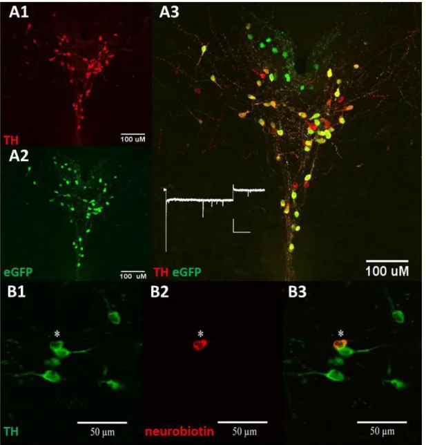
Figure 2.1: The tyrosine hydroxylase-eGFP transgenic mouse model identifies dopamine neurons via fluorescence in the vPAG. (A) Immunohistochemistry can be used to label and verify validity of the reporter via (1) tyrosine hydroxylase fluorescence, (2) eGFP fluorescence, and the (3) co-localization of eGFP and tyrosine hydroxylase in dual-positive cells. No significant Ih current was observed in vPAG DA neurons (B) (1) eGFP and tyrosine hydroxylase-positive cells can be visualized under the microscope to selectively record from. Staining showed that the recorded cells loaded with (2) neurobiotin via diffusion from the patching pipette,
are (3) tyrosine hydroxylase-positive. ... 38 Figure 2.2: The dopamine neurons in the VTA and in the vPAG display
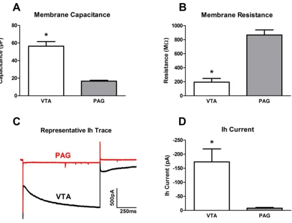
different electrophysiological membrane properties. (A) vPAG dopamine neurons have significantly lower membrane capacitance (p < 0.0001) than VTA dopamine neurons. (B) vPAG dopamine neurons have significantly higher membrane resistance (p < 0.0001) than VTA dopamine neurons. (C), (D) vPAG dopamine neurons display little to no hyperpolarization current (Ih) comparing to VTA
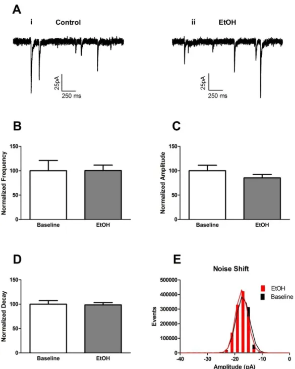
dopamine neurons. ... 39 Figure 2.3: Acute bath-applied alcohol had no effect on mini inhibitory
post synaptic currents (mIPSCs) in vPAG dopamine neurons. (A) Representative mIPSC traces of baseline control (i) and after 10 minutes of alcohol wash-on (ii). (B) Acute alcohol had no effect on mIPSC frequency. (C) Acute alcohol had no effects on mIPSC amplitude. (D) Acute alcohol had no effect on mIPSC decay. (E)
Acute alcohol had no effect on mIPSC noise. ... 40 Figure 2.4: Acute bath-applied alcohol had no effects on spontaneous
inhibitory post synaptic current (sIPSC) frequency in vPAG dopamine neurons. Representative sIPSC traces of (A) baseline control and (B) after 10 minutes of alcohol wash-on. (C) Acute alcohol had no effects on sIPSC frequency. (D) Acute alcohol
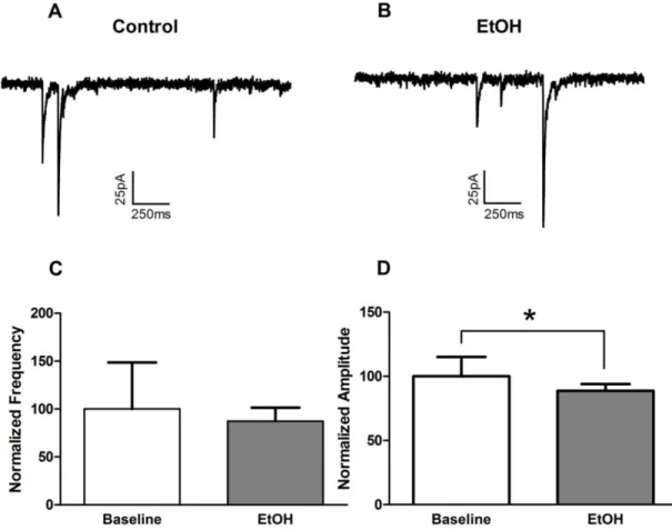
significantly decreased sIPSC amplitude. ... 41 Figure 2.5: Acute bath-applied alcohol increased mini excitatory post
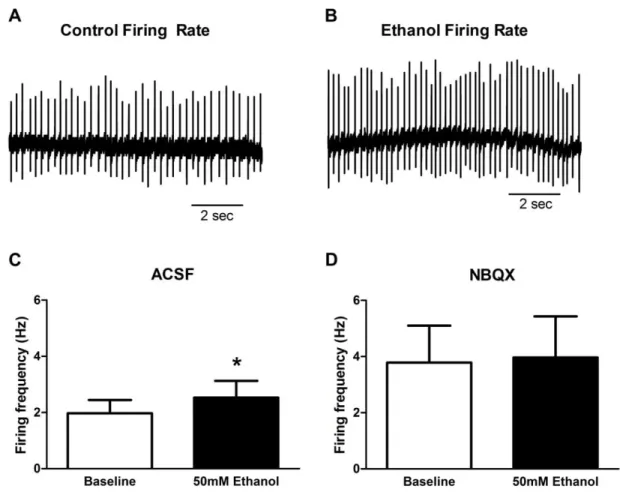
Figure 2.6: Acute bath-applied alcohol (50mM) increased firing rate of vPAG dopamine neurons in the cell-attached recording configuration. (A) Representative firing rate trace of baseline control. (B) Representative firing rate trace in the same neuron after 10 minutes of alcohol wash-on. (C) Alcohol increased firing rate in vPAG DA neurons. (D) Alcohol had no significant effect on firing rate in the
presence of NBQX (10µM). ... 43 Figure 2.7: Chronic intermittent alcohol vapor exposure did not affect
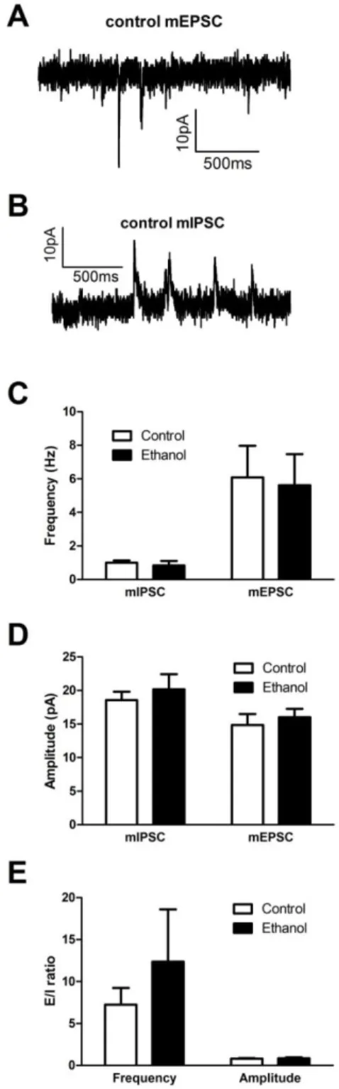
inhibitory and excitatory synaptic properties. (A) Representative mEPSC trace. (B) Representative mIPSC trace. (C) Chronic intermittent alcohol had no effect on mini frequency. (D) Chronic intermittent alcohol had no effect on mini amplitude. (E) Chronic intermittent alcohol had no effect on mini excitatory/inhibitory
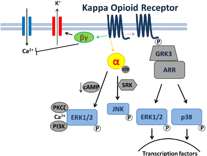
transmission ratio. ... 44 Figure 3.1: KOR-mediated signal transduction. Receptor activation by a
KOR-selective ligand can result in activation of several kinase cascades. Arrows refer to activation steps, T line refer to blockers or inhibition of functions. Abbreviations are as follows: α, G-protein alpha subunit; βγ, G-protein beta-gamma subunit; cAMP, cyclic adenosine monophosphate; ERK1/2, extra-cellular signal-regulated kinase; GRK3, G-protein coupled receptor kinase3; JNK, c-Jun N-terminal Kinase; p38, p38 MAPK; p, phosphorylation;PI3K,
phosphoinositol 3-kinase; PKCζ, protein kinase C zeta. ... 63 Figure 3.2: KOR agonist, dynorphin A (300nM), decreases mIPSCs in
vPAG DA neurons via KOR actions. (A) Representative mIPSC traces of baseline control (i) and after 10 minutes of 300nM dynorphin A wash-on (ii). (B) Dynorphin A significantly decreased mIPSC frequency. (C) Dynorphin A had no effect on mIPSC amplitude. (D) Cumulative frequency of mIPSC was shifted towards longer interevent intervals by Dynorphin A. (E) Cumulative probability of mIPSC amplitude was not affected by dynrophin A. (F) Dynorphin A no longer decreased mIPSC frequency in the presence of nor-BNI (100nM). (G) Dynorphin A had no effect on mIPSC amplitude in the presence of nor-BNI. (H) nor-BNI alone had no effect on mIPSC frequency. (I) nor-BNI alone had no effect on
mIPSC amplitude. ... 64 Figure 3.3: The postsynaptic GPCR inhibitor, GDPβs (4mM), defused
towards longer interevent intervals. (D) Dynorphin A had no effect
on mIPSC amplitude cumulative probability. ... 66 Figure 3.4: The ERK1/2 inhibitor SL327 (10 µM) and the p38 inhibitor
SB203580 (20 µM) did not block dynorphin A from attenuating mIPSC. (A) Dynorphin A significantly decreased mIPSC frequency in the presence of SL327. (B) Dynorphin A had no effects on mIPSC amplitude in the presence of SL327. (C) Dynorphin A significantly decreased mIPSC frequency in the presence of SB203580. (D) Dynorphin A significantly decreased mIPSC amplitude in the
presence of SB203580. ... 67 Figure 3.5: KOR inhibition does not require calcium and potassium ion
channels. The GPCR βγ subunit inhibitor blocked dynorphin A from attenuating mIPSC. (A) KOR/dynorphin A inhibition persisted even when all Ca2+ was removed by incubating slices in 0mM Ca2+/4mM Mg2+, 100µM EGTA with 50µM BAPTA-AM. (B) KOR/dynorphin A inhibition persisted even when all Ca2+ was removed and K+ channels are blocked with 4AP (100µM). (C) Gallein (100µM) prevented dynorphin A from inhibiting mIPSC frequency. mIPSC
amplitude remained unaffected. ... 68 Figure 3.6: The PI3kinase inhibitor wortmannin (1µM) and the PKA
inhibitor RP-camps (10µM) did not block dynorphin A from attenuating mIPSC. (A) Dynorphin A significantly decreased mIPSC frequency but had no effect on amplitude in the presence of wortmannin. (B) Dynorphin A decreased mIPSC frequency and had
no effect on amplitude in the presence of RP-camps. ... 69 Figure 3.7: KOR presynaptically inhibit GABA release potentially via the
disruption of functions in SNARE complex of the release machinery
by the βγ subunit of the KOR GPCR. ... 70 Figure 4.1: BNST receives dopamine projections from both the VTA and
vPAG, while vPAG DA neurons co-expressing vGlut2 project to a sub-region of the dorsal BNST. Injection of the retrograde tracer fluorogold (FG) into the BNST demonstrated that tyrosine hydroxylase (TH)-positive cells in the (A) VTA and (B) vPAG project to the BNST. Next, we stained for TH in a vGlut2-Ai3 reporter mouse to illustrate that (C) dopamine neurons in the vPAG are also (D) vGlut2-positive. (E) BNST images of vGlut2-Ai3 mice revealed that vGlut2-positive neurons terminals populated a
sub-region of the BNST that highly resembles the oval nucleus. ... 90 Figure 4.2: GABAergic and dopaminergic cell body distribution in the
and GABAergic neurons are situated laterally to the aqueduct. Further, the GABAergic and dopaminergic neurons are
distinct from one another and do not co-express. ... 91 Figure 4.3: Light-evoked action potential in the vPAG DA neurons. A
vGlut2-cre mouse was injected with a viral vector of double inverted channelrhodopsin(ChR2)-mcherry into the vPAG. Transfected cell bodies in the vPAG that expressed mcherry were visualized and recorded from. Blue light stimulation pulses varying in frequency were presented while the vPAG dopamine neurons fired in a time-locked manner at corresponding frequencies of 1, 5, 10, 20, and
40Hz. ... 93 Figure 4.4: Light-evoked glutamatergic current in the BNST neurons. A
vGlut2-cre mouse was injected with a viral vector of double inverted channelrhodopsin(ChR2)-mcherry into the vPAG. Transfected vGlut2 terminals projected from the vPAG expressed mcherry and could be visualized in the BNST. Neurons in proximity to clusters of mcherry-expressing terminals were recorded from. Blue light stimulation pulses varying in frequency were presented while glutamatergic current were evoked in BNST neurons at
corresponding frequencies of 1, 5, 10, 20, and 40Hz. ... 95 Figure 4.5: Light-evoked GABAergic and glutamatergic currents in the
BNST neurons. A TH-cre mouse was injected with a viral vector of double inverted channelrhodopsin(ChR2)-mcherry into the vPAG. Transfected TH projections from the vPAG expressed mcherry and could be visualized in the BNST. Neurons in proximity to clusters of mcherry-expressing terminals were recorded from. Blue light stimulation pulses were presented and both (A) evoked-GABAergic and (B) evoked-glutamatergic currents were
observed in BNST neurons. ... 96 Figure 4.6: Activation of vPAG TH-positive dopamine neurons produced
a robust anti-nociceptive effect. TH-cre and wild type animals were injected with viral vectors of double inverted Gq-coupled DREADD into the vPAG. Upon activation of vPAG dopamine neurons via the injection of CNO prior to the Von Frey hyperalgesia test, TH-cre animals displayed a significant decrease in response to the
hyperalgesia test than the wild type animals. ... 97 Figure 4.7: Activation of vPAG TH-positive dopamine neurons had no
TH-Figure 4.8: Activation of vPAG TH-positive dopamine neurons had no significant effects on elevated plus maze assay. Upon activation of vPAG dopamine neurons via the injection of CNO prior to the open test, there was no significant differences in A) time spent in open arms, B) entry number to open arms, C) time spent in closed arms, and D) entry number to closed arms between TH-cre and wild type
animals. ... 99 Figure 4.9: Activation of vPAG TH-positive dopamine neurons had no
significant effects on light/dark box assay. Upon activation of vPAG dopamine neurons via the injection of CNO prior to the open test, there was no significant differences in A) time spent in light compartment, B) entry number to light compartment, C) time spent in dark compartment, and D) entry number to dark compartment
LIST OF ABBREVIATIONS
BNST bed nucleus of the stria terminalis cAMP cyclic adenosine monophosphate
DA dopamine
ERK extracellular signal-regulated kinase
GABA gamma-aminobutyric acid
GRK3 G-protein coupled receptor kinase3 JNK c-Jun N-terminal Kinase
mEPSC mini excitatory post-synaptic current mIPSC mini inhibitory post-synaptic current PI3K phosphoinositide 3-kinase
PKA protein kinase A
PKCζ protein kinase C zeta SEM standard error of the mean
sIPSC spontaneous inhibitory post-synaptic current
TH tyrosine hydroxylase
CHAPTER 1. GENERAL INTRODUCTION
DOPAMINE SIGNALING
For well over three decades, dopamine (DA) signaling has been known to play a critical role in reward and motivation, as well as the establishment of stimulus-reward association (1-3). Dopamine release in the striatal structures can be increased by reward, such as food, drug intake and sexual behavior, while dopamine antagonists can prevent these behaviors (4-7). Reward association, such as the ability to be trained to lever-press for naturally rewarding things as food, water or sexual contact, requires dopamine signaling, as reward association cannot be learned when dopamine function is impaired (8). Additionally, previously acquired stimulus-reward associative learning cannot be continued or maintained when dopamine systems are blocked (2, 9, 10). The DA pathways that modulate behavioral responses to environmental stimuli are central to the signaling of reward.
in the rewarding properties of these stimuli. Further, selective chemical lesions of NAc-projecting dopamine neurons lead to the attenuation of the rewarding effects of cocaine and amphetamine (14), while this effect was not found in animals with ablation of norepinephrine-expressing neurons. Dopamine pathways regulate motivational behaviors, demonstrated by motivational deficits in feeding and water drinking caused by dopamine lesions in the lateral hypothalamus (15). Striatal pathways regulate movement and reward: the dopamine D1-mediated direct pathway promotes movement and reward while the dopamine D2-mediated indirect pathway activation increases immobility and appears to serve as punishment (16, 17).
Dopamine is generally thought to play a critical role in the formation and maintenance of addictive behaviors in drug use (18-21), but despite the large body of evidence supporting this notion, the necessity of dopamine signaling in all drug reward remains controversial. Several drugs of abuse such as phencyclidine (22), morphine (23), and nicotine (24) have demonstrated both dependent and dopamine-independent rewarding effects. Taken together, the importance of dopamine in reward-related behavior is undeniable; however, dopamine might not be required or limited to reward behaviors as studies find dopamine involvement in other complex emotional behaviors and conditions.
Mental Disorders (DSM-IV-TR2000) by deficits in reward function, coupled with heightened punishment from negative consequences. Depressive individuals often display psychomotor retardation, consisting of slowing of speech, eye and body movement, as well as abnormal body posture and facial expressions (26, 27), overlapping with the symptoms of Parkinson’s disease caused by dopamine deficiencies. Interestingly, these motor abnormalities caused by dopamine deficiencies in Parkinson’s disease can be alleviated by dopamine agonists (28). In addition, medications for Parkinson’s disease that improve motor deficits, such as dopamine agonists and monoamine oxide inhibitors, have also been found to relieve Parkinson-associated depression (29-31).
Depression is one of many negative emotional disorders modulated by dopamine; studies have shown that dopamine neurons can also modulate negative affective states such as anxiety. In these studies, select inhibition of VTA dopamine neurons induced depressive-like phenotype (32), and the ablation of kappa opioid receptors in dopamine neurons decreased anxiety-like effects in the open field and light/dark box behavioral assays (33). Additionally, dopamine D3 knockout animals display a decrease in depression-like behavior after immobilization stress (34). This evidence demonstrates the involvement of dopamine signaling in negative affective disorders, suggesting a regulatory role of dopamine signaling in not only reward, but also the development and maintenance of negative emotions.
affective disorders. The following section discusses the periaqueductal gray and its role in dopamine signaling.
PERIAQUEDUCTAL GRAY
The periaqueductal gray is a highly heterogeneous brain region; in addition to the dopamine neurons, there are large populations of GABAergic and glutamatergic neurons (35). Due to the diversity of its cell types, the PAG also has a variety of functional outputs that modulate a wide array of behaviors, including reward and autonomic functions, as well as negative affective behaviors such as panic and aggression. The PAG’s complex composition presents difficulties when trying to isolate neural networks and examine the functions of specific cell types. With the advent of transgenic animal models, as well as viral tools, the roles of periaqueductal gray neurons are slowly being unraveled.
(38). Other studies have shown that modulation in the PAG can influence emotional behavior: antagonism of the GABA receptors in the PAG increases anxiety-like behavior (39). Further, the sensitization of the PAG has been shown to suppress positive affect in rats (40), suggesting that increased activity in the PAG is heavily engaged during production of negative emotional behaviors and the maintenance of negative affective states. The PAG is also an important site in the ascending nociceptive control (ANC) pathway projecting to the rostral ventral medulla and there by regulates pain-related behaviors (41). There is evidence that dopamine neurons in the PAG may potentially play a role in the ANC as opiate anti-nociception is attenuated upon chemical lesion of PAG dopamine neurons (42). The PAG DA-mediated negative emotional behaviors mentioned above often display high comorbidity with one another, as stress (43), anxiety (44), and depression (45), have all been shown to exacerbate pain perception, and vice versa, suggesting that the regulation of these negative affects and their projections could
be modulated in a similar fashion by certain groups of PAG DA neurons.
affective disorders. Other behaviors mediated by PAG includes defensive behaviors (51), cardiovascular functions via its projections to the hypothalamus (52, 53), and lordosis behavior in sexual courtship (54).
As mentioned earlier, dopamine signaling mediates the rewarding properties of drugs of abuse, including alcohol. An abundance of evidence highlight the importance of the role the PAG plays in the negative affect specifically associated with alcohol abuse. Withdrawal following chronic ethanol exposure can induce hyperalgesia (55), suggesting a link between the PAG ascending nociceptive pathway and alcohol abuse. Further, dorsal PAG stimulation-induced freezing and escape was sensitized during ethanol withdrawal (56), and neurons in the PAG are critically involved in the neural circuitry for audiogenic ethanol withdrawal seizures (57). Taken together, this evidence illustrates that PAG-mediated behaviors can be altered by prolonged alcohol exposure. Additionally, alcohol can affect neural activity in the PAG, causing increased action potential frequency and hyper-excitability in PAG neurons during ethanol withdrawal (58). These studies provide essential information that links PAG function to alcohol abuse and related negative affects. It is important to note that the PAG has also been implicated in the withdrawal from diazepam (59) and opiates (60), both of which are popular substances of abuse. Given the recent findings that dopamine neurons in the PAG project to areas sensitive to drugs of abuse, it is compelling to investigate the modulation and functions of these A10dc dopamine neurons.
and central amygdala), a number of studies have demonstrated projections from the extended amygdala onto the PAG in regulating negative emotional behaviors as well (61-63). These extended amygdala projections to the PAG have been demonstrated to be GABA-mediated inhibitory inputs and modulate anxiety-like behaviors (38, 64, 65). In addition, studies have demonstrated a population of tonically active GABAergic neurons in the lateral portion of the PAG (66). Although local GABAergic interneurons have been shown to inhibit dopamine projection neurons in other dopamine-rich brain regions such as the VTA (67), the role of this local population of GABAergic neurons on vPAG dopamine neurons is unclear. The PAG also receives glutamatergic inputs from the lateral habenula (68, 69) that are associated with pain (70, 71), defensive behaviors (72), and sleep (73), and other projections to the PAG have also been found from the prefrontal cortex (74) and the brain stem (75, 76). These studies identified some projections that could contribute to the modulation of PAG-mediated behaviors; however, despite all the anatomical and synaptic studies conducted in the PAG, specific dopamine neuron-modulation has yet to be investigated due to the heterogeneity of the PAG. The investigation of how drugs of abuse modulate the PAG requires a thorough understanding of the actions of drugs, as well as their effects on neural networks and substrate alteration. The following section focuses on the effects of acute and chronic alcohol.
ALCOHOL
Alcohol addiction
1994): tolerance, withdrawal, drinking a larger amount or longer period than intended, inability to control use, a large amount of time spent in alcohol-seeking and related activities, missing important social/occupational/recreational activities due to alcohol use, as well as the continuation of use despite knowledge of having persistent physical or psychological problems caused by alcohol. Initially, alcohol consumption seems to be driven largely by its positive reinforcing effects (i.e. the euphoric effects of ethanol) (77). Over prolonged exposure, there is a proposed shift to a negative reinforcement, as negative affect is increased due to withdrawal. It is believed that the negative affective state drives an individual’s consumption of ethanol to alleviate the symptoms (78), creating a vicious cycle strengthened by repeated exposure. The negative affective disorders induced by ethanol exposure include depression (79, 80), anxiety (81), panic disorder (82), stress (83-85), and pain (86). The manifestation of these psychiatric disorders has been shown to render an individual more prone to relapse, and could cause relapse even long after the withdrawal period (87, 88). Additionally, repeated ethanol withdrawal episodes enhance negative affective symptoms (89). Due to the causative relationship between negative mood states and ethanol use, individuals easily fall into a cyclic addictive behavior pattern. The high comorbidity of alcohol addiction and negative mood disorders often presents difficulties in the treatment of alcohol abuse; therefore, understanding the mechanisms underlying the formation of these negative affective symptoms and their modulation is critical for the treatment of alcoholism. Acute Alcohol
Opposite from the impact on inhibitory transmission, acute alcohol has consistently been shown to exert inhibitory actions on excitatory cation-permeable ionotropic glutamate receptors: the AMPA receptors, the NMDA receptors, and the kainite receptors (119). Alcohol-inhibition on NMDARs has been observed in many brain regions, from the hippocampus to the amygdala, as well as the cerebellum (120-123). Much like the inhibitory receptors, alcohol also affects NMDA receptors differentially in a subunit-dependent manner. NMDA receptors are tetrameric and are composed of an obligatory NR1 subunit, in combination with at least one NR2 or NR3 subunit. The combination of different splice variations of the NR1 and accompanying NR2 can account for alcohol sensitivity (124), and although receptors containing NR3 subunit are relatively insensitive, the combination with NR2B could overcome the presence of NR3-insensitivity and enhance alcohol-induced inhibition (124). Glutamatergic AMPA receptors are inhibited by alcohol as well; however the impact of inhibition of AMPA receptors is not as robust as NMDA receptors (125-127). In addition, subunit composition of AMPA receptors has minimal influences on alcohol sensitivity (128), which could be due to alcohol-induced receptor desensitization (129). The above evidence suggests that acute alcohol has a consistent effect on ionotropic glutamatergic receptors and decreases glutamatergic currents.
the potential of acute alcohol to induce synaptic plasticity in areas known to regulate learning and memory, as well as negative emotional behaviors, all of which are highly correlated with alcohol addiction (133).
Chronic Alcohol
Prolonged alcohol exposure often results in two behavioral alterations: tolerance and dependence. These changes make the treatment of alcoholism more difficult, as tolerance encourages overuse due to the physiological habituation to lower doses, and dependence is diagnosed during withdrawal when individuals experience negative affect such as pain, anxiety, sleep loss, and seizure in the absence of alcohol. Both of these behavioral modifications encourage continuing alcohol usage to achieve the desired intoxication level, as well as to avoid the negative affect of withdrawal. Changes in behavior arise due to shifts in synaptic transmission in neural pathways in a variety of brain regions. In this section, the effects of chronic alcohol are addressed in the context of the balance between inhibitory and excitatory transmission. It is important to note however, that there are many different chronic alcohol exposure and withdrawal paradigms. Depending on the brain region, experimental design, length of exposure, number of repeated withdrawals, as well as the time point during withdrawal, a large discrepancy can be observed in the results.
is an increase in evoked GABA release, as well as an enhancement of mIPSC in CeA neurons after chronic alcohol. Further, dependent animals show dramatically increased GABA release in the CeA upon in vivo microinjection of alcohol directly into the CeA relative to naïve animals, suggesting an increase GABAergic tone after chronic alcohol exposure (135, 136). In contrast, some studies show that voluntary drinking is associated with a significant increase in paired-pulse ratio, suggesting a decrease in GABA release probability in the dentate gyrus neurons of monkeys (137). Together, these studies demonstrate that chronic alcohol can alter presynaptic GABA release.
α1 levels remained unchanged (144). In addition to the alteration in receptor subunit expression, other proteins could play important modulatory roles in receptor initiation and trafficking, altering receptor functions after chronic alcohol. These modulatory proteins include clathrin and adaptor complex, which are crucial for GABAA receptor internalization (145-147). Protein kinases such as PKA, PKC, and fyn could regulate GABAA receptor trafficking, and chronic alcohol has been shown to decrease associate of PKCγ with the α1 GABAA subunit, and increase associate with α4 in the cerebral cortex (148). This, however, was not observed in the hippocampus (145). The association between PKCγ and the α4 subunit could lead to GABAA receptor phosphorylation and disrupt recognition by the adaptor complex, thus preventing its internalization (147).
suggest that both pre- and post-synaptic GABA transmission can be altered by chronic alcohol exposure.
In contrast to acute alcohol’s effects on excitatory inputs, chronic alcohol exposure leads to a general increase in glutamatergic transmission (151, 152). However, similar to the acute effects of alcohol, NMDA receptors are modulated by chronic alcohol in a more robust fashion than other types of glutamatergic receptors (153, 154). Both in vivo and ex vivo chronic alcohol exposures induce increases in NMDAR functions, and
been shown to increase mGluR-mediated signaling, as well as protein expression, particularly mGluR1 and mGluR5 (162, 171, 172).
The above section detailed chronic alcohol exposure’s ability to modulate both inhibitory and excitatory transmission, as well as illustrated how the balance between inhibitory and excitatory inputs could be altered by chronic alcohol in brain region- and duration- dependent manners. It is clear that more studies are needed to further isolate the regulation of specific pathways and projections in order to better link them to the behavioral changes accompanied by chronic alcohol use in humans. The following section discusses the kappa opioid receptor and how it can modulate drinking behaviors. KAPPA OPIOID RECEPTORS
and serotonin (182-185). KORs have been shown to locate presynaptically on dopaminergic input to the amygdala, NAc, and PFC (186-188); on GABAergic inputs to the NAc and the extended amygdala (central nucleus of the amygdala and BNST) (189); on glutamatergic inputs to the VTA and NAc (189) and on serotonergic inputs to the NAc (182), as well as on noadrenergic inputs to the PFC (190). The KORs have also been found on the cell bodies of dorsal raphe serotonin (5-HT) neurons (182), locus coeruleus norepinephrine neurons (191), as well as VTA mesocortical dopamine neurons (192). Mechanistically, it has been shown that kappa opioid receptor activation has the ability to modulate dopaminergic neurons in the VTA. More specifically, a KOR agonist can inhibit the VTA DA neurons projecting to the prefrontal cortex, but not projections to the amygdala (192). Interestingly, while isolating GABA-mediated inhibitory post-synaptic currents (IPSCs) in the VTA dopamine neurons, the same research group found that opioid agonists also inhibited GABAergic transmission. The KOR system is widely distributed in the central nervous system and has been implicated in numerous pathophysiological disorders associated with mood and motivation (193-196), pointing to its potential as a putative therapeutic target for neuropsychiatric disorders.
regulation seem to play a critical role in these negative affective disorders. Studies have shown that KOR activation in the dorsal raphe, which also contains A10dc dopamine neurons and is just ventral of the vPAG, produces conditioned place aversion (202), and mediates the aversive effects of stress, and reinstates drug-seeking (182). In addition, KOR knockouts and conditional knockouts in dopamine-containing neurons produced anxiolytic-like effects and enhanced cocaine-induced plasticity (33). Depressive-like effects of the KOR agonist salvinorin A have also been shown associated with decreased phasic dopamine release in the NAc (203). Further, anti-nociception induced by oxytocin can be blocked by KOR antagonism in the PAG (204). The above evidence suggests that KOR actions can modulate negative emotions and drug-seeking behaviors, some of which are mediated via dopamine signaling. As mentioned earlier, dopamine signaling plays a critical role in reward and some negative emotional behaviors. In addition, the PAG is a brain region highly implicated in both drug addiction and negative affect. Given the evidence that kappa opioid receptors are distributed widely in the PAG (205), and that KOR actions can modulate dopamine signaling, the PAG becomes a critical brain region to study to better understand the mechanisms underlying drug addiction-associated mood disorders. More research on KOR modulation of neural activity in the PAG will allow us to better adapt strategies to develop treatment for negative mood disorders and alcoholism.
KAPPA OPIOID RECEPTORS AND ALCOHOLISM
signaling at the receptor level, as well as the ligand level (206). Microdialysis studies show that acute alcohol can stimulate the release of opiates, including dynorphin, in regions implicated in reward and affective disorders such as the VTA, the amygdala, and the nucleus accumbens (207-211). In addition, radioimmunoassay studies demonstrated that a 6-week chronic alcohol exposure significantly increased dynorphin B levels in the hypothalamus and substantia nigra, while it was decreased in the prefrontal cortex (212). Vice versa, KOR functions have also been shown to modulate alcohol-related behaviors.
alcohol exposure. Long-term alcohol exposure elevated foot-shock-induced c-Fos expression in the basolateral amygdala in wild type mice, but had the opposite effect in dynorphin-knockout mice (217). Just as opioid-inhibition of presynaptic GABAergic inputs onto VTA dopamine neurons can lead to the activation of dopamine neurons (67, 176), similar effects have been demonstrated by acute ethanol. In both the VTA (218) and the substantia nigra (SN) (219), it has been shown that acute ethanol stimulates the firing rate of dopamine neurons and inhibits GABAergic inhibitory currents. Recent data has suggested that ethanol differentially impacts populations of dopamine neurons by brain region. In a chronic vapor ethanol exposure paradigm, studies have shown that chronic alcohol exposure alters dopamine transporter (DAT) expression in the NAc, but not the striatum or the BNST (220), suggesting differential modulatory systems for dopamine neurons of different projections. Given the important role that KOR signaling plays in the development and maintenance of alcoholism, understanding the mechanisms underlying KOR modulation of dopamine signaling could shed some light onto the development of treatments for alcohol abuse, as well as battle against the vicious cycle encouraged by withdrawal-induced negative affects.
DISSERTATION
behavior circuits could shed light onto the development of more effective treatments. The dopamine signaling system mediates the rewarding properties of drugs of abuse. Evidence shows that alcohol exposure induces alterations in the signaling of dopamine-rich brain regions, such as the VTA. While the kappa opioid system is known to mediate negative affect, a rather novel population of dopamine neurons in the vPAG has been shown to project to regions regulating negative emotions such as stress, anxiety, and depression. Thus, it is likely that prolonged exposure to alcohol could increase vPAG dopamine signaling, consequently exciting stress and anxiety nuclei, and inducing stress- and anxiety-like behaviors.
CHAPTER 2. EFFECTS OF ALCOHOL ON VPAG DOPAMINE NEURON
INTRODUCTION
Alcohol use disorders are an enormous public health problem, and understanding the neurochemical systems involved in regulating the actions of alcohol can provide insight for the development of more effective treatments. Numerous reports have suggested that the subjective response to acute alcohol is related to the risk of development of alcohol use disorders (221). As such, understanding the actions of acute alcohol is an important area of investigation. Animal studies have shown that acute alcohol can induce locomotor stimulation (222-224) and is also anxiolytic (225, 226). Alcohol also modulates pain perception and the anti-nociceptive effects of opiates (227). Chronic alcohol exposure produces much different behavioral effects during withdrawal. Locomotor activity has been reported to decrease (228), and anxiety-like behaviors are observed (229) during withdrawal period. Other withdrawal-induced behaviors include aggression (230, 231), anhedonia (232), insomnia (233), and increased sensitivity to pain (55, 234). Given the diversity of these behavioral outcomes, there are likely multiple brain regions and their projection regions that are modulated by alcohol exposure.
A large body of evidence suggests that dopamine (DA) signaling plays a critical role in mediating the rewarding aspects of acute alcohol. Much of this research has
focused on the mesolimbic DA system, which is composed of DA neurons in the ventral tegmental area (VTA) that project to the nucleus accumbens (235, 236). However, recent studies have shown that dopamine signaling in other brain regions critical to alcohol abuse, such as the extended amygdala (237), can play an important role in alcohol reward. Interestingly, the extended amygdala receives a strong dopaminergic projection from the A10dc DA neurons, located in the ventral periaqueductal gray (vPAG), and dorsal raphe nucleus (DRN) (38, 238). The vPAG is particularly relevant as a target for the acute actions of alcohol as it has been implicated in the regulation of arousal (231, 239), anxiety (240-242), sleep (46), pain (42, 243-245), and opiate reward (246). Despite the potential behavioral relevance of vPAG DA neurons to the actions of alcohol, there have been no studies examining either the properties of these neurons, or the ability of alcohol to modulate their function. In this study, we utilized a TH-eGFP transgenic mouse to selectively record from DA neurons in the vPAG and VTA for cell property comparison, as well as evaluating the impact of both acute and chronic alcohol on vPAG DA synaptic transmission.
MATERIALS AND METHODS
Animals and Husbandry
Adult male TH-eGFP Swiss Webster mice aged between 5 to 9 weeks were bred and used in accordance with an animal use protocol approved by the University of North Carolina – Chapel Hill (IACUC). Mice were group-housed in our colony room under a 12:12-hour light cycle, with lights on at 7:00 AM daily. Mice were given ad libitum access to rodent chow and water. Mating pairs of mice were created by GENSAT and obtained from the Mutant Mouse Regional Resource Center in North Carolina. In the TH-eGFP mouse line, the genome was modified to contain multiple copies of a modified BAC in which an eGFP reporter gene was inserted immediately upstream of the coding sequence of the gene for tyrosine hydroxylase (TH). Data presented here were obtained from the transgenic mice maintained in-house.
Electrophysiology Brain Slice Preparation and Slice Whole-Cell Electrophysiology
Na2GPT. Signals were acquired via a Multiclamp 700B amplifier (Molecular Devices, Sunnyvale, California), digitized at 20 kHz, filtered at 3 kHz, and analyzed using Clampfit 10.2 software (Molecular Devices). Input resistance and access resistance were continuously monitored during experiments. Experiments in which changes in access resistance were greater than 20% were not included in the data analysis.
Chronic Intermittent Alcohol Vapor Paradigm
Adult mice were exposed to alcohol vapor or air in La Jolla Alcohol Research chambers for 16 hours per day (16 hours on, 8 hours off; from 4pm to 8am the following day) for 2 cycles of 4 days on, and 3 days off. Alcohol vapor was created by bubbling 95% alcohol in a vaporizer at a rate of 4-6 L/min air; the vapor was administered at a flow rate of approximately 15 L/min air to each individually sealed cage. Alcohol levels were adjusted to reach animal blood alcohol concentration (BAC) above 200 mg/dL, and monitored using a breathalyzer at the beginning and end of every exposure. To achieve intoxication BAC levels, alcohol-exposed mice were injected with 10ml/kg of 68.1mg/kg of the alcohol dehydrogenase inhibitor pyrazole combined with 1.5g/kg 20% (v/v) alcohol in saline; air-exposed mice received 10 ml/kg of 68.1mg/kg pyrazole in saline. Electrophysiology was performed 48 hours after exiting the chamber from the last cycle. Immunohistochemistry
Microsystems, Nussloch, Germany) and stored in a 50% glycerol solution at -20˚C until immunohistochemistry was performed. Slices were rinsed for 5 minutes in chilled PBS, followed by a 10-minute incubation in 0.1% Triton X-100 in PBS solution, two 5-minute PBS washes, and a 30-minute incubation in 0.5% Triton X-100 in PBS solution. After a 1-hour incubation in a blocking solution made of 0.1% Triton X-100/10% Normal Donkey Serum in PBS, the tissue was then incubated for 48 hours at 4°C with their respective primary antibodies diluted in 0.1% Triton X-100/10% Normal Donkey Serum in PBS blocking solution (anti-Tyrosine Hydroxylase, [1:500], Pel-Freez P60101-0, Lot 28632; anti-Green Fluorescent Protein, [1:500], Aves Laboratories, GFP-1020). Slices were rinsed three times for 10 minutes in chilled PBS before incubating for 24 hours at 4°C in their respective secondary antibodies diluted in PBS (Alexa Fluor 647 Donkey anti-Sheep, [1:800], TH; Alexa Fluor 488 Donkey anti-Chicken, [1:200], GFP; Jackson Immuno Research). Tissue was rinsed four times for 10 minutes in chilled PBS, and mounted using Vecta-Shield Mounting Medium (Vector Laboratories, Burlingame, CA, H-1000) prior to image collection.
Statistics
RESULTS
Basal Electrophysiology Properties: vPAG vs. VTA
We first wanted to confirm that the TH-eGFP mouse line would report correctly for DA neurons in the vPAG. To examine this, we performed dual label immunofluorescence and looked for overlap of TH and eGFP in the vPAG of the reporter (Fig2.1). We found that 69.6±4.5% of eGFP positive neurons were co-localized withTH (n =2 animals). Having determined the fidelity of this TH-eGFP reporter line for DA neurons in the vPAG, we then examined the membrane capacitance and resistance in eGFP-positive neurons in the vPAG.
neurons in the vPAG had a significantly smaller Ih current (-8.1±2.4 pA, n=36) (Fig2.2C and D), with most neurons in the vPAG lacking any measurable hyperpolarization current.
Acute alcohol modulation of GABAergic transmission in the vPAG
We found that 10-minute bath application of 50mM alcohol (n=10) significantly (p<0.05) decreased the amplitude (88.7±15.5% of baseline. Fig2.4D) of spontaneous inhibitory post-synaptic current (sIPSC), but had no effect on frequency (87.3±42.2%. Fig2.4C). Acute alcohol modulation of miniature excitatory post-synaptic current in the vPAG
Having established that acute alcohol had minimal effects on inhibitory inputs to vPAG DA neurons, we next examined the impact of acute alcohol on excitatory glutamatergic synaptic function. We found that a 10-minute bath application of 50mM alcohol (n=7) significantly (p=0.02) increased mEPSC frequency (144.9±17.7% of baseline. Fig2.5C), but had no effects on amplitude (93.3±4.6% of baseline, Fig2.5D). Interestingly, this effect did not appear to reverse over the course of the 10-minute washout. Taken together, these results demonstrate that alcohol has no effects on inhibitory inputs, but facilitates excitatory inputs onto vPAG dopamine neurons via a presynaptic mechanism.
Acute alcohol modulation of vPAG dopamine neuron cell firing rate
(n=7. Fig2.6D) on firing rate in the presence of the excitatory AMPA receptor antagonist NBQX (10uM). These results suggest that acute alcohol increases the firing of vPAG DA neurons via an enhancement of excitatory inputs.
Chronic intermittent alcohol vapor exposure modulation of synaptic transmission in the
vPAG
DISCUSSION
express vGlut2. Future studies using optogenetic approaches will more clearly examine this interesting possibility.
Guan et al., that alcohol caused a significant but minimal decrease in spontaneous inhibitory transmission; however, the decrease in amplitude we observed appears less robust than the effect in the posterior VTA. This could be due to some differences in effects of alcohol on the surrounding network and inputs. Taken together, these results suggest that vPAG DA neurons could be similarly modulated by alcohol as the posterior VTA DA neurons, and may have a partially overlapping set of GABA inputs, either from local interneurons or distal projections such as the BNST and the central amygdala.
although a direct network effect was not observed, chronic intermittent alcohol exposure could have an impact on modulatory peptides in the vPAG and how they regulate synaptic transmission.
CONCLUSIONS
FIGURES AND TABLES
Figure 2.1: The tyrosine hydroxylase-eGFP transgenic mouse model identifies
dopamine neurons via fluorescence in the vPAG. (A) Immunohistochemistry can be used to label and verify validity of the reporter via (1) tyrosine hydroxylase fluorescence, (2) eGFP fluorescence, and the (3) co-localization of eGFP and tyrosine hydroxylase in dual-positive cells. No significant Ih current was observed in vPAG DA neurons (B) (1) eGFP and tyrosine hydroxylase-positive cells can be visualized under the microscope to
Figure 2.2: The dopamine neurons in the VTA and in the vPAG display different electrophysiological membrane properties. (A) vPAG dopamine neurons have
CHAPTER 3. MODULATION OF KAPPA OPIOID RECEPTORS ON VPAG DOPAMINE NEURONS
INTRODUCTION
The PAG is a heterogeneous region in cell types, as well as in its projection targets implicated in various behaviors. For instance, the projection from the PAG to the prefrontal cortex (46), hypothalamus (73), and the dorsal raphe (46) has been shown to modulate sleeping behavior. Projections to the pons regulate cardiovascular and respiratory functions (277); projections to the hypothalamus regulate cardiovascular functions (52), as well as defense and fear conditioning (278); it as well as projects to the rostral ventral medulla that regulates nociception (279). Despite the large body of studies that have investigated PAG projections and functions, few have demonstrated cell-type specific properties. In this study, we took advantage of transgenic mouse lines, to specifically target vPAG dopamine neurons to examine how their synaptic transmission is modulated.
opiate anti-nociception is attenuated upon chemical lesion of vPAG dopamine neurons (42), also suggesting a role of dopamine signaling in the mediation of opioid actions in the vPAG. Another PAG dopamine-mediated behavior heavily linked to negative emotional state is sleep, as sleep deprivation has been shown link to stress (47), anxiety (48), and depression (49). Evidence illustrated the critical role of PAG dopamine projections to the prefrontal cortex in sleeping behavior, as selective chemical lesion of dopamine neurons in the PAG leads to increase in sleep time (46). These studies show that the vPAG DA signaling is heavily engaged in negative affect, which overlaps with those behaviors previously identified to encourage drug relapse.
As mentioned in chapter I, dopamine signaling has been closely associated with the rewarding properties of drugs of abuse, and the kappa opioid receptor activity can modulate drinking behavior, as well as negative emotions. Kappa opioid receptor (KOR) activation mediates aversive effects produced by alcohol challenge (213), and systemic antagonism of KOR also attenuates dependence-induced excessive ethanol self-administration in rats (214). It would also seem like KOR systems can modulate dopamine mediated reward-seeking behavior, as acute KOR activation decreases the rewarding properties of alcohol (215) while decreasing operant responding to ethanol (215). Together, these studies demonstrated that the kappa opioid receptor (KOR) system plays a critical role in negative emotional disorders, as well as drug abuse, possibility via the modulation of dopamine signaling.
Physiologically, the activation of receptors of the opioid family putatively inhibits presynaptic transmission. Opioid receptor-activation inhibits glutamate transmission presynaptic to neurons in the hypothalamus (181), as well as presynaptic to the brainstem catecholamine neurons (280). Opioid receptor-activation has also been shown to presynaptically inhibit GABAergic transmission in the VTA, resulting in disinhibition of the VTA dopamine neurons, increasing their activity (67, 281). The above evidence raises the possibility that kappa opioid receptor activation could modulate vPAG dopamine neurons, as well as the activities of their output regions. With the knowledge that vPAG DA neurons project to the BNST that regulates stress and negative affect, understanding of kappa modulation on these neurons could provide insight onto the regulation of emotional behaviors. In this study, we utilized a tyrosine hydroxylase (TH)-eGFP transgenic mouse to selectively record from DA neurons in the vPAG and pharmacologically evaluated the impact and mechanisms of KOR activation on GABA-mediated inhibitory inputs onto vPAG dopamine neurons.
MATERIALS AND METHODS
Animals and Husbandry
Adult male TH-eGFP mice on a Swiss Webster background (aged between 5 to 9 weeks) were bred and used in accordance with an animal use protocol approved by the University of North Carolina – Chapel Hill (IACUC). Mice were group-housed in our colony room under a 12:12- hour light cycle, with lights on at 7:00 AM daily. Mice were given ad libitum access to rodent chow and water. Mating pairs of mice were created by GENSAT and obtained from the Mutant Mouse Regional Resource Center in North Carolina. In the TH-eGFP mouse line, the genome was modified to contain multiple copies of a modified BAC in which an eGFP reporter gene was inserted immediately upstream of the coding sequence of the gene for tyrosine hydroxylase (TH). Data presented here were obtained from the transgenic mice maintained in-house.
Electrophysiology Brain Slice Preparation
Mice were decapitated under isoflurane anesthesia and their brains were rapidly removed and placed in ice-cold sucrose artificial cerebrospinal fluid (ACSF): (in mM) 194 sucrose, 20 NaCl, 4.4 KCl, 2 CaCl2, 1 MgCl2, 1.2 NaH2PO4, 10.0 glucose, and 26.0 NaHCO3 saturated with 95% O2/5% CO2. Three hundred micron slices were prepared using a Leica VT1200 vibratome (Wetzlar, Germany).
Slice Whole-Cell Electrophysiology
maintained at approximately 30°C (Warner Instruments, Hamden, Connecticut) Recording electrodes (3–5 MΩ) were pulled with a Flaming-Brown Micropipette Puller (Sutter Instruments, Novato, CA), using thin-walled borosilicate glass capillaries. During inhibitory transmission experiments, recording electrodes were filled with (in mmol/L) 70 KCl, 65 K+-gluconate, 5 NaCl, 10 4-(2-hydroxyethyl)-1-piperazineethanesulfonic acid, 2 QX-314, .6 EGTA, 4 ATP, .4 GTP, pH 7.4, 290 to 295 mOsmol. In experiments where post-synaptic GPCR signaling was blocked, GDPβs was used to replace GTP in the internal solution. All experiment were conducted under the voltage clamp configuration, cells were held at −70 mV and inhibitory post-synaptic currents (IPSCs) were pharmacologically isolated with 3 mmol/L kynurenic acid, to block α-amino-3- hydroxy-5-methyl-4-isoxazole-propionic acid (AMPA) and N-methyl-D-aspartate (NMDA) receptor-dependent synaptic current. To isolate miniature inhibitory post-synaptic currents (mIPSCs), tetrodotoxin (0.5 μmol/L) was added to the perfusing ACSF solutions described above. Signals were acquired via a Multiclamp 700B amplifier (Molecular Devices, Sunnyvale, California), digitized at 20 kHz, filtered at 3 kHz, and analyzed using Clampfit 10.2 software (Molecular Devices). Input resistance and access resistance were continuously monitored during experiments. Experiments in which changes in access resistance were greater than 20% were not included in the data analysis.
Statistics
periods using paired Students t-tests. The effects of antagonists/blockers on the ability of drugs to modulate synaptic transmission were compared using t-tests during the washout period. All values given for drug effects throughout the article are presented as mean ± SEM.
Drugs
RESULTS
Endogenous KOR agonist Dynorphin A attenuates GABAergic input onto vPAG DA
neurons via presynaptic mechanisms
Fig3.3A,C), but not amplitude (97.8±11.3% of baseline, Fig3.3B, D), providing additional support that dynorphin attenuates inhibitory input onto vPAG DA neurons via a pre-synaptic mechanism. Unpublished results from the Kash lab have shown GDPβs effective in blocking the postsynaptic effects of a NPY Y1 agonist (Leu-pro NPY) in the BNST.
Dynorphin effects on GABA are not mediated through MAP kinase signaling
We next investigate the role of MAP kinases ERK1/2 and p38 in KOR modulation of GABAergic transmission. As mentioned earlier, ERK1/2 antagonist has been shown to mediate KOR activation-induced attenuation in evoked IPSC response in the BNST (178). We incubated the slices in either a selective MEK inhibitor (SL327, 10 µM, n=6), or p38 inhibitor (SB203580, 20 µM, n=5) for 40 min before and during dynorphin wash-on. In the presence of the MEK inhibitor SL327, dynorphin A significantly decreased mIPSC frequency (67.2±10.6% of baseline, p=0.04, Fig3.4A), but not amplitude (97.1±4.3% of baseline); In the presence of the p38 inhibitor SB203580, dynorphin A significantly decreased mIPSC frequency (46.6±9.9% of baseline, p<0.05, Fig3.4B) and amplitude (83.2±3.6% of baseline, p=0.02, Fig3.4B). Together, these results suggest that the dynorphin-induced attenuation of GABAergic input onto vPAG DA neurons was not mediated through MAP kinase signaling.
Dynorphin effects on GABA are not mediated through calcium and potassium ion
channel conductance
eliminate the role of calcium channels, we incubated the slices in calcium-free ACSF and the selective calcium chelators BAPTA-AM (50µM) and EGTA (100µM) for 1-2 hours before recording, and continued to record from the slice in calcium-free ACSF in the presence of just EGTA, or EGTA plus 4-AP (100µM) to block potassium channels. In calcium-free experiments, dynorphin A significantly decreased mIPSC frequency (71.9±8.6% of baseline, p=0.02, n=6, Fig3.5A), but not amplitude (102.3±9.6% of baseline, Fig3.5A). In calcium-free experiments where potassium channels were blocked with 4-AP, dynorphin A significantly decreased mIPSC frequency (77.8±8.0% of baseline, p<0.05, n=8, Fig3.5B), but not amplitude (107.5±5.7% of baseline, Fig3.5B). These data suggest that the change in conductance of these ion channels does not mediate the KOR activation effect on IPSCs. To further investigate the role of KOR βγ subunits, we incubated the slices in gallein (100µM), an inhibitor of G protein βγ subunit-dependent signaling. The KOR activation-induced attenuation of GABA transmission was blocked in the presence of gallein (n=6), with no significant difference in mIPSC frequency (107.1±7.1% of baseline, Fig3.5C) or amplitude (102.8±7.4% of baseline, Fig3.5C). Together these data suggest that the effects of KOR on GABAergic transmission were mediated via βγ subunit signaling, but not the ion channels.
Dynorphin effects on GABA are not mediated through PI3 kinase or PKA signaling
DISCUSSION
Ventral PAG dopamine neurons have been implicated in a variety of emotional behaviors. How these mid-brain dopamine neurons are modulated by kappa opioid functions could be a crucial piece of information in understanding the regulation of addiction, as well as the associated negative affect that often leads to relapse. This study focused on determining the effects of kappa opioid receptor-activation on the inhibitory synaptic transmission onto vPAG dopamine neurons, in addition, the downstream signaling mechanisms through which the effects take place.
achieved by the addition of GDPβs in the patching pipette, filling the recorded cell by diffusion. GDPβs is a non-hydrolyzable GDP analogue; it blocks postsynaptic GPCR signaling by preventing the exchange of GDP to GTP, thus preventing G protein activation. We found that the attenuation of mIPSCs caused by dynohpin A persisted in the presence of postsynaptic GDPβs, suggesting that the postsynaptic KOR does not play a role in the effects seen. Using GDPβs to block postsynaptic GPCRs has been shown effective in the Kash lab where postsynaptic NPY Y1 receptor agonist effects were blocked in its presence. The above evidence demonstrated KOR mediates inhibitory inputs onto vPAG DA neurons presynaptically. This is not surprising as KOR agonists have been shown to inhibit presynaptic GABA release in other brain regions, such as the BNST (178), and in the hypothalamus (293).
(182, 289). We hypothesized KOR activation attenuates inhibitory transmission input, allowing dopamine neuron activity to be enhanced, increasing in signaling in projection areas such as the BNST and amygdala to increase anxiety- and stress-like behaviors, making p38 a likely candidate for the attribution of decreased inhibition. However, in the presence of the p38 inhibitor SB203580, diminished GABAergic current persisted. Curiously, both mIPSC frequency and amplitude decreased upon application of the KOR agonist dynorphin A, implying perhaps SB203580 modulated postsynaptic properties that makes vPAG dopamine neurons sensitive to dynorphin A.
occurs downstream of calcium entry and is calcium-independent. These results raise the possibility that dynorphin A could be activating the KOR and directly affect the presynaptic release machinery in the GABAergic inputs onto the vPAG dopamine neurons. This data is similar to studies on KOR presynaptic inhibition of glutamatergic inputs in the hypothalamus (181). Although we did not identify the molecular target through which dynorphin inhibits presynaptic GABA, our results are consistent with studies proposing direct modulation of the exocytosis release machinery by the βγ subunit of the Gi/o-coupled GPCR (300, 301). In addition, we continued to rule out the possibility of the actions of phosphorylated KOR, inhibiting PKA and the generation of cAMP. We found a close to significant (p=0.051) decrease in mIPSC frequency upon the application of dynorphin A, consistent with other results that suggest KOR does not inhibit through that actions of PKA.
FIGURES AND TABLES
Figure 3.3: The postsynaptic GPCR inhibitor, GDPβs (4mM), defused into the recorded vPAG DA neuron did not block dynorphin A from attenuating mIPSC, suggesting a presynaptic mechanism. (A) Inhibition of postsynaptic GPCR did not block dynorphin A from attenuating mIPSC frequency. (B) Dynorphin A had no effects on mIPSC
amplitude in the presence of GDPβs. (C) GDPβs did not block dynorphin A’s ability to shift the mIPSC cumulative frequency towards longer interevent intervals. (D)
Figure 3.5: KOR inhibition does not require calcium and potassium ion channels. The GPCR βγ subunit inhibitor blocked dynorphin A from attenuating mIPSC. (A)
KOR/dynorphin A inhibition persisted even when all Ca2+ was removed by incubating slices in 0mM Ca2+/4mM Mg2+, 100µM EGTA with 50µM BAPTA-AM. (B)
CHAPTER 4. VPAG DOPAMINE MODULATION ON PROJECTION AREA AND ITS BEHAVIORAL IMPLICATIONS
INTRODUCTION
membrane capacitance and significantly higher resistance. In addition, the commonly observed hyperpolarization-activated Ih current in the VTA DA neurons was not observed in the vPAG DA neurons. Based on these differences in cell properties, these two distinct groups of DA neurons could potentially modulate activity in the projection region differentially.
cutting edge chemical genetic tools – optogenetics and designer receptors exclusively activated by designer drugs (DREADD), to address the modulatory role of the vPAG dopamine neurons in the BNST.
Optogenetics utilizes light-sensitive proteins found in algae and bacteria to activate or inhibit neural activity. The follow three types of proteins provide activation and inhibition in sub-second time scale and are most commonly incorporated: the neural-activating cation channel channelrhodopsin-2 (ChR2) (308, 309), the neural-inhibiting halorhodopsin (NpHR) (310, 311), the neural-silencing proton pump archaerhodopsin (Arch) (312). These proteins change the excitability of cells by regulating membrane potential according to their protein properties, and are activated by exposure to light of different wavelengths. In neurons, a large current is required to depolarize the cell to trigger an action potential, thus functional ChR2 must be expressed at a high level in order to reach a cumulative current large enough to reach action potential. Such a high density expression can be achieved via the infection of an adeno-associated virus (AAV). In vivo studies have demonstrated effective control of activation or inhibition of neural networks, as well as behavior, via optogenetics (313).
demonstrated the lack of appreciable affinity to any receptor but the modified hM1D-hM4D (316). In addition, studies have shown that not only can CNO activate Gα signaling; the activation of downstream signaling pathways that involve multiple effectors is also preserved in the modified human muscarinic DREADD receptors, such as β-arrestin and downstream MAP kinase signaling (315).
MATERIALS AND METHODS
Animals and Husbandry
Adult male TH-cre mice on a C57 (aged between 5 to 9 weeks) were bred and used in accordance with an animal use protocol approved by the University of North Carolina – Chapel Hill (IACUC). Mice were group-housed in our colony room under a 12:12- hour light cycle, with lights on at 7:00 AM daily. Mice were given ad libitum access to rodent chow and water. Mating pairs of mice were purchased from The Jackson Laboratory. In the TH-cre male mouse line, the tyrosine hydroxylase (TH) promoter directs expression of Cre recombinase to catecholaminergic cells; in the vesicular glutamate transporter 2 (vGlut2-cre) mouse line, the vGlut2 transporter promoter directs expression of Cre recombinase to a subset of glutamatergic neurons. Data presented here were obtained from the transgenic mice maintained in-house.
Immunohistochemistry
1-hour incubation in a blocking solution made of 0.1% Triton X-100/10% Normal Donkey Serum in PBS, the tissue was then incubated overnight (~18 hours) at room temperature with their respective primary antibodies diluted in 0.1% Triton X-100/10% Normal Donkey Serum in PBS blocking solution (anti-Tyrosine Hydroxylase, [1:1000], Immunostar 22941-907001; anti-Green Fluorescent Protein, [1:500], Aves Laboratories, GFP-1020). Slices were rinsed three times for 10 minutes in chilled PBS before incubating for 2 hours in their respective secondary antibodies diluted in PBS (Alexa Fluor 647 Donkey anti-Mouse, [1:200], TH; Alexa Fluor 488 Donkey anti-Chicken, [1:200], GFP; Jackson Immuno Research). Tissue was rinsed four times for 10 minutes in chilled PBS, and mounted using Vecta-Shield Mounting Medium (Vector Laboratories, Burlingame, CA, H-1000) prior to image collection.
Stereotaxic Surgery
infection and protein expression before DREADD behavioral assays, and 4-6 weeks prior to electrophysiological experiments.
Viral vectors
DNA plasmids coding double floxed-ChR2-mcherry were obtained from the laboratory of Karl Deisseroth. Plasmid DNA were grown and collected using a standard plasmid maxiprep kit (Qiagen, Hilden, Germany). Following plasmid purification and restriction digest, and sequencing to assure DNA fidelity, purified AAV was produced, using calcium phosphate precipitation methods by the UNC Vector Core facilities (AAV serotype 2; University of North Carolina at Chapel Hill). Viral titers used were > 1012 g.c. (genome copies) /mL. DREADD virus was obtained from Dr. Bryan Roth at UNC-Chapel Hill, who developed the construct.
Electrophysiology Brain Slice Preparation
Mice were decapitated under isoflurane anesthesia and their brains were rapidly removed and placed in ice-cold sucrose artificial cerebrospinal fluid (ACSF): (in mM) 194 sucrose, 20 NaCl, 4.4 KCl, 2 CaCl2, 1 MgCl2, 1.2 NaH2PO4, 10.0 glucose, and 26.0 NaHCO3 saturated with 95% O2/5% CO2. Three hundred micron slices were prepared using a Leica VT1200 vibratome (Wetzlar, Germany).
Slice Whole-Cell Electrophysiology