https://openaccess.leidenuniv.nl
License: Article 25fa pilot End User Agreement
This publication is distributed under the terms of Article 25fa of the Dutch Copyright Act (Auteurswet) with explicit consent by the author. Dutch law entitles the maker of a short scientific work funded either wholly or partially by Dutch public funds to make that work publicly available for no consideration following a reasonable period of time after the work was first published, provided that clear reference is made to the source of the first publication of the work.
This publication is distributed under The Association of Universities in the Netherlands (VSNU) ‘Article 25fa implementation’ pilot project. In this pilot research outputs of researchers employed by Dutch Universities that comply with the legal requirements of Article 25fa of the Dutch Copyright Act are distributed online and free of cost or other barriers in institutional repositories. Research outputs are distributed six months after their first online publication in the original published version and with proper attribution to the source of the original publication.
You are permitted to download and use the publication for personal purposes. All rights remain with the author(s) and/or copyrights owner(s) of this work. Any use of the publication other than authorised under this licence or copyright law is prohibited.
If you believe that digital publication of certain material infringes any of your rights or (privacy) interests, please let the Library know, stating your reasons. In case of a legitimate complaint, the Library will make the material inaccessible and/or remove it from the website. Please contact the Library through email: [email protected]
Article details
Rooden E.J. van, Bakker A.T., Overkleeft H.S. & Stelt M. van der (2018), Activity-based protein profiling. In: eLS.: John Wiley & Sons, Ltd.
Activity-based Protein
Profiling
Eva J van Rooden,
Leiden Institute of Chemistry, Leiden University, Leiden, The NetherlandsAlexander T Bakker,
Leiden Institute of Chemistry, Leiden University, Leiden, The NetherlandsHerman S Overkleeft,
Leiden Institute of Chemistry, Leiden University, Leiden, The NetherlandsMario van der Stelt,
Leiden Institute of Chemistry, Leiden University, Leiden, The NetherlandsArticle Contents •Introduction
•Labelling •Analytical Platforms •Applications •Future Prospects
Online posting date: 22ndJanuary 2018
Activity-based protein profiling is a method to study a subset of the enzymatically active pro-teome. This method uses chemical probes that covalently react with active enzymes. These labelled proteins can subsequently be analysed by means of a detection tag on the probe. A diverse set of probes has been developed for many enzyme classes, such as serine hydrolases, proteases, glycosidases and kinases. Different analytical techniques are currently available to visualise, identify and quantify probe-labelled pro-teins with high efficiency. Activity-based protein profiling has well-developed applications in discov-ering new drug targets and in profiling inhibitors for potency and selectivity. Activity-based protein profiling will, therefore, continue to aid research both in fundamental biology and drug discovery.
Introduction
Activity-based protein profiling (ABPP) is a method to study the abundance of active enzymes in complex proteomes. ABPP uses chemical tools, termed activity-based probes (ABPs), which covalently and irreversibly react with a nucleophile in the active site of targeted proteins. Because only active enzymes are labelled by a probe, ABPP measures the abundance of active enzymes. This can differ from the total abundance of an enzyme, considering the activity of enzymes is regulated by posttranslational modifications See also: Proteins: Post-synthetic Modification – Function and Physical Analysis.
eLS subject area:Biochemistry
How to cite:
van Rooden, Eva J; Bakker, Alexander T; Overkleeft, Herman S; and van der Stelt, Mario (January 2018) Activity-based Protein Profiling. In: eLS. John Wiley & Sons, Ltd: Chichester. DOI: 10.1002/9780470015902.a0023406
This makes ABPP a unique and powerful method. Increasingly, ABPP is called activity-based or chemical proteomics (Simon and Cravatt, 2010), complementing abundance-based proteomics
See also:Shotgun Proteomics. ABPP can be used to compare activity of certain enzymes between different proteomes, for example between healthy and diseased tissue, which enables drug target discovery. Furthermore, ABPP can be applied to characterise inhibitors and drug candidates for both potency and selectivity in a native physiological context, aiding the selection of therapeutically relevant compounds.
Every ABPP experiment consists of two parts: an activity-dependent labelling part and an analytical part to visualise and characterise this labelling event. This general view of ABPP shows it is a multidisciplinary endeavour: organic chemistry is needed to synthesise and characterise ABPs, analyt-ical chemistry to provide the read-out of the labelling event, and biology to understand the proteomes being studied.
In this article, we will start with the first part: the labelling of active proteins using an ABP. The design of an ABP will be explained and several examples of probes and their enzyme targets will be discussed. In the second section, an overview is provided of the analytical platforms available to visualise the labelled proteome. Finally, in the third section, the applications of ABPP will be reviewed, focussing on comparative ABPP and competitive ABPP experiments.
Labelling
Activity-based Protein Profiling
H
H O
O P
O N H NH
O O
S HN
NH
F O O
– HN N HO P O O
R F
O OH N NH O
P O− O R
F
O OH N NH O
P O O R
Labelling BOC
(a)
(b)
(c)
Proteome Labelled proteome
(two-step probe)
Trap Linker Tag
Labelled proteome (one-step probe)
Figure 1 Labelling enzymes with an activity-based probe. (a) General activity-based probe design, with fluorophosphonate-biotin as example. (b) Probe labelling cartoon: two-step labelling using bioorthogonal chemistry (BOC) is optional for probes equipped with a suitable tag. (c) Mechanism of serine hydrolase labelling: catalytic triad reacting with the fluorophosphonate trap.
is used to enrich or purify probe-labelled enzymes (pulldown), a radioactive label or a ligation handle for a two-step labelling procedure (Speerset al.,2003).
In the labelling part (Figure1b), the ABP binds covalently to the target enzyme. This labelling event can take place in lysates, intact cells, tissues or living organisms (Blumet al.,2007). There are two types of probes for the detection of active proteins (Figure
1b): (1) one-step probes make use of a compound with a detection tag already installed and (2) two-step probes rely on a ligation handle, which can be used to install the detection tag after the probe has reacted with the protein. One-step labelling is fast and efficient, but the large tag can decrease the affinity and selectiv-ity of the probe for the target enzymes and/or may interfere with cell permeability. Two-step probes may circumvent these issues, but are less efficient in the workflow. Key is that the ligation han-dle and the detection tag react in a bioorthogonal manner, which means that the biological system does not interfere with the cou-pling reaction (Willemset al.,2011). The most commonly used bioorthogonal reaction is the ‘click’ reaction where an alkyne moiety reacts with an azide moiety in a copper(I)-catalysed cycli-sation (Tornøeet al.,2002). For an extensive review on different types of bioorthogonal chemistry, see Pattersonet al. (2014).
InTable1, several examples of ABPs for different enzyme
classes are depicted. For a comprehensive overview, the reader is referred to excellent reviews (Evans and Cravatt,2006; Nodwell and Sieber, 2012). Here, predominantly ABP design will be discussed using enzyme class specific examples to explain the different methods of probe design.
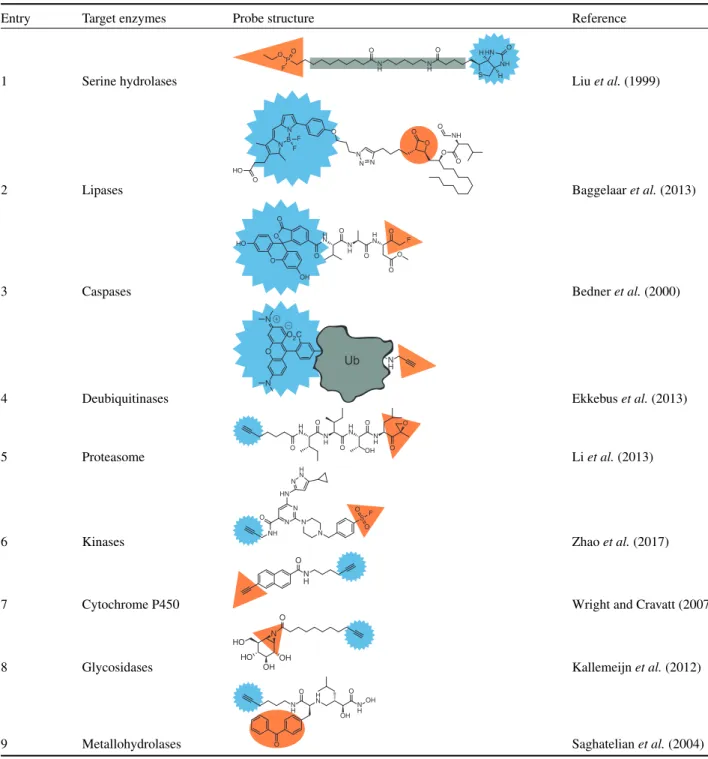
Serine hydrolases
Probe1(Table1) is a broad-spectrum probe, which is designed to react with any serine hydrolase. The hydrophobic linker between the electrophilic trap and the biotin group does not contain any
side chains that can provide extra interactions with selected mem-bers of the hydrolases, thus providing no specificity for a particu-lar serine hydrolase. The mechanism of covalent bond formation between a fluorophosphonate probe and the catalytic triad of a serine hydrolase is depicted inFigure1c(Liuet al.,1999). The aspartic acid and histidine residues form a charge relay system with the serine, increasing its nucleophilicity. The catalytically active serine nucleophile of the hydrolase attacks the electrophilic fluorophosphonate, which results in expulsion of a fluoride ion and concurrent covalent binding of the enzyme with the probe. The formed covalent bond is stable and the active site is occu-pied, rendering the enzyme inactive. Probe2is an example of a tailored probe, used for profiling of the lipase DAGL-α (diacyl-glycerol lipase alpha) and other related proteins (Baggelaaret al., 2013). The design of this probe is based on the anti-obesity drug Orlistat, which has an irreversible covalent binding mechanism, with a lactone as electrophilic trap. This example highlights one method of ABP design: using a known covalent inhibitor as a template. The tag used for probe2is a fluorophore.
Cysteine proteases
Table 1 Enzyme classes and reported activity-based probes specific to that class (orange trap and blue tag as inFigure 1)
Entry Target enzymes Probe structure Reference
1 Serine hydrolases
H H O O P O N H O N H O S HN NH F
Liuet al.(1999)
2 Lipases N B N F F HO O O N N N O O O O NH O
Baggelaaret al.(2013)
3 Caspases F H N N H H N O O O O O O O HO OH O O
Bedneret al.(2000)
4 Deubiquitinases
Ub N H O2C O N N Ub N H Ub
Ekkebuset al.(2013)
5 Proteasome N H H N H N N H O O O O O O OH
Liet al.(2013)
6 Kinases NH
O N N N N S O O F HN H N N
Zhaoet al.(2017)
7 Cytochrome P450
N H O
Wright and Cravatt (2007)
8 Glycosidases N OH O HO OH HO
Kallemeijnet al.(2012)
9 Metallohydrolases O N H H N N H OH O OH
O Saghatelianet al.(2004)
selective caspase-specific recognition (Bedneret al.,2000). The reaction of a terminal alkyne trap with the active site cysteines in deubiquinating enzymes is an example of the importance of the recognition element in the activity profile of an ABP (Ekkebus et al.,2013). Normally, alkyne moieties are considered unreac-tive towards nucleophiles, however, when attached to the protein ubiquitin (Ub, probe4,Table1), the alkyne is able to function as electrophilic trap.
Threonine proteases
Activity-based Protein Profiling
subunits. Probe5(Table1) is based on epoxomicin, containing an epoxyketone electrophilic trap, which reacts with both the threo-nine nucleophile and theN-terminal amine base in the active site. Probe5is equipped with an alkyne tag, which can be used for two-step labelling.
Kinases
Kinases comprise one of the largest enzyme families and are a common target for cancer drugs as well. Generally, kinases catalyse the phosphorylation of their substrate using ATP (adeno-sine triphosphate). These enzymes lack a nucleophilic catalytic residue and, therefore, development of probes for kinases has been challenging. Recently, probe6(Table1) was reported as a broad-spectrum kinase ABP (Zhao et al.,2017). This probe contains a sulfonyl fluoride trap that targets a conserved lysine residue in the ATP-binding site of kinases.
Cytochrome P450s
Cytochrome P450s are a family of enzymes that metabolise a wide variety of substrates, including drug molecules. For this enzyme family alkyne-containing probes have been developed (probe7,Table1) (Wright and Cravatt,2007). P450 enzymes oxidise the alkyne to a highly reactive ketene species, which forms a covalent bond in the active site. Interestingly, probe7 contains two alkynes, and the enzyme will only oxidise the conjugated alkyne group, leaving the other alkyne group available as a ligation handle.
Glycosidases
Glycosidases catalyse the hydrolysis of glycosidic bonds and thereby this enzyme family degrades a wide variety of substrates: saccharides, glycolipids and glycoproteins.
For glycosidases, ABPs have been developed based on the natural product cyclophellitol, an irreversible inhibitor with an epoxide electrophilic trap. Probe 8 is an example of these cyclophellitol-inspired probes, with an aziridine trap and an alkyne tag and is used to profile the retainingβ-exoglucosidase subfamily of glycosidases (Kallemeijnet al.,2012).
Photoaffinity probes
Not all enzymes have a suitable nucleophile in the active site that can be targeted with an electrophilic trap. These enzymes can sometimes be labelled with probes bearing a photoreactive trap
(Geurinket al.,2012). These photoaffinity probes form covalent bonds by UV (ultraviolet) irradiation of the photoreactive group. For example, metallohydrolases have been targeted using probe9
(Table1) (Saghatelianet al.,2004). A metal ion in the active site
is chelated to the hydroxamine group of the probe and covalent linkage is induced upon UV irradiation of the benzophenone as photoreactive group.
In summary, both the choice of trap and the linker determine the type of enzymes that will be labelled by the probe. The nature of the tag determines the means of detection, which will be discussed in the following sections.
Analytical Platforms
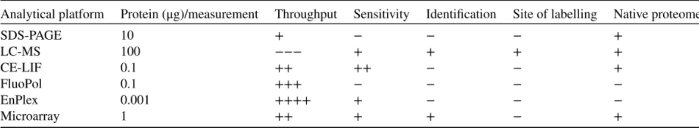
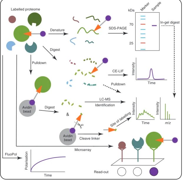
The purpose of the second analytical part of an ABPP experi-ment is to visualise the labelling event (Sieber and Cravatt,2006). Of note, ABPP does not measure catalytic activity, meaning the turnover of substrate(s) to product(s) in a certain amount of time. Instead, ABPP measures the amount of available active sites of a certain enzyme and thereby reports on the functional state of this protein. In general, the tag of the probe determines the read-out technology to be used (Tables2and3). Sodium dode-cyl sulfate polyacrylamide gel electrophoresis (SDS-PAGE) and liquid chromatography-mass spectrometry (LC-MS) are the most used analytical orthogonal platforms. In the following section, the advantages and disadvantages of these analytical platforms will be discussed (Figure2).
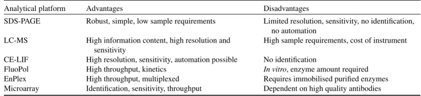
In gel-based experiments, the labelled proteins are sepa-rated and characterised by molecular weight. First, proteins are denatured using the detergent SDS, loaded on a polyacry-lamide gel and subsequently separated using gel electrophoresis (SDS-PAGE). Proteins labelled by one-step fluorescent ABPs are visualised with in-gel fluorescence scanning. Alternatively, ABPs with a biotin can be visualised using streptavidin-horseradish peroxidase (HRP) in a western blot experiment. This technique is robust, simple, has a high throughput and can be performed directly using lysates. To assign the identity of the fluores-cently labelled proteins, specific inhibitors or genetic deletion of the gene is required. Disadvantages of the gel-based ABPP include a limited resolution and sensitivity. Also, the identity of the measured proteins sometimes remains ambiguous and the possibility for automation is limited (Patricelliet al.,2001).
For LC-MS-based ABPP experiments, proteins are labelled with a biotinylated ABP, enriched using (strept)avidin chro-matography (pulldown) and digested with a protease. The
Table 2 Comparison of ABPP analytical platforms
Analytical platform Protein (μg)/measurement Throughput Sensitivity Identification Site of labelling Native proteome
SDS-PAGE 10 + − − − +
LC-MS 100 −−− + + + +
CE-LIF 0.1 ++ ++ − − +
FluoPol 0.1 +++ − − − −
EnPlex 0.001 ++++ + − − −
Table 3 Main advantages and disadvantages of each ABPP analytical platform
Analytical platform Advantages Disadvantages
SDS-PAGE Robust, simple, low sample requirements Limited resolution, sensitivity, no identification, no automation
LC-MS High information content, high resolution and sensitivity
High sample requirements, cost of instrument
CE-LIF High resolution, sensitivity, automation possible No identification
FluoPol High throughput, kinetics In vitro, enzyme amount required EnPlex High throughput, multiplexed Requires immobilised purified enzymes Microarray Identification, sensitivity, throughput Dependent on high quality antibodies
resulting peptides are separated with liquid chromatography and measured using mass spectrometry (Liet al.,2013). The mea-sured peptides will allow the identification of the labelled pro-teins. The peptides are sequenced using MS/MS experiments, and these peptide sequences are searched against a database of protein sequences. If a cleavable linker is used, the site of modification can be identified by releasing the probe-labelled peptide from the avidin bead and measuring the specific probe-peptide conju-gate (Weerapanaet al.,2007; Yanget al.,2013). This provides direct evidence that a probe has covalently labelled a protein. LC-MS-based ABPP has high resolution, sensitivity and informa-tion content. However, the throughput is low, elaborate sample preparation is needed and pulldown experiments commonly suffer from high background of abundant unlabelled proteins.
To improve the resolution, sensitivity and automation pos-sibilities for SDS-PAGE, capillary electrophoresis coupled to laser-induced fluorescence scanning (CE-LIF) has been devel-oped (Okerberget al.,2005). Proteomes labelled with a fluores-cent probe are digested with a protease and the resulting peptides separated using capillary electrophoresis. The fluorescence signal arising from probe-labelled peptides is measured. This distin-guishes proteins with similar molecular weight, which comigrate on an SDS-PAGE gel.
Fluorescence polarisation (FluoPol)-ABPP has been developed to perform high-throughput screens and to assess inhibitor kinet-ics (Bachovchinet al.,2009; Lahavet al.,2017). FluoPol mea-sures the apparent size of a molecule, because a small fluorescent probe rotates quickly in solution resulting in low polarisation of light, while a large probe-protein adduct rotates slowly giving rise to a high polarisation signal. The advantage of FluoPol com-pared to substrate assays is that it can be used to find inhibitors for poorly characterised enzymes of which the substrate is unknown. Recently, FluoPol has also been applied in cellular imaging where free and bound probe could be distinguished, thereby separat-ing the background signal from free fluorescent probes (Dubach et al.,2014). Interestingly, FluoPol can also be performed with noncovalent probes. A potential disadvantage of FluoPol is the requirement of purified or overexpressed enzyme. Typically, Flu-oPol assays only measure the potency of inhibitors against one enzyme. Recently, EnPlex was developed, a technique that makes it possible to assess both potency and selectivity of inhibitors (Bachovchinet al.,2014). Multiple purified enzymes are immo-bilised on coloured Luminex beads, with a different colour for each enzyme. These beads are mixed, incubated with inhibitor
and subsequently labelled with a biotinylated ABP, which is stained with coloured streptavidin. The bead mixture is mea-sured by flow cytometry, detecting both the identity (bead colour) and activity (streptavidin colour) of each enzyme. Owing to the requirement of multiple purified enzymes, this platform is elabo-rate to set up, but once available has the highest throughput.
A technique that has the identification advantage of LC-MS but with higher throughput is microarray ABPP (Sieberet al., 2004). The probe-labelled proteome is incubated with an antibody microarray and a fluorescence signal is measured for the probe-labelled proteins. This technique is dependent on high-quality antibodies and prior knowledge of the probe targets is required (there is no discovery possibility as with LC-MS).
Figure2andTables2and3summarise the analytical platforms
that can be coupled to ABPP. Various techniques can be combined with each other, such as SDS-PAGE and CE-LIF, which can be coupled to LC-MS to identify the tagged proteins (Bachovchin et al.,2010). In short, protein bands from SDS-PAGE can be excised and digested with a protease or using an in-gel diges-tion and the resulting peptides will be measured by LC-MS. The probe-labelled peptides from CE-LIF can be enriched using anti-fluorophore antibodies and also identified with LC-MS.
Applications
Over the last two decades, ABPP has been developed into a mature method. The labelling methods and analytical platforms have become well established. Therefore, ABPP is increasingly applied to answer biological questions by exploiting the unique ability of ABPP to directly report on enzyme activity in living biological systems. Two types of experimental setups have been widely used: comparative and competitive ABPP (Cravattet al., 2008).
Activity-based Protein Profiling
Avidin bead
Denature SDS-PAGE
kDa Marker Sample
70
25
Digest
CE-LIF
Time
Intensity
Pulldown
Digest
LC-MS
Time
Intensity
m/z
Intensity
Cleave linker
&
Identification
Site of labelling
FluoPol
Time
Polarisation
Microarray
Read-out
Pulldown
In-gel digest
Avidin bead Labelled proteome
Figure 2 Visualisation of ABPP analytical platforms: SDS-PAGE, CE-LIF, LC-MS, microarray and FluoPol.
of potential new drug targets. For example, monoacylglycerol lipase was found to more active in aggressive versus nonaggres-sive human cancer cell lines, thereby nominating this enzyme as a potential pharmacological target for cancer therapy (Nomura et al.,2010a,b). Comparative ABPP has been used in many bio-logical processes, such as host–virus interactions (Blais et al., 2010,2012), microbial virulence factors (Puriet al.,2010) and diet-induced obesity (Sadler et al.,2012). Furthermore, ABPP can be used to identify novel enzymes, such as PLA2G4E as a calcium-dependentN-acyltransferase (Oguraet al.,2016).
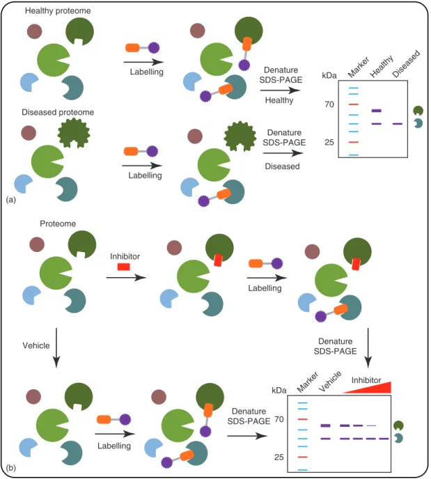
Inhibitor potency and selectivity can be simultaneously eval-uated in a competitive ABPP experiment using broad-spectrum ABPs (Figure3b) (Leunget al.,2003). ABPP efficiently guides the hit and lead optimisation process, thereby shortening the drug discovery process. Interestingly, there is also a chance for serendipitous discoveries, such as identifying novel hits for other
enzymes. In competitive ABPP a sample is pretreated with an inhibitor before the ABP is added to label residual enzyme activ-ities. A decrease in fluorescence intensity of the bands will indi-cate whether the compound interacted with a protein. Competitive ABPP is also an excellent way to confirm target engagement of an enzyme in a cellular or animal model. For example, probe1
(Table1) was used to screen a library of compounds against a
Denature
SDS-PAGE kDa Marker Healthy
70
25 Healthy proteome
Labelling
Diseased proteome
Diseased
Proteome
Inhibitor
kDa Marker Vehicle
70
25
Inhibitor Labelling
(a)
(b)
Denature SDS-PAGE Labelling
Denature SDS-PAGE
Healthy
Diseased
Vehicle
Labelling
Denature SDS-PAGE
Figure 3 ABPP experiments. (a) Comparative ABPP. (b) Competitive ABPP.
which were not identified by the classical selectivity screening assays. It is, therefore, recommended that preclinical drug dis-covery should include (competitive) ABPP to profile the drug candidate on human tissues and cells.
Competitive ABPP is, however, restricted to profiling enzyme activities identified by the probe. For an ideal drug target pro-filing study, the drug candidate itself should be converted into an ABP (Kallemeijnet al.,2012). This is, however, difficult to realise if the inhibitor does not contain a protein reactive func-tionality. A combination of broad-spectrum ABPs targeting var-ious enzyme families would therefore be ideal to get a broad overview of the selectivity profile of the drug candidate. Other
chemical proteomics techniques such as cellular thermal shift assays (CETSA) (Reinhardet al.,2015) and drug affinity respon-sive target stability (DARTS) (Lomenicket al.,2009) are used to get a proteome-wide selectivity profile; however, these are not necessarily activity-based and should be used only as comple-mentary techniques.
Future Prospects
Activity-based Protein Profiling
to enable further exploration of the enzymatically active subset of the proteome. Furthermore, new analytical platforms should be developed to enhance the sensitivity and resolution of the ABPP technique to detect low abundant enzymes and to study the effects of posttranslational modifications on the proteins. Increas-ing the throughput of ABPP experiments by usIncreas-ing automation is another desired feature. Organic chemists should develop novel probes to target novel enzyme classes and further develop cleav-able linkers to identify the site of modification with novel frag-mentation techniques such as electron transfer dissociation (Syka et al., 2004). Importantly, biologists could benefit a lot from the current ABPP toolbox. Recent examples of online, search-able databases, such as chemicalprobes.org and probes-drugs.org (Skuta et al.,2017; Arrowsmithet al.,2015), aid scientists in selecting the optimal probes. The ABPP-field could benefit from adding the best probes to these open data resources and making well-characterised probes available. ABPP will continue to play an important role in elucidating the function of proteins and the discovery and development of novel drugs.
Glossary
Covalent bond A bond that is based on the sharing of electrons and forms a stable chemical linkage.
Enzyme A protein that catalyses a chemical reaction in a biological setting.
Inhibitor A compound that blocks the activity of an enzyme. Pulldown Assay to pull certain proteins out of a solution. Proteome All the proteins expressed in a cell at a certain
moment in time.
References
Arrowsmith CH, Audia JE, Austin C, et al.(2015) The promise
and peril of chemical probes. Nature Chemical Biology11(8):
536–541.
Bachovchin DA, Brown SJ, Rosen H and Cravatt BF (2009) Iden-tification of selective inhibitors of uncharacterized enzymes by high-throughput screening with fluorescent activity-based probes. Nature Biotechnology27(4): 387–394.
Bachovchin DA, Ji T, Li W,et al.(2010) Superfamily-wide portrait
of serine hydrolase inhibition achieved by library-versus-library
screening.Proceedings of the National Academy of Sciences of the
United States of America107(49): 20941–20946.
Bachovchin DA, Koblan LW, Wu W,et al.(2014) A high-throughput,
multiplexed assay for superfamily-wide profiling of enzyme activ-ity.Nature Chemical Biology10(8): 656–663.
Baggelaar MP, Janssen FJ, Vanesbroeck ACM,et al.(2013)
Develop-ment of an activity-based probe andin silicodesign reveal highly
selective inhibitors for diacylglycerol lipase-𝛼 in brain.
Ange-wandte Chemie International Edition52(46): 12081–12085. Bedner E, Smolewski P, Amstad P and Darzynkiewicz Z
(2000) Activation of caspases measured in situ by binding of fluorochrome-labeled inhibitors of caspases (FLICA):
correla-tion with DNA fragmentacorrela-tion.Experimental Cell Research259:
308–313.
Blais DR, Lyn RK, Joyce MA,et al.(2010) Activity-based protein
profiling identifies a host enzyme, carboxylesterase 1, which is
differentially active during hepatitis C virus replication.Journal
of Biological Chemistry285(33): 25602–25612.
Blais DR, Nasheri N, McKay CS, Legault MCB and Pezacki JP (2012) Activity-based protein profiling of host-virus interactions. Trends in Biotechnology30(2): 89–99.
Blum G, von Degenfeld G, Merchant MJ, Blau HM and Bogyo M (2007) Noninvasive optical imaging of cysteine protease activity
using fluorescently quenched activity-based probes.Nature
Chem-ical Biology3(10): 668–677.
Cravatt BF, Wright AT and Kozarich JW (2008) Activity-based protein profiling: from enzyme chemistry to proteomic chemistry. Annual Review of Biochemistry77(1): 383–414.
Dubach JM, Vinegoni C, Mazitschek R,et al.(2014)In vivoimaging
of specific drug–target binding at subcellular resolution.Nature
Communications5(May): 1–9.
Ekkebus R, Van Kasteren SI, Kulathu Y,et al.(2013) On terminal
alkynes that can react with active-site cysteine nucleophiles in
pro-teases.Journal of the American Chemical Society135: 2867–2870.
van Esbroeck ACM, Janssen APA, Cognetta AB, et al. (2017)
Activity-based protein profiling reveals off-target proteins of
the FAAH inhibitor BIA 10–2474. Science 356: 1084–1087.
http://science.sciencemag.org/content/356/6342/1084/tab-pdf. Evans MJ and Cravatt BF (2006) Mechanism-based profiling of
enzyme families.Chemical Reviews106(8): 3279–3301.
Geurink PP, Prely LM, Van Der Marel GA, Bischoff R and Overkleeft HS (2012) Photoaffinity labeling in activity-based protein profil-ing.Topics in Current Chemistry324: 85–114.
Kallemeijn WW, K-y L, Witte MD, et al. (2012) Novel
activity-based probes for broad-spectrum profiling of
retain-ing B -exoglucosidasesin situandin vivo.Angewandte Chemie
International Edition8: 12529–12533.
Kato D, Boatright KM, Berger AB, et al.(2005) Activity-based
probes that target diverse cysteine protease families.Nature
Chem-ical Biology1(1): 33–38.
Lahav D, Liu B, Van Den Berg RJBHN,et al.(2017) A fluorescence
polarization activity-based protein profiling assay in the discov-ery of potent, selective inhibitors for human nonlysosomal
glu-cosylceramidase.Journal of the American Chemical Society139:
14192–14197.
Leung D, Hardouin C, Boger DL and Cravatt BF (2003) Discovering potent and selective reversible inhibitors of enzymes in complex
proteomes.Nature Biotechnology21(6): 687–691.
Li N, Kuo C-L, Paniagua G,et al. (2013) Relative quantification
of proteasome activity by activity-based protein profiling and
LC-MS/MS.Nature Protocols8(6): 1155–1168.
Liu Y, Patricelli MP and Cravatt BF (1999) Activity-based
pro-tein profiling: the serine hydrolases.Proceedings of the National
Academy of Sciences96(26): 14694–14699.
Lomenick B, Hao R, Jonai N,et al.(2009) Target identification using
drug affinity responsive target stability (DARTS).Proceedings of
the National Academy of Sciences106: 21984–21989.
Nodwell MB and Sieber SA (2012) ABPP methodology: introduction
and overview.Topics in Current Chemistry324: 1–42.
Nomura DK, Dix MM and Cravatt BF (2010a) Activity-based protein
profiling for biochemical pathway discovery in cancer. Nature
Reviews Cancer10(9): 630–638.
Nomura DK, Long JZ, Niessen S,et al.(2010b) Monoacylglycerol
lipase regulates a fatty acid network that promotes cancer
patho-genesis.Cell140(1): 49–61.
Ogura Y, Parsons WH, Kamat SS and Cravatt BF (2016)
phosphatidylethanolamines.Nature Chemical Biology12(July): 1–5.
Okerberg ES, Wu J, Zhang B,et al.(2005) High-resolution functional
proteomics by active-site peptide profiling.Proceedings of the
National Academy of Sciences of the United States of America102
(14): 4996–5001.
Patricelli MP, Giang DK, Stamp LM and Burbaum JJ (2001) Direct visualization of serine hydrolase activities in complex proteomes
using fluorescent active site-directed probes.Proteomics 1 (9):
1067–1071.
Patterson DM, Nazarova LA and Prescher JA (2014) Finding the right
(bioorthogonal) chemistry.ACS Chemical Biology9: 592–605.
Puri AW, Lupardus PJ, Deu E,et al. (2010) Rational design of
inhibitors and activity-based probes targeting clostridium difficile
virulence factor TcdB.Chemistry and Biology17(11): 1201–1211.
Reinhard FBM, Eberhard D, Werner T,et al.(2015) Thermal
pro-teome profiling monitors ligand interactions with cellular
mem-brane proteins.Nature Methods12(12): 1129–1131.
Sadler NC, Angel TE, Lewis MP,et al.(2012) Activity-based protein
profiling reveals mitochondrial oxidative enzyme impairment and
restoration in diet-induced obese mice.PLoS One7(10): 1–10.
Saghatelian A, Jessani N, Joseph A, Humphrey M and Cravatt BF (2004) Activity-based probes for the proteomic profiling of
metal-loproteases.Proceedings of the National Academy of Sciences of
the United States of America101(27): 10000–10005.
Shannon DA and Weerapana E (2015) Covalent protein modification:
the current landscape of residue-specific electrophiles. Current
Opinion in Chemical Biology24: 18–26.
Sieber SA and Cravatt BF (2006) Analytical platforms for activity-based protein profiling – exploiting the versatility of
chem-istry for functional proteomics.Chemical Communications(22):
2311–2319.
Sieber SA, Mondala TS, Head SR and Cravatt BF (2004) Microarray platform for profiling enzyme activities in complex proteomes. Journal of the American Chemical Society126(48): 15640–15641. Simon GM and Cravatt BF (2010) Activity-based proteomics of
enzyme superfamilies: serine hydrolases as a case study.Journal
of Biological Chemistry285(15): 11051–11055.
Skuta C, Popr M, Muller T,et al. (2017) Probes & drugs portal:
an interactive, open data resource for chemical biology.Nature
Methods14(8): 759–760.
Speers AE, Adam GC and Cravatt BF (2003) Activity-based protein
profilingin vivousing a copper (I) -catalyzed azide-alkyne [3+2]
cycloaddition.Journal of the American Chemical Society125(16):
4686–4687.
Syka JEP, Coon JJ, Schroeder MJ, Shabanowitz J and Hunt DF (2004) Peptide and protein sequence analysis by electron
trans-fer dissociation mass spectrometry.Proceedings of the National
Academy of Sciences of the United States of America101(26): 9528–9533.
Tornøe CW, Christensen C and Meldal M (2002) Peptidotriazoles on solid phase: [1,2,3] -triazoles by regiospecific copper (I) -catalyzed
1, 3-dipolar cycloadditions of terminal alkynes to azides.Journal
of Organic Chemistry67: 3057–3064.
Weerapana E, Speers AE and Cravatt BF (2007) Tandem orthogonal proteolysis-activity-based protein profiling (TOP-ABPP) – a gen-eral method for mapping sites of probe modification in proteomes. Nature Protocols2(6): 1414–1425.
Willems LI, Van Der Linden WA, Li N,et al.(2011)
Bioorthog-onal chemistry: applications in activity-based protein profiling. Accounts of Chemical Research44(9): 718–729.
Wright AT and Cravatt BF (2007) Chemical proteomic probes for
profiling cytochrome P450 activities and drug interactionsin vivo.
Chemistry and Biology14(9): 1043–1051.
Yang Y, Hahne H, Kuster B and Verhelst SHL (2013) A simple and effective cleavable linker for chemical proteomics applications. Molecular & Cellular Proteomics: MCP12(1): 237–244.
Zhao Q, Ouyang X, Wan X,et al.(2017) Broad-spectrum kinase
profiling in live cells with lysine-targeted sulfonyl fluoride probes. Journal of the American Chemical Society139: 680–685.
Further Reading
Book on methods
Overkleeft H and Florea BI (eds) (2017)Activity-Based Proteomics,
Methods and Protocols, 1st edn. New York: Humana Press.
Review comparing ABPP to other chemical proteomics techniques
Ziegler S, Pries V, Hedberg C and Waldmann H (2013) Target identification for small bioactive molecules: finding the needle in
the haystack.Angewandte Chemie International Edition52(10):
2744–2792.
Example of novel proteomes being studied
Zweerink S, Kallnik V, Ninck S,et al.(2017) Activity-based protein
profiling as a robust method for enzyme identification and
screen-ing in extremophilic archaea.Nature Communications8(May):
15352.
Covalent probes for ligandability instead of activity
Backus KM, Correia BE, Lum KM,et al.(2016) Proteome-wide
covalent ligand discovery in native biological systems.Nature534
(7608): 570–574.
Overview how to confirm probe targets
Kovács J and van der Hoorn RAL (2016) Twelve ways to confirm
tar-gets of activity-based probes in plants.Bioorganic and Medicinal