Original Research Article
Radio-surgical correlation of chronic suppurative otitis media
Yogeesha B. S., Nagaraj Maradi*, Ravi Shekhar, Rohini D. Urs
INTRODUCTION
Acute or recurrent infection of the middle ear cleft may cause irreversible inflammatory changes termed as chronic otomastoiditis.1 Chronic Otitis media is one of the most common infectious diseases with dreaded complications and considerable morbidity. This is of particular importance in a developing country such as India with its huge disease prevalence, wherein it translates to significant amount of loss of labour, leading to financial burden on the society. Hence it becomes important to detect the disease in its earliest stages and treat it accordingly. This is particularly true in cases of
cholesteatoma because of the nature of the disease and its associated complications.
With the advances in the field of radiology the role of
modern multi-detector, multi-row computerized
tomography scanning systems in evaluating
cholesteatoma and its complications is indispensible.2 High resolution computerized tomography (HRCT) of temporal bone also gives an idea about the anatomical variations helping the surgeon prepare for the surgery. But it cannot differentiate between cholesteatoma, granulation tissue, scar tissue or secretions in the middle ear. The role of turbo spin echo diffusion weighted Department of Otorhinolaryngology and Head and Neck Surgery, SSIMS and RC, Jnanashankara Campus, Davangere, Karnataka, India
Received: 30 August 2019
Revised: 10 November 2019
Accepted: 14 November 2019
*Correspondence:
Dr. Nagaraj Maradi,
E-mail: nagarajmaradi@gmail.com
Copyright: © the author(s), publisher and licensee Medip Academy. This is an open-access article distributed under the terms of the Creative Commons Attribution Non-Commercial License, which permits unrestricted non-commercial use, distribution, and reproduction in any medium, provided the original work is properly cited.
ABSTRACT
Background: Chronic suppurative otitis media (CSOM) because of disease nature and location of vital structures like middle ear ossicles, facial nerve, and lateral semicircular canal poses clinical as well as radiological challenge in diagnosis, especially the squamosal variety. Hence this study to evaluate radio-surgical correlation in cases of CSOM.
Methods: We retrospectively analysed 92 case records who met the inclusion criteria. Their pre-operative high-resolution computed tomography (HRCT) temporal bone imaging was evaluated for erosion of the ossicular chain and the fallopian canal. This was correlated with the surgical findings noted intra-operatively. The appropriate statistical analysis was carried out. The radio-surgical correlation was evaluated by Cohen’s kappa value.
Results: The kappa value for status of ossicular chain was 0.805 and 0.384 for status of fallopian canal. HRCT imaging had a positive predictive value and negative predictive value of 94.3% and 85.3% respectively, in detecting ossicular chain erosion. In detecting fallopian canal erosion, HRCT showed a sensitivity of 33.3%. Analysing the individual ossicles, we found kappa to be 0.266 for malleus, 0.463 for incus and 0.827 for stapes.
Conclusions: There was excellent radio-surgical correlation for ossicular chain erosion while it was poor for fallopian canal erosion. HRCT showed excellent radio-surgical correlation for Stapes, moderate for Incus and poor for Malleus. In-spite of its shortcomings in differentiating cholesteatoma and non-cholesteatomatous pathologies of the middle ear cleft, HRCT imaging plays a key role in assessing the status of the ossicles and fallopian canal.
Keywords: Fallopian canal erosion, High resolution computerised tomography temporal bone, Ossicular erosion, Radio-surgical correlation
magnetic resonance imaging (MRI) is emerging to overcome this inherent disadvantage of HRCT imaging.3 HRCT temporal bone imaging is also the imaging modality of choice to evaluate ossicular chain status and bony abnormalities afflicting the temporal bone like otosclerosis, osteoma, exostosis etc.
We attempt to evaluate the accuracy of HRCT in assessing ossicular and fallopian canal status in cases of chronic suppurative otitis media.
METHODS
This is a retrospective analysis of patients from June 2015 to August 2017 who were treated at ENT department in SSIMS and RC, a tertiary care referral hospital in Davanagere, Karnataka. Patients who attended ENT out-patient department and were ordered HRCT temporal bone imaging were searched in the database. Only patients who were diagnosed to have chronic suppurative otitis media were included in the study. Their pre-operative HRCT findings and intra-operative findings were correlated with respect to the status of the ossicles and status of the facial nerve canal were intact/eroded.
Type of the surgery was determined by the clinical examination, computerized tomography scan findings and intraoperative findings.
Statistical analysis was done using SPSS statistical analysis software (version 23). Correlation of HRCT and surgical findings was determined by the kappa coefficient (κ) using SPSS software. The Cohen’s kappa coefficient4 ranged from 0-1; with 0 indicating no correlation and 1 indicating complete correlation between the two factors. Kappa coefficients in the range 0-0.4, >0.4-0.6, >0.6-0.8, and >0.8-1.0 indicated poor, moderate, good, and excellent correlation, respectively.
RESULTS
A total of 100 patients who underwent HRCT temporal bone scanning from the department of ENT were searched. 92 of them were diagnosed to have chronic otitis media. 7 patients had otoscerosis, 1 patient had right external auditory canal osteoma and were excluded from the study.
Among the 92 patients, 59 were male and 33 were female. The youngest patient was 6 years old, while the
oldest was 58 years old. The mean age of the study population was 27.17 years±13.8 years Figure 1.
Among 92 patients, 82 patients were diagnosed with chronic suppurative otitis media, attico-antral disease (CSOM-AAD), (40 among the 82 having right sided disease, 34 having left sided disease, 8 having bilateral disease), 9 patients were diagnosed as aural polyp (5 right, 4 left) and 1 had chronic suppurative otitis media, tubo-tympanic disease (CSOM-TTD) Figure 2.
Figure 1: Number of patients with different diagnoses undergoing HRCT temporal bone imaging.
RT: Right, LT: Left, CSOM: Chronic suppurative otitis media, AAD: Attico-antral disease, TTD: Tubo-tympanic disease.
Figure 2: Various procedures performed and the number of cases.
CM: Cortical mastoidectomy, MRM: Modified radical mastoidectomy.
Table 1: Correlation between HRCT temporal bone and intra-operative findings.
Variable True
positive True negative
False positive
False negative
Sensitivity %
Specificity %
PPV %
NPV %
Kappa value
Ossicular chain
erosion 50 29 3 5 90.9 90.6 94.3 85.3 0.805
Fallopian canal
erosion 1 88 1 2 33.3 98.9 50 97.8 0.384
8
1
4
34
5
40
0 5 10 15 20 25 30 35 40 45
BIL CSOM AAD
BIL CSOM TTD
LT AURAL POLYP
LT CSOM AAD
RT AURAL POLYP
RT CSOM AAD
NO OF PATIENTS Column1 Column2
1
41
1 1
47
1 0
5 10 15 20 25 30 35 40 45 50
Table 2: Radio-surgical correlation for individual ossicles.
Variable True
positive
True negative
False positive
False
negative Sensitivity % Specificity %
Kappa value
Malleus 24 1 3 1 96 25 0.266
Incus 26 1 1 1 96.3 50 0.463
Stapes 20 7 1 1 95.2 87.5 0.827
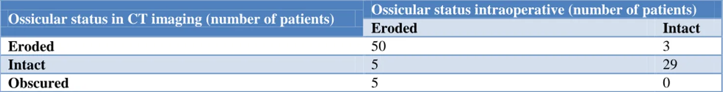
Table 3: Correlation of ossicular status in HRCT temporal bone and intraoperative (number of patients).
Ossicular status in CT imaging (number of patients) Ossicular status intraoperative (number of patients)
Eroded Intact
Eroded 50 3
Intact 5 29
Obscured 5 0
Surgery wise, 88 out of 92 patients underwent Modified Radical Mastoidectomy, (47 right, 41 left), 2 patients underwent atticotomy (1 each of right and left), 1 patient had cortical mastoidectomy and 1 had tympanoplasty Table 1.
The above table shows the results in terms of statistical analysis. HRCT temporal bone imaging had a sensitivity of 90.9%, specificity of 90.6% in detecting the ossicular chain erosion. HRCT temporal bone could detect ossicular erosion in 50 out of 55 patients with a positive predictive value of 94.3%, and it could successfully rule out ossicular erosion in 29 out of 32 patients with a negative predictive value of 85.3%. Kappa value for inter-test reliability was 0.805.
While studying the integrity of fallopian canal on HRCT Temporal bone, the sensitivity was 33.3% and specificity was 98.9%. Of the 92 patients, 3 patients had erosion of the fallopian canal as noted intra-operatively, among which 1 was detected and 2 were missed on HRCT temporal bone pre-operatively. And among the 89 patients with an intact fallopian canal, 1 was falsely reported as eroded by HRCT temporal bone. Kappa value for inter-test reliability was 0.384 Table 2.
The sensitivity and specificity of HRCT temporal bone in predicting erosion of malleus, incus, and stapes is 96%, 96.3%, 95.2% and 25%, 50%, 87.5% respectively. While the Kappa values for correlation are 0.266 for malleus, 0.463 for Incus, 0.827 for stapes indicating correlation being poor for malleus, moderate for Incus and excellent for stapes Table 3.
This table shows that among 53 patients who were diagnosed to have ossicular erosion on HRCT Temporal bone imaging, 50 had ossicular erosion intra-operatively while 3 had an intact ossicular chain. Also, among the 34 patients who were diagnosed to have an intact ossicular chain by HRCT Temporal bone, 29 had intact chain while 5 were found with eroded ossicular chain intra-operatively. The 5 patients whose ossicular status was
reported as obscured by the radiologist in fact had ossicular erosion intraoperatively.
DISCUSSION
Chronic suppurative otitis media with its varied
presentation presents a challenge to the
otorhinolaryngologist in its evaluation and management. The role of HRCT Temporal bone in cases of cholesteatoma is already well established. But its routine use in chronic suppurative otitis media of tubo-tympanic variety is not warranted. By virtue of its projection advantage and superior contrast resolution compared to conventional tomography, HRCT imaging can accurately detail the anatomy as well as demonstrate the pathology in the middle ear cleft.2
In the present study all the cases of chronic suppurative otitis media who underwent HRCT temporal bone imaging were retrospectively analysed. We found 92 such cases. The pre-operative HRCT findings were noted with respect to the status of the ossicular chain- intact/eroded, intactness of the fallopian canal. The case files were reviewed for the surgery performed and intra-operative findings and these were then correlated with HRCT findings statistically. There were 59 males and 33 females in our study with a male: female ratio of 1.79:1. This is similar to the ratio described in the literature. Majeed et al, reported a ratio of 1.77:1, which was comparable to the study done by Vlastarakos et al.2,5
If we analyses the patients according to the disease category among the 92 patients, 82 had chronic suppurative otitis media attico-antral disease, 9 were diagnosed to have aural polyp and 1 had chronic suppurative otitis media, tubo-tympanic disease (Figure 1). As explained, HRCT has a crucial role in describing the anatomy and evaluating the pathology in chronic suppurative otitis media attico-antral disease cases. All 82 cases of chronic suppurative otitis media attico-antral disease underwent either atticotomy if the disease was limited (2 cases) or modified radical mastoidectomy (MRM) if the disease was extensive (80 cases). Among the 9 cases of aural polyp, 8 cases were found to have underlying cholesteatoma and underwent MRM and 1 patient underwent cortical mastoidectomy. The lone Chronic suppurative otitis media, tubo-tympanic disease case underwent tympanoplasty (Figure 2).
Despite its limitations in accurately predicting the middle ear cleft soft tissue density as cholesteatoma, HRCT Temporal bone has a pivotal role in such cases because of its ability to detect the bone erosion, ossicular erosion, lateral semicircular canal defects, and erosion of the fallopian canal. In our study the clinical evidence of cholesteatoma correlated well with imaging evidence as all those clinically diagnosed cases showed bony erosion associated with soft tissue density in the middle ear cleft on HRCT imaging. This is comparable to the findings noted in the study by Majeed et al, and Sirigiri et al, who demonstrated 100% sensitivity and 90% specificity in
detecting cholesteatoma through computerized
tomography.2,7 Blunting of scutum is the first sign of attic cholesteatoma on HRCT and as such can detect the early stages of cholesteatoma, which can be beneficial to the patients as a limited surgery like atticotomy will clear the disease and preserve hearing. Rocher and colleagues proved this point by demonstrating excellent correlation for the scutum erosion on HRCT with a κ>0.7.8
Ossicular erosion is picked up well by HRCT temporal bone imaging. Studies have documented that the common ossicles to be affected are the long process of the incus, stapes supra-structure and handle of the malleus, in that order.2,9 Visualizing the malleus and incus is easier in HRCT especially in coronal axis, whereas incudo-stapedial joint and stapes, which are the common structures eroded by the pathology, are hard to visualize. This is because of the relatively thinner cross-sectional area of these ossicles compared to malleus or incus and the middle ear pathology appearing as soft tissue attenuation on HRCT. But Jackler et al in their study opined that the axial view of HRCT can best detect the Stapes and the long process of Incus.10
Our study showed high sensitivity for HRCT in detecting erosions of all the ossicles although there was poor radio-surgical correlation for Malleus (κ=0.266) while there was moderate correlation for Incus (κ=0.463), excellent correlation for Stapes (κ=0.827). This finding is similar to that reported by Chee et al, who showed excellent
radio-surgical correlation for malleus (κ=0.83) and stapes (κ=0.94), good radio-surgical correlation for incus (κ=0.62).11 Also, Devasamudra et al, showed high
sensitivity for HRCT in detecting erosions of incus (85%), stapes (82.3%) and low sensitivity for Malleus (68.75%).12 In contrast, Rogha et al, reported good radio-surgical correlation for Malleus (κ=0.61), poor for Incus (κ=0.36) and stapes (κ=0.27).12
In our study, the HRCT reporting with respect to the status of the ossicles was not uniform. 29 out of the 53 reports having ossicular erosion documented the status of the individual ossicles. 24 reports just mentioned about the ossicular discontinuity without mentioning the individual ossicular status. Thus, we could analyse the sensitivity, specificity, PPV, NPV and kappa values for the 29 cases only (Table 2). So, the emphasis must be on uniform reporting of the HRCT Temporal scans by the radiologists. It will be better if the radiologist and the otorhinolaryngologist come together and devise a reporting protocol so that data archiving and retrieval will be easier and comprehensive. Such said protocol shall mention the important anatomical and surgical landmarks with emphasis on ossicles, fallopian canal, lateral semicircular canal, tegmen plate, sigmoid plate, which can guide the surgeon in planning and executing the surgery. The 5 HRCT reports which mentioned the status of the ossicles as obscured when correlated surgically were found to have eroded ossicles, mainly Incus and Stapes (Table 3). This is indicative of the soft tissue attenuation property of the cholesteatoma or any other middle ear pathology like granulations, secretions, polypoidal middle ear mucosa which obscures precise identification of the ossicles. Hence this finding on HRCT should alert the surgeon to possibility of an extensive disease.
Facial nerve palsy is a dreaded complication of the surgery for the middle ear cleft. Pre-operative knowledge of the status of the fallopian canal is an invaluable tool for any otorhinolaryngologist. Unfortunately, the radio-surgical correlation is poor for the fallopian canal erosion. The tympanic segment of the facial nerve is the commonly affected part. Both coronal and axial cross sections are important to evaluate the fallopian canal. Coronal views are better for evaluating tympanic segment while axial sections show mastoid segment dehiscence more clearly.10
CONCLUSION
HRCT temporal bone imaging is an essential and indispensable investigative tool in pre-operative evaluation of chronic suppurative otitis media attico-antral disease cases. In spite of its shortcomings in identifying and segregating cholesteatoma from non-cholesteatomatous middle ear cleft pathologies; it shows radio-surgical correlation which is excellent for detecting stapes erosion, moderate for incus erosion and poor for malleus and fallopian canal erosion. The turbo-spin echo diffusion weighted MRI sequences can be used to identify cholesteatoma more precisely. Also, it is imperative that the reporting of HRCT Temporal bone should be uniform, mentioning key anatomical and surgical landmarks so as to avoid confusion and help in data archiving for future references.
ACKNOWLEDGEMENTS
The authors would like to thank department of radiology for their help and guidance during this study. Authors extend their sincere gratitude to the staff of the department of otorhinolaryngology and administration of the SSIMS and RC for their support.
Funding: No funding sources Conflict of interest: None declared Ethical approval: Not required
REFERENCES
1. Biswas A. Pure tone audiometry. In: Biswas A (ed) Clinical audio-vestibulometry, 4th edn. Bhalani publishing house, Mumbai; 2009:5-16.
2. Majeed J, Reddy LS. Role of CT mastoids in the diagnosis and surgical managementof chronic inflammatory ear diseases. Indian J Otolaryngol Head Neck Surg. 2017;69(1):113-20.
3. Vaid S, Kamble Y, Vaid N, Bhatti S, Rawat S, Nanivadekar A, et al. Role of magnetic resonance imaging in cholesteatoma: the Indian experience. Indian journal of otolaryngology and head and neck surgery: official publication of the Association of Otolaryngologists of India, 2011;65(Suppl3):485-92.
4. Cohen J. A coefficient of agreement for nominal scales. Educ Psychol Meas. 1960;20:37-46.
5. Vlastarakos PV, Kiprouli C, Pappas S, Xenelis J, Maragoudakis P, Troupis G, et al. CT scan versus surgery: how reliable is the pre-operative radiological assessment in patients with chronic otitis media. Eur Arch Otorhinolaryngol. 2012;269:81-6.
6. Paparella MM, Kim CS. Mastoidectomy update. Laryngoscope.1977;87:88.
7. Sirigiri RR, Dwaraknath K. Correlative study of HRCT inattico-antral disease. Indian J Otolaryngol Head Neck Surg. 2011;63:155-8.
8. Rocher P, Carlier R, Attal P, Doyon D, Bobin S. Contribution and role of the scanner in the preoperative evaluation of chronic otitis. Radiosurgical correlation apropos of 85 cases. Ann Otolaryngol Chir Cervicofac. 1995;112(7):317-23. 9. Gül A, Akdağ M, Kiniş V, Yilmaz B, Şengül E,
Teke M, et al. Radiologic and surgical findings in chronic suppurative otitis media. J Craniofac Surg. 2014;25(6):2027-9.
10. Jackler RK, Dillon WP, Schindler RA. Computed tomography in suppurative ear disease: A correlation of surgical and radiographic findings. The Laryngoscope. 1984;94:746-52.
11. Chee NW, Tan TY. The value of pre-operative high-resolution CT scans in cholesteatoma surgery. Singapore Med J. 2001;42(4):155-9.
12. Rogha M, Hashemi SM, Mokhtarinejad F,
Eshaghian A, Dadgostar A. Comparison of preoperative temporal bone CT with intraoperative findings in patients with cholesteatoma. Iran J Otorhinolaryngol. 2014;26(74):7-12.
13. Rocher P, Carlier R, Attal P, Doyon D, Bobin S. Contribution and role of the scanner in the preoperative evaluation of chronic otitis. Radiosurgical correlation apropos of 85 cases. Ann Otolaryngol Chir Cervicofac. 1995;112(7):317-23.