ARTIGO ORIGINAL
Carotid Atherosclerosis and White Matter
Hypodensities: a Controversial Relationship
Aterosclerose Carotídea e Hipodensidades da Substância Branca: uma Relação
Controversa
1. Departamento de Imagem Médica. Centro Hospitalar e Universitário de Coimbra. Coimbra. Portugal. 2. Departamento de Neurologia. Centro Hospitalar e Universitário de Coimbra. Coimbra. Portugal. 3. Faculdade de Medicina da Universidade de Coimbra. Coimbra. Portugal.
Recebido: 24 de Dezembro de 2013 - Aceite: 21 de Abril de 2014 | Copyright © Ordem dos Médicos 2014
Ricardo FÉLIX-MORAIS1, João SARGENTO-FREITAS2, Fernando SILVA2, Gustavo CORDEIRO2, César NUNES1, Joana
RIBEIRO3, Miguel CORDEIRO1, Cristina MOURA1, Luís CUNHA2
Acta Med Port 2014 Sep-Oct;27(5):581-586
RESUMO
Introdução: As hipodensidades da substância branca de provável etiologia vascular, são uma causa importante de morbilidade, condi-cionando deterioração cognitiva. No entanto, numerosas dúvidas persistem quanto à sua fisiopatologia. O objectivo deste estudo é clarificar o papel da aterosclerose carotídea e outros factores de risco vascular no desenvolvimento das hipodensidades da substância branca de provável etiologia vascular.
Material e Métodos: Realizou-se uma avaliação imagiológica, por tomografia computadorizada crânio-encefálica e ecografia carotí-dea, com menos de um mês de intervalo. Procedeu-se à recolha de informação sobre os factores de risco vascular. Determinámos associações independentes entre hipodensidades da substância branca de provável etiologia vascular, espessura da íntima média carotídea, estenose carotídea ateromatosa e factores de risco vascular.
Resultados: Foram incluídos 472 doentes, idade média de 67,32 (DP: 14.75), 274 (58,1%) sexo masculino. Os preditores indepen-dentes da hipodensidades da substância branca de provável etiologia vascular foram: idade (OR: 1,067, 95% IC: 1,049 – 1,086,
p < 0,001) e a hipertensão (OR: 1,726, 95% IC: 1,097 – 2,715, p = 0,018). Não foi encontrada uma associação entre a espessura da intima média carotídea (OR: 2,613, 95% IC: 0,886 – 7,708, p = 0,082) ou grau de estenose carotídea (OR: 1,021, 95% IC: 0,785 – 1,328,
p = 0,877) e hipodensidades da substância branca de provável etiologia vascular.
Discussão: Dos diversos factores de risco analisados, apenas a idade e hipertensão se associaram de forma independente às hipodensidades da substância branca de provável etiologia vascular. Não foi encontrada uma relação entre a aterosclerose extra-craniana, expressa pela espessura do complexo intima-média ou grau de estenose, com o desenvolvimento de hipodensidades da substância branca de provável etiologia vascular. Sendo a aterosclerose um fenómeno sistémico, estes achados sugerem que as hi-podensidades da substância branca de provável etiologia vascular, tenham um mecanismo alternativo ou concorrente à aterosclerose no seu desenvolvimento.
Conclusão: Os dados deste estudo, sugerem que a idade e hipertensão sejam os principais factores de risco no desenvolvimento de hipodensidades da substância branca de provável etiologia vascular. Não foi encontrada uma associação independente entre a aterosclerose carotídea e as hipodensidades da substância branca de provável etiologia vascular.
Palavras-chave: Cerébro; Doença Carotídea.
ABSTRACT
Introduction: White matter hypodensities of presumed vascular origin, are recognized as an important cause of morbidity with estab-lished clinical and cognitive consequences. Nonetheless, many doubts remain on its physiopathology. Our goal is to clarify the potential role of carotid atherosclerosis and other vascular risk factors in the development of white matter hypodensities of presumed vascular origin.
Material and Methods: We included patients that underwent CT brain scan and neurosonologic evaluation within a one-month period. Full assessment of vascular risks factors was performed. We seek to find independent associations between white matter hypodensi-ties of presumed vascular origin, carotid intima-media thickness and vascular risk factors.
Results: 472 patients were included, mean age was 67.32 (SD: 14.75), 274 (58.1%) were male. The independent predictors of white matter hypodensities of presumed vascular origin were age (OR: 1.067, 95% IC: 1.049 – 1.086, p < 0.001) and hypertension (OR: 1.726, 95% IC: 1.097 – 2.715, p = 0.018). No association was found between IMT (OR: 2.613, 95% IC: 0.886 – 7.708, p = 0.082) or carotid artery stenosis (OR: 1.021, 95% IC: 0.785 – 1.328, p = 0.877) and white matter hypodensities of presumed vascular origin. Discussion: Only age and hypertension proved to have an independent association with white matter hypodensities of presumed vascular origin. Carotid atherosclerosis, evaluated by IMT and the degree of carotid artery stenosis, showed no association with white matter hypodensities of presumed vascular origin. Since atherosclerosis is a systemic pathology, these results suggest that alternative mechanisms are responsible for the development of white matter hypodensities of presumed vascular origin.
Conclusion: Age and hypertension seem to be the main factors in the development of white matter hypodensities of presumed vas-cular origin. No association was found between carotid atherosclerosis and white matter hypodensities of presumed vasvas-cular origin. Keywords: Brain; Carotid Artery Disease.
35
35 ano
s a p
rom ov
er
as
ciê
CA
P
ORT
UGUE
SA
1979 - 2014
INTRODUCTION
White matter abnormalities are present in brain imag-ing in 11-21% of adults aged 64 years of age, increas-ing up to 94% at age 821 and may have different names.
In 1987, Hachinski2 introduced the term leukoaraiosis (from
ARTIGO ORIGINAL on CT-scan and/or MRI. These abnormalities are shown as hyperintensities on T2, proton density (PD) and fluid
attenu-ated inversion recovery (FLAIR) sequences and hypoin-tensities on T1-weighted MRI sequences, FLAIR being the most sensitive for its detection. On CT-scan imaging, these are shown as hypointensity areas with undefined margins.3
Other terms have been used, namely ischaemic leukoen-cephalopathy, white matter hyperintensities or age-depen-dent white matter abnormalities. These different terms show the persisting doubts regarding its physiopathology and its controversial clinical-radiological correlation. A recent inter-national consensus has standardised the terminology and these abnormalities should now be referred to as white mat-ter hypointensities of presumed vascular aetiology (WMH-PVE) on CT-scan imaging or as white matter hyperintensi-ties on MRI imaging.3 Different studies show that white
mat-ter abnormalities are associated to a cognitive decline4,5 and
their extension and progression are key-factors for this im-pairment. They are also related to gait disturbances, urinary incontinence, depression and falls,6 as well as to increased
stroke risk.7 Other studies have found an association
be-tween the presence of extensive white matter lesions and an increase in mortality.8 White matter abnormalities are
probably based on multifactorial pathophysiological factors. The almost endemic prevalence of WMHPVE in this age group and their association to an increase in co-morbidity and mortality highlights the importance of identification of risk factors for their development, in order to design primary and/or secondary prevention actions. Some studies sug-gest that atherosclerosis has a pathophysiological role in this pathology.9 Common carotid artery intima-media
thick-ness (IMT) and the presence of atheromatous plaques in carotid arteries are validated and largely used markers to evaluate extracranial atherosclerosis, having been related to cerebrovascular and coronary complications,10 providing
the rationale for the study of the relationship between extra-cranial atherosclerosis and the development of white matter abnormalities.
Our study aimed to clarify the differential contribution of several cerebrovascular risk factors as well as carotid
atherosclerosis to the development of WMHPVE.
MATERIAL AND METHODS
Our study involved patients who were consecutively referred to the Neurology Department at the Hospital and University Centre of Coimbra and underwent brain CT-scan and carotid ultrasound less than one month apart, between January and August 2011. An evaluation of the risk factors was carried out, including the patient’s age as well as the presence of hypertension, dyslipidemia, diabetes mellitus, atrial fibrillation, congestive heart failure, smoking habit and obesity. The patients presenting with pathologies showing severe brain abnormalities on imaging like tumours or extensive intracranial haemorrhage were excluded from the study, as these would restrict the correct evaluation of any white matter abnormality. Our study was submitted and approved by the hospital’s Ethics Committee.
Imaging - Age-Related White Matter Changes (ARWMC) classification: The imaging evaluation of white matter abnormalities was independently carried out by two similarly experienced neuro-radiologists blinded to clinical information. A semi-quantitative classification of white matter abnormalities used the Age-Related White Matter Changes rating scale11 where 0 score corresponds to the
absence of abnormalities, 1 to focal abnormalities, 2 to initial confluence of lesions and3 to confluent lesions. When the assigned score was different between both neuro-radiologists, the final score was defined by consensus. The hypodensities located to the basal ganglia and to the brainstem were not included in the evaluation, according to the new consensus rules.3 Lacunar-type vascular
lesions were excluded, described by round 3-15 diameter lesions with a well-defined margin and the same density as cerebrospinal fluid.3,11
All brain CT-scan exams were performed on 64-detector GE Light Speed equipment, with the following technical parameters: 512 x 512 matrix image, SFOV 25-cm reconstruction, CTDlvol 76 mGy, 330 mAs, 120 kV. A 2.5mm slice thickness was used for the infra-tentorial region and
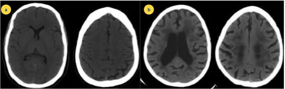
Figure 1 – ARWMC grading scale. a) – Axial CT scan slices: the absence of white matter abnormalities may be observed, ARWMC rating score 0. b) – Axial CT scan slices: presence of periventricular white matter abnormalities with extension to the deep and subcortical white matter– ARWMC rating score 3.
ARTIGO ORIGINAL
5mm thickness for supra-tentorial region.
Carotid ultrasound: The haemodynamic evaluation was
out by two neuro-sonologists with identical experience, using a Logiq 7 model General Electric® ultrasound system,
with a 7.5 MHz linear probe for extracranial evaluation. Each patient was also evaluated by one of two neuro-sonologists with identical experience. Imaging was obtained with the patient lying in the decubitus position, with the neck slightly extended and the head in contralateral rotation to the examined side. IMT was measured at the common carotid artery, 10mm proximal to its bifurcation, at optimal angles of Doppler insonation.10 Manual measurement of IMT was
obtained in both common carotid arteries and the average value was used for statistical analysis. The quantification of atheromatous stenosis was obtained at the origin of the internal carotid artery (ICA), according to Grant 2003 criteria.10 Every patient’s ICA was bilaterally classified as:
normal (no visible atheromatous plaque), < 50% stenosis (presence of atheromatous plaques with no haemodynamic meaning), 50-69% stenosis, > 70% stenosis to near occlusion, near occlusion and total occlusion. The highest degree of stenosis visible on one ICA for each patient was used for statistical analysis (for instance: in the presence of <50% stenosis in the left and 50-69% stenosis in the right, the latter was considered for analysis). Non-atheromatous pathologies such as dissection, vasculitis or cardiac embolism, were excluded from the study.
Vascular risk factors
We considered the following vascular risk factors in our study: patent’s age, hypertension, dyslipidemia, type-1 and 2 diabetes mellitus (DM) (both forms were considered together in statistical analysis), obesity (defined as body mass index > 30), congestive heart failure (CHF), atrial fibrillation (AF) and smoking habit (active or former); these were identified using a questionnaire supplied by the patient’s GP, who had requested the imaging procedures and confirmed by analysis of the clinical records.
Statistical analysis
SPSS Statistics, version 17 software was used for statistical analysis. A cross-sectional study involving all patients was carried out. Upon characterisation of the group of patients, including the average (standard deviation) for continuous variables and the absolute number (percentage) for categorical variables, two models of multivariate ordinal regression were carried out including all studied vascular risk factors, the IMT or degree of carotid atheromatous stenosis. Given the large size of our group of patients, the number of variables and the aims of the study, we included all the studied variables in the models of regression. Statistical significance was considered when p < 0.05.
RESULTS Population
Our study involved 472 patients. The demographic characteristics of our group of patients as well as the prevalence of vascular risk factors and IMT measurement are shown in Table 1.
Risk factors
The results of the ordinal regression for the identification of independent WMHPVE predictive factors, including all vascular risk factors in the study and the maximum degree of carotid stenosis are shown in Table 2.
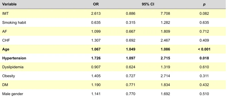
The results of the ordinal regression for the identification of independent WMHPVE predictive factors, including all vascular risk factors in the study and carotid artery IMT are shown in Table 3.
The analysis of the relationship between vascular risk factors and the WMHPVE showed a strong independent association between the presence of WMHPVE and the patient’s age (OR: 1.072, 95% CI: 1.054-1.090, p < 0.001) together with the presence of hypertension (OR: 1.797, 95% CI: 1.163-7.776, p = 0.008). We did not find any independent association between the IMT or carotid stenosis and the presence of WMHPVE.
Figure 2 - IMT measurement. IMT was measured by manual technique in both common carotid arteries (a and b); IMT is increased in b.
ARTIGO ORIGINAL DISCUSSION The association between the WMHPVE and
cerebrovascular risk factors is controversial.12,13 In our study,
we did not find any association between the presence of WMHPVE and dyslipidemia, smoking habit, atrial fibrillation, obesity, diabetes or congestive heart failure.
The characteristics of our group of patients (Table 1) reveal that patients with more intense WMHPVE also display more intense carotid stenosis and higher IMT. However, in the multivariate analysis (Table 2 and 3) the presence of carotid atherosclerosis was not statistically significantly correlated with WMHPVE, suggesting that carotid atherosclerosis is more present in the elderly and
hypertensive patients but in isolation is not a risk factor for the development of WMHPVE. Atherosclerosis is a systemic process,14 and a correlation between WMHPVE
and extracranial atherosclerosis, using the degree of carotid stenosis and IMT would be expected. The lack of correlation found in our study suggests that even though this one of the most used markers in current literature,10 the
presence of an alternative or contributing mechanism to the development of atherosclerosis should be sought. One of these mechanisms is the disruption of blood-brain barrier (BBB) by a process secondary to endothelial dysfunction of small intracranial blood vessels.14-16
Table 1 - Vascular risk factors and carotid IMT on our group of patients
Variable Total population (n = 472) No white-matter hypodensities (n = 160) White-matter hypodensities (n = 312)
Age 67.32 (14.75) 58.33 (15.16) 71.93 (12.21)
Male gender 274 (58.1%) 103 (64.4%) 171 (54.8%) Hypertension 331 (70.1%) 90 (56.3%) 241 (77.2%)
DM 112 (23.7%) 27 (16.9%) 85 (27.2%)
Dyslipidemia 219 (46.4%) 72 (45.0%) 147 (47.1%)
Smoking habit 42 (8.9%) 21 (13.1%) 21 (6.7%)
AF 88 (18.6%) 15 (9.4%) 73 (23.4%)
Obesity 41 (8.7%) 11 (6.9%) 30 (9.6%)
CHF 46 (9.7%) 10 (6.3%) 36 (11.5%)
IMT 0.79 (0.19) 0.71 (0.20) 0.83 (0.19)
Presence of carotid plaques 179 (37.9%) 78 (48.8%) 254 (81.4%)
AF: Atrial fibrillation; DM: Diabetes Mellitus; CHF: Congestive heart failure; IMT – Common carotid artery intima-media thickness.
Table 2 - Ordinal regression for identification of independent predictive factors for white-matter hypodensities, including vascular risk factors and the highest degree of carotid stenosis
Variable OR 95% CI p
Carotid stenosis 1.021 0.785 1.328 0.877
Smoking habit 0.724 0.368 1.423 0.349
AF 1.142 0.707 1.846 0.587
CHF 1.260 0.681 2.329 0.462
Age 1.072 1.054 1.090 < 0.001
Hypertension 1.797 1.163 2.776 0.008
Dyslipidemia 0.941 0.651 1.359 0.745
Obesity 1.277 0.702 2.323 0.424
DM 1.190 0.778 1.818 0.422
Male gender 1.125 0.771 1.642 0.540
ARTIGO ORIGINAL
Analysis of positive associations
Hypertension: Both systolic and diastolic hypertension
have been associated to white matter abnormalities.17,18
Hypertension leads to medial fibrinoid necrosis and lipohialinosis in small-diameter arteries, contributing to narrowing and possible occlusion of small arteries and perforating arterioles supplying the deep/subcortical white matter.15 Perforating arteries have a relatively poor
anastomotic system, as a result, dependent white matter is highly vulnerable to a lesion in these vessels. Hypertension also leads to changes in BBB integrity and for this reason the white matter is highly susceptible to oedema, enzyme destruction and astrocyte activation15,19 being considered a
direct risk factor to the induction of endothelial dysfunction. In our study, the positive association between the presence of hypertension and WMHPVE may be based on these events. However, we should note that new studies have shown a genetic ground for the association between hypertension and white matter abnormalities.20,21 This may
explain less satisfactory results than would be expected, when antihypertensive drugs are used in WMHPVE prevention.15
Age: Grade-1 ARWMC are the most common white
matter abnormalities in elderly patients, found in more than half of asymptomatic patients aged above 5522
and becoming endemic in older age groups, with an approximate 92% prevalence,1,13 explaining why these
abnormalities are sometimes called age-related white-matter abnormalities. Cerebrovascular endothelial patency increases exponentially with age23 like in most age-related
events, the loss of cerebrovascular endothelial integrity may start in different ages and follow different courses, although age remains one the major predictive factors for endothelial dysfunction. It is also interesting to note that an
Table 3 - Ordinal regression for the identification of predictive factors of white-matter hypodensities, including vascular risk factors and common carotid artery IMT
Variable OR 95% CI p
IMT 2.613 0.886 7.708 0.082
Smoking habit 0.635 0.315 1.282 0.635
AF 1.099 0.667 1.809 0.712
CHF 1.307 0.692 2.467 0.409
Age 1.067 1.049 1.086 < 0.001
Hypertension 1.726 1.097 2.715 0.018
Dyslipidemia 0.907 0.624 1.319 0.610
Obesity 1.405 0.727 2.714 0.311
DM 1.190 0.771 1.834 0.432
Male gender 1.141 0.770 1.692 0.510
AF: Atrial fibrillation; DM: Diabetes Mellitus; CHF: Congestive heart failure; IMT – Common carotid artery intima-media thickness.
increase in BBB patency has been observed23 in patients
with Alzheimer’s disease.
Study limitations
This study has several limitations such as a possible bias introduced by its monocentric nature and patient selection having been based on the need of medical healthcare and neuroimaging evaluation. The duration and severity of risk factors, as well as therapy aimed to control those risk factors were not considered in this study. This option was caused by difficulty in obtaining reliable retrospective data. The use of brain CT-scan for the evaluation of the degree of WMHPVE may be regarded as a limitation although the qualitative evaluation using neuroimaging grading scales confer a higher validity; the main advantage of MRI imaging is mainly due to a higher detection of lower-degree lesions, which becomes more important in the quantitative analysis.11
CONCLUSIONS
The results found have shown that WMHPVE were mainly based on patient’s age and the presence of hypertension. We were not able to find any independent association between carotid atherosclerosis and WMHPVE, suggesting that this is not its main aetiological factor; however, further studies are needed to evaluate the impact of macrocirculatory changes on WMHPVE aetiology.
CONFLICTS OF INTEREST
The authors declared there were no conflicts of interest in the writing of this manuscript.
FINANCIAL SOURCES
ARTIGO ORIGINAL REFERENCES1. American Psychiatric Association. Diagnostic and statistical manual of mental disorders. 4th ed. Chicago: APA; 1994.
2. Hachinski VC, Potter P, Merskey H. Leuko-araiosis. Arch Neurol. 1987;44:21-3.
3. Wardlaw JM, Smith EE, Biessels GJ, Cordonnier C, Fazekas F, Frayne R, et al. Neuroimaging standards for research into small vessel disease and its contribution to ageing and neurodegeneration. Lancet Neurol. 2013;12:822–38.
4. Mok VC, Wong A, Lam WW, Fan YH, Tang WK, Kwok T, et al. Cognitive impairment and functional outcome after stroke associated with small vessel disease. J Neurol Neurosurg Psychiatry. 2004;75:560–6. 5. van der Flier WM, Van Straaten EC, Barkhof F, Verdelho A, Madureira S,
Pantoni S, et al. Small vessel disease and general cognitive function in nondisabled elderly: the LADIS study. Stroke. 2005;36:2116–20. 6. van der Flier WM, van Straaten EC, Barkhof F, Verdelho A, Madureira S,
Pantoni L, et al. Cerebral white matter lesions, gait, and the risk of incident falls: a prospective population-based study. Stroke. 2009;40:175–180. 7. Buyck JF, Dufouil C, Mazoyer B, Maillard P, Ducimetière P, Alpérovitch
A, et al. Cerebral white matter lesions are associated with the risk of stroke but not with other vascular events: the 3-city dijon study. Stroke. 2009;40:2327–31.
8. Oksala NK, Oksala A, Pohjasvaara T, Vataja R, Kaste M, Karhunen PJ, et al. Age related white matter changes predict stroke death in long term follow-up. J Neurol Neurosurg Psychiatry. 2009;80:762–6.
9. Jong G, Kessels F, Lodder J. Two types of lacunar infarcts: further arguments from a study on prognosis. Stroke. 2002;33:2072–6. 10. Grant EG, Benson CB, Moneta GL, Alexandrov AV, Baker JD, Bluth
EI, et al. Carotid artery stenosis: gray-scale and Doppler US diagnosis - Society of Radiologists in Ultrasound Consensus Conference. Radiology. 2003;229:340-6.
11. Wahlund LO, Barkhof F, Fazekas F, Bronge L, Augustin M, Sjögren M, et al. European Task Force on Age-Related White Matter Changes. A new rating scale for age-related white matter changes applicable to MRI and CT. Stroke. 2001;32:1318-22.
12. Gouw AA, van der Flier WM, Pantoni L, Inzitari D, Erkinjuntti T, Wahlund LO, et al. On the etiology of incident brain lacunes: longitudinal
observations from the LADIS study. Stroke. 2008;39:3083-5.
13. de Leeuw FE, de Groot JC, Achten E, Oudkerk M, Ramos LM, Heijboer R, et al. Prevalence of cerebral white matter lesions in elderly people: a population based magnetic resonance imaging study. The Rotterdam Scan Study. J Neurol Neurosurg Psychiatry. 2001;70:9-14.
14. Stevenson SF, Doubal FN, Shuler K, Wardlaw JM. A systematic review of dynamic cerebral and peripheral endothelial function in lacunar stroke versus controls. Stroke. 2010;41:434–42.
15. Wardlaw JM, Smith C, Dichgans M. Mechanisms of sporadic cerebral small vessel disease: insights from neuroimaging. Lancet Neurol. 2013;12:483–97.
16. Fazekas F, Ropele S, Enzinger C, Gorani F, Seewann A, Petrovic K, et al. MTI of white matter hyperintensities. Brain. 2005;128:2926–32. 17. Longstreth WT Jr, Manolio TA, Arnold A, Burke GL, Bryan N, Jungreis
CA, et al. Clinical correlates of white matter findings on cranial magnetic resonance imaging of 3301 elderly people: the Cardiovascular Health Study. Stroke. 1996;27:1274–82.
18. Breteler MM, van Swieten JC, Bots ML, Grobbee DE, Claus JJ, van den Hout JH, et al. Cerebral white matter lesions, vascular risk factors, and cognitive function in a population-based study: the Rotterdam Study. Neurology. 1994;44:1246–52.
19. Schmidt R, Schmidt H, Haybaeck J, Loitfelder M, Weis S, Cavalieri M, et al. Heterogeneity in age-related white matter changes. Acta Neuropathol. 2011;122:171-85.
20. Kochunov P, Glahn D, Lancaster J, Winkler A, Kent JW Jr, Olvera RL, et al. Whole brain and regional hyperintense white matter volume and blood pressure: overlap of genetic loci produced by bivariate, whole-genome linkage analyses. Stroke. 2010;41:2137-42.
21. Kochunov P, Glahn DC, Lancaster J, Winkler A, Karlsgodt K, Olvera RL, et al. Blood pressure and cerebral white matter share common genetic factors in Mexican Americans. Hypertension. 2011;57:330-5.
22. Xiong YY, Mok V. Age-related white matter changes. J Aging Res. 2011;2011:617927.
Joana RIBEIRO, Miguel CORDEIRO, Cristina MOURA, Luís CUNHA