Available online at http://pubs.sciepub.com/bse/4/2/1 ©Science and Education Publishing
DOI:10.12691/bse-4-2-1
Significance of Bilateral Coactivation Ratio for Analysis
of Neuromuscular Fatigue of Selected Knee Extensor
Muscles during Isometric Contractions at 0º in
Sportspersons
Sanjeev Nara1, Manvinder Kaur1, Dhananjoy Shaw2, Dinesh Bhatia3,*
1
Department of Biomedical Engineering, Deenbandhu Chhotu Ram University of Science & Technology, Haryana, India
2
Indira Gandhi Institute of Physical Education and Sports Sciences, University of Delhi, Delhi, India
3
Biomedical Engineering Department, North Eastern Hill University Shillong, India *Corresponding author: [email protected]
Abstract
Muscle coactivation is the activation of two or more muscles simultaneously around a joint. Coactivation of knee muscles especially quadriceps is considered to be an important phenomenon for the stabilization of patellofemoral joint. The purpose of this study was to investigate the Coactivation ratio of selected knee extensor muscles as measure of neuromuscular fatigue in relation to gender, performance and side (right and left) of male (n1=19) and female (n2=8) during isometric contraction. The isometric contraction consisted of performing knee extension with an angle between 0º to 10º with a load of 30 repetition maximum (RM) on the CYBEX exerciser. The statistical analysis applied were ANOVA with post hoc analysis to determine the influence of fatigue in terms of gender, performance and side (right and left). The results showed significant decrease in the coactivation ratio of the selected muscles pair during isometric contraction with progression of fatigue (time). It also showed male dominance behavior over females in coactivation of Vastus Medialis (VM) and Vastus Lateralis (VL) muscles.. The results of this study would help to better understand the changes in activation strategies that can provide valuable information regarding the mechanisms that alter neuromuscular activity.Keywords: coactivation, ratio, fatigue, gender, isometric, bilateral
Cite This Article:
Sanjeev Nara, Manvinder Kaur, Dhananjoy Shaw, and Dinesh Bhatia, “Significance of Bilateral Coactivation Ratio for Analysis of Neuromuscular Fatigue of Selected Knee Extensor Muscles during Isometric Contractions at 0º in Sportspersons.” Biomedical Science and Engineering, vol. 4, no. 2 (2016): 31-36. doi: 10.12691/bse-4-2-1.1. Introduction
Coactivation or cocontraction is the activation of two or more things simultaneously. The simultaneous activity of the muscles acting around a joint is known as muscle coactivation [1]. It is an important phenomenon considered for joint stabilization that minimizes the effects of potential internal and external disturbances and helps in regulating joint load thereby preventing injury [2]. Fatigue is basically the inability of the muscle to undergo sustained muscle contraction over a period of time [3]. Missenard et. al. (2008) reported that various neuromuscular factors of fatigue results in the decrease of muscle force and affect the pattern of motor control during exercise [4]. This change in the pattern of motor control leads to decreased coactivation levels that alter the joint stability. Semmler et. al. (2013) explained that the muscle fatigue cause changes in central nervous system (CNS) regulations that in turn reduce muscular coactivation [5]. Silva et al. (2014) proposed that the neuromuscular fatigue does not alter muscle cocontraction between Vastus medialis and Vastus lateralis in maximum voluntary contraction tests [6].
and manage the rehabilitation related to patellofemoral joint injuries. Changes in activation strategies of muscles can provide valuable information regarding the mechanisms that alter neuromuscular strength of fatigued muscles.
2. Materials and Methods
2.1. Participants
Thirty healthy sportsperson engaged in different sports were recruited in the study. Out of these, three were removed for having past symptoms of knee injury. The study population thus consisted of male (n1=19) and female (n2=8) sportsperson. Male subjects were of age 19(±2) years, height 164 (±5) centimeters and weight 68.6(±6)kg/666.8 (±58.8) Newton. The age of female subjects were 19(±2) years, height 152(±3) centimeters and weight 54(±5)kg/529.5(±49) Newton. EMG activity was recorded simultaneously from the selected muscles of both (right and left) the lower limbs of all subjects during isometric contraction.
The excluding criteria of subjects were any medical conditions/ contractures/ deformities in the knee/ankle joint or suffering from skin problem, which might impede the fixation of the electrodes on the body surface. Before participating in the study each participant was explained about the purpose and protocol to be followed for the study. Written informed consent was obtained from each participant as per ethical requirements before participating in the study.
2.2. Instrumentation
Cybex 6000 device (Cybex, Division of Lumex Inc., Ronkonkoma, USA) was used for isometric contractions
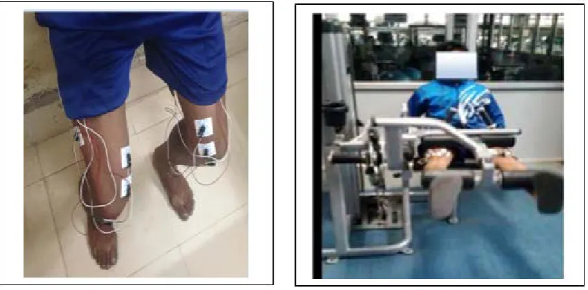
of selected muscles. EMG signals were acquired using four-channel wireless EMG BIOPAC Inc. (CMRR: 110dB at 50/60 Hz, Maximum Sampling rate: 200K samples/sec and Gain: 5-50,000, Input Impedance: 2 MΩ). The skin was rubbed with cotton containing alcohol to minimize the skin impedance, thereby improving the quality of signal acquisition. Disposable electrodes (44 x 32 x 1 mm) were placed on the subject’s selected muscles following standard protocols [11] to acquire EMG simultaneously from both the legs as shown in Figure 1(a). The inter-electrode distance was 20 mm, center to center.
2.3. Data Acquisition
After the subject preparation was done, subject was seated on the end of the Cybex bench (Cybex 6000, Cybex, Division of Lumex Inc., Ronkonkoma, USA) with the padded edge of the bench against the posterior surface of the knee joint, the feet hooked behind the padded rollers and the hands grasping the bench just behind their buttocks shown in Figure 1(b). This was done to achieve comfortable posture of the subject for leg extension during the data acquisition. Each selected subjects were then assigned to 30 RM load and they were asked to sustain it for maximum duration of time (recorded in seconds) independently for males and females. 30 RM was opted for the study because 25-30 RM is the most recommended training and testing load for developing and measuring muscular endurance in athletes [12]. Isometric contractions of knee extensor muscles VM and VL were performed at an angle between 0º to 10º to emphasize full extension of legs taking in regard the comfort of subjects as well. Sampling rate during acquisition was set to 2000Hz as per Nyquist criteria. The collected data was stored using AcqKnowledge 4.3 software (BIOPAC Inc., USA).
Figure 1. a) Electrode Placement, b) Experimental Setup
2.4. Data Segmentation
The raw EMG data is further segmented into 30, 60 and 90 seconds for each subject, as all subjects were able to perform minimum of 90 seconds isometric contraction. Further the subjects were divided into high and low performance group based on median split method. Median
2.5. Data Processing
The raw EMG signals acquired from the subjects were quantified with the help of MATLAB. For processing of EMG signal, notch filter was applied to remove 50 Hz noise interference from the signal. Cascaded low-pass (20 Hz) and a high-pass filter (450 Hz) were subsequently applied in order to remove other noise sources from the signal. The filtered EMG signals were rectified in order to calculate the RMS values. RMS is the square root of the arithmetic mean of the squares of the set of values. Let Xt
represent the rectified EMG signal, then RMS value is represented by equation 1:
2 t 1
(X ) RMS
T
t
T t
=
−
∑
(1)where, Xt is the rectified signal and T, t are the two time
intervals at which signal acquisition takes place. It is most frequently used parameter for the EMG analysis because during muscle contractions, it reflects the level of physiological activity in the motor unit [11].
2.6. Determination of Coactivation Ratio
The cocontraction between VM and VL muscles as indicated by the surface EMG signal was calculated as shown in equation 2.
Normalized value of VM VM : VL
Normalized value of VL Winter, 2009; Begalle, Distefano,
. Blackburn & Padua, 2012
=
(2)
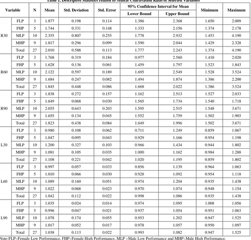
Table 1. Descriptive Statistics related to Muscle Coactivation Ratio of Selected Variables
Variable N Mean Std. Deviation Std. Error 95% Confidence Interval for Mean Minimum Maximum
Lower Bound Upper Bound
R30
FLP 3 1.877 0.198 0.114 1.386 2.368 1.650 2.009
FHP 5 1.744 0.331 0.148 1.333 2.156 1.374 2.178
MLP 10 2.355 0.807 0.255 1.778 2.932 1.453 4.190
MHP 9 1.817 0.296 0.099 1.590 2.044 1.429 2.328
Total 27 2.010 0.588 0.113 1.777 2.243 1.374 4.190
R60
FLP 3 1.768 0.319 0.184 0.977 2.560 1.410 2.020
FHP 5 1.628 0.136 0.061 1.459 1.797 1.523 1.843
MLP 10 2.122 0.597 0.189 1.695 2.549 1.528 3.524
MHP 9 1.684 0.247 0.082 1.494 1.874 1.386 2.200
Total 27 1.845 0.448 0.086 1.668 2.022 1.386 3.524
R90
FLP 3 1.838 0.272 0.157 1.162 2.513 1.527 2.033
FHP 5 1.649 0.068 0.030 1.565 1.734 1.540 1.718
MLP 10 2.055 0.643 0.203 1.595 2.515 1.548 3.671
MHP 9 1.655 0.134 0.045 1.552 1.759 1.502 1.903
Total 27 1.823 0.438 0.084 1.649 1.996 1.502 3.671
L30
FLP 3 0.980 0.108 0.062 0.711 1.249 0.859 1.067
FHP 5 1.047 0.095 0.043 0.929 1.166 0.954 1.198
MLP 10 1.200 0.327 0.103 0.966 1.434 0.944 1.802
MHP 9 1.081 0.105 0.035 1.000 1.162 0.984 1.288
Total 27 1.108 0.221 0.042 1.020 1.195 0.859 1.802
L60
FLP 3 0.997 0.057 0.033 0.856 1.139 0.964 1.063
FHP 5 1.010 0.066 0.030 0.928 1.092 0.954 1.118
MLP 10 1.089 0.160 0.051 0.974 1.204 0.935 1.438
MHP 9 1.022 0.068 0.023 0.970 1.074 0.948 1.154
Total 27 1.042 0.112 0.022 0.998 1.086 0.935 1.438
L90
FLP 3 1.035 0.024 0.014 0.974 1.095 1.008 1.056
FHP 5 0.996 0.047 0.021 0.937 1.054 0.951 1.063
MLP 10 1.078 0.174 0.055 0.953 1.202 0.947 1.525
MHP 9 1.017 0.052 0.017 0.978 1.057 0.950 1.097
Total 27 1.038 0.113 0.022 0.993 1.082 0.947 1.525
Note-FLP=Female Low Performance, FHP=Female High Performance, MLP =Male Low Performance and MHP=Male High Performance.
2.7. Statistical Analysis
SPSS software (IBM) was used for statistical analysis. A One-Way ANOVA with Least Significant differences (LSD) were carried out to determine the differences
between the gender, performance and side (right and left) on cocontraction ratio with progression of time related to VM:VL ratio. The findings have been shown in
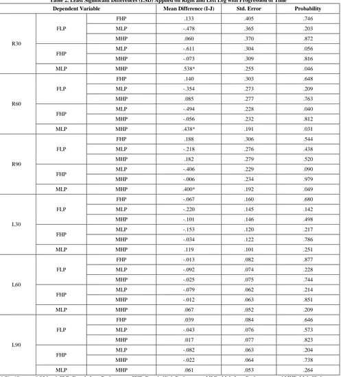
Table 2. Least Significant Differences (LSD) Applied on Right and Left Leg with Progression of Time
Dependent Variable Mean Difference (I-J) Std. Error Probability
R30
FLP
FHP .133 .405 .746
MLP -.478 .365 .203
MHP .060 .370 .872
FHP MLP -.611 .304 .056
MHP -.073 .309 .816
MLP MHP .538* .255 .046
R60
FLP
FHP .140 .303 .648
MLP -.354 .273 .209
MHP .085 .277 .763
FHP MLP -.494 .228 .040
MHP -.056 .232 .812
MLP MHP .438* .191 .031
R90
FLP
FHP .188 .306 .544
MLP -.218 .276 .438
MHP .182 .279 .520
FHP MLP -.406 .229 .090
MHP -.006 .234 .979
MLP MHP .400* .192 .049
L30
FLP
FHP -.067 .160 .680
MLP -.220 .145 .142
MHP -.101 .146 .498
FHP MLP -.153 .120 .217
MHP -.034 .122 .786
MLP MHP .119 .101 .251
L60
FLP
FHP -.013 .082 .877
MLP -.092 .074 .228
MHP -.025 .075 .744
FHP MLP -.079 .062 .214
MHP -.012 .063 .851
MLP MHP .067 .052 .209
L90
FLP
FHP .039 .084 .646
MLP -.043 .076 .573
MHP .017 .077 .823
FHP MLP -.082 .063 .204
MHP -.022 .064 .738
MLP MHP .061 .053 .264
* Significant at 0.05 level. FLP=Female Low Performance, FHP=Female High Performance, MLP =Male Low Performance and MHP=Male High Performance.
3. Results
The summary of statistical findings from Table 2 is as follows:
1) During first 30 seconds (0 to 30 seconds), statistically significant difference (p<0.046) was found in coactivation ratio between male low and high performance sportspersons in right leg.
2) During second 30 seconds (30 to 60 seconds), statistically significant difference (p<0.031) was found in coactivation ratio between male low and high performance sportspersons in right leg.
3) During third 30 seconds (60 to 90 seconds), statistically significant difference (p<0.049) was found in coactivation ratio between male low and high performance sportspersons in right leg.
4) At any point of time there were no sex differences. 5) Significant differences between male low and high performance at were found only in the right side of leg.
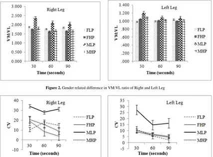
6) Overall there was male dominating phenomenon Further analysis of gender related differences in the VM:VL ratio with progression of fatigue (time) during isometric contraction in right and left leg is shown in
MLP=Male Low Performance and MHP=Male High Performance. It clearly demonstrated that during isometric contraction, coactivation ratio of males dominated females. For both males and females, the ratio decreases with
progression of fatigue (time). It is also depicted that the male low performance sportspersons have higher values of coactivation ratio followed by female low performance sportspersons.
Figure 2. Gender related difference in VM:VL ratio of Right and Left Leg
Figure 3. Coefficient of variation of VM:VL ratio of Right and Left Leg Table 3. Coefficient of Variation of Right and Left Leg with Progression of Time
Variable R30 R60 R90 L30 L60 L90
FLP 10.5148071 18.04285 14.8042 11.02866 5.687853 2.368313
FHP 19.00625896 8.37408 4.125389 9.129929 6.529956 4.728885
MLP 34.24987881 28.11297 31.30116 27.22564 14.69783 16.14713 MHP 20.68966837 15.67236 8.953088 10.23939 6.981432 5.418507 Note-FLP=Female Low Performance, FHP=Female High Performance, MLP =Male Low Performance and MHP=Male High Performance.
The coefficient of variation in both right and left leg is demonstrated in Table 3. From the findings of Table 3 as illustrated in Figure 3 it is evident that Male Low Performance (MLP) has higher coactivation ratio of VM and VL muscles than that of Female Low Performance (FLP), Male High Performance (MHP) and Female High Performance (FHP) at both right and left legs. This implied that MLP having higher chances of injuries. Attribution factor may be higher muscular development asymmetry in MLP. Cocontraction variations are superior at right leg, as it is the dominant leg.
The following points are evident from the results of coefficient of variation:
1) There is higher asymmetricity in MLP group because of lack of training.
2) There is less asymmetricity in MHP group because of training adaptation.
3) In FLP group because of lack of strength development in the muscles of both legs, asymmetricity is comparatively lower than that in males.
4) In FHP group, training improved the strength in both muscles equally, thus asymmetricity is less.
4. Discussion of Findings
Muscle coactivation is an important phenomenon for regulation of joint load that minimizes the external and internal disturbances around the joint thus preventing injury. Our results demonstrated the decrease in the ratio with progression of fatigue (time) which is in concordance with the results reported by researchers related to fatigue
of VM and VL muscles. The findings of the study collectively demonstrated gender, performance and side (right and left) related differences in cocontraction ratio. It has been found that male sportspersons have higher VM:VL ratio than that of female sportspersons during isometric contraction. It is also demonstrated that low performance sportspersons have higher values of cocontraction than that of high performance sportspersons. This indicates that male low performance sportspersons followed by female low performance sportspersons are prone to the higher risks of picking up patellofemoral injuries than compared to male and female of high performance sportspersons. The findings suggested that the isometric exercises employed by clinicians for rehabilitation of sportsperson from patellofemoral injuries must be designed according to gender, performance and side (right and left) rather than common strategies normally followed for both the genders. These results would provide important inputs in prevention of possible training related injuries in patellofemoral joint. It would also provide crucial information that would help in adjusting training protocol of sportspersons by their trainers.
5. Conclusions
The following are the conclusive remarks from the study:
1) The study found gender related differences with regard to cocontraction ratio of VM and VL muscles during isometric contraction in sportspersons.
2) Cocontraction of males is found to be superior in comparison to female sportspersons across the experimental protocol.
3) Performance related differences are found with regards to Cocontraction ratio of VM and VL muscles during isometric contraction in sportspersons.
4) Predominance of right leg over left leg was found across the protocol.
5) Decrease in coactivation ratio is found with progression of fatigue (time).
These results would help in the understanding the significance of the gender, performance and side (right and left) related differences in the co activation ratio and its relation to individual member of the muscular group. Such understanding is important for the clinicians and trainers to evaluate and manage the rehabilitation related to patellofemoral joint injuries. Changes in activation strategies of muscles can provide valuable information
regarding the mechanisms that alter neuromuscular strength of fatigued muscles.
Acknowledgement
The authors acknowledge all volunteers and sportspersons from the Indira Gandhi Institute of Physical Education and Sports Sciences, University of Delhi for their voluntary participation in the study.
References
[1] Falconer, K. & Winter, D.A. (1985).Quantitative assessment of co-contraction at the ankle joint in walking. Electromyogr Clin Neurophysiol., 25 (2-3), 135-149.
[2] Lloyd, D.G. & Buchanan, T.S. (2001).Strategies of muscular support of varus and valgus isometric loads at the human knee. J Biomech., 34(10), 1257-1267.
[3] Sakurai, T., Toda, M., Sakurazawa, S., Akita, J., Kondo, K. and Nakamura, Y. (2010). Detection of Muscle Fatigue by the Surface Electromyogram and its Application. 9th IEEE/ACIS International Conference on Computer and Information Science, 43-47.
[4] Missenard, O., Mottet, D. & Perrey, S. (2008). The role of co-contraction in the impairment of movement accuracy with fatigue. Experimental Brain Research, 185(1), 151-156.
[5] Semmler, J.G., Ebert, S.A. & Amarasena, J. (2013). Eccentric muscle damage increases intermuscular coherence during a fatiguing isometric contraction. Actaphysiologica, 208(1), 716-725.
[6] Silva, C.R. et al. (2014). Influence of neuromuscular fatigue on co-contraction between vastus medialis and vastus lateralis during isometric contractions. Kinesiology, 46(2), 179-185.
[7] Rainoldi, A., et al. (2008). Mechanical and EMG Responses of Vastus lateralis and changes in biochemical variables to isokinetic exercise in endurance and power athletes. J Sports Sci., 26(3), 321-331.
[8] Marson, R.A. (2011). Study of muscular fatigue by EMG analysis during isometric exercise. Biosignals and Biorobotics Conference (BRC), 1-4.
[9] Choi, H. (2003). Quantitative assessment of co-contraction in cervical musculature. Medical Engineering & Physics, 25(2), 133-140.
[10] Heiden, T.L., Lloyd, D.G. & Ackland, T.R. (2009). Knee joint kinematics, kinetics and muscle co-contraction in knee osteoarthritis patient gait. Clinical Biomechanics, 24(10), 833-841.
[11] Konard, P. (2005). The ABC of EMG: A Practical Introduction to Kinesiological Electromyography. Noraxon Inc. USA, version 1.0.
[12] Shaw, D. (2000). Encyclopeadia of Sports Injuries and Indian Sports Persons. K.S.K. Publishers & Distributors.
[13] Winter, D. (2009). Biomechanics and motor control of human movement. New Jersey: John Wiley & Sons, 4th Ed.