VOL. 6, NO. 3, pp. 196 - 204, September, 2016 Submitted May 2016; Revised August 2016; Accepted September 2016
Agarose Coated Culture Plate in Tumorsphere Culture of Cervical Cancer Cell Line HeLa: An
Alternative to Non Adhesive Culture Plate
I Putu Juniartha1*, Muhammad Rasjad Indra2, Hidayat Sujuti3, Diana Lyrawati4, Tatit Nurseta5,6
1Department of Biomedical Sciences, Faculty of Medicine, Brawijaya University, Malang, Indonesia 2Department of Physiology and Molecular, Faculty of Medicine, Brawijaya University, Malang, Indonesia 3Department of Biochemistry and Biomolecular, Faculty of Medicine, Brawijaya University, Malang, Indonesia 4Department of Pharmacy, Department of Biomedical Sciences, Faculty of Medicine, Brawijaya University, Malang, Indonesia
5Department of Obstetry and Gynecology, Faculty of Medicine, Brawijaya University, Malang, Indonesia 6Department of Obstetry and Gynecology, dr. Saiful Anwar Public Hospital, Malang, Indonesia
ABSTRACT
Cervical cancer recurs in 90% cases and linked to cancer stem cells that able to self-renew and responsible for recurrence, metastasis, and mortality of cancer. Isolation and identification of cancer stem cells using serum-free medium needs expensive growth factors and consume time. This study try to grow tumor sphere using culture plate coated with 1% agarose as an efficient and economical alternative to non-adhesive culture plate. HeLa cell line was grew in culture plate coated with 1% agarose and non-adhesive culture plate using similar medium and culture condition. Tumor spheres morphology was observed and the colonies were counted in 7 days followed by single cell assay. Tumor spheres then counted for CD133+, CD34+, and Sox2 expression using flowcytometry. Culture plate coated with 1% agarose can be used as an economic and efficient alternative to culture tumor sphere. Using culture plate coated with 1% agarose, the tumor spheres formed in 7 days with similar morphology to non-adhesive culture plate. Tumorsphere had three dimensional – sphere shape that tightly attached, colonized, and overlapped. The tumor sphere colony counts of two plates were statistically have no significant difference (p=0,667). Single cell assay of a tumor sphere shows that it can grow new tumor spheres with similar morphology. The tumor sphere from culture plate coated with 1% agarose express CD133+ and CD34+ as much as 8.78% ± 2.14 and Sox2 as much as 35.30% ± 23.82 whereas tumor sphere from non-adhesive culture plate express CD133+ and CD34+ as much as 62.36% ± 1.06 and Sox2 as much as 98.86% ± 0.56 (p = 0000).
Keywords: Cancer stem cell, tumorsphere, agarose, non-adhesive culture plate, Sox2
Cervical cancer is the second most common cancer in women after breast cancer. In every year, approxi-mately 500.000 women diagnosed with invasive cervical cancer and half of them die [1, 2].
Recurrent cervical cancer still remains a problem that requires proper treatment. Recurrency happened in almost 90% cases in 2 years after therapy and 20 – 70% are in the early stage and local advanced stage [3]. Until now, cervical cancer is treated by chemotherapy, surgery, and genetic based therapy that targets tumor
oncogene and gene suppressor, but the cancer still re-curs [4].
Recent studies shows that a tumor grew from a small population in tumor cells called cancer stem cell that have several properties such as self-renewal, chemoresistance, and able to differentiate into cancer with tumorigenic properties. These properties were considered to take part in cancer recurrence, metasta-sis, and high mortality [5, 6, 7, 8].
Several study had been done to identify cancer stem cell in cervical cancer. Feng et al., (2009) successfully
JTLS | J. Trop. Life. Science 196 Volume 6 | Number 3 | September | 2016
INTRODUCTION
*Corresponding author: I Putu Juniartha
Department of Biomedical Science Faculty of Medicine, Brawijaya University
Jalan Veteran, Malang 65145, Indonesia Email: koasputu@gmail.com
How to cite:
characterized cancer stem-like cells from primary cervi-cal cancer by tumor sphere culture [1]. Wang et al., (2014) successfully enriched and characterized stem-like cell from cervical cancer cell line HeLa [5].
Sox2 is a regulator of transcription factor and im-portant in regulating stem cell phenotype. Sox2 had been detected in several tumors and an increase in its expression is associated with carcinogenesis [1, 5, 9].
Cancer stem cell from primary cervical cancer and HeLa cells isolated using different method to grow tu-mor sphere. Primary culture use fresh cervical cancer sample from operating room and supplemented with growth factor that is expensive, consume time, and sometimes not giving appropriate result. Study from Wang et al., (2014) use non-adhesive culture system to grow tumor sphere. This method is simple and eco-nomic with appropiate result but still use non-adhesive plate with agarose coating that is expensive [5].
This study is aimed to search for an alternative method to grow tumor sphere from HeLa cells using culture plate coated with agarose as a substitute to non-adhesive culture plate. In this study, agarose is used to prevent attachment of cells in the plate bottom. As an alternative to non-adhesive culture plate, this study will compare the morphology, colony count, and several markers of tumor sphere grow on the two plates. If the results of two plates are consistent, then culture plate coated with agarose can be used as a sub-stitute to non-adhesive culture plate. Using this method, hopefully cell line can be used as attractive al-ternative cancer stem cell model to study the treatment of cancer stem cell.
Ethics
This study had been approved by Ethics Committee of Medical Faculty Brawijaya University.
Cells
HeLa cells were obtained from Biomedical Labora-tory, Medical Faculty of Brawijaya University.
Monolayer HeLa culture (as control)
HeLa cells were grew in 24 well plastic culture plate (Corning Incorporated #CL53527, NY) using Mini-mum Essentials Media (Gibco #11095-072, USA) sup-plemented with 10% Fetal Bovine Serum (Gibco #10437-077, USA), 1,25% Penicillin & Streptomycin, 0,44% (Sigma #P4333, USA), and also Sodium Bicar-bonate (Merck #106329, USA) in a humidified atmos-phere (5% CO2 at 37°C).
Culture medium and agarose preparations
1 × RPMI solutions were made from 5 mL RPMI (Gibco, USA) + 0.02 g Sodium Bicarbonate (Merck #106329, USA). 2 × RPMI solutions were made from 100ml RPMI (Gibco #11875-085, USA) + 0.2 grams Sodium Bicarbonate (Merck, USA). Both solutions were supplemented with 1 – 5 drops HCl until system pH reach 7.2 – 7.4, 10% Penicillin & Streptomycin (Sigma, USA) + Amphotericin (Sigma #15290018, USA), 0,5% Fetal Bovine Serum (Gibco, USA). 1% agarose were made by diluting 0.004 g agarose (Low Gelling Temperature Culture Grade Agarose, Sigma® #A6560, USA) in 4 cc distilled water then melted with microwave at 400 Hz for 10 seconds then placed to 40°C water bath.
Tumorsphere culture
Monolayer HeLa cells were washed using 1 × RPMI for 2 times, then enzymatically disaggregated with 0.05 µL trypsin EDTA (Sigma #T4299, USA) and incubated in a humidified atmosphere (5% CO2 at 37°C) for 3 minutes, then add 1 × RPMI, centrifuge at 800 RPM for 8 minutes, then add 1 mL 2 × RPMI.
Cell were plated in three different plates condition: the first, using 24 well plastic culture plate (Corning, USA), the second using 24 well plastic culture plate (Corning, USA) coated with 1% agarose, and the third using Nunc® Ultra Low Cell Binding Surface 24 well culture plate (Sigma #Z721077). The cells were plated to each well as much as 2000 cells/ well. For plate that uses agarose, add 1 cc agarose to each well. Tumor-sphere culture were repeated 6 times (in 6 well) for each plate conditions.
Morphology analysis
Tumorsphere formation were observed everyday to compare the morphology and size for each culture plate.
Tumorpshere colony formation
The numbers of tumor sphere formed in each cul-ture plate well were counted in the day - 7.
Single cell assay
Single cell assay use modified hemocytometer mi-cropipette to make micro capillary tip end. Hemocy-tometer micropipette was heated and pulled away to decrease the size of tip end [10]. The micropipette then connected with yellow tip to get single cell. Tumor sphere were harvested and disaggregated with 0.05 µL Trypsin EDTA (Sigma, USA) and incubated in a
midified atmosphere (5% CO2 at 37°C) for 3 minutes, then add 1 × RPMI, then centrifuge at 800 RPM for 8 minutes, then add 2 ×RPMI. Single cell were plated using micropipette to three different plate used in tu-mor sphere culture. Colony formation from single cell were observed and captured every day from day 1 until day 7.
Flowcytometry analysis
Cells were harvested from each plate and disaggre-gated with Trypsin EDTA, then cold centrifuge (4°C) at 1500 RPM for 5 minutes except the culture plate coated with agarose use centrifuge with room tempera-ture. Wash the cell with 200 µL Phosphate Buffer Saline (Sigma #P4417, USA), then centrifuge at 1500RPM for 15 minutes, repeats 2 times. Add 5 µL FITC CD133 antibody (Invitrogen #PA5-38014, USA), FITC - anti CD34 antibody (Invitrogen #CD34-581-01, USA), and PerCP/Cy Sox2 antibody (Invitrogen #PA1-094, USA) to each cell and incubate for 15 – 20 min-utes in dark room. Add 200 µl Cell Staining Buffer (Bi-olegend #420201, USA) then analyze the expression of CD133, CD34, and Sox2 using FacsCalibur and Cel-lQuest software.
Data analysis
Data analysis was performed by descriptive and in-ferential. Descriptive analysis was done by calculating mean and standard deviation. The data were presented in tables and diagrams. Inferential analysis was con-ducted by dependent variable data comparisons be-tween groups. Statistical analysis for CD133, 34, and Sox2 were performed by Two – Way ANOVA, fol-lowed by Tukey HSD. P < 0.05 was considered statisti-cally significant. A post test control groups design was employed in this research.
Morphology analysis
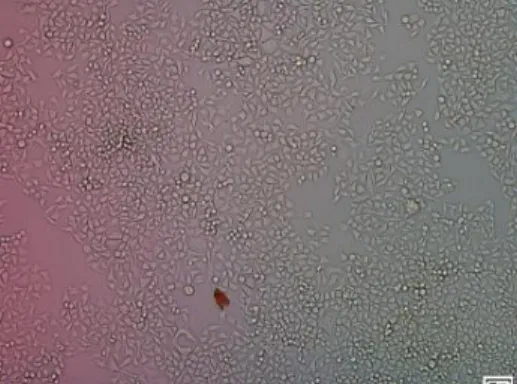
Microscopically, HeLa cells that grew in plastic cul-ture plate have different morphology with HeLa cells that grew on 1% agarose – coated culture plate and non-adhesive culture plate. On plastic culture plate, HeLa cell grew as spindle – shaped monolayer cell that attached to the plate bottom and form junction with another cell (Figure 1).
On culture plate coated with 1% agarose, HeLa cell grew as three dimensional tumor spheres on the medium but some still grew as monolayer on the plate bottom. During observation, the tumor spheres were not moving, indicating that tumorspheres grew in the
Figure 1. HeLa cells cultured with 24 well plastic culture plate. HeLa cells grew as spindle – shaped mono-layer cell that attached to the plate bottom.
Figure 2. Tumor sphere grew on: 4th day (a) 24 well plastic culture plate coated with 1% agarose (b) non adhe-sive plate. Both tumorspheres grows as three di-mensional tumorspheres that tightly attached, colo-nized, and overlapped. 7th day (c) 24 well culture plate coated with 1% agarose (d) non adhesive plate. Tumor sphere increased in size from ±v20 µm in day-1 to ±100 µm in day-7. Tumorsphere became more tightly attached and overlapped.
agarose (Figure 2a and c).
On non-adhesive culture plate, HeLa cell also grew three dimensional tumor spheres that floating over the medium with initial size ± 20 µm in day-1 and become ± 100 µm in day-7 (Figure 2b and d). Tumorspheres looks tightly attached, colonized, and overlap in three dimensional configuration. It also moves dynamically to do self-renewal and producing new colony.
Basically tumor spheres that grew on agarose – coated culture plate and non-adhesive plate are mor-phologically similar, but the presence of agarose makes the cells trapped in it. Only some HeLa cells that have cancer stem cell properties can grew on the agarose and form tumorsphere, the rest still grew as monolayer
JTLS | J. Trop. Life. Science 197 Volume 6 | Number 3 | September | 2016
RESULTS AND DISCUSSION
Figure 3. Micro drop method using modified hemocytometer micropipette. In preliminary study, we took single cell at volume 40, 30, and 20 µL, but the single cell population were still “crowded” at the culture plate. Using microdrop method, we could take less single cell with distant gap to other cells so we can ob-serve single cell growth clearly.
Figure 4. Single Cell Assay from day-1 until day-7. Single cell grew with binary manner and forming three dimen-sional, sphere – like, and overlapped tumorsphere with identical morphology and properties to its parental tumorsphere.
on the plate bottom. On non-adhesive culture plate, HeLa cells tend to colonize first and then grew to be-come tumor sphere. Different from that, agarose make single HeLa cells can directly grow into tumorsphere.
Colony formation
In this study, two single cells already counts as tu-mor sphere colonies. Monolayer HeLa cells did not form colony (as control), while culture plate coated with 1% agarose form 1245 ± 139.931 tumor sphere colonies, and non-adhesive plate form 1272.83 ± 64.129 colonies in 7 days (Table 1). We can conclude that cul-ture plate coated with 1% agarose have higher colony formation compared to non-adhesive culture plate.
Single cell assay
Single cell assay can be used to proof that single
cell of tumorsphere have self renewal properties by producing daughter cells with similar morphology and properties to its parental tumorsphere. Tumor spheres were disaggregated with Trypsin EDTA to form single cell then re-plated to 24 well culture plates with micro drop technique using modified hemocytometer mi-cropipette (Figure 3). Single cell were cultured as much as 50 cell per well with distant gap so we can observe the cell growth clearly during 8 days. Single cell grew with binary manner and form three dimensional, sphere-like, and overlapped tumorsphere with identical morphology and properties to its parental tumorsphere (Figure 4).
Flowcytometry analysis of CD133, CD34, and Sox2 Tumorsphere colonies that formed from single cell assay were analyzed for surface marker CD133 and CD34, and also stemness protein Sox2 (Table 2).
CD133, CD34, and Sox2 expression from control sam-ples
As much as 96.04% ± 0.57 control cells did not ex-press CD133 and CD34 markers but 0.89% ± 3.09 cells express double positive markers CD133+ CD34+. 94.22% ± 3.06 cells that express double positive mark-ers also express Sox2 protein (Figure 5a).
CD133, CD34, and Sox2 Expression of Tumor sphere Cultured with Culture plate coated with 1% agarose
As much as 8.78% ± 2.14 tumor sphere cells ex-press double positive markers CD133+ CD34+ and 35.30% ± 23.82 of those cells also express Sox2 protein (Figure 5b).
CD133, CD34, and Sox2 expression from tumorsphere cultured with non-adhesive culture plate
As much as 62.36% ± 1.06 cells express double pos-itive markers CD133+ CD34+ and 98.86% ± 0.56 cells also express Sox2 protein (Figure 5c).
Flowcytometry analysis show that HeLa cells con-tains cancer stem cell that express double positive markers CD133+ CD34+ that can be used as surface markers. Tumor sphere that grew from culture plate coated with 1% agarose were difficult to isolate because they grow beneath the agarose layer. Besides that, agarose have gel phase that make the tumor sphere dif-ficult to be deposited during centrifugation and the re-sult become invalid because most cells read by flowcy-tometer are debris.
JTLS | J. Trop. Life. Science 197 Volume 6 | Number 3 | September | 2016 Table 1. Tumorsphere colony formation in culture plate coated with 1% agarose and non adhesive plate (day-7)
Well 1 Well 2 Well 3 Well 4 Well 5 Well 6 Mean ± SD P Culture plate coated with 1% agarose 1446 1349 1064 1279 1154 1178 1245 ± 139.931 0.667 Non adhesive culture plate 1267 1245 1320 1170 1356 1279 1272.83 ± 64.129
Table 2. Expression of CD133, CD34, and Sox2 from tumorsphere derived from control, culture plate coated with agarose and non-adhesive culture plate
Plate CD133-CD34- CD133+CD34+ Sox2
Control 96.04% ± 0,57 0.89% ± 3.09 94.22% ± 306
Culture Plate Coated with 1% Agarose 4.29% ± 2.71 8.78% ± 2.14 35.30% ± 23.82 Non Adhesive Culture Plate 29.11% ± 0.78 62.36% ± 1.06 98.86% ± 0.56
p (ANOVA) p = 0.000 p = 0.000
p (Tukey) p = 0.000 p = 0.038
Figure 5. Flowcytometry analysis of (a) Control samples. Most cells (96.04% ± 0.57) did not express CD133 and CD34 markers. A small number of cells express double positive markers CD133+ CD34+ (0.89% ± 3.09) and also express Sox2 protein (94.22% ± 3.06). (b) Culture plate coated with 1% agarose. A small number of cells express double positive markers CD133+ CD34+ (8.78% ± 2.14) and also express Sox2 protein (35.30% ± 23.82). (c) Non-adhesive culture plate. Most cells express double positive markers CD133+ CD34+ (62.36% ± 1.06) and also express Sox2 protein (98.86% ± 0.56).
CD34+ also express Sox2 dominantly. In mono-layer HeLa cells, small amount of cells that express CD133+ CD34+ and Sox2 show that there are small amount of HeLa cells that originally contain cancer stem cell that considered to support the immortals properties.
Until now, cancer stem cells are isolated by side population, specific surface markers, and tumorsphere culture. From previous study, cancer stem cells culture can be identified by its three dimensional sphere-like morphology and several surface marker of tumorsphere [11]. In this study we had identified the tumor sphere or cancer stem cells that grew from HeLa cells on two kind of plates, the first culture plate coated with 1% agarose and the second non-adhesive culture plate. As a positive control we grew monolayer HeLa cells using culture plate without any coating.
Morphology characterization of HeLa cells
This study had identified cancer stem cell from tu-mor sphere culture using non-adhesive culture method. This method enables cells to grow in non-adherent condition and form tumor sphere. Non-adhesive cul-ture method uses plastic culcul-ture plate coated with 1% agarose or non-adhesive culture plate to prevent the at-tachment of cells to the plate bottom. Non-adhesive culture plate made of polystyrene with hydro cell sur-face which have super hydrophobic polymer properties that covalently binds so the cells can not bind to plate bottom.
Figure 1 show that HeLa cells on plastic culture plate without agarose grew as monolayer cells that at-tached to plate bottom, whereas on culture plate coated with 1% agarose and non-adhesive culture plate, the HeLa cells grew as three dimensional floating sphere colonies. This shows that non adhesive culture method can be used to isolate cancer stem cell from HeLa cells. Tumor sphere from HeLa cells morphologically similar to sphere that isolated from primary cervical cancer cells. The size of spheres increased progressively from ±20 to ±100 µm in 7 days.
Several cell lines had been successfully isolated us-ing tumor sphere culture such as primary breast cancer cells from transgenic mice Her – 2/ neu, MCF 7, BT474, and HCC1954. Previous study also success-fully isolate several cell line using non – adhesive method such as oral squamous cell carcinoma cell line (SAS, OECM – 1, Cal 27, SCC25, and Ca922), cell line from head and neck (Fadu and TW 205), colon (HT29 and COLO320), lungs (NCI-H23 and NCIH661) [11] and also HeLa [11, 12]. Wang et al.
(2014) also successfully identified cancer stem cell from HeLa cells using non adhesive culture plate coated with agarose [5]. The growth of the sphere in non-ad-hesive culture initially suspended and detached from its parental cells because it cannot attach to the plate bot-tom and also cannot aggregate with other cells so it differentiates into spheroids that floating on the medium and form small clusters.
In this condition, the cells will have decreased cell to cell interaction, cell to matrix interaction, and also abil-ity to attach and induce anoikis phenomena that in-duce apoptosis response [9]. In anoikis condition, the spheres still attempt to attach but non-adhesive condi-tion in culture plate triggers survival signal. The sur-vival signal make the spheres able to survive and prolif-erate as floating tumor cells that do not have normal solid – phase scaffolding that form the microenviron-ment [9].
Non-adhesive condition also induces Epithelial to Mesenchymal Transition (EMT) that mediated by WNT, Sonic Hedgehog, Snail/ Slug, and Notch signal-ing pathway. There is a connection between EMT and Cancer Stem Cells shown by morphological growth and motility alteration of cells. It supports that non ad-hesive culture method can be used to isolate cancer stem cell from HeLa cells [9].
Tumorsphere culture from HeLa cells considered to be more economic and time-efficient than primary cer-vical cancer cell culture because it is ethic problem-free and do not use expensive growth factor like eFGF and bFGF. It also have similar result that can represent cancer stem cell of cervical cancer.
Tumor sphere colony formation from HeLa cells Culture plate coated with 1% agarose grew approx-imately 1245 ± 139.931 tumor sphere colonies in 7 days whereas non-adhesive culture plate grew approximately 1272.83 ± 64.129 tumor sphere colonies in 7 days. Sta-tistical analysis shows no significant difference of colony formation between two plates (p = 0.667) (Table 1).
optimize tumor sphere culture, agar condition, culture plate, and culture medium were adjusted to fulfill tu-mor sphere’s need. In this study we use low melting point agarose – culture grade, RPMI culture medium, and Fetal Bovine Serum.
Agarose is a polymer that composed of galactose subunit made from the cell wall component of red al-gae species. Agarose have three different phases: gel at room temperature, rigid at 65°C, and melt at 85°C. This study use 1% agarose to change culture plate con-dition from high attachment to low attachment to sup-port the growth of tumorsphere.
Single cell assay of tumorsphere
Using micro drop method, we can grew approxi-mately 50 cells per well (in 24 well culture plate) with distant space between cells to observe single cell growth to become tumor sphere clearly [10]. Single cell grew into tumor sphere in binary manner from a cell into 2, 4, and so on in 7 – 8 days, forming three di-mensional spheres-like tumor sphere with size 100 µm.
Single cell assay were easier to observe in culture plate coated with 1% agarose because the cells grew be-neath agarose and the position is fixed, while single cell on non-adhesive culture plate were difficult to ob-serve because it is floating and moving dynamically. CD133 and CD34 expression
Several cancer stem cell markers have been used to isolate cancer stem cells, for example CD133, CD44, and ALDH1. This study found that tumor sphere from HeLa cells express CD133+ CD34+ (double positive) surface marker in large amount. Double positive mark-ers were expressed in 62.36% ± 1.06 tumor spheres from culture plate coated with 1% agarose and ex-pressed in 8.78% ± 4.29 tumor spheres from non-adhe-sive culture plate. In control samples, only 0.89% ± 3.09 cells express double positive markers.
Statistical analysis shows that there is significant differences of CD133+ CD34+ expression between cul-ture plate coated with 1% agarose and non-adhesive culture plate (p = 0.000). This study are not consistent with study of Chen et al. (2012) that found CD133 ex-pression were increased 3 – 4% in tumor sphere com-pared to its parental cells because 96.04% ± 0.57 (al-most all) parental HeLa cells did not express CD133 nor CD34. Using immunofluorescence method, Chen also found that CD133 are expressed in cell membrane [7].
Wang et al. (2013) use side population method to isolate cancer and found that only 3.64% cells express
CD133. Compared to sphere culture, it can isolate more cancer stem cell than side population [4]. Single CD133 or CD34 marker only cannot be used to specify the target cancer stem cell because of the heterogeneity of tumor and its derivatives cannot be predicted [7]. Sudiarta et al. (2015) shows that tumor sphere from primary cervical cancer in the 3rd stadium only express CD44+ but do not express CD34+ in all samples, so we can conclude that tumor sphere from cervical cancer cell line express different markers to primary cancer cell line [13].
Lopez et al. (2012) also successfully identify cancer stem cell from human cervical cancer cell line HeLa, SiHa, Ca Ski, and C-4 by sphere culture using serum free – medium and EGF + bFGF growth factor. The tumor spheres of the cell lines expresses CD133+ CD34+ markers [12].
CD133 or Prominin – 1 have 37 exons located on Chromosome 4 with length 152 kb and is a glycopro-tein with 865 amino acids and have molecular weight 120 kDa. Sing et al., were the first to report that CD133 can be used as a surface marker of brain cancer stem cell. CD133 show high ability to proliferate, self-renew, and differentiate to tumor that have similar phenotype to brain tumor [14]. Olempska et al., and Hermann et al., also found that cancer stem cell that express CD133 marker shows self-renewal, differentia-tion, and proliferation potency in vitro. This shows that CD133 can be used as a cancer stem cell specific marker, especially solid tumors, and as a target of effec-tive anti-cancer therapy [14].
CD34 is a trans membrane phospoglycoprotein, firstly identified in 1984 on stem cell and hematopoi-etic progenitor cells with molecular weight 115 kDa. In hematopoietic cells, CD34 have a role in cytoadhesion and also regulate differentiation and proliferation of cells [15]. CD34 also expressed by stromal, epithelial, and endothelial cells so CD34 alone cannot be used as an exclusive marker of a cell.
Sox2 expression
This study found that tumor sphere from cervical cancer cell line express double positive CD133+CD34+ that also express Sox2 protein as much as 35.30% ± 23.82 in culture plate coated with 1% agarose and 98.86% ± 0,56 in non-adhesive culture plate. Statistical analysis shows that there is no significant difference of Sox2 expression between control, culture plate coated with agarose, and non-adhesive culture plate (p = 0.000). Tukey HSD test shows that there is significant difference between culture plate coated with 1%
agarose and non-adhesive culture plate (p = 0.038). Sox2 is a transcription factor and also a regulator of transcription factor that play important role in regulat-ing gene expression both in normal growth and can-cer12. Sox2 is a key transcription factor in embryo and important in regulating normal stem cell phenotype. Together with Oct – 4 and Nanog, Sox2 control self-renewal and differentiation process by coordinated transcription process. Sox2 have been detected in hu-man tumor and show a potential function in tumorige-nesis. When Sox2 stably expressed in cervical cancer cells, cells that express Sox2 will increase its prolifera-tion, clonogenicity, and tumorigenicity in vitro and in vivo compared to the control. This shows that Sox2 may take part in carcinogenesis of cervical cancer thus can be a molecular target therapy [7, 16.]
Overexpression and amplification of Sox2 also linked to squamous cell carcinoma of many tissues like lungs, esophagus, cervix, penis, and skin [17]. Prasad et al. (2005) reported that Sox2 play a role in initiation of carcinogenesis and expressed in 80% cervical squa-mous cell carcinoma which should be expressed only in 25% normal cells [18]. Sudiarta et al., in 2005 also found that primary cervical cancer cell in the 3rd sta-dium that express CD44+CD34- markers also express Sox2 protein as much as 68% [13]. On the other hand, Wang et al. (2014) also confirm the expression of Sox2 in tumor sphere from non-adhesive culture plate using western blot method [15].
Role of MicroRNA and Sox2 in cancer stem cell self renewal
Self-renewal defined as a process where stem cell produce one (asymmetrical division) and two (symmet-rical division) of daughter cell that have similar growth potential to parental cell. Abnormality self-renewal mechanism of stem cell can induce cancer growth. Self-renewal occurs by cell divisions and controlled by a va-riety of Cyclin Dependent Kinases (CDK) that only ac-tivates upon binding to specific cyclin. CDK also regu-lated by a variety of modulator and inhibitor protein as a response to different environment condition. Those proteins also regulated post transcriptionally by mi-croRNA and other transcription factor [14]. Cancer cells have altered expression of transcription factor that induce uncontrolled proliferation as a hallmark of can-cer so it will have faster G1/S transition phase com-pared to normal condition [14].
MicroRNA (miRNA) is a 29 – 22 long non – cod-ing RNA that inhibits gene expression in post – tran-scription level. miRNA is an important regulator in
proliferation, differentiation, and maintainance of stemness properties and it is disregulated in carcino-genesis. miRNA 302 – 367 expressed in a high number in embryonic stem cell but not in other somatic stem cell [20, 21, 22]. miRNA 302 – 367 regulated by spe-cific embryonic stem cell transcription factor Oct3/4, Sox2, and Nanog which were the key regulator in maintaining the stemness properties of embryonic stem cell [23, 24, 25].
Normally embryonic stem cell have shorter G1 phase, accumulation on S phase, and there is no check-point on DNA damage in G1 phase. miRNA 302 inhi-bition can induce G1 arrest and induce the cell to dif-ferentiation, but overexpression of miRNA will induce the cell to exit the G1 phase. CyclinD1 and Cdk4 also regulated by miRNA 302 – 367 post transcriptionally. In the cell nucleus, miRNA 302 – 367 genes are acti-vated by Oct3/4 and Sox2. miRNA 302 – 367 posi-tively regulate self-renewal process by inhibiting cy-clinD1/ Cdk4 and induce the cell to enter the S phase. On the other hand, miRNA 302 – 367 is also a posi-tive regulator of Nodal/ Activin pathway that maintain cell pluripotency [23, 24, 25].
Culture plate coated with 1% agarose can be used as an economic and eficient alternative to grow tumors phere with similar morphology, colony counts, but lower marker CD133, CD34, and Sox2 expression compared to non-adhesive culture plate.
We thank to Diana Lyrawati, Ph.D. (Department of Pharmacy; Department of Biomedical Sciences, Faculty of Medicine, Brawijaya University, Malang, Indonesia) and Dr. Tatit Nurseta (Department of Obstetry and Gynecology, Faculty of Medicine, Brawijaya University, Malang, Indonesia) for helpful comments and critical reading of this paper. This study was supported by private funding.
1. Feng D, Cheng F, Cairong L et al (2009) Identification and Characterization of Cancer Stem – Like Cells from Primary Carcinoma of the Cervix Uteri. Oncology Reports. 22: 1129 – 1134.
2. Siegel R, Carol D, Katherine V et al (2012) Cancer Treatment and Survivor Statistics. American Cancer Society. 1-22.
3. Chiva LM, Fernando L, Lucia GC et al (2008) Surgical Treatments of Reccurent Cervical Cancer: State of Art and
ACKNOWLEDGMENT
New Achievements. Gynecologic Oncology. 110 (3): S60 – S66.
4. Wang K, Hanfang Z, Lijing L et al (2013) Identification of a Cancer Stem Cell – Like Side Population in the HeLa Human Cervical Carcinoma Cell Line. Oncology Letters. 6: 1673 – 1680.
5. Wang L, Huijie G, Caiyu L et al (2014) Enrichment and Characterization of Cancer Stem – Like Cells from a Cervical Cancer Cell Line. Molecular Medicine Reports. 9: 2117 – 2123.
6. Baumann M, Krause M (2008) Tumor’s Biology Impact on Clinical Cure Rates in The Impact of Tumor Biology on Cancer Treatment and Multidisciplinary Strategies. Springer. 1: 324 – 325.
7. Ji J, Peng SZ (2010) Expression of Sox2 in Human Cervical Carcinogenesis. Human Pathology. 41: 1438 – 1447.
8. Calvet CY, Franck MA, Luis MM (2014) The Culture of Cancer Cell Lines as Tumorspheres Does Not Systematically Result in Cancer Stem Cell Enrichment. PloS ONE. Hal. 1 – 9.
9. Chen SF, Yun CC, Shin N et al (2012) Nonadhesive Culture System as a Model of Rapid Sphere Formation with Cancer Stem Cell Properties. PloS ONE. 7 (2): e31864.
10. Yongjian D, Jiang Q, Yu L et al (2012) A Simple Method for Isolating and Culturing Single Cancer Stem Cell. Journal of Southern Medical University. 32 (6): 802 – 806. 11. Zhang SL, Yi SW, Tong Z et al (2012) Isolation and Characterization of Cancer Stem Cells from Cervical Cancer HeLa Cells. Cytotechnology. 64: 477-484. 12. Lopez J, Adela P, Veverly M et al (2012) Cancer Initiating
Cells Derived from Established Cervical Cell Lines Exhibit Stem Cell Markers and Increased Radioresistance. Biomed Central. 12 (48):1 – 14.
13. Sudiarta KE, Satuman, Wibi R et al (2015) The Efficacy of Taraxacum Officinale Leaves Extract in Regulate Apoptosis, RAR 2 Gene, and Sox2 Expression onβ Primary Culture Human Cervical Cancer Stem Cells. International Journal of Pharmtech Research. 8 (4): 535-544.
14. Zhang H, Su YL (2010) Research Progression of CD133 as a Marker of Cancer Stem Cells. Chinese Journal of Cancer. 29 (3): 243 – 247.
15. Sidney LE, Matthew JB, Siobhan ED et al (2014) Concise Review: Evidence for CD34 as a Common Marker for Diverse Progenitors. Stem Cell Express. 32: 1380 – 1389. 16. Roy UB, Eunjeong S, Lalitha R et al (2012) Sox2
Maintains Self – Renewal of Tumor Initiating Cells in Osteosarcomas. Oncogene. 31 (18): 2270 – 2282. 17. Liu KC, Bao SL, Meng Z et al (2013) The Multiple Role
for Sox2 in Stem Cell Maintenance and Tumorigenesis. Cell Signal. 25 (5): 1 – 19.
18. Prasad NB, Biankin AV, Fukushima N et al (2005) Gene Expression Profiles in Pancreatic Epitehelial Neoplasia Reflects the Effects of Hedgehog Signaling on Pancreatic Ductal Epithelial Cells. Cancer Res. 65.1619 – 1626. 19. Wang Y, Robert B (2011) Cell Cycle Regulation by Micro
RNAs in Stem Cells. Springer. 459 – 471.
20. Navarro A, Mariano M (2010) MicroRNAs in Human Embryonic and Cancer Stem Cells. Yonse Medicine Journal. 51 (5): 622 – 632.
21. Liu C, Dean GT (2011) MicroRNA Regulation of Cancer Stem Cells. Clinical Cancer Research. 71 (18): 5950 – 5954.
22. Peter ME (2009) Let-7 and Micro-200 MicroRNAs: Guardians Against Pluripotency and Cancer Progression. Cell Cycle. 8 (6): 843 – 852.
23. Jesus AB, Gema LA, Pablo M (2009) The miR 302 – 367 Cluster as a Potential Stemness Regulator in ESCs. Cell Cycle. 8 (3): 394 – 398.
24. Card DAG, Pratibha BH, Leping L et al (2008) Oct4/ Sox2 Regulated miR – 302 Targets Cyclin D1 in Human Embryonic Stem Cells. Molecular and Cellular Biology. 28 (20): 6426 – 6438.
25. Takahashi R, Hiroaki M, Takahiro O (2014) The Role of microRNAs in the Regulation of Cancer Stem Cells. Frontiers in Genetics. 4: 1-11
26. Rizzino A (2013) Concise Review: The Sox2 – Oct4 Connection: Critical Players in a Much Larger Independent Network Integrated at Multiple Levels. Stem Cell Express. 31: 1033 – 1039.