Original Research Article
Nasal endoscopy: an excellent tool for the novice
Visweswara Rao Suraneni, Suneel Kudamala*, Karishma Begum
INTRODUCTION
According to Draf, Hirschmann first attempted nasal and sinus endoscopy using a modified cystoscope in 1901.1 In the middle of the twentieth century, Professor Hopkins developed the rod optic telescope following which endoscopy of the upper airway progressed at a rapid pace.2 Prior to the advent of endoscopes intranasal surgery was done in a radical manner. The developments in endoscopy and the detailed examination of nose and paranasal sinuses prompted surgeons like Messerklinger and Stammberger to explore the field of endoscopic sinus surgery.3,4
Nasal endoscopy has revolutionized the ease of diagnosing nasal pathologies over the recent years. It offers the advantage of detailed examination of the intra-nasal architecture and precise identification of the pathology. It is an excellent adjunct to first-hand information obtained by anterior and posterior rhinoscopy. It is cost effective and also saves the patient from going for more costly investigations like CT, MRI when not required.
The major advantage of nasal endoscopy is in the better illumination and magnification it provides. It helps to objectively show the exact pathology to the patient and explain the management strategy. It also serves as an excellent tool for teaching purpose and documentation.
ABSTRACT
Background: Nasal endoscopy has revolutionized the ease of diagnosing nasal pathologies over the recent years. It offers the advantage of detailed examination of the intra-nasal architecture and precise identification of the pathology. It is cost effective and a useful adjunct for performing minor procedures in the office setting. Thus, nasal endoscopy has evolved as a tool with multiple roles of diagnosing the disease, planning of treatment and post treatment examination.
Methods: This is a prospective study conducted at a tertiary care hospital during a period of 2 years on 100 patients to evaluate the role of nasal endoscopy in diagnosing the disease and planning the management. 60 cases were managed conservatively depending on the initial finding on nasal endoscopy while the other 40 cases underwent surgery. Pre-treatment symptoms and post treatment outcomes were observed objectively on nasal endoscopy.
Results: Majority of the patients (60%) were managed conservatively and were labeled as cured after enough evidence was obtained both subjectively from the patient and objectively as seen on endoscopy. The remainder of patients (40%) underwent surgery and follow up endoscopy was done in all of them.
Conclusions: Diagnostic nasal endoscopy is of immense help to the novice surgeon in understanding and managing various nasal pathologies. Through this study, it can be concluded that most of the nasal symptoms can be managed conservatively and surgery advised for absolute indication, with the help of nasal endoscopy.
Keywords: Diagnostic nasal endoscopy, Computed tomography, Functional endoscopic sinus surgery Department of Otorhinolaryngology, MIMS, Nellimarla, Andhra Pradesh, India
Received: 09 October 2018
Revised: 13 December 2018
Accepted: 18 December 2018
*Correspondence:
Dr. Suneel Kudamala,
E-mail: suneel88doesit@gmail.com
Copyright: © the author(s), publisher and licensee Medip Academy. This is an open-access article distributed under the terms of the Creative Commons Attribution Non-Commercial License, which permits unrestricted non-commercial use, distribution, and reproduction in any medium, provided the original work is properly cited.
With the innovation of angled endoscopes viz. 300, 450, 700 and 900 the exact intranasal anatomy in correlation with the pathology was made possible. It is also useful in taking biopsies of suspected lesions in the office setting. Thus, nasal endoscopy has evolved as a tool with multiple roles of diagnosing the disease, planning of treatment and post treatment examination.
The present study was conducted at a tertiary care hospital to evaluate the role of nasal endoscopy in correct identification of nasal pathology, establishing the diagnosis, planning the appropriate treatment and examining the intranasal condition post treatment.
Aims and objectives
To study efficacy of nasal endoscopy in diagnosing nasal pathology over clinical examination.
To observe the nasal pathology prior to treatment, following treatment and to thereby decide the need for further investigations or surgery.
To evaluate the various applications of nasal endoscopy in addition to its diagnostic abilities.
METHODS
This prospective study was conducted at Otorhinolaryngology department, MIMS Hospital, Nellimarla, during a period of 2 years from September 2016 to September 2018. Total of 100 patients were studied.
Inclusion criteria
Patients presenting to the ENT OPD with nasal complaints above 7 years of age were included in the study.
Exclusion criteria
Patients with less than 7 years of age, history of trauma, acute infections of nose and paranasal sinuses and those who are unwilling and uncooperative were excluded from the study.
The study was carried out after approval by the Institutional ethics committee.
A detailed history of the symptoms was taken from each patient according to a pre-designed proforma. A thorough examination of the nose including posterior rhinoscopy was done. Ear and throat examination was undertaken. Every patient was subjected to diagnostic nasal endoscopy after obtaining consent. The procedure was done under local anaesthesia with topical application of packs soaked in 4% xylocaine with adrenaline (1:1000). Initially 00 endoscope was used and if required 300 endoscope was employed. For adults 4mm scope was used and for children, 2.7 mm. A meticulous examination of the entire intra-nasal condition was done from roof to
floor and anterior to posterior paying importance to the three defined passes of nasal endoscopy. The objective findings were recorded and documented.
According to the pathology observed, the patients were assigned to two groups. One group was advised conservative medical treatment; the other group was advised further investigative procedure like biopsies of doubtful lesions or CT, MRI or directly advised surgical management. The patients in the medical management group were observed after adequate treatment and were subjected to additional investigation or surgery if no response was observed. Follow up nasal endoscopy was done for both the medical and surgical management group at 1 week, 2 weeks, 1 month and 2 months post treatment. All the results were documented and outcomes analysed.
RESULTS
In the study, total of 100 patients were studied. The age ranged from 7 years to 75 years. Most number of patients belonged to the age range 21-30 years which contribute 30% of total patients. The age distribution wise number is depicted in Table 1. The total number of males was 70 (70%) and females were 30 (30%). Male to female ratio was 2.3:1.
Table 1: Age wise distribution.
Age group (in years) Percentage of patients (%)
0-10 02
11-20 18
21-30 30
31-40 20
41-50 13
51-60 07
61-70 06
71-80 04
Conservative management group
Out of the 100 patients observed 60 (60%) were prescribed conservative medical management based on the initial observation on diagnostic nasal endoscopy.
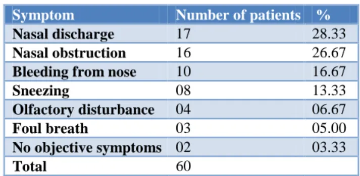
Table 2: Symptomatology wise distribution in conservative management group.
Symptom Number of patients %
Nasal discharge 17 28.33
Nasal obstruction 16 26.67
Bleeding from nose 10 16.67
Sneezing 08 13.33
Olfactory disturbance 04 06.67
Foul breath 03 05.00
No objective symptoms 02 03.33
The predominant symptoms in this group were nasal discharge seen in 17 patients followed by nasal obstruction in 16 patients. Symptomatology wise distribution of cases in conservative management group is depicted in Table 2.
Out of 17 patients with nasal discharge, mucopurulent discharge was found in middle meatus suggestive of chronic rhinosinusitis in 15 patients and in 2 patients polypoidal uncinate and middle turbinate was found. In 16 patients with nasal obstruction, mild adenoid hypertrophy was found in 8 patients, small bilateral ethmoidal polyps were found in 7 patients and in 1 patient a mass in the nasopharynx was observed. In 10 patients with epistaxis, 6 patients had anterior epistaxis and posterior epistaxis was found in 3. In 1 patient, maggots were seen bilaterally and removal was done under endoscopic guidance in the office set up and turpentine impregnated gauze packing done. All 8 patients with sneezing had Allergic rhinitis. In 4 patients with olfactory disturbance, greenish black crusts with atrophied turbinates suggestive of atrophic rhinitis were found. Out of 3 patients with foul breath, chronic rhinosinusitis was found in 2 and atrophic rhinitis was found in 1. 2 patients had no objective finding was seen on endoscopy and were managed symptomatically.
Chronic rhinosinusitis cases were prescribed systemic antibiotics, topical decongestants and oral antihistamines. Topical steroid spray was given to adenoid hypertrophy patients and for ethmoidal polyps systemic steroids were given along with topical steroids. The sole patient of mass in the nasopharynx was identified as a case of nasopharyngeal carcinoma after histopathological confirmation and was referred to radiotherapy. Allergic rhinitis was managed by topical steroid spray and oral antihistamines. In Atrophic rhinitis, crusts were removed endoscopically and saline nasal douches were prescribed.
Figure 1: Pus in middle meatus.
All the patients were followed up at 1 week, 2 weeks, 1 month and 2 months and were evaluated for treatment response at each visit. After adequate understanding about the complete resolution of symptoms objectively as seen on diagnostic nasal endoscopy (DNE), the patients were labeled as cured. The DNE pictures of some of the conditions managed conservatively are depicted in Figures 1 and 2.
Figure 2: (A) Nasal myiasis; (B) Nasopharyngeal carcinoma; (C) Polypoidal uncinate; (D) Atrophic
rhinitis.
Figure 3: (A) Antro choanal polyp; (B) Adenoid hypertrophy; (C) Ethmoidal polyps; (D) Spur.
Figure 4: (A) Septal perforation; (B) Synechiae.
Figure 5: (A) Postoperative middle meatal antrostomy; (B) Postoperative FESS.
A
D C
B
A
D C
B
A B
Surgical management group
40 patients were treated surgically after the initial diagnostic nasal endoscopy.
The predominant symptom in this group was nasal obstruction seen in 34 patients, followed by nasal discharge in 5 patients and 1 patient had a complaint of whistling noise on respiration. Symptomatology wise distribution of cases in Surgical Management Group is depicted in Table 3.
Table 3: Symptomatology wise distribution in surgical management group.
Symptom Number of patients %
Nasal obstruction 34 85.0
Nasal discharge 05 12.5
Whistling noise on
respiration 01 2.5
Total 40 100.0
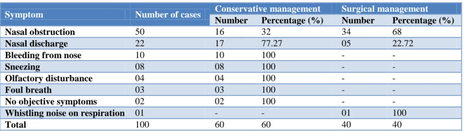
Table 4: Treatment wise management of different symptoms.
Symptom Number of cases Conservative management Surgical management
Number Percentage (%) Number Percentage (%)
Nasal obstruction 50 16 32 34 68
Nasal discharge 22 17 77.27 05 22.72
Bleeding from nose 10 10 100 - -
Sneezing 08 08 100 - -
Olfactory disturbance 04 04 100 - -
Foul breath 03 03 100 - -
No objective symptoms 02 02 100 - -
Whistling noise on respiration 01 - - 01 100
Total 100 60 60 40 40
In 34 patients with nasal obstruction , deviated nasal septum was observed in 13 , extensive nasal polyposis in 10 , rhinolith in 3 , synechiae in 2 , adenoid hypertrophy in 2 patients, inferior turbinate hypertrophy in 2, inverted papilloma in 1 and respiratory epithelial adenomatoid hamartoma (REAH) in 1 patient. Out of 13 cases of DNS, septoplasty was done in 10. In the remaining 3 patients, coexistent external nasal deformity was present in which septorhinoplasty was done. In 10 cases of nasal polyposis, the disease was extensive as seen on initial DNE and CT of the PNS was ordered. All the cases underwent FESS. In 3 patients of rhinolith, removal was done under endoscopic guidance. In 2 patients of synechiae, release of synechiae was done. In the 2 patients of inferior turbinate hypertrophy, turbinectomy was done. 2 patients of adenoid hypertrophy underwent adenoidectomy. In 1 patient of inverted papilloma, after confirmation by histopathology, removal under endoscopic guidance was done. In 1 patient of REAH, polypoidal mass attached to septum was seen and removal was done after further confirmation by histopathology. All 5 patients with nasal discharge had chronic rhinosinusitis and underwent FESS. The sole patient of whistling noise while breathing had septal perforation and it was closed by unilateral septal flap.
All the patients were followed up at similar intervals as done for the conservative management group and the treatment outcomes evaluated with endoscopy at each visit. The DNE pictures of some of the conditions managed surgically are depicted in Figures 3-4 and some follow up endoscopy pictures are depicted in Figure 5.
The treatment wise management of different symptoms is shown in Table 4.
DISCUSSION
In this study, maximum number of patients were in 21-30 years age group (30%). This observation is similar to study of Sheetal et al,in which majority of patients are in the age group of 20 to 40 years and to the study conducted by Zojaji et al, who observed the mean age of the patients to be 33 years.5,6 While in study conducted by Sancheti et al, maximum number of patients were in 36-40 years age group.7
In this study, most of the patients were males (70%). This is similar to the study of Sancheti et al where the majority of patients were males (58.3%). However, in the study conducted by Geminiani et al, the number of males and females were almost similar.8
In this study, the most common symptom overall was nasal obstruction seen in 50 patients (50%). This is similar to study conducted by Zojaji et al. However, in the study conducted by Kirtane et al, nasal discharge was the most common symptom seen in 61% patients, followed by nasal obstruction in 59% patients. 9
conditions.10 They observed that nasal endoscopy could diagnose chronic rhinosinusitis in 70 of 122 patients.
In the present study, out of the 25 medically managed cases, 8 cases were managed as allergic rhinitis depending on inferior turbinate hypertrophy seen on initial nasal endoscopy. This finding is similar to the work of Kumar et al who observed inferior turbinate hypertrophy as a common finding on 1st pass of nasal endoscopy in their study of 56 cases of Allergic rhinitis.11
In the patients with epistaxis, nasal endoscopy was very useful in identifying the exact bleeding point. This finding is supported by the study of Babu et al who studied the role of rigid nasal endoscopy in the diagnosis and management of epistaxis in 50 patients.12
In this study, 4 patients had olfactory disturbance and atrophic rhinitis diagnosed in all. Crust removal was done with the guidance of endoscopy. Similar study was conducted by Ari et al. who observed the benefit of endoscopic removal of crusts in cases of atrophic rhinitis.13
In 50 patients of nasal obstruction, 34 cases were surgically managed, out of which majority (13 cases) were having deviated nasal septum. This finding is in similar lines of the study conducted by Pullarat et al who observed that nasal septum deviation was the most common sign on nasal endoscopy among the people with complaint of nasal block and discharge.14
10 patients had adenoid hypertrophy out of which 8 cases were managed conservatively and in 2 patient’s adenoidectomy was performed. This is similar to study conducted by Baldassari et al who concluded that in children presenting with upper airway obstruction and suspected adenoid hypertrophy, flexible nasal endoscopy is the best initial choice for evaluation of adenoid size.15
In the present study, 17% had nasal polyps as observed on nasal endoscopy out of which 7 were medically managed and the rest underwent surgery. Whereas in a study conducted by Tegnoor et al, nasal polyps were found on DNE in 32 % cases and they have also concluded that DNE is of more diagnostic value in evaluating polyps.16
Rhinolith was observed in 3 cases and removal was achieved with the help of nasal endoscopy. This finding is supported by the work of Singh et al who have concluded that rigid endoscopy has important role in establishing a diagnosis and in evaluating the posterior extent of a rhinolith without providing any risk of radiation exposure.17
CONCLUSION
Nasal endoscopy is of immense help to the novice surgeon in understanding and managing various nasal pathologies. Through this study, it can be concluded that
most of the nasal symptoms can be managed conservatively instead of going for unnecessary surgical procedures, if carefully followed by nasal endoscopy prior and after treatment. Some persistent nasal pathologies treated conservatively can be postponed for surgical intervention provided, careful follow up with nasal endoscopy is done during the symptom free interval. Furthermore, post-operative surveillance to assess the surgical outcome can be done with the help of nasal endoscopy. It is also effective as a teaching tool and for documentation purposes. Thus, nasal endoscopy has proved to be an important tool in the management of nasal pathology and has stood the test of time.
ACKNOWLEDGEMENTS
The authors acknowledge the support rendered by MIMS for conducting this research work.
Funding: No funding sources Conflict of interest: None declared
Ethical approval: The study was approved by the Institutional Ethics Committee
REFERENCES
1. Draf W. Endoscopy of the Paranasal sinuses. Newyork: Springer–Verlag; 1983: 24.
2. Messerklinger W. Endoscopy of the nose. Baltimore: Urban Schwarzenberg. 1978;1(1)100-1. 3. Govindaraj S, Adappa ND, Kennedy DW.
Endoscopic sinus surgery: Evolution and technical innovations. J Laryngol Otol. 2010;124:242–50. 4. Chandra RK, Conley DB, Kern RC. Evolution of
the endoscope and endoscopic sinus surgery. Otolaryngol Clin N Am. 2009;42:747–52.
5. Sheetal D, Devan PP, Manjunath P, Martin P, Satish Kumar K. CT pns - Do we really require before fess? J Clin Diagn Res. 2011;5:179-81.
6. Zojaji R, Mirzadeh M, Naghibi S. Comparative evaluation of preoperative CT scan and intraoperative endoscopic sinus surgery findings in patients with chronic rhinosinusitis. Iran J Radiol. 2008;5:77-82.
7. Sancheti P, Velankar HK, Setty YK, Mathew MS, Dabholkar YG. Diagnostic Nasal Endoscopy or CT Scan of Paranasal Sinuses: Which One First? Res J Ear Nose Throat. 2017;1(7):1-4.
8. Rafael JG, Rodrigo FV, Adriano BM, Henrique PDC, Joao JDS. Comparison between computed tomography scan and nasal endoscopy in diagnosis of chronic rhinosinusitis. Intl Arch Otorhinolaryngol. 2007;11:402-5.
9. Kirtane MV. Functional endoscopic sinus surgery (A preliminary study). Indian J Otolaryngol. 1991;43:126-9.
Conditions. J Evol Med Dent Sci. 2014;3(14):3695-703.
11. Kumar S, Singh R, Singh M, Nag S. Nasal Endoscopic Findings in Allergic Rhinitis: A Prospective Study. IOSR J Dent Med Sci. 2014;13(5):45-8.
12. Mahesh babu MM, gowda B, Satish HS. Role of Rigid Nasal Endoscopy in the Diagnosis and Management of Epistaxis. J Dent Med Sci. 2014;13(3):40-5.
13. Sevil AY, Koksal YB, Fatih Y. A forgotten difficult entity: Ozaena Report of two cases. Eastern J Med. 2010;15:114.
14. Pullarat AN, Kottayil S, Raj G, Basheer NK. A comparative analysis of CT scan versus diagnostic nasal endoscopy in chronic rhino sinusitis. Int J Otorhinolaryngol Head Neck Surg. 2018;4:930-4.
15. Cristina M. Baldassari, Sukgi Choi.Assessing Adenoid Hypertrophy in Children: X-Ray or Nasal Endoscopy? Laryngoscope. 2014;124(7):1507-734. 16. Tegnoor MS, George JW, George W, Joshi R.
Comparative study between diagnostic nasal endoscopy and computed tomography of PNS in sino nasal diseases. Int J Otorhinolaryngol Head Neck Surg. 2017;3:972-8.
17. Singh R, Galagali JR , Santosh Kumar. A Large Rhinolith and Importance of Nasal Endoscopy: A Case Report. IOSR J Pharm Bio Sci. 2015;10(4):23-5.