Kolesnyk Yu. M., Isachenko M. I. Pathogenetic features of morphodensitometric characteristics of cardiomyocytes and marker profile of the left ventricular remodeling in rats with experimental intermittent hypoxia of different duration. Journal of Education, Health and Sport. 2020;10(9):752-762. eISSN 2391-8306. DOI http://dx.doi.org/10.12775/JEHS.2020.10.09.091
https://apcz.umk.pl/czasopisma/index.php/JEHS/article/view/JEHS.2020.10.09.091 https://zenodo.org/record/4051648
The journal has had 5 points in Ministry of Science and Higher Education parametric evaluation. § 8. 2) and § 12. 1. 2) 22.02.2019. © The Authors 2020;
This article is published with open access at Licensee Open Journal Systems of Nicolaus Copernicus University in Torun, Poland
Open Access. This article is distributed under the terms of the Creative Commons Attribution Noncommercial License which permits any noncommercial use, distribution, and reproduction in any medium, provided the original author (s) and source are credited. This is an open access article licensed under the terms of the Creative Commons Attribution Non commercial license Share alike.
(http://creativecommons.org/licenses/by-nc-sa/4.0/) which permits unrestricted, non commercial use, distribution and reproduction in any medium, provided the work is properly cited. The authors declare that there is no conflict of interests regarding the publication of this paper.
Received: 20.08.2020. Revised: 01.09.2020. Accepted: 25.09.2020.
UDC: 616.12-018.1+616.124-007.61]:616.831-008.64:612.22]-092.9
PATHOGENETIC FEATURES OF MORPHODENSITOMETRIC
CHARACTERISTICS OF CARDIOMYOCYTES AND MARKER PROFILE OF THE
LEFT VENTRICULAR REMODELING IN RATS WITH EXPERIMENTAL
INTERMITTENT HYPOXIA OF DIFFERENT DURATION
Yu. M. Kolesnyk, M. I. Isachenko
Zaporizhzhia State Medical University
Department of Pathophysiology
Kolesnik Yu. M., MD, PhD, DSc, Professor, Rector of Zaporizhzhia State Medical University, Honored Scientist and Engineer of Ukraine.
ORCID ID: 0000-0002-1556-5085
Isachenko M. I., MD, Post-graduate student of the Department of Pathophysiology, Zaporizhzhia State Medical University, Zaporizhzhia, Ukraine.
ORCID ID: 0000-0002-3026-1012, [email protected], +380973029038
Abstract
Myocardial remodeling is considered as a three-component complex process,
including cardiomyocyte hypertrophy, their apoptosis, interstitial and perivascular fibrosis. At
the same time, the nature of remodeling and the direction of restructuring in the myocardium
depend on the prevalence of one of these processes, the degree of their severity and the impact
duration. Therefore, to understand the effect of short- and long-term intermittent hypoxia on
the direction of myocardial remodeling and its type, the aim of this study was to determine
the marker profile of left ventricular myocardial remodeling in the rats with experimental
intermittent hypoxia durated 15 and 60 days.
Material and methods. A total of 30 male Wistar rats aging 6-10 months were used
for the experiment and assigned to 3 experimental groups of 10 animals each: control; rats
exposed to 15-day hypoxia; rats exposed to 60-day hypoxia. The object of the study was the
left ventricular myocardium. The number of nuclei in an image, their average linear size and
density (the ratio of the total area of nuclei to the area of the cytoplasm), and the
concentration of RNA in the nucleus and cytoplasm were measured in the sections stained by
Einarson via morphodensitometric method. An immunofluorescence method was used to
study the content of immunoreactive material to markers of remodeling (cardiotrophin-1, type
I collagen, titin, annexin V). As a result of the study, it was found that intermittent hypoxia
led to a decrease in the number of nuclei in cardiomyocytes in both experimental groups
compared to the control. Such changes in the number of nuclei were accompanied by
significantly larger nuclear size compared to the control (in rats with 15-day hypoxia by
52.9 %, in rats with 60-day hypoxia - by 153.4 %) and a decrease in the density of nuclei in
relation to the cytoplasm by 19.5 % and 21.2 %, respectively. In both groups, a significantly
lower control concentration of RNA in the nuclei of cardiomyocytes was found alongside
with an increase in their area. Such changes were accompanied by a significantly higher
concentration of RNA by 23.2 % in the cytoplasm in case of long-term intermittent hypoxia.
The marker profile of remodeling parameters in rats exposed to 15-day hypoxia was
characterized by a higher content of cardiotrophin-1 and titin by 11.5 % and 23.1 %,
respectively, as compared to the control. The content of type I collagen was 19.9 % higher,
while the content of annexin V did not change significantly. In rats with long-term 60-day
hypoxia, the content of cardiotrophin-1 was higher by 73.6 %, titin - by 124.9 %, type I
collagen - by 41.9 %, and annexin V - by 95.9 % in comparison to the control. When
calculating the titin / collagen ratio in rats based on their content, a significant increase in
myocardial stiffness was determined in the 60-day hypoxia group which was 1.44, while it
was 0.91 in the control, and in rats with 15-day short-term hypoxia – 0.93.
Conclusions. Intermittent hypoxic effects, regardless of duration, result in
morpho-structural rearrangements of the myocardium, are characterized by an increase in viscoelastic
mechanical properties and the development of hypertrophy, but with an increase in the
duration of exposure (60 days) – by an additional formation of interstitial fibrosis and
apoptosis of cardiomyocytes. Short-term hypoxia forms a hypertrophic type of myocardial
and a moderate decrease in RNA concentration. The marker profile of remodeling changes in
a physiological manner and is characterized by an increase in the content of cardiotrophin-1,
type I collagen and titin, while maintaining the titin / collagen ratio and normal apoptotic rate.
Long-term hypoxia causes a fibro-apoptotic type of pathological myocardial remodeling
including an almost two-fold decrease in the number of nuclei with an increase in their size
and a decrease in RNA concentration. The marker profile of remodeling is characterized by an
increase in all 4 components, a significant increase in the titin / collagen ratio and high
apoptotic rate of cardiomyocytes.
Key words: titin, annexin V; cardiotrophin-1; type I collagen; myocardium; left
ventricle of the heart; intermittent hypoxia; Wistar rats.
Introduction
Analysis of the results of large-scale studies has shown that various impacts on the
cardiovascular system (CCC) result in myocardium remodeling, the component ratio of which
depends on the severity and duration of driving factors. The left ventricular myocardium is
sensitive to pressure overload and oxygenation levels, so it is the first to adapt through
physiological myocardial remodeling (PhMR) [1]. In a case of overstimulation, its
long-lasting effect or ineffective adaptation, the physiological effect becomes pathological, and
remodeling turns pathological (PMR). A demonstrative example of this action is hypoxia.
Short-term intermittent stimuli have a therapeutic effect, causing PhMR, while long-term
influence and overload tend to PMR. Most often, researchers believe that intermittent hypoxia
at last causes physiological changes in the myocardium, but all of these studies were
short-term estimating a limited number of parameters without a comprehensive description of
morpho-structural changes in the heart "geometry" [2].
Today, myocardial remodeling is considered as a three-component complex process,
including hypertrophy of cardiomyocytes, their apoptosis, interstitial and perivascular
fibrosis. The nature of remodeling and the direction of restructuring in the heart muscle
depend on the prevalence of one of these processes and their severity [3].
In this regard, in recent years, in accordance with cardiological and pathoanatomical
guidelines for the purpose of determining the component ratio of myocardial remodeling and
evaluating morpho-structural changes in it, it is recommended to study a panel of biomarkers
consisting of cardiotrophin-1 – a marker of hypertrophy [4], titin – a protein that is a part of
the sarcomere for modeling the myocardial passive elasticity [5], type I collagen – a key
impaired degradation leads to the development of fibrosis [6], and annexin V – as a marker of
cardiomyocyte apoptosis [7].
Therefore, to understand the influence of short- and long-term intermittent hypoxia
(IH) on the direction of myocardial remodeling and to establish its type, the aim of the study
was to determine the pathogenetic features of morphodensitometric characteristics of
cardiomyocytes and marker profile of the left ventricular remodeling in rats with experimental
intermittent hypoxia of different duration (15 and 60 days).
Materials and methods. The experiment was conducted on the basis of the Training
Medical Laboratory Center of the Zaporizhzhia State Medical University (Certificate of
technical qualification 033/18 of 12.12.2018, valid until 25.12 2023). All devices used for
study were certified and underwent annual metrological control (Laboratory of Experimental
Pathophysiology, License 2CK2 YMK2 T6PB SG5N SJLS4).
The experimental part of the study was carried out exactly in accordance with the
National “Common Ethical Principles of Animal Experiments” (Ukraine, 2001), in agreement
with the Directive 2010/63EU of the European Parliament and of the Council of 22
September 2010 on the protection of animals used for scientific purposes [8].
To detect the pathogenetic features of the marker profile in left ventricular remodeling,
a total of 30 male rats, weighing 220-290 g, 6-10-month-old were divided into 3 experimental
groups: the 1st – the control group (10 intact male Wistar rats); the 2nd – 10 male Wistar rats
exposed to 15-day intermittent hypoxia (IH 15), the 3rd – 10 male Wistar rats exposed to
60-day intermittent hypoxia (IH 60).
Hypoxic training was performed in a ventilated pressure chamber with a volume of 1.0
cubic meter (m3) at an altitude of 6000 m (pO2 = 9.8 %) using a model widely used for the
studies at the Department of Pathophysiology of ZSMU [9]. The animals were housed in the
pressure chamber daily from 10 am to 4 pm. The training was conducted in the following
mode: an oxygen tension in the pressure chamber corresponded to the altitude of 1 km on the
1st day, 2 km - on the 2nd day, 3 km – on the 3rd, 4 km - on the 4th, 5 km - on the 5th, 6 km -
on the 6th day and in the days that followed. Hypoxic training at the altitude of 6 km was
performed for 10 days in 15-day hypoxia and for 55 days in 60-day hypoxia. [10].
The animals were euthanized via rapid decapitation after thiopental anesthesia (45
mg/kg body weight, intraperitoneally). The study objects in the experimental animals was
fragments of the left ventricle, which were embedded in paraplast blocks after standard
histological preparation and then sectioned into 5 μm-thick slices using a rotary microtome
The images of sections stained by Einarson via morphodensitometric method were
taken. AxioScope (Carl Zeiss, Germany) microscope images were analyzed in an interactive
mode in AxioVision 40 V 4.8.2.0 software program (License No. 3005339) using ImageJ
software (National Institutes of Health, USA); the number of cardiomyocyte nuclei, the
average nuclear linear size (μm), the nuclear density (a ratio of the total nuclear area to the
cytoplasmic area), the concentration of RNA in the nucleus and cytoplasm (OD) were
calculated.
The study of the immunoreactive material (IRM) to remodeling markers content (IF)
was carried out using the immunofluorescence technique in accordance with the protocol of
the immunohistochemical study [11]. Goat polyclonal antibodies Cardiotrophin-1 were used
to detect cardiotrophin-1, (N-20): sc-20867; primary goat polyclonal antibodies COL1A1
(C-18): sc-8784 - type I collagen; goat polyclonal antibodies Annexin V (R-20): sc-1929 -
annexin V. FITC-conjugated murine anti-goat IgG: sc-2356 were used as secondary
antibodies to the above mentioned markers. Primary murine monoclonal antibodies Titin
(E-2): sc-271946 and secondary FITC-conjugated murine anti-rabbit IgG: sc-2359 were used to
detect titin. All antibodies, produced by Santa Cruz Biotechnology, Inc., were applied at a
dilution of 1:200.
To understand the degree of fibrosis development in the experimental rats, the
titin/collagen ratio was calculated according to their content.
All statistical computations were performed in the Microsoft Excel 2016 table
processor (Microsoft Corp., USA). For all parameters, the arithmetic mean (M), its dispersion
and mean error (m) were calculated. To determine the significance of differences between the
study results among the experimental and control groups of rats, the Student’s coefficient (t)
was calculated, followed by a verification of significant differences between the samples (p)
and the confidence interval of the mean according to the Student’s distribution tables. A PSt
value < 0.05 were considered statistically significant [12].
Results. As a study result, it was found that intermittent hypoxia led to a decrease in
the number of nuclei in cardiomyocytes in both experimental groups compared to the control.
In IH60 rats, this indicator was significantly lower than that in the control by 39% (Table 1).
Such changes in the number of nuclei were accompanied by a significant increase in
their size as compared to the control (in IH15 rats by 52.9%, in IH60 rats – by 153.4%) and a
decrease in the nuclear density compared to the cytoplasm by 19.5% in IH15 and 21.2% in
Table 1 – Morphodensitometric characteristics of the myocardium in rats with
experimental hypoxia, (М±m)
Parameters Control IH15 IH60
Number of the nuclei 1069,1±24,2 1013,8±23,1 651,8±15,3*
The nuclear size, μm 6,1±0,2 9,3±0,2* 15,5±0,3*
The nuclear density, μm 21,9±0,5 17,6±0,5* 17,2±0,5*
RNA concentration in the nucleus, OD 0,241±0,007 0,157±0,002* 0,140±0,003*
RNA concentration in the cytoplasm, OD 0,052±0,001 0,053±0,000 0,064±*0,001
Note. * – significance of the differences in comparison with the control group, Р<0.05.
An interesting pattern was found in both groups as a significantly lower RNA
concentration in the nuclei of cardiomyocytes with an increase in their area. Thus, this
indicator was 34.9% lower in animals with IH15 than the control one, while in IH60 rats – by
41.9% (Table 1). Such changes were accompanied by 23.2% higher concentration of RNA in
the cytoplasm in IH60, and this indicator was characterized only by a tendency to increase in
IH15 rats (Table 1).
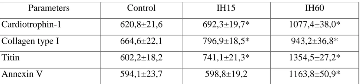
The marker profile of remodeling parameters in rats of the experimental groups
changed in a way different from the morphodensitometric parameters. The content of
cardiotrophin-1 and titin in IH15 rats was significantly higher by 11.5% and by 23.1% than
that in the control. The content of collagen type I showed significant changes in the direction
of increase by 19.9% in comparison with the control, while the content of annexin V did not
change significantly compared to the control values (Table 2).
Table 2 – The content of immunoreactive material to markers of the left ventricular
myocardium remodeling in the rats with experimental hypoxia, (М±m)
Parameters Control IH15 IH60
Cardiotrophin-1 620,8±21,6 692,3±19,7* 1077,4±38,0*
Collagen type I 664,6±22,1 796,9±18,5* 943,2±36,8*
Titin 602,2±18,2 741,1±21,3* 1354,5±27,2*
Annexin V 594,1±23,7 598,8±19,2 1163,8±50,9*
Note. * – significance of the differences in comparison with the control group Р<0.05.
In rats with long-term hypoxia IH60, the content of cardiotrophin-1 was higher by
control index by 41.9%, and annexin V – by 95.9% (Table 2). Calculated the titin/collagen
ratio revealed a significant increase in myocardial stiffness in the group of rats with IH60,
which amounted to 1.44, while it was 0.91 in the control, and 0.93 – in IH15.
Discussion. The experimental study showed that the hypoxic effect, regardless of its
duration, was accompanied by changes in the left ventricle myocardium and a decrease in the
number of nuclei with a compensatory increase in their size, especially in IH60 rats (2.5 times
larger than that in the control). However, the number of nuclei in IH60 rats was less by 35.7%
than that in IH15. Due to changes in the total area of the nuclei and cytoplasm of
cardiomyocytes, the nuclear density showed the downward trend. Such morpho-structural
changes of nuclei and their ratio to the cytoplasm are a sign of the hypertrophy development
as a response to hypoxic effects. It was expected to identify major changes in the myocardium
of rats with long-term hypoxia – IH60 (Table 1 and 2). Similar facts have been proven by
Antonio F. Corno et al. in the study confirming the formation of myocardial hypertrophy in
response to chronic and intermittent hypoxia [13].
The identified lower RNA concentration in the nuclei of cardiomyocytes in the rats of
both experimental groups should be considered as a result of the increase in their area while
maintaining the total RNA content in the first case of IH15, and as a possible nuclear
dysfunction - in the second case of IH60 [14].
The determined nature of the marker profile has enabled to assess the type of
remodeling and its direction towards physiological or pathological, as well as to identify the
dependence on the hypoxic exposure duration. Thus, under hypoxic effects in rats, an increase
in the marker of myocardial hypertrophy cardiotrophin-1 was observed. Its content was
quantitatively dependent on the exposure duration and IH60 index exceeded IH15 by 2 times.
According to Mirtschink P. and others, this increase could be initiated by HIF-factor, which is
the main regulator of key indicators in response mediated by gene transcription at low oxygen
levels [15].
At the present stage, it is impossible to characterize the morpho-structural
rearrangements in the myocardium and describe the nature of fibrotic changes without taking
into account the content of titin and collagen type I, the increase in expression of which was
reliably found in rats of both experimental groups. However, it is important to note that in rats
with IH15, the ratio remained as close as possible to the control values (0.93:0.91) amid the
moderate increase in their content. Probably, the 15-day hypoxic effect helps to increase the
elasticity of the matrix skeleton as an element of adaptation to new functioning conditions,
Zadnipryany I. V. obtained significant manifestations of myocardial remodeling with the
prevalence of fibrosis in 15-day exposure at 3.5 km that developed due to the activation of
fibroblasts and reduced collagen degradation. But in their study, the authors did not
differentiate the type of collagen, which provides no confidence that its accumulation is
pathological [16].
Prolonged 60-day hypoxic effect resulted in a significant increase in the content of
titin and collagen with the almost two-fold increase in their ratio compared to the control
(1.44:0.91) demonstrating the development of fibrosis amid the compensatory increase in
elastic properties of the heart muscle. The researchers suggest that such changes in titin
content were associated with oxidative modification of all three filamentous systems of the
sarcomere and titin isoforms imbalance towards increasing a more severe form of N2B. These
changes were especially pronounced in oxidative stress, which have been developed in
prolonged hypoxia [17].
Determination of the annexin V content as the marker of cardiomyocyte apoptosis
showed that there were no significant changes in 15-day hypoxic exposure. This fact
suggested the physiological mode of cardiac muscle remodeling in rats of this group. In the
case of long-term hypoxia, the content of annexin V was found to be 124.8 % higher than that
in the control, which was a sign of significant cell death processes with the development of
so-called myogenic dilation of the heart [18].
Conclusions
1. Intermittent hypoxic effects, regardless of duration, result in morpho-structural
rearrangements of the myocardium, are characterized by an increase in viscoelastic
mechanical properties and the development of hypertrophy, but with an increase in the
duration of exposure (60 days) – by an additional formation of interstitial fibrosis and
apoptosis of cardiomyocytes.
2. Short-term hypoxia forms a hypertrophic type of myocardial remodeling with a
decrease in the number of nuclei alongside with an increase in their size and a moderate
decrease in RNA concentration. The marker profile of remodeling changes in a physiological
manner and is characterized by an increase in the content of cardiotrophin-1, type I collagen
and titin, while maintaining the titin / collagen ratio and normal apoptotic rate.
3. Long-term hypoxia causes a fibro-apoptotic type of pathological myocardial
remodeling including an almost two-fold decrease in the number of nuclei with an increase in
characterized by an increase in all 4 components, a significant increase in the titin / collagen
ratio and high apoptotic rate of cardiomyocytes.
References:
1. Yalçin, F., Kucukler, N., Cingolani, O., Mbiyangandu, B., Sorensen, L., Pinherio,
A., Abraham, M. R., & Abraham, T. P. (2019). Evolution of ventricular hypertrophy and
myocardial mechanics in physiological and pathological hypertrophy. Journal of applied
physiology (Bethesda, Md. : 1985), 126(2), 354–362.
https://doi.org/10.1152/japplphysiol.00199.2016.
2. Tham, Y. K., Huynh, K., Mellett, N. A., Henstridge, D. C., Kiriazis, H., Ooi, J.,
Matsumoto, A., Patterson, N. L., Sadoshima, J., Meikle, P. J., & McMullen, J. R. (2018).
Distinct lipidomic profiles in models of physiological and pathological cardiac remodeling,
and potential therapeutic strategies. Biochimica et biophysica acta. Molecular and cell biology
of lipids, 1863(3), 219–234. https://doi.org/10.1016/j.bbalip.2017.12.003.
3. Kolesnyk, Yu.M., Kolesnyk, M.I., Abramov, A.V., Tumanskyi, V.O., Hancheva,
O.V. (2015) Imunohistokhimichna i morfo-densytometrychna diahnostyka patolohichnoho
remodeliuvannia miokarda pry arterialnoi hipertenzii ta tsukrovomu diabeti (Metodychni
rekomendatsii). Kyiv, 25s. (Ukrainian).
4. Shimizu, I., & Minamino, T. (2016). Physiological and pathological cardiac
hypertrophy. Journal of molecular and cellular cardiology, 97, 245–262.
https://doi.org/10.1016/j.yjmcc.2016.06.001.
5. Hong, J., Chu, M., Qian, L., Wang, J., Guo, Y., & Xu, D. (2017). Fibrillar Type I
Collagen Enhances the Differentiation and Proliferation of Myofibroblasts by Lowering α2β1
Integrin Expression in Cardiac Fibrosis. BioMed research international, 2017, 1790808.
https://doi.org/10.1155/2017/1790808.
6. Ding, Y., Wang, Y., Jia, Q., Wang, X., Lu, Y., Zhang, A., Lv, S., & Zhang, J.
(2020). Morphological and Functional Characteristics of Animal Models of Myocardial
Fibrosis Induced by Pressure Overload. International journal of hypertension, 2020, 3014693.
https://doi.org/10.1155/2020/3014693.
7. Galluzzi, L., Vitale, I., Aaronson, S. A., Abrams, J. M., Adam, D., Agostinis, P.,
Alnemri, E. S., Altucci, L., Amelio, I., Andrews, D. W., Annicchiarico-Petruzzelli, M.,
Antonov, A. V., Arama, E., Baehrecke, E. H., Barlev, N. A., Bazan, N. G., Bernassola, F.,
Bertrand, M., Bianchi, K., Blagosklonny, M. V., Kroemer, G. (2018). Molecular mechanisms
of cell death: recommendations of the Nomenclature Committee on Cell Death 2018. Cell
8. Zakon Ukrainy № 3447-IV «Pro zakhyst tvaryn vid zhorstkoho povodzhennia».
Vidomosti Verkhovnoi Rady Ukrainy.2006;№ 27:s230. (Ukrainian).
9. Kolesnyk, Yu.M., Hancheva, O.V., Abramov, A.V., Ivanenko, T.V., Fedotova,
M.I., Danukalo, M.V., (2016) inventors; Zaporizhzhia State Medical University, assignee.
Method of modeling physiological myocardial remodeling in small rodents. Ukraina pat.
112290. Dec 12. (Ukrainian).
10. Kolesnyk, Y., Isachenko, M., & Melnikova, O. (2019). The features of the nitric
oxide system in the left ventricle myocardium in the rats with experimental intermittent
hypoxia of different duration. Pathologia, 0(3). doi: 10.14739/2310-1237.2019.3.188783.
11. Fedotova, M.I., Kovalov, M.M., Zhulinskyi, V.O., Kadzharian, Ye.V. (2017)
Osoblyvosti ekspresii izoform syntazy oksydu azotu u miokardi livoho shlunochka shchuriv
pry arterialnii hipertenzii riznoho henezu. Aktualni problemy suchasnoi medytsyny: Visnyk
Ukrainskoi medychnoi stomatolohichnoi akademii.;17,4(60): 91−95. (Ukrainian).
12. Zaitsev VM, Lyfliandskyi VH, Marynkyn VY. (2006) Prykladnaia medytsynskaia
statystyka. Sankt-Peterburh:Folyant.432 s.(Russian).
13. Corno, A. F., Milano, G., Morel, S., Tozzi, P., Genton, C. Y., Samaja, M., & von
Segesser, L. K. (2004). Hypoxia: unique myocardial morphology?. The Journal of thoracic
and cardiovascular surgery, 127(5), 1301–1308. https://doi.org/10.1016/j.jtcvs.2003.06.012.
14. Zhong, Z., Hu, J. Q., Wu, X. D., Sun, Y., & Jiang, J. (2016). Anti-apoptotic effects
of myocardin-related transcription factor-A on rat cardiomyocytes following hypoxia-induced
injury. Canadian journal of physiology and pharmacology, 94(4), 379–387.
https://doi.org/10.1139/cjpp-2014-0461.
15. Mirtschink, P., & Krek, W. (2016). Hypoxia-driven glycolytic and fructolytic
metabolic programs: Pivotal to hypertrophic heart disease. Biochimica et biophysica
acta, 1863(7 Pt B), 1822–1828. https://doi.org/10.1016/j.bbamcr.2016.02.011.
16. Sataeva, T. P., & Zadnipryany, I. V. (2018). Issledovanie markerov
remodelirovaniia miokarda pri éksperimental'noĭ gipobaricheskoĭ gipoksii na fone korrektsii
preparatom tsitoflavin [Investigation of markers for myocardial remodeling during
experimental hypobaric hypoxia in the correction with cytoflavin]. Arkhiv patologii, 80(6),
35–42. https://doi.org/10.17116/patol20188006135. (Russian).
17. Koser, F., Loescher, C., & Linke, W. A. (2019). Posttranslational modifications of
titin from cardiac muscle: how, where, and what for?. The FEBS journal, 286(12), 2240–
18. de Jong, R., Pluijmert, N. J., de Vries, M. R., Pettersson, K., Atsma, D. E., Jukema,
J. W., & Quax, P. (2018). Annexin A5 reduces infarct size and improves cardiac function
after myocardial ischemia-reperfusion injury by suppression of the cardiac inflammatory