Original Research Article
Improved sensitivity of direct microscopy for Mycobacterium
tuberculosis in lymph nodes: comparison of fluorescent microscopy and
modified bleach method ZN microscopy along with conventional ZN
staining method
Gul Aalmeen, Sangeeta Sharma*, Sneha Vishwasrao, Rani Bansal, Anjali Khare
INTRODUCTION
Lymphadenopathy is the most common presentation of extra pulmonary tuberculosis (EPTB). Tuberculous lymphadenitis is seen in nearly 35% of EPTB.1 It arises
as a result of lymphatic spread from a primary focus. Most often it involves the cervical group of lymph nodes attributed to the rich lymphatic supply of the region and
most commonly seen in second and third decades of life.2
The precise cause of enlarged lymph nodes is often difficult to establish by history, physical examination, and radiographic studies alone.3
Fine needle aspiration cytology (FNAC) has assumed an important role in the evaluation of peripheral lymphadenopathy as a possible minimally invasive
ABSTRACT
Background: Extra pulmonary tuberculosis arises as a result of lymphatic spread from a primary focus. Fine needle aspiration cytology has assumed an important role in the evaluation of peripheral lymphadenopathy as a possible minimally invasive alternative to excisional biopsy. In most low-income countries; the only practically available bacteriologic method for diagnosing EPTB is direct smear microscopy for acid fast bacilli from the sample of the lesion. There are various methods of staining and concentration for improving sensitivity of direct microscopy for detection of tubercle bacilli in specimen.
Methods: This prospective study was carried out in the Department of Pathology, Subharti Medical College and associated Chattrapati Shivaji Subharti Hospital, Meerut for a period of 2 years from July 2016 - August 2018 in 151 patients with clinical suspicion of TB and significant lymphadenopathy.
Results: AFB positivity increased from 40.39% on conventional ZN stain to 48.34% on modified bleach method ZN stain and to 56.29% on Auramine-O fluorescent stain. Taking fluorescent microscopy (Auramine-O) as reference method the sensitivity, specificity, positive predictive value, negative predictive value, and diagnostic accuracy of conventional ZN stain and modified bleach method ZN stain were calculated as 71.8%, 100%, 100%, 73.33%, 84.10% and 85.33%,100%,100% ,84.61% ,92.05%, respectively.
Conclusions: The addition of fluorescent microscopy (Auramine-O) and modified bleach method ZN microscopy along with conventional ZN staining method would be an important adjunct to improve the microscopic detection of
Mycobacterium tuberculosis in fine-needle aspirates of lymph nodes.
Keywords: Auramine-O stain, Acid fast bacilli, Bleach modified ZN stain, Convention Ziehl-Neelson stain, Extra pulmonary tuberculosis, Fine needle aspiration cytology
Department of Pathology, Subharti Medical College, Meerut, Utter Pradesh, India
Received: 01 February 2019
Accepted: 07 February 2019
*Correspondence:
Dr. Sangeeta Sharma,
E-mail: [email protected]
Copyright: © the author(s), publisher and licensee Medip Academy. This is an open-access article distributed under the terms of the Creative Commons Attribution Non-Commercial License, which permits unrestricted non-commercial use, distribution, and reproduction in any medium, provided the original work is properly cited.
alternative to excisional biopsy.1 In most low-income
countries, the only practically available bacteriologic method for diagnosing extra pulmonary TB is direct smear microscopy for acid fast bacilli from the sample of the lesion.4 Mycobacterium culture is the gold standard
for detection of tubercle bacilli, but it is time consuming and false negativity is high in extra pulmonary cases. It is expensive and may not be readily available in all centres.5,6
In recent times, PCR has been found to be the most sensitive technique for rapid diagnosis of M. tuberculosis. However, it is not applicable in routine use because of cost implications.7 Due to the above-mentioned
limitations, various attempts have been made to improve the sensitivity of ZN microscopy.8 There are various
concentration methods for improving sensitivity of direct microscopy for detection of tubercle bacilli in specimen. Among these, the bleach concentration method is one of the safest concentration methods which improve the sensitivity of detection of acid fast bacilli.9
METHODS
This prospective study was carried out in the Department of Pathology, Subharti Medical College and associated Chattrapati Shivaji Subharti Hospital, Meerut for a period of 2 years from July 2016 - August 2018 in 151 patients with clinical suspicion of TB and significant lymphadenopathy (irrespective of age and sex).
At first smears were prepared with aspirate and then the remaining aspirated material was used for bleach method. Smears were stained with:
• Leishman- Giemsa stain,
• ZN stain and Auramine –O stain.
Sensitivity, specificity, diagnostic accuracy of conventional ZN and modified bleach method ZN were calculated by comparing test diagnosis with fluorescence microscopy (Auramine-O) as reference method.
Modified bleach method ZN stain
Rinsed the syringe/needle hub with 1 ml normal saline and transferred into 5ml sterile disposable, conical screw-capped tubes. To this conical tube, 2ml of 5% NaOCl was added. The mixture was incubated at room temperature for 15 min by shaking at regular intervals. Then the conical tube containing the mixture was centrifuged at 3000rpm for 15 min after addition of 2ml of distilled water. The supernatant was discarded carefully, and the sediment was transferred with a sterile pipette on to a clean sterile slide. Air dry the slide, heat fixed and stained by the ZN method.
As a control, 2ml of distilled water was centrifuged and stained the sediment by ZN staining to rule out any error due to contamination while testing each specimen.
Auramine-O fluorescence acid-fast stain
Fixed smears were flooded with auramine-phenol. Then allowed to stand for 10 minutes. Smears were then rinsed with water. Decolorized with 1% acid-alcohol. And left to stand for 5 minutes.
A second rinse with water was followed by counterstaining with 0.1% potassium permanganate for 10 seconds. Slides were finally rinsed with water and air dried
Examined using Motic BA 210 microscope with fluorescent attachment and wide band blue excitation filter (450-480nm) for the presence of bright fluorescent bacilli. Using x40 objective-scanned the stained smear from one side to another and back. At least one length was scanned before reporting negative. Slides were examined under x400 magnification for identification of AFB. In case of appearance of bacilli; pictures were clicked immediately with attached Moticam 1080.
For every batch of slides freshly stained positive control was checked first.
RESULTS
Age of patients in present study ranged from 18 months to 65 years with mean age of 23.7 years. Maximum cases, 35.76% (54/151) were in 3rd decade followed by 25.83%
(39/151) in 2nd decade of life. There was female
preponderance with male: female ratio was of 1:1.47.
Most common lymph node group involved was the cervical group- anterior and posterior triangles (64.23%), followed by sub mandibular (11.92%), supraclavicular (11.25%) axillary nodes (6.62%), inguinal nodes (2.64%). Few cases also involved abdomen (1.98%), sternal notch (0.66%) and lumbar (0.66%) regions.
Aspirates of 151 clinically suspicious tubercular cases were analyzed and for confirmation of tubercular etiology different staining methods, namely, conventional Zeihl- Neelsen, Modified Bleach method Zeihl-Neelsen and Auramine- O fluorescent stains were done and compared.
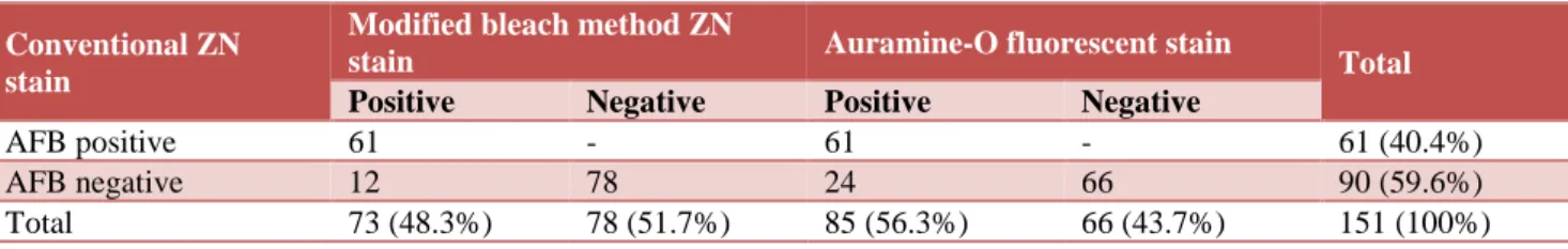
Total of 61/151 cases were positive for AFB with conventional ZN stain, 73/151 were positive with modified bleach method ZN and 85/151 were positive with Auramine –O fluorescent stain, as shown in Table 1. The smear Positivity of all the cases (151) increased from 40.39% on conventional ZN stain to 48.34% on modified bleach method ZN stain and to 56.29% on Auramine- O fluorescent stain.
Sensitivity, specificity and diagnostic accuracy
As Auramine- O fluorescent stain had highest AFB positivity (Table 1), it was taken as reference method for statistical analysis as shown in Table 2. The sensitivity,
specificity, positive predictive value and negative predictive value of conventional ZN stain was 71.8%, 100%, 100% and 73.33%, respectively. The diagnostic accuracy was 84.10% as shown in Table 3.
Table 1: Comparison of different staining methods for detection of AFB.
Conventional ZN stain
Modified bleach method ZN
stain Auramine-O fluorescent stain Total
Positive Negative Positive Negative
AFB positive 61 - 61 - 61 (40.4%)
AFB negative 12 78 24 66 90 (59.6%)
Total 73 (48.3%) 78 (51.7%) 85 (56.3%) 66 (43.7%) 151 (100%)
The sensitivity, specificity, positive predictive value and negative predictive value of modified bleach method ZN stain was 85.33%,100%,100% and 84.61%, respectively. The diagnostic accuracy was 92.05% as shown in Table 3.
Table 2:Comparative chart of results for detection of AFB by conventional ZN stain and modified bleach
method ZN stain.
Statistical indices Conventional
ZN stain
Modified bleach method ZN stain
True positive (TP) 61 73
True negative (TN) 66 66
False positive (FP) - -
False negative (FN) 24 12
Table 3: Taking fluorescent microscopy (Auramine-O) as reference method statistical values of conventional ZN stain and modified bleach method
ZN stain.
Statistical values Conventional
ZN stain
Modified bleach method ZN stain
Sensitivity 71.8% 85.88%
Specificity 100% 100%
Positive predictive
value (PPV) 100% 100%
Negative predictive value (NPV)
73.33% 84.61%
Diagnostic
accuracy 84.10% 92.05%
DISCUSSION
In present study, majority of cases, i.e.35.76% (54/151) were in the 3rd decade followed 25.82% (39/151) cases in
2nd of life. Similar findings were seen in studies by
Krishna M et al, and Dhawan I et al, et al with maximum cases in 2nd and 3rd decades and 2nd decade of life,
respectively.1,11 There was female preponderance in our
study with male: female ratio of 1:1.47. This was in accordance to studies conducted by Dhawan I et al, and Khare M et al.11,12 In the present study, the most common
lymph node group involved was cervical group with 97/151 cases. Similar distribution of lymph node involvement was seen by various other authors in their studies where cervical group was most commonly involved.11-13 This may be due to easy accessibility of
cervical lymph nodes for examination and evaluation.
In the present study, AFB positivity increased from 40.39% (61/151) on conventional ZN stain to 48.34% (73/151) on modified bleach method ZN stain and to 56.29% (85/151) on Auramine-O fluorescent stain. This was comparable to findings by various other authors in their studies as shown in Table 4.14-19
Aspirates which were once positive for AFB by conventional ZN remained positive for AFB by modified bleach method ZN and Auramine O. Similarly, all the cases once AFB positive by modified bleach method ZN remained positive by Auramine O staining method.
Though Mycobacterium culture is the gold standard for detection of tubercle bacilli, but it is time consuming and false negativity is high in extra pulmonary case. As Auramine- O fluorescent stain had highest AFB positivity it was taken as reference method for statistical analysis in present study.
was 85.33%,100%,100%,84.61% and 92.05% respectively.
Roiko JN et al, found lower sensitivity (20%) of conventional ZN staining method in their study but specificity was 100% in reference to fluorescent microscopy as seen in present study.10 Thakur B et al, in
their study observed that with culture as reference method, slightly higher sensitivity of conventional ZN (80%) as compared to present study findings (71.8%) were seen while specificity was lower (93.85%) than our findings (100%).6 Arora et al, reported 75% positivity on
fluorescence microscopy in culture-positive cases of tuberculous lymphadenitis.20 In a study by Joshi P et al,
taking culture as the gold standard, ZN staining showed a sensitivity of 62.50%, specificity of 87.50%, PPV of 83.33%, and NPV of 70%.5 On the other hand, it
increased with autofluorescence revealing a sensitivity of 95%, specificity of 81.81%, positive predictive value (PPV) of 82.6% and negative predictive value (NPV) of 94.7%.
However, direct comparison is a challenge owing to the fact that different techniques are used as gold standard techniques and specimen types used are different
While Auramine-O fluorescent microscopy has the advantage of speed, ease of screening, and reduces observer fatigue, modified bleach method ZN microscopy increases the concentration of aspirate and decreases necrosis in the background for better demonstration of AFB. In smaller centers, a specialized set up with fluorescence microscope attachments is required. In such cases the addition of modified bleach method ZN microscopy along with conventional ZN staining method can be an important adjunct to improve the microscopic detection of Mycobacterium tuberculosis in fine-needle aspirates of lymph nodes. Hence, authors concluded that Auramine-O fluorescent staining method and modified bleach method ZN staining have obvious advantage over conventional ZN staining method.
Funding: No funding sources Conflict of interest: None declared
Ethical approval: The study was approved by the Institutional Ethics Committee
REFERENCES
1. Krishna M, Gole SG. Comparison of conventional Ziehl-Neelsen method of acid fast bacilli with
modified bleach method in tuberculous
lymphadenitis. J Cytol. 2017; 34(4):188-92.
2. Kochhar AK, Patel KB, Shah M. Pattern of lymphadenopathy on fine needle aspiration cytology of superficial lymph nodes (a study of 150 cases). J Adv Res Biol Sci. 2012;4:288-92.
3. Pakalpaty S, Lakshmi GSR, Jahnavi I, Nagamani K, Prashanthi K, Soujanya KN, et al. A Study of Early
and Accurate Detection of Mycobacterium
tuberculosis among meningitis cases by using conventional saining methods. Acta Biomedica Scientica. 2017;4(2):1-5.
4. Khubnani H, Munjal K. Application of bleach
method in diagnosis of extra-pulmonary
tuberculosis. Indian J Pathol Microbiol. 2005 Oct;48(4):546-50.
5. Joshi P, Singh M, Bhargava A, Singh M, Mehrotra R. Autofluorescence-an important ancillary technique for the detection of Mycobacterium tuberculosis: revisited. Diag Cytopathol. 2013 Apr;41(4):330-4.
6. Thakur B, Mehrotra R, Nigam JS. Correlation of various techniques in diagnosis of tuberculous lymphadenitis on fine needle aspiration cytology. Pathol Res Int. 2013;1-4.
7. Farnia P, Mohammadi F, Zarifi Z, Tabatabee DJ, Ganavi J, Ghazisaeedi K, et al. Improving sensitivity of direct microscopy for detection of acid-fast bacilli in sputum: use of chitin in mucus digestion. J Clin Microbiol. 2002 Feb 1;40(2):508-11.
8. Gong G, Lee H, Hoon Kang G, Shim YH, Huh J, Kwang Khang S. Nested PCR for diagnosis of tuberculous lymphadenitis and PCR‐SSCP for identification of rifampicin resistance in fine‐needle aspirates. Diag Cytopathol. 2002 Apr;26(4):228-31.
9. Aung WW, Nyein MM, Ti T, Maung W. Improved
method of direct microscopy for detection of acid-fast bacilli in sputum. Southeast Asian J Trop Med Public Health. 2001;32(2):390-3.
10. Rioki JN, Ndungu J, Rogena EA, Jaoko W. Comparative evaluation of direct Ziehl-Neelsen (ZN) smear and modified ZN against fluorescent technique in the detection of acid-alcohol fast bacilli in lymph node aspirates. East Africa J Pathol. 2014;1:19-22.
11. Dhawan I, Gupta O, Kaushik A, Ranga S, Kasana D, Gupta PK. Cytomorphological spectrum and Ziehl-Nelson staining in suspected tuberculous lymphadenitis: A study of 100 cases. Ann Pathol Lab Med. 2015 Feb 6;2(1):A36-41.
12. Khare M, Kaushal M. Utility of modified bleach method technique for the demonstration of acid fast
bacilli in the diagnosis of tuberculous
lymphadenopathy in comparison to routine Ziehl-Neelsen staining. Ann Pathol Lab Med. 2016; 3(4);329-32.
13. Gangane N, Anshu, Singh R. Role of modified bleach method in staining of acid-fast bacilli in lymph node aspirates. Acta Cytol. 2008;52(3):325-28.
14. Bhardwaj S, Kashyap I, Kumari R. Comparative evaluation of two staining techniques for detection of Tubercular bacilli in lymph nodal aspirates. Int J Health Sci Res. 2015;5(7):115-21.
15. Annam V, Kulkarni MH, Puranik RB. Comparison
of the modified fluorescent method and
of acidfast bacilli in lymphnode aspirates. Cyto J. 2009;6-13.
16. Patel MM, Patel K, Italiya SL, Kaptan KR. Improved diagnosis of tuberculosis in lymph node cytology by bleach method for detection of acid fast bacilli in comparison to conventional Ziehl Neelsen staining method. Int J Med Sci Public Health. 2013;2(4):935-39.
17. Dwivedi G, Mathur C. Modified bleach method – improving microscopic detection of acid fast bacilli in fine needle aspiration smears of lymph nodes. JMSCR. 2013;1(4):176-81.
18. Akhter S, Fernandes H, Goyal G. Comparision of auramine-O stain with Ziehl Neelson stain in suspected cases of tubercular lymphadenitis. Int J Recent Trends Sci Technol. 2015;15(3):524-5. 19. Krishna M, Kumar A. Tuberculous mycobacteria
bacilli fluorescence and compare with
Ziehl-Neelsen stain in fine-needle aspiration cytology of tubercular lymphnode. Int J Otorhinolaryngol Head Neck Surg. 2016 Apr 8;2(2):66-9.
20. Arora B, Arora DR. Fine needle aspiration cytology in diagnosis of tuberculous lymphadenitis. Indian J Med Res. 1990;91:189-92.
Cite this article as: Aalmeen G, Sharma S, Vishwasrao S, Bansal R, Khare A.Improved sensitivity of direct microscopy for Mycobacterium tuberculosis in lymph nodes: comparison of