Journal of Feline Medicine and Surgery Open Reports
1 –7
© The Author(s) 2016 Reprints and permissions:
sagepub.co.uk/journalsPermissions.nav DOI: 10.1177/2055116916638681 jfmsopenreports.com
Creative Commons Non Commercial CC-BY-NC: This article is distributed under the terms of the Creative Commons Attribution-NonCommercial 3.0 License (http://www.creativecommons.org/licenses/by-nc/3.0/) which permits non-commercial use, reproduction and distribution of the work without further permission provided the original work is attributed as specified on the SAGE and Open Access pages (https://us.sagepub.com/en-us/nam/open-access-at-sage).
Introduction
Fibrosarcoma is a common cutaneous neoplasm of cats, representing 15% of cases in one study of 340 cats.1 The
hepatic form is reported much less frequently.2,3 One
series documented hepatic fibrosarcoma in only 1/41 cases of non-lymphomatous hepatobiliary masses.3
Peritoneopericardial diaphragmatic hernia (PPDH) is a structural abnormality resulting in communication between the pericardial sac and peritoneal cavity. The embryogenesis of PPDH is unknown but may include malformation of the pleuroperitoneal folds and septum transversum.4–6 It is often discovered as an incidental
finding in cats and, consequently, when clinical signs are absent, surgical correction is difficult to justify.7,8
Previous case reports have documented a possible association between incarcerated hepatic tissue in PPDH and the development of hepatic myelolipomas and hepatic cysts.6,9–12 This report describes a hepatic
fibrosarcoma incarcerated in a PPDH in a cat. To our knowledge no such association has previously been documented in this species.
Case description
A 14-year-old, female neutered domestic shorthair (body weight 4.5 kg) was referred following a 1 day history of dyspnoea. The cat spent time outdoors, but no history of trauma was reported. The cat had a stable body weight. Immediately prior to referral a cumulative dose of furo-semide 8 mg/kg was administered intramuscularly.
On physical examination the cat was hypothermic (36.7°C), had an inspiratory dyspnoea, reduced lung
Hepatic fibrosarcoma incarcerated in
a peritoneopericardial diaphragmatic
hernia in a cat
Michael Linton
1, Lydia Tong
2, Adrian Simon
1, Eugene Buffa
1,
Ross McGregor
1, Julien Labruyére
3and Darren Foster
1Abstract
Case summary A 14-year-old, female neutered domestic shorthair presented for dyspnoea. Thoracic ultrasonography and radiography showed that a heterogeneous mass was present within the pericardial sac, and the mass continued caudally with the mesenteric fat. On CT, the outline of the diaphragm was not continuous and there was an obvious defect with diaphragmatic thickening present at the mid-level of the liver. A pleural effusion and a small-volume pericardial effusion were also present. A ventral midline coeliotomy and median sternotomy revealed a 5 × 6 × 7 cm firm, irregular, tan-coloured soft tissue mass within the pericardial sac attached to both the diaphragmatic defect and liver. The mass was carefully dissected away from the heart and the diaphragmatic defect was repaired with primary closure. Postoperatively, the cat had a persistent pneumothorax that required continuous pleural suction for 41 h. The cat died 44 h postoperatively. Histopathology and immunohistochemistry confirmed the mass to be a hepatic fibrosarcoma incarcerated in a peritoneopericardial diaphragmatic hernia (PPDH).
Relevance and novel information This is the first reported case of metaplastic transformation of liver into a sarcoma in a cat with PPDH. In addition, hepatic fibrosarcoma is a rarely reported location for fibrosarcoma in this species.
Accepted: 16 February 2016
1 Eastside Veterinary Emergency and Specialists, Rose Bay, NSW,
Australia
2 Faculty of Veterinary Science, The University of Sydney, Sydney,
Australia
3 VetCT Consultants in Telemedicine, St John’s Innovation Centre,
Cowley Road Cambridge, England CB4 OW
Corresponding author:
Michael Linton BVSc, MACVSc (Small Animal Medicine), Eastside Veterinary Emergency Specialists, Sydney, 10 Newcastle Street, Rose Bay, Sydney, NSW 2029, Australia
Email: michael.linton@eastsidevets.com.au
that diagnostic tests to differentiate these differentials were performed first. These included dorsoventral radi-ography of the thorax and a brief transthoracic ultra-sound (CX50; Philips) while the cat was in sternal recumbency. Mask inhalation oxygen was administered during these procedures. No sedation was required.
A bilateral pleural effusion was noted radiographically and was anechoic in appearance ultrasonographically. For diagnostic and therapeutic reasons, ultrasound-guided thoracocentesis was performed and 250 ml of serous fluid was removed. Protein concentration of the effusion was 37 g/l (reference interval [RI] 0–25 g/l). Nucleated cell count was 1.7 × 10⁹/l (RI 0–1.5 × 10⁹/l).
Red cell count was 0.1 × 10¹²/l (RI 0 × 1012/l). These values
are consistent with a modified transudate. Cytology and cell counts of the fluid revealed 24% mature neutrophils, 14% small lymphocytes and 62% vacuolated and phago-cytic macrophages. No other significant cell population or microorganisms were identified.
Echocardiography (ECG; right parasternal) demon-strated a 1 mm pericardial effusion. Hypertrophy of the myocardium (left ventricular wall at end diastole 7.5 mm [normal <5.5 mm]) was evident. No other ECG abnormalities were noted. Specifically, the left atrium: aorta diameter ratio was 1.1 (RI <1.5). There was no ultrasonographic evidence of cardiac tamponade. Transthoracic ultrasonography identified a 4.27 cm het-erogeneous mass (measured in a medial to lateral plane) caudal to the heart (Figure 1). The mass appeared to be closely associated with the diaphragm. Abdominal ultrasound revealed diminished hepatic volume. Abdominal hepatic tissue was homogeneous.
A mild non- or pre-regenerative anaemia (haemato-crit 26% [RI 30.3–52.3%]) was present on haematology screening (IDEXX ProCyte Dx Haematology Analyser). A stress leukogram was present. Red and white cell mor-phology was normal.
Serum biochemistry (IDEXX Catalyst Dx Chemistry Analyser) revealed a mild hypokalaemia 3.6 mmol/l (RI 3.7–5.4 mmol/l), mild hyperglycaemia 11.0 mmol/l (RI
3.2–7.6 mmol/l), mild azotaemia (urea 18.0 mmol/l [RI 5.0–15.0 mmol/l]) and a mild panhypoproteinaemia (albumin 24 g/l [RI 25–38 g/l] and globulin 30 g/l [RI 31–52 g/l]). Total thyroxine (T4) was within the normal range (44 nmol/l [RI 10–60 nmol/l]). Urine specific grav-ity (USG) was isosthenuric (1.010).
The patient’s azotaemia, hypokalaemia and isos-thenuria was considered to be caused by a combination of reduced appetite and thirst prior to presentation, fol-lowed by dehydration from furosemide administration. Mild chronic renal failure was another differential; how-ever, the recent diuretic administration made the inter-pretation of USG as ‘appropriate’ or not difficult to determine.
CT was performed (Aquilion 64; Toshiba Medical Systems) after the cat was sedated with methadone 0.9 mg SC (Methone; Ceva), induced with alfaxalone 5 mg IV (Alfaxan; Jurox Pty) and oxygen supplementa-tion provided. Iohexol (Omnipaque; GE Healthcare) 1800 mg IV was administered for the postcontrast series. The CT scan demonstrated that the intrathoracic mass was continuous with the intra-abdominal lobes of the liver and extended cranially through a large dia-phragmatic defect into the pericardial space (Figure 2). The mass (7 cm) had a heterogeneous pattern of enhance-ment compared with the homogeneous intra-abdominal hepatic tissue. Lymphodenomegaly of the sternal lymph nodes was evident.
A diagnosis of a PPDH was made. Differentials for the intrathoracic mass included granulomatous disease of viral and non-viral causes, migrating foreign body, bacterial empyema with abscessation and neoplasia (of hepatic and non-hepatic origin). The sternal lymphade-nopathy was presumed to represent localised inflamma-tion or metastasis.
Thoracic ultrasonography demonstrating the lesion in cross-section: the mass has a heterogeneous echogenicity and is surrounded by hypoechoic free fluid caudal to the heart
A cranioventral midline coeliotomy was performed in order to repair the diaphragm and remove the intratho-racic lesion, if possible. The cat was premedicated with methadone 0.9 mg SC and induced with alfaxalone 5 mg IV. Fentanyl 10 μg/kg/h IV constant rate infusion (CRI) (DBL Fentanyl Injection; Hospira) was administered throughout the surgery. Isoflurane (0.5–1.5%) was used for maintenance. Lactated Ringer’s solution (Hartmanns Compound Sodium Lactate; Baxter Viaflex) was admin-istered at a rate of 5–10 ml/kg/h.
The coeliotomy was extended cranially and a median sternotomy was performed using a sagittal saw. A 3–4 cm radial diaphragmatic hernia was identified at the xiphoid cartilage of the sternebrae, within which the liver lobe (right lateral) and herniated omentum were present. The hernia contents were manually reduced into the abdomen. A biopsy of the left liver lobe (present in the abdomen) was taken using the guillotine method with 3/0 Polydiaxonone (PDS; Ethicon Johnson & Johnson).
A 5 × 6 × 7 cm firm, irregular, tan-coloured soft tissue mass was visualised surrounding the heart on the right side and within the pericardial cavity (Figure 3). There were satellite nodules of similar appearance adherent to the pericardium (considered possible metastases). These were biopsied. The mass was adhered to the diaphrag-matic defect and liver by fibrous strands. The heart was free of macroscopic tumour. The mass was carefully resected with blunt and sharp dissection and bipolar cautery was used to maintain haemostasis. A subtotal pericardectomy (subphrenic) was performed to reduce the risk of a pericardial effusion redeveloping at a later period.
The diaphragmatic hernia edges were debrided and closed with continuous 2/0 polydiaxonone. The medial sternotomy was closed with a single 0.8 mm orthopaedic wire (Roth Medical) and multiple 1/0 polydiaxonone loops around the sternebrae. Polydiaxonone was used owing to insufficient orthopaedic wire available at the time of the procedure. The remaining thoracotomy and laparotomy incision was closed in a three-layered approach. Specifically, the linea alba and subcutaneous tissue was sutured with 2/0 and 3/0 PDS in a simple continuous pattern and skin with 3/0 nylon (Riverlon; Riverpoint) in a forward interlocking pattern. Two drains (SurgiVet Chest Drainage Tubes, 12 Fr 42 cm) were placed in the dorsolateral aspect of the eighth inter-costal space at the left and right thoracic wall. Negative thoracic pressure was achieved. A 14 Fr oesophageal feeding tube was placed (Esophagostomy Tube, Feline [14 Fr, 33 cm]; MILA International).
The cat required mechanical ventilation throughout the anaesthesia. Intraoperative ECG revealed one short period of ventricular tachycardia (30 s) and the occa-sional ventricular premature complex that did not war-rant therapy. Anaesthesia was otherwise uneventful. Postoperative analgesia was provided with a combina-tion of a fentanyl intravenous CRI (5–7.5 μg/kg/h) and Figure 2 CT angiography of the thorax, sagittal
reconstruction: the mass is present within the pericardial sac and caudal to the contrast-enhancing heart. The mass has a mild and heterogeneous pattern of contrast enhancement and is in continuity caudally with the mesenteric fat
Figure 3 Image of the mass dissected away from the heart during exploratory thoracotomy
(Thora-seal iii; Medline) from the pleura performed for approximately 41 h. At this time, the pneumothorax appeared to resolve and oxygen saturation remained
>95% without pleural suction. Over 41 h, 75 ml sero-sanginous fluid was removed from the pleural space. A continuous ECG identified the occasional ventricular premature complex that did not warrant therapy. Forty-four hours postsurgery the cat was found to be deceased; 5 mins prior to death the cat was noted to have normal vital signs and an oxygen saturation of 96%.
The owners declined post-mortem examination. We speculate that either recurrence of pneumothorax or a cerebrovascular accident were the most likely differen-tials for the cause of death.
Histological examination of the mass revealed a malignant, markedly pleomorphic spindle cell prolifera-tion that invaded and compressed small regions of hepatic parenchyma embedded within the mass (Figure 4). Some of the cells formed a collagenous matrix (con-firmed with Van Gieson’s staining) (Figure 5); however, the majority showed no differentiation. Cytoplasm was a moderate-to-dark eosinophilic grey colour. Nuclei exhib-ited marked anisokaryosis, with bizarre round-, ovoid-, triangular-, cigar- and spindle-shaped nuclei observed. The chromatin ranged from dark smudged to pale and vesiculate, and some had prominent and multiple nucle-oli. The mitotic rate was 12 per 10 high-power fields (HPFs) and there were many bizarre mitoses.
Isolated islands of hepatic tissue within the mass had essentially normal hepatic architecture, although there was invasion of the sinusoids by the malignancy (Figure 6).
Sections of adjacent pericardium were also examined, revealing moderate reactive pericarditis. Sections of non-herniated normal liver showed a moderate centri-lobular fatty change and mild chronic cholangitis, with no evidence of a neoplasm.
Immunohistochemical staining was performed to determine the origin of the tumour using antivimentin antibody (stains mesenchymal, mesothelial and round cells [hepatocytes stain negative]) (Figure 7). Antipan-cytokeratin antibodies (AE1/AE3), which stain epithe-lial cells and well-differentiated bile ducts, were used
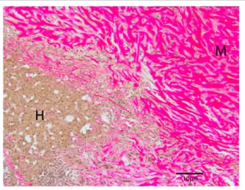
Figure 5 Marked collagenous matrix production by the mass (M) staining pink, adjacent to and invading a small island of hepatocytes (H) (Van Gieson’s, × 100)
Figure 6 Invasion of malignant spindle cells within sinusoids causing attenuation and atrophy of hepatocytes. The isolated islands of hepatic tissue within the mass had essentially normal hepatic architecture, although there was invasion of the sinusoids by the malignancy (haematoxylin and eosin, × 200)
Figure 4 Histopathology of the lesion – malignant, markedly pleomorphic spindle cell proliferation invading small islands of hepatic tissue (H) (haematoxylin and eosin, × 40)
instead of CK19 as a feline-validated CK19 marker could not be sourced in Australia.13,14 One hundred percent of
the proliferating spindle cells stained strongly positive for vimentin. The proliferating cells in the mass were negative for AE1/AE3, although multifocal sparsely dis-tributed bile ducts within the mass stained strongly posi-tive (Figure 8). The differentials mesothelioma or scirrhous hepatocellular/biliary carcinomas could be excluded because the proliferating cells were AE1/AE3-negative.14 In the mass the proliferative marker Ki-67
was positive in 12 per every 100 cells examined.
In comparison, staining of sections of pericardium (location of satellite nodules) revealed that 50% of non-neoplastic pericardial mesothelial cells were positive for AE1/AE3. This is an expected staining pattern for mesothelium.
A combination of histological morphology of the neo-plasm and immunohistochemical properties demon-strated that the vast majority of proliferating cells were mesenchymal in origin, warranting the diagnosis of sar-coma. As there was a significant component of collagen production (confirmed with Van Gieson’s staining) the mass was classified as a fibrosarcoma.
Our clinicopathological assessment was that the cat had a PPDH with associated liver lobe incarceration. This liver lobe subsequently underwent neoplastic trans-formation to produce a hepatic fibrosarcoma.
Discussion
Feline fibrosarcoma is a malignant neoplasia that in some cats is associated with sites of inflammation, par-ticularly vaccination injection sites.15–17 Cats have an
increased tendency to respond to inflammation by fibro-blast proliferation and, ultimately, tumour genesis com-pared with other species.17 Transition zones that pass
from inflammation to sarcoma have been microscopi-cally observed and support this theory.18 Hence, it is
thought that any source of inflammation can lead to sar-coma formation in the cat. Occasionally, the tumour appears spontaneously without a known source of inflammation.19 Hepatic fibrosarcoma is one such
exam-ple (albeit rare) in the cat.2,3
In cats intrapericardial hepatic cysts and myelipomas have been linked with PPHA in numerous case reports.6,9,10,12 In these reports it has been speculated that
incarceration of hepatic tissue in the PPDH led to vascu-lar and lymphatic congestion and hypoxia of hepatic tis-sue predisposing to formation of cysts or myelipomas. In people, it has been suggested that as a result of incom-plete vascular and/or ductal systems, ectopic livers are predisposed to hepatocarigonenesis due to longer expo-sure of ectopic liver tissues to carcinogenic substances.20,21
The authors of this mentioned study speculate that incarceration of liver led to vascular and lymphatic con-striction predisposing to neoplastic transformation to hepatic fibrosarcoma. Presumably, the mechanisms described above would have the potential to elicit neo-plastic transformation in hepatic mesenchymal cells, as well as hepatocytes.
In this case, multiple negative prognostic markers were apparent on histopathology and immunohisto-chemistry. Specifically, nuclei exhibited marked anisokaryosis, with multiple bizarre nuclei identified within cells. The mitotic rate (12 per 10 HPFs) was high. In canine sarcomas, mitotic rate index was prognostic for development of metastasis.22 Additionally, in this case,
the high Ki-67 index of the mass could have been associ-ated with a poor long-term prognosis.23 In feline
mam-mary carcinomas, Ki-67 was significantly increased in Figure 7 Immunohistochemical staining with vimentin
antibody shows strong positive staining of malignant mass cells (M) and negative staining of hepatocytes (H) (vimentin,
× 100)
Figure 8 Immunohistochemical staining with cytokeratin AE1/AE3 antibody shows negative staining of malignant mass cells, negative staining of hepatocytes and strong positive staining of occasional remnant biliary structures (arrow) (cytokeratin AE1/AE3, × 200)
The exact cause of death in this cat will remain unknown as post-mortem examination was declined. Complications that result in severe clinical deterioration and mortality following intrathoracic surgery include pneumothorax, re-expansion pulmonary oedema, haemorrhage and malignant cardiac arrhythmias.7,8,26
Additionally, cerebrovascular accident is a reported postoperative complication in people and cats and there-fore should be considered as a plausible cause of patient mortality.27,28 Although these complications do not
nec-essarily result in death in every instance, animals under-going thoracic surgery may be further compromised by changes in breathing patterns brought on by surgery, narcotic pain relief and recumbency, thus affecting their ability to handle these challenges. In our opinion, the per-acute nature of the cat’s deterioration makes pneu-mothorax or cerebrovascular accident the most likely possibilities.
Specifically, pneumothorax has been reported follow-ing diaphragmatic hernia repair in dogs and cats.26
Apparent or non-apparent iatrogenic lung lacerations may seal temporarily only for the laceration to open spontaneously during recovery as respiratory excur-sions and patient mobility increase. In this cat, although it appeared that the pneumothorax had resolved, prema-ture discontinuation of active pleural suction may have lead to a fatal recurrent pneumothorax.
Although single isolated periods of ventricular tachycardia were identified intra- and postoperatively, ECG recording immediately prior to the cat’s death demonstrated normal sinus rhythm, suggesting that a malignant arrhythmia was an unlikely cause of death. Exsanguination and pulmonary oedema were improba-ble as intraoperative haemorrhage was not appreciated during surgery, nor were there any markers of haemor-rhage or pulmonary oedema on examination shortly before death.
Conclusions
This is the first reported feline case of PPDH where metaplastic transformation of hepatic tissue into a
2 Brito e Silva MSd, Faria AM, Andraschko MM, et al. Biliary
duct fibrosarcoma in cat. Acta Sci Vet 2009; 37: 69–71.
3 Lawrence HJ, Erb HN and Harvey HJ. Nonlymphomatous
hepatobiliary masses in cats: 41 cases (1972 to 1991). Vet
Surg 1994; 23: 365–368.
4 Berry CR, Koblik PD and Ticer JW. Dorsal
peritoneoperi-cardial mesothelial remnant as an aid to the diagnosis of feline congenital peritoneopericardial diaphragmatic-hernia. Vet Radiol 1990; 31: 239–245.
5 Wallace J, Mullen HS and Lesser MB. A technique for
sur-gical-correction of peritoneal pericardial diaphragmatic-hernia in dogs and cats. J Am Anim Hosp Assoc 1992; 28: 503–510.
6 Liptak JM, Bissett SA, Allan GS, et al. Hepatic cysts
incar-cerated in a peritoneopericardial diaphragmatic hernia.
J Feline Med Surg 2002; 4: 123–125.
7 Burns CG, Bergh MS and McLoughlin MA. Surgical and
nonsurgical treatment of peritoneopericardial diaphrag-matic hernia in dogs and cats: 58 cases (1999–2008). J Am Vet Med Assoc 2013; 242: 643–650.
8 Reimer SB, Kyles AE, Filipowicz DE, et al. Long-term
outcome of cats treated conservatively or surgically for peritoneopericardial diaphragmatic hernia: 66 cases (1987–2002). J Am Vet Med Assoc 2004; 224: 728–732.
9 Wouda RM, Chalkley MD, Fraser AR, et al. Hepatic
myelo-lipoma incarcerated in a peritoneopericardial diaphrag-matic hernia in a cat. Aust Vet J 2010; 88: 231–235.
10 Schuh JCL. Hepatic nodular myelolipomatosis
(myelo-lipomas) associated with a peritoneao-pericardial dia-phragmatic-hernia in a cat. J Comp Pathol 1987; 97: 231–235.
11 Gourley IM, Popp JA and Park RD. Myelolipomas of the liver
in a domestic cat. J Am Vet Med Assoc 1971; 158: 2053–2057.
12 Scruggs SM and Bright JM. Chronic cardiac tamponade in
a cat caused by an intrapericardial biliary cyst. J Feline Med
Surg 2010; 12: 338–340.
13 Thway K, Nicholson AG, Lawson K, et al. Primary
pulmo-nary myxoid sarcoma with EWSR1-CREB1 fusion: a new tumor entity. Am J Surg Pathol 2011; 35: 1722–1732.
14 Bacci B, Morandi F, De Meo M, et al. Ten cases of feline
mesothelioma: an immunohistochemical and ultrastruc-tural study. J Comp Pathol 2006; 134: 347–354.
15 Hendrick MJ, Shofer FS, Goldschmidt MH, et al.
Compari-son of fibrosarcomas that developed at vaccination sites and at nonvaccination sites in cats – 239 cases (1991–1992).
16 Lester S, Clemett T and Burt A. Vaccine site-associated sar-comas in cats: clinical experience and a laboratory review (1982-1993). J Am Anim Hosp Assoc 1996; 32: 91–95.
17 Ladlow J. Injection site-associated sarcoma in the cat:
treatment recommendations and results to date. J Feline Med Surg 2013; 15: 409–418.
18 Hendrick MJ, Goldschmidt MH, Shofer FS, et al.
Postvacci-nal sarcomas in the cat: epidemiology and electron probe microanalytical identification of aluminum. Cancer Res
1992; 52: 5391–5394.
19 Doddy FD, Glickman LT, Glickman NW, et al. Feline
fibro-sarcomas at vaccination sites and non-vaccination sites.
J Comp Pathol 1996; 114: 165–174.
20 Collan Y, Hakkiluoto A and Hastbacka J. Ectopic liver. Ann
Chir Gynaecol 1978; 67: 27–29.
21 Arakawa M, Kimura Y, Sakata K, et al. Propensity of
ectopic liver to hepatocarcinogenesis: case reports and a review of the literature. Hepatology 1999; 29: 57–61.
22 Kuntz CA, Dernell WS, Powers BE, et al. Prognostic factors
for surgical treatment of soft-tissue sarcomas in dogs: 75 cases (1986–1996). J Am Vet Med Assoc 1997; 211: 1147–1151.
23 Ettinger SN, Scase TJ, Oberthaler KT, et al. Association of
argyrophilic nucleolar organizing regions, Ki-67, and proliferating cell nuclear antigen scores with histologic grade and survival in dogs with soft tissue sarcomas: 60 cases (1996–2002). J Am Vet Med Assoc 2006; 228: 1053–1062.
24 Rasotto R, Caliari D, Castagnaro M, et al. An
Immunohis-tochemical study of HER-2 expression in feline mammary tumours. J Comp Pathol 2011; 144: 170–179.
25 Campbell FE and Kittleson MD. The effect of hydration
status on the echocardiographic measurements of normal cats. J Vet Intern Med 2007; 21: 1008–1015.
26 Minihan AC, Berg J and Evans KL. Chronic diaphragmatic
hernia in 34 dogs and 16 cats. J Am Anim Hosp Assoc 2004; 40: 51–63.
27 Limburg M, Wijdicks EF and Li H. Ischemic stroke after
surgical procedures: clinical features, neuroimaging, and risk factors. Neurology 1998; 50: 895–901.
28 Altay UM, Skerritt GC, Hilbe M, et al. Feline
cerebrovas-cular disease: clinical and histopathologic findings in 16 cats. J Am Anim Hosp Assoc 2011; 47: 89–97.


![Methyl 2 methyl 7 oxo 1,2,4 triazolo[3,2 b] 1,3 thiazine 5 carboxylate](data:image/gif;base64,R0lGODlhAQABAIAAAP///wAAACH5BAEAAAAALAAAAAABAAEAAAICRAEAOw==)