Received: August 10, 2016.
Accepted: January 12, 2017.
Pre-published: January 19, 2017.
©2017 Ferrata Storti Foundation
Material published in Haematologica is covered by copyright. All rights are reserved to the Ferrata Storti Foundation. Use of published material is allowed under the following terms and conditions:
https://creativecommons.org/licenses/by-nc/4.0/legalcode. Copies of published material are allowed for personal or inter-nal use. Sharing published material for non-commercial pur-poses is subject to the following conditions:
https://creativecommons.org/licenses/by-nc/4.0/legalcode, sect. 3. Reproducing and sharing published material for com-mercial purposes is not allowed without permission in writing from the publisher.
Correspondence:
Ferrata Storti Foundation EUROPEAN
HEMATOLOGY ASSOCIATION
Haematologica
2017
Volume 102(4):626-636
doi:10.3324/haematol.2016.153791
Check the online version for the most updated information on this article, online supplements, and information on authorship & disclosures: www.haematologica.org/content/102/4/626
Introduction
Sickle cell disease (SCD) is characterized by multi-organ morbidity and an increased risk of early death. Several studies show that the survival of children with SCD has improved over the last decades.1This improved survival is attributed to the implementation of newborn screening, use of prophylactic penicillin, and vac-cinations against Haemophilus influenzatype b and Streptococcus pneumonia,2-4 with possible contributions from advances in red blood cell transfusion medicine, iron chelation therapy, and transcranial Doppler screening to identify those at increased risk of stroke.5,6
The Cooperative Study of Sickle Cell Disease (CSSCD), a multicenter natural his-tory study of SCD conducted between 1978 and 1988, reported median ages at death during the study period of 42 and 48 years, respectively, for male and female HbSS patients, and median ages at death of 60 and 68 years, respectively, for male and female HbSC patients.7While many patients died following acute episodes of pain, acute chest syndrome or stroke, statistical modeling revealed that the acute chest syndrome, renal failure, seizures, high white blood cell count (WBC) and low level of fetal hemoglobin (HbF) were associated with an increased risk of early
A
lthough recent studies show an improved survival of children
with sickle cell disease in the US and Europe, for adult patients
mortality remains high. This study was conducted to evaluate the
factors associated with mortality in adult patients following the approval
of hydroxyurea. We first evaluated the association between selected
variables and mortality at an academic center (University of North
Carolina). Data sources were then searched for publications from 1998
to June 2016, with meta-analysis of eligible studies conducted in North
America and Europe to evaluate the associations of selected variables
with mortality in adult patients. Nine studies, combined with the UNC
cohort (total n=3257 patients) met the eligibility criteria. Mortality was
significantly associated with age (per 10-year increase in age) [7 studies,
2306 participants; hazard ratio (HR): 1.28; 95% confidence interval (CI):
1.10-1.50], tricuspid regurgitant jet velocity 2.5 m/s or more (5 studies,
1577 participants; HR: 3.03; 95%CI: 2.0-4.60), reticulocyte count (3
stud-ies, 1050 participants; HR: 1.05; 95%CI: 1.01-1.10),
log(N-terminal-pro-brain natriuretic peptide) (3 studies, 800 participants; HR: 1.68; 95%CI:
1.48-1.90), and fetal hemoglobin (7 studies, 2477 participants; HR: 0.97;
95%CI: 0.94-1.0). This study identifies variables associated with
mortal-ity in adult patients with sickle cell disease in the hydroxyurea era.
Risk factors for mortality in adult patients
with sickle cell disease: a meta-analysis
of studies in North America and Europe
Poulami Maitra,1Melissa Caughey,2Laura Robinson,3Payal C. Desai,4Susan Jones,5Mehdi Nouraie,6Mark T. Gladwin,7Alan Hinderliter,3Jianwen Cai1and Kenneth I. Ataga5
1Department of Biostatistics, University of North Carolina, Chapel Hill; 2Division of
Cardiology, University of North Carolina, Chapel Hill; 3Children’s Hospital of Philadelphia;
4Division of Hematology, The Ohio State University, Columbus; 5Division of
Hematology/Oncology, University of North Carolina, Chapel Hill; 6Department of
Medicine, Howard University, Washington, DC and 7Department of Medicine, University
of Pittsburgh, PA, USA
death in HbSS patients.7However, this prospective study was conducted prior to the approval of hydroxyurea for the treatment of sickle cell anemia. More recent studies show associations of elevated echocardiography-derived tricuspid regurgitant jet velocity (TRV),8,9 pulmonary hypertension (PHT),10,11elevated levels of N-terminal pro-brain natriuretic peptide (NT-pro-BNP),12 history of asth-ma and/or wheezing,13 end-stage renal disease requiring dialysis,14severity of hemolysis,15and prolongation of QTc interval16with an increased risk of death in patients with SCD.
Hydroxyurea, approved by the US Food and Drug Administration (FDA) in 1998, remains the only drug that has been shown to alter the natural history of SCD in peer-reviewed studies. In two randomized, placebo-con-trolled studies, hydroxyurea was shown to decrease the frequency of acute pain episodes, acute chest syndrome, red blood cell (RBC) transfusion and hospitalization rates in both adults and children with sickle cell anemia.17,18 Despite the studies showing an improved survival in adult patients with SCD following treatment with hydrox-yurea,19-21the mortality rate remains high for patients aged 18 years or older following transition to adult care,22 pos-sibly reflecting a lack of access to high-quality care. The present study evaluated the association of selected clinical and laboratory variables with mortality in a cohort of adult patients with SCD followed at an academic medical center. With the relatively small number of patients evalu-ated in this single center study, we have conducted com-prehensive meta-analyses of contemporary prospective and retrospective studies to evaluate the factors associated with mortality in adult patients with SCD following the approval of hydroxyurea. Our goal is to uncover the underlying causes of mortality in SCD.
Methods
Patients and study design
We evaluated a subset of patients followed at the Comprehensive Sickle Cell Clinic at the University of North Carolina (UNC), Chapel Hill between August 2004 and December 2014 (subsequently referred to as the UNC cohort). The data were collected as part of a prospective study of the natural history of pulmonary hypertension in SCD.9 The patients were evaluated
while in a non-crisis, 'steady state'; they had not experienced an episode of acute chest syndrome in the four weeks preceding enrollment, and had no clinical evidence of congestive heart fail-ure. This study was approved by the Institutional Review Board at UNC, Chapel Hill, and all subjects gave written informed consent to participate in accordance with the Declaration of Helsinki.
Sickle cell disease-related clinical complications and
laboratory variables
The presence or history of clinical complications, including the number of acute pain episodes (or vaso-occlusive crises) in the past year, history of acute chest syndrome, and use of hydroxyurea, was ascertained at the time of evaluation as well as through a review of the patients’ medical records. Acute pain episodes, acute chest syndrome, and other SCD-related complications were defined using accepted definitions.19,23 SCD genotypes were
dichotomized into clinically severe (HbSS, HbSβ0thalassemia and
HbSD) and less severe (HbSC and HbSβ+thalassemia) groups. The
TRV was measured by continuous wave Doppler echocardiogra-phy. Multiple views were obtained to record optimal tricuspid
flow signals. Measurements were performed on at least three waveforms with well-defined velocity envelopes and an average value was used for data analysis. Patients with non-quantifiable TRVs were assumed to have normal estimated PASP. The echocar-diograms were interpreted by a cardiologist blinded to all patient data.
Laboratory measurements obtained in study subjects include complete blood counts with reticulocyte counts, serum creatinine, lactate dehydrogenase, total bilirubin, direct bilirubin, indirect bilirubin, D-dimer and NT-pro-BNP. Hemoglobin analysis was performed by high performance liquid chromatography to con-firm the SCD diagnosis and ascertain HbF levels.
Search strategy and covariate abstraction
A meta-analysis was conducted in accordance with the Preferred Reporting Items for Systematic Reviews and Meta-Analyses guidelines and Meta-Analysis of Observational Studies in Epidemiology Group criteria.24Published articles were identified
using MEDLINE (PubMed), Web of Science and EMBASE search engines between January 1, 1998, and June 30, 2016. The search on PubMed was performed using the Medical Subject Headings (MeSH) “anemia, sickle cell [mesh] OR “sickle cell disease” OR "sickle cell anemia" AND “mortality OR survival.” The search on Web of Science was performed using the search terms “sickle cell” AND “mortality OR survival” and the search on EMBASE was per-formed using the search terms “sickle cell anemia OR sickle cell disease” AND “mortality OR survival”. In addition, we interrogat-ed references from relevant original papers and review articles to identify further relevant studies. Reviews, editorials, duplicate citations, non-human studies, highly selected study populations, retrospective analyses of large administrative datasets and articles written in languages other than English were excluded. We limited our analysis to prospective and retrospective studies conducted in North America and Europe and published since 1998, the year hydroxyurea was approved by the FDA for treatment of sickle cell anemia. Multiple publications from the same patient population were narrowed down to the single publication with the most complete information. Eligible studies were combined with the UNC cohort for meta-analyses.
Statistical analysis
Median, range and interquartile range for continuous variables and frequencies for categorical variables were provided in the UNC cohort. NT-pro-BNP was log-transformed based on previous literature.25We evaluated the Spearman correlation coefficient of
each pair of variables to check the collinearity. Median ages at death were calculated for those individuals who died during the study. Median survival age was calculated based on Kaplan-Meier estimates for the survival probability using age as the time scale taking into account the left truncation. Log-rank tests were con-ducted to compare survival probabilities among different groups. Cox proportional hazards regression was fit to the data and vari-ables were selected by backward elimination at 5% level of signif-icance to obtain the final model. Schoenfeld residuals were used to check the proportional hazards assumption. Hazard ratios (HRs) were calculated in the UNC cohort, controlling for age and sex.
Mortality HRs were meta-analyzed using DerSimonian and Laird26random effects models26,27due to the diversity of individual
studies and the small number of studies included. The Knapp and Hartung27method was used to adjust the standard errors of the
estimated coefficients. Unadjusted HRs in the studies were used for our meta-analyses whenever they were reported. The degree of heterogeneity across the studies was examined using I2values,28
HRs were available from a minimum of 3 studies. We considered the Voskaridou et al. study21 to contribute two separate HRs
depending on whether patients received hydroxyurea. Similarly, from the Elmariah et al.study,25two estimates were used based on
the use of hydroxyurea. All the analyses were performed using SAS statistical software v.9.4 (Cary, NC, USA). The graphs for the meta-analyses were made using the R Project for Statistical Computing.
Results
Study population
One hundred and sixty-one subjects in the UNC cohort [female: 97 (60.3%)] with SCD (SS: 119; SC 19; Sβ0: 12; Sβ+: 10; SD: 1), and a median age of 36 years (range 18-71 years) (Table 1) were followed for a median duration of 7.2 years (range 0.06-10.3 years) and a total of 954.0 per-son-years. The majority of the subjects [159 (99%)] were self-reported African Americans. There were 29 (18%) deaths during the period of follow up (SS: 20, SC: 4, Sβ0: 3,
Sβ+: 2). Amongst those who died during the study, the median age of death was 48.0 years (range 29.8-70.2 years), while the median age in the subjects alive at the time of last contact was 38.7 years (range 22.6-79.4 years). When the subjects were grouped based on presumed dis-ease severity (SS/Sβ0/SD vs. SC/Sβ+), the median age at the time of death in the SS/Sβ0/SD group was 44.7 years (range 24.1-79.4 years), while the median age at the time of death in the SC/Sβ+group was 59.4 years (range 29.8-69.2 years). The median survival age for all subjects in the UNC cohort was 50.2 years [95% confidence interval (CI): 45.2-62.3 years) (Figure 1), with a median survival age in the SS/Sβ0/SD group of 49.0 years (95%CI: 44.9-68.6 years). With the small total number of subjects and deceased subjects in the SC/Sβ+group, the median survival age in this group cannot be reliably estimated.
Factors associated with mortality in UNC cohort
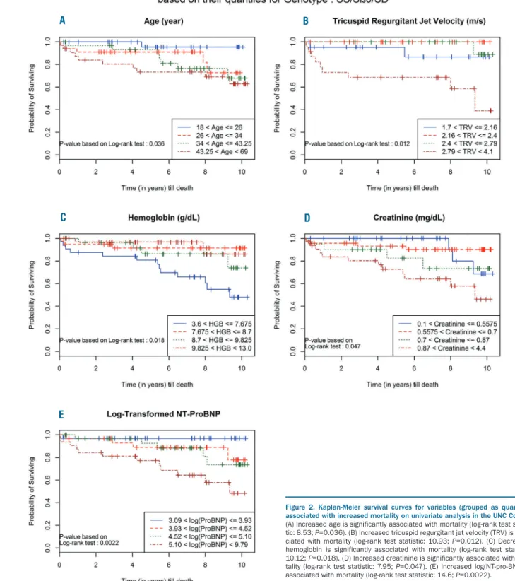
Deaths in the UNC cohort were ascertained from hospi-tal records as well as vihospi-tal statistics data from the North Carolina Center for Health Statistics. The variables in the initial model are shown in Table 2. When covariates were grouped based on quartiles, age (P=0.036), TRV (P=0.012), hemoglobin (P=0.018), creatinine (P=0.047) and log(NT-pro-BNP) (P=0.0022) were associated with an increased risk of death (Figure 2A-E) in univariate analysis of a sub-set in this cohort (SS/Sβ0/SD group). There were no asso-ciations between WBC, platelet count, reticulocyte count, HbF, lactate dehydrogenase, the number of acute pain episodes in the previous year, history of acute chest syn-drome, or hemoglobin genotype with mortality in univari-ate analysis of patients in the SS/Sβ0/SD group.Multivariable analysis
We found that the correlation coefficient between each pair of variables was less than 0.6 and, hence, the variables are not collinear (Online Supplementary Table S1). Carrying out the usual backward variable selection, TRV [hazard ratio (HR): 22.7, 95%CI: 5.88-87.5; P<0.0001], frequency of acute pain episodes in the previous year (HR: 1.2, 95%CI: 1.06-1.35; P=0.0038) and D-dimer (HR: 1.24 for 1000 ng/mL increment in D-dimer, 95%CI: 1.07-1.44; P=0.0039) were
Table 1. Baseline demographic and laboratory characteristics of UNC cohort.
Characteristic Number Number (%)/median (interquartile range)
Age (years) 161 36 (27-44)
Sex 161
Male 64 (40%)
Female 97 (60%)
Race 161
Black 159 (99%)
Hispanic 2 (1%)
Genotype 161
SS 119 (73.9%)
Sβ0 12 (7.5%)
Sβ+ 10 (6.2%)
SC 19 (11.8%)
Other (SD) 1 (0.6%)
Use of hydroxyurea 161
Yes 93 (58%)
No 68 (42%)
White blood cell count (x109/L) 161 9.3 (7.2-11.2)
Hemoglobin (g/dL) 161 8.9 (7.9-10.3)
Platelet count (x109/L) 159 408 (303-498)
Reticulocyte count (%) 159 6.3 (4.3-9.2)
Absolute reticulocyte count 158 176.35 (125.4-241.6)
Fetal hemoglobin (%) 158 5.6 (2.6-10.7)
Creatinine (mg/dL) 161 0.7 (0.6-0.9)
Lactate dehydrogenase (U/L) 158 847.5 (644-1136)
Total bilirubin (mg/dL) 161 1.9(1.1-3.0)
Indirect bilirubin (mg/dL) 158 1.8(1.1-2.7)
Direct bilirubin (mg/dL) 158 0.1(0.09-.1)
D-dimer (ng/mL) 121 1234 (740-2023.3)
Tricuspid regurgitant 110 2.4(2.16-2.8)
jet velocity (m/s)
Number of pain episodes 161 3 (1-5)
in previous year
History of acute chest syndrome 161
Yes 135(84%)
No 26(16%)
found to be associated with an increased risk of death. The HR estimates indicate that after controlling for the other variables, the hazard of dying was approximately 23 times higher for every 1 m/s increase in the TRV; for each addi-tional pain episode requiring an emergency department visit or hospitalization, the hazard of dying was increased by 20%, and the hazard of dying was approximately 1.24 times higher for every 1000 ng/mL increase in the D-dimer. When a sensitivity analysis was performed with an assigned TRV value of 1.69 m/s (lower than the lowest measurable TRV value in the cohort) for all missing values, TRV was no longer significantly associated with an increased risk of death. When D-dimer was excluded from the model due to missing values in some subjects, age (HR: 1.04, 95%CI: 1.009-1.073; P=0.012), reticulocyte count (HR: 1.09, 95%CI: 1.015-1.17; P=0.018) and log(NT-pro-BT) (HR: 1.62, 95%CI: 1.21-2.17; P=0.001) were associated with an increased risk of death.
Meta-analysis
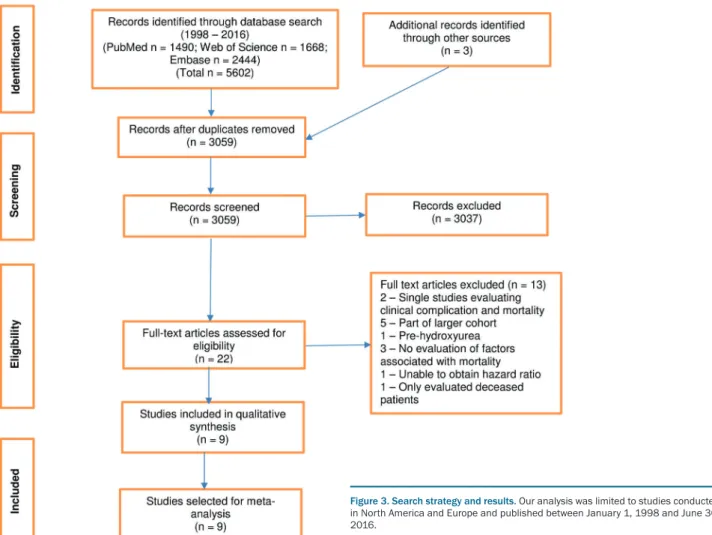
Our initial search retrieved 1490 articles from PubMed, 1668 articles from Web of Science and 2444 articles from
EMBASE (Figure 3). Nine studies11,21,24,29-34 met our inclu-sion criteria and were analyzed together with the UNC cohort for a total of 3257 participants. The quality of individual studies was assessed using the Newcastle-Ottawa Scale (Table 3). We were unable to evaluate pub-lication bias due to the small number of studies included in the meta-analysis.35Because the Walk-PHaSST cohort31 included patients as young as 12 years old, we restricted our analyses to only those patients who were at least 18 years old. The number of patients in the meta-analyses of the selected variables are shown in Table 3. The num-ber of studies included in the meta-analyses varied from 3 to 9 depending on what had been reported for each variable in the publications. The median age of death of deceased subjects in these studies (available in only 5 of the 9 published studies) ranged between 39.7 and 53 years. The median age of survival for the subjects with severe SCD genotypes (SS/Sβ0/SD), available in only 3 studies (2 of the published 9 studies plus the UNC cohort), ranged between 49.0 and 60.8 years. We found statistically significant associations of age (per 10-year increase in age) (HR: 1.28; 95%CI: 1.10-1.50), TRV 2.5
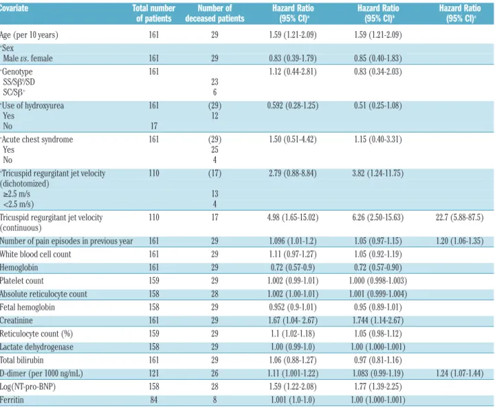
Table 2. Univariate and multivariate Cox proportional hazards regression analysis of mortality for demographic, laboratory and clinical variables in the UNC cohort.
Covariate Total number Number of Hazard Ratio Hazard Ratio Hazard Ratio of patients deceased patients (95% CI)a (95% CI)b (95% CI)c
Age (per 10 years) 161 29 1.59 (1.21-2.09) 1.59 (1.21-2.09)
+Sex
Male vs. female 161 29 0.83 (0.39-1.79) 0.85 (0.40-1.83)
+Genotype 161 1.12 (0.44-2.81) 0.83 (0.34-2.03)
SS/Sβ0/SD 23
SC/Sβ+ 6
+Use of hydroxyurea 161 (29) 0.592 (0.28-1.25) 0.51 (0.25-1.08)
Yes 12
No 17
+Acute chest syndrome 161 (29) 1.50 (0.51-4.42) 1.15 (0.40-3.31)
Yes 25
No 4
+Tricuspid regurgitant jet velocity 110 (17) 2.79 (0.88-8.84) 3.82 (1.24-11.75)
(dichotomized)
≥2.5 m/s 13
<2.5 m/s) 4
Tricuspid regurgitant jet velocity 110 17 4.98 (1.65-15.02) 6.26 (2.50-15.63) 22.7 (5.88-87.5)
(continuous)
Number of pain episodes in previous year 161 29 1.096 (1.01-1.2) 1.05 (0.97-1.15) 1.20 (1.06-1.35)
White blood cell count 161 29 1.11 (0.97-1.27) 1.05 (0.92-1.19)
Hemoglobin 161 29 0.72 (0.57-0.9) 0.72 (0.57-0.90)
Platelet count 159 29 1.002 (0.99-1.01) 1.000 (0.998-1.003)
Absolute reticulocyte count 158 28 1.002 (1.00-1.01) 1.001 (0.999-1.004)
Fetal hemoglobin 158 29 0.952 (0.9-1.01) 0.95 (0.89-1.01)
Creatinine 161 29 1.67 (1.04- 2.67) 1.744 (1.14-2.67)
Reticulocyte count (%) 159 29 1.1 (1.02-1.18) 1.05 (0.98-1.12)
Lactate dehydrogenase 158 29 1.00 (0.99-1.0) 1.00 (1.000-1.001)
Total bilirubin 161 29 1.06 (0.88-1.27) 0.97 (0.81-1.16)
D-dimer (per 1000 ng/mL) 121 26 1.11 (1.001-1.22) 1.083 (0.99-1.19) 1.24 (1.07-1.44)
Log(NT-pro-BNP) 158 28 1.59 (1.22-2.08) 1.77 (1.39-2.25)
Ferritin 84 8 1.001 (1.0-1.0) 1.00 (1.000-1.001)
m/s or more (HR: 3.03; 95%CI: 2.0-4.60), reticulocyte count (HR: 1.05; 95%CI: 1.01-1.10), HbF (HR: 0.97; 95%CI: 0.94-1.00), and log(NT-pro-BNP) (HR: 1.68; 95%CI: 1.48-1.90) with mortality (Figure 4A-E). This means that the weighted hazard of dying was approxi-mately 30% higher for every 10-year increase in age, approximately 200% higher for TRV 2.5 m/s or more compared to lower values, approximately 5% higher for every 1% increase in reticulocyte count; approximately
3% lower for every 1% increase in HbF, and approxi-mately 100% higher for every 1 unit increase in log(NT-pro-BNP).
No statistically significant associations were found between WBC (HR: 1.05; 95%CI: 0.97-1.14), hemoglobin (HR: 0.86; 95%CI: 0.68-1.09), platelet count (HR: 1.00; 95%CI: 1.00-1.00), ferritin (HR: 1.00; 95%CI: 0.99-1.01), creatinine (HR: 1.20; 95%CI: 0.95-1.52), lactate dehydro-genase (HR: 1.00; 95%CI: 1.00-1.00), total bilirubin (HR:
Figure 2. Kaplan-Meier survival curves for variables (grouped as quartiles) associated with increased mortality on univariate analysis in the UNC Cohort.
(A) Increased age is significantly associated with mortality (log-rank test statis-tic: 8.53; P=0.036). (B) Increased tricuspid regurgitant jet velocity (TRV) is asso-ciated with mortality (log-rank test statistic: 10.93; P=0.012). (C) Decreased hemoglobin is significantly associated with mortality (log-rank test statistic: 10.12; P=0.018). (D) Increased creatinine is significantly associated with mor-tality (log-rank test statistic: 7.95; P=0.047). (E) Increased log(NT-pro-BNP) is associated with mortality (log-rank test statistic: 14.6; P=0.0022).
A B
C D
1.08; 95%CI: 0.93-1.25), use of hydroxyurea (HR: 0.70; 95%CI: 0.41-1.22), history of acute chest syndrome (HR: 1.14; 95%CI: 0.50-2.59), and male sex (HR: 1.47; 95%CI: 0.41-5.31) with mortality (Online Supplementary Figure S1A-J).
All of the studies, except that of Karacaoglu et al.,34used the time from entry to event as the response variable. We performed a sensitivity analysis by removing this study from the meta-analysis. The results are similar [with and without: WBC 1.05 (0.97, 1.14) vs.1.03 (0.93, 1.14); hemo-globin 0.87 (0.61, 1.25) vs.0.92 (0.62, 1.36); and HbF 0.97 (0.94, 1.00) vs.0.97 (0.94, 1.00)]. The study was, therefore, maintained in the meta-analysis.
Discussion
As patients with SCD age, they begin to manifest evi-dence of end-organ damage which contributes to increased morbidity and early mortality. In random effects meta-analyses, we find statistically significant associations of increased reticulocyte count, TRV and log(NT-pro-BNP) with increased mortality, whereas decreased HbF was associated with increased mortality. While no significant associations were seen between either hemoglobin or lac-tate dehydrogenase and the risk of death, the association of increased reticulocyte count with mortality suggests that hemolysis may contribute to the increased mortality
observed in SCD. The intensity of hemolytic anemia, eval-uated using a composite variable derived from several individual markers of hemolysis (reticulocyte count, serum lactate dehydrogenase, aspartate aminotransferase and total bilirubin concentrations), was associated with an increased risk of death in a previous study of patients with SCD.15 Hemolysis with the subsequent release of cell-free hemoglobin results in the generation of reactive oxygen species which is a potent scavenger of nitric oxide.36 This appears to predispose patients to a vasculopathy, charac-terized by systemic and pulmonary hypertension, endothelial dysfunction, and proliferative changes in the intima and smooth muscle of blood vessels.37In addition to their relationship with hemolysis, reticulocytes are more adhesive than mature erythrocytes due to increased expression of the integrin complex, α4β1(which binds to both fibronectin and vascular cell adhesion molecule-1, an adhesion receptor expressed on the surface of endothelial cells) as well as expression of CD36 by a subpopulation of sickle reticulocytes.38The degree of adhesiveness is report-edly correlated with the severity of disease in patients with SCD.39
We further confirm the association of both elevated echocardiography-derived TRV and NT-pro-BNP levels with increased mortality in adult SCD patients. As increased TRV is not sufficiently specific to confidently establish the presence of PHT,10 a right heart catheteriza-tion (RHC) is required in patients with elevated TRV
Table 3. Summary of publications in meta-analysis.
Publication # of Sickle cell Variable with Hazard Ratio Median/mean age Median age Duration of Quality subjects disease genotypes at death of deceased of survival follow up appraisal
subjects (years) of SS/Sβ0/SD
(Newcastle-subjects Ottawa Scale
(years) [NOS])
*Voskaridou et al., 201021 131 HbSS, HbSβ0, HbSβ+ Age (Median: 33.0, SD: 11.2, HR: 0.99; N/A N/A 5 years 6
0.94-1.05) (range: 0.1-18)
LDH (Mean: 753.88, SD: 413.62, HR: 0.99; 0.99-1.0)
Reticulocyte count (%) (Mean: 10.03, SD: 7.32, HR: 1.01; 0.94-1.09) Hemoglobin (Mean: 9.10, SD: 1.39, HR: 0.88; 0.60-1.3)
HbF (Mean: 7.09, SD: 5.26, HR: 0.95; 0.82-1.09)
Bilirubin (Mean: 1.92, SD: 3.27, HR : 1.09; 0.99-1.2)
**Voskaridou et al., 201021 199 HbSS, HbSβ0, HbSβ+ Age (Median: 35.0, SD: 12.8, N/A N/A 8 years 6
HR: 0.98; 0.93-1.04) (range: 0.1-17)
LDH (Mean: 701.16, SD: 330.16, HR: 1.0; 1.0-1.0)
Reticulocyte count (%) (Mean: 6.88, SD: 6.38, HR: 1.06; 1.02-1.09) Hemoglobin (Mean: 9.43, SD: 1.67, HR: 0.74; 0.62-0.89)
HbF (Mean: 5.58, SD: 6.22, HR: 0.92; 0.86-0.99) Bilirubin (Mean: 1.80, SD: 1.61, HR: 1.31; 1.16-1.49)
Saraf et al., 201129 306 HbSS, HbSβ0 Age (HR: 1.03; 1.0-1.05) N/A N/A 1/1/1997 - 8
Creatinine (HR: 1.14; 0.99-1.3) 6/30/2009
HbF (HR: 0.99; 0.95-1.04) WBC (HR: 0.94; 0.87-1.01)
Mehari et al., 201311 84 HbSS, HbSC, HbSβ0, Age (HR: 1.0; 0.97-1.03) N/A N/A 4.7 years 6
HbSβ+, other Creatinine (HR: 1.09; 0.71-1.67) (maximum
genotypes not defined Ferritin (HR: 1.26; 1.04-1.53) follow up of 11 years)
Cabrita et al., 201332 164 HbSS, HbSC, HbSβ0, Age (Median: 42.3, IQR: 33, 50, 49 (range: 25-82) N/A 68.1 months 7
HbSβ+, HbSD, HbS/HPFH HR: 1.04; 1.0-1.08) (IQR: 48-78)
Creatinine (μmol/L) (Median: 70, IQR: 59, 85, HR: 5.24; 0.41-67.9)
Platelets (HR: 0.99; 0.99-1.0)
LDH (Median: 606, IQR: 385, 879, HR: 0.99; 0.99-1.0)
Hemoglobin (g/L) (Median: 94, IQR: 80, 110, HR: 0.62; 0.45-0.86)
HbF (HR: 1.04; 0.9-1.2) WBC (Median: 9.1, IQR: 7.4, 10.9, HR: 0.93; 0.77-1.13)
TRV (≥2.5 m/s: 48/164 {29.27%}, HR: 2.71; 0.98-7.5)
Elmariah et al., 201424 542 HbSS, HbSC, HbSβ0, Creatinine (HR: 1.39; 1.25-1.53) 45 (range: 20-86) 58 9.3 years 7
HbSβ+ **Platelets (HR: 1.0; 0.95-1.05) (range: 2.7-10.5)
Use of hydroxyureaa (Yes: 184/441 {41.7%}, HR: 0.82; 0.55-1.22)
History of ACS (Yes: 354/475 {74.5%}, HR: 1.27; 0.81-1.99)
**Hemoglobin (HR: 1.21; 1.06-1.38) *Hemoglobin (HR: 1.36; 1.07-1.72) **WBC (HR: 1.06; 1.0-1.11) Bilirubin (HR: 1.01; 0.88-1.16) Log(NT-pro-BNP) (HR: 1.62; 0.66-3.97)
Gladwin et al., 201431 605 HbSS, HbSC Age (Mean: 38.0, SD: 12.57, 40.8 (range: 21.3-65.3) 60.8 2.4 years 8
HR: 1.03; 0.99-1.06) (range: 0.04-3.4)
Creatinine (Mean: 17.07, SD: 35.45, HR: 0.99; 0.97-1.01)
Platelets (Mean: 350.63, SD: 134.85,
Use of hydroxyurea (Yes: 230/605 {38.02%}, HR: 1.62; 0.70-3.73)
History of ACS (Yes: 384/605 {63.47%}, HR: 0.67; 0.29-1.55) Ferritin (Mean: 607.88, SD: 1297.61, HR: 1.0; 1.0-1.0) LDH (Mean: 451.82, SD: 298.79, HR: 1.0; 1.0-1.0) Sex (Male: 278/605 {45.95%} HR: 2.52; 1.03-6.19)
Reticulocyte count (Mean: 8.91, SD: 5.99, HR: 1.03; 0.96-1.1) Hemoglobin (Mean: 9.31, SD: 1.92, HR: 0.9; 0.71-1.13) HbF (Mean: 7.30, SD: 7.64, HR: 0.96; 0.9-1.04) WBC (Mean: 9.68, SD: 3.66, HR: 1.1; 1.0-1.23) Bilirubin (Mean: 14.66, SD: 31.73, HR: 0.97; 0.93-1.01) Log(NT-pro-BNP) (Mean: 4.24, SD: 1.56,
HR: 1.73; 1.41-2.12)
TRV (≥2.5 m/s: 347/562 {61.74%}, HR: 3.5; 1.03-11.9)
Schimmel et al., 201533 85 HbSS, HbSC, HbSβ0, TRV (≥2.5 m/s: 25/81 {30.86%}, 53 (IQR: 37-60) N/A 82 months 7
HbSβ+ HR: 1.1; 0.3-3.7) (IQR: 75-85)
Damy et al., 201630 656 HbSS, HbSβ0 Age (HR: 1.03; 0.99-1.06) N/A N/A 48 months 7
Sex (HR: 1.60; 0.91-2.75) (range: 32-59)
Hemoglobin(g/L) (HR: 1.44; 0.52-3.97) HbF (HR: 0.97; 0.86-1.10)
TRV (HR: 3.92; 1.97-7.78)
Karacaoglu et al., 201634 324 HbSS, HbSβ0, HbSβ+ HbF (HR: 0.94; 0.74-1.2) 36.6 ± 13 N/A 66 ± 44 months 7
Hemoglobin (HR: 0.62; 0.45-0.83) (range: 3 – 148 months)
WBC (HR: 1.10; 1.05-1.16)
UNC cohort, 2015 161 HbSS, HbSC, HbSβ0, Age (Mean: 36.83, SD: 12.37, 48.0 49.0 6.4 years 7
HbSβ+, HbSD HR: 1.05; 1.02-1.08) (range: (95% CI: (range:
Creatinine (Mean: 0.85, SD: 0.52, 29.8-70.2) 44.9-68.6) 0.06-10.3) HR: 1.67; 1.04-2.67)
Platelets (Mean: 411.45, SD: 156.80, HR: 1.0; 0.99-1.01)
Use of hydroxyurea (Yes: 93/161 {57.76%}, HR: 0.59; 0.28-1.25)
History of ACS (Yes: 135/161 {83.85%}, HR: 1.49; 0.51-4.42)
Ferritin (Mean: 631.76, SD: 813.42, HR: 1.0; 1.0-1.0)
LDH (Mean: 984.03, SD: 496.54, HR: 1.0; 0.99-1.0)
Gender (Male: 64/161 {39.75%}, HR: 0.83; 0.39-1.79)
Reticulocyte count (Mean: 7.38, SD: 4.68, HR: 1.1; 1.02-1.18) Hemoglobin (Mean: 9.15, SD: 1.75, HR: 0.72; 0.57-0.9)
HbF (Mean: 8.24, SD: 7.52, HR: 0.95; 0.9-1.0) WBC (Mean: 9.40, SD: 2.81, HR: 1.1; 0.97-1.27) Bilirubin (Mean: 2.49, SD: 2.34, HR: 1.06; 0.88-1.27) Log(NT-proBNP) (Mean: 4.74, SD: 1.09, HR: 1.59; 1.22-2.08) TRV (≥2.5 m/s): 47/110 {42.73%}, HR: 2.79; 0.88-8.83)
*With Hydroxyurea; ** Without Hydroxyurea; LDH: lactate dehydrogenase; HbF: fetal hemoglobin; HR: hazard ratio; Pain Crisis: frequency of Pain Crisis; TRV: tricuspid regurgitant jet velocity; IQR: interquartile range; CI: confidence interval. *The paper by Voskariduo et al.21reports OR for Cox PH model: we have treated them as HRs. Here, reported median is presumed to be the same as the
mean; data have been pooled to obtain overall mean. aOnly HbSS/HbSβ0thalassemia patients evaluated.
continued from the previous page
ues to confirm the diagnosis. With the strength of data showing that increased TRV and PHT are associated with mortality in SCD, we recommend that patients undergo periodic echocardiographic screening for assessment of mortality risk, consistent with recently published clinical practice guidelines.40NT-pro-BNP levels assess ventricular strain and levels greater than 160 pg/mL are reported to be an independent risk factor for mortality in SCD.41,42 The utility of NT-pro-BNP alone as a screening tool for PHT has not been studied, although in combination with TRV
and 6-minute walk distance, it appears to increase the pre-dictive capacity for PHT.10
significant in univariable analysis and it became much stronger in the multivariable model.
This meta-analysis provides additional evidence that decreased HbF level is associated with increased mortality in SCD. There is strong epidemiological and clinical evi-dence that elevated HbF level is associated with relatively mild clinical manifestations of SCD.7The reduction of dis-ease severity by elevated levels of HbF is the basis for the development of HbF-inducing drugs. The beneficial effect of hydroxyurea in SCD is thought to be largely due to the induction of HbF following ‘stress erythropoiesis’, although the production of nitric oxide and the soluble guanylyl cyclase and cGMP-dependent protein kinase
pathway may play a role in induced expression of the γ -globin gene.43 Although no significant association was observed between the use of hydroxyurea and mortality in the meta-analysis, there are not sufficient data on adherence to hydroxyurea or the doses of hydroxyurea in the individual studies.
Sickle cell disease has been described as an inflammato-ry disease.44While not statistically significant, there was a trend towards an association between WBC and mortality in the meta-analysis. Although leukocytes contribute to disease pathophysiology,45it remains uncertain whether a decrease in the leukocyte count is associated with clinical benefit.17,19 It has been suggested that the association
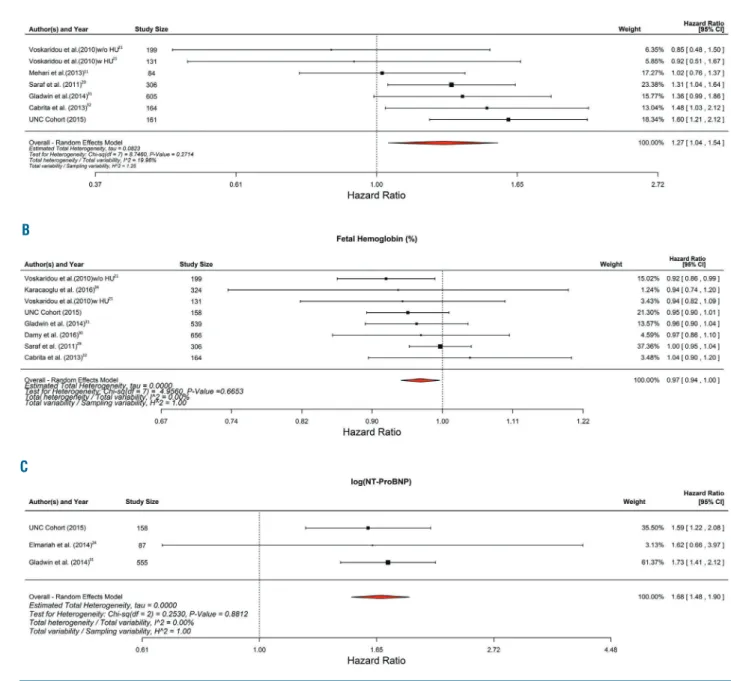
Figure 4. Forest plots of hazard ratios for variables significantly associated with mortality in random effects meta-analyses (continued on next page). (A) Age (per 10-year increase in age) was significantly associated with mortality [hazard ratio (HR): 1.28; 95% confidence interval (CI): 1.10-1.50]. (B) TRV ≥ 2.5 m/s (HR: 3.03; 95%CI: 2.0-4.60). (C) Reticulocyte count was significantly associated with mortality (HR: 1.05; 95%CI: 1.01-1.10);
A
B
between neutropenia and the frequency of pain episodes in the Multicenter Study of Hydroxyurea (MSH)17 was 'forced' due to the study design requiring a titration of hydroxyurea to maximum tolerated dose. This appears to be supported by the MSH follow-up study, during which patients received doses of hydroxyurea that were less than the maximum tolerated doses, with no notable effects on leukocyte counts.19In addition to its inflammatory nature, SCD is characterized by hypercoagulability.46,47 There is increasing evidence that coagulation activation plays a role in disease pathophysiology46and may be associated with complications of SCD.47,48Consistent with the hypercoag-ulable state in SCD, venous thromboembolism is common and is associated with increased mortality in this patient population.49 Further studies are required to define the contribution of coagulation activation to SCD pathogene-sis as well as its effects on adverse outcomes.
The present study appears to confirm previous reports of early death in patients with SCD. It is conceivable that the current practice of commencing hydroxyurea in young chil-dren, possibly combined with subsequent decreases in both morbidity and end-organ damage, may result in greater longevity of SCD patients. A prospective, multicenter study will be required to confirm whether the survival of SCD patients has improved in the hydroxyurea era.
This study has several limitations. The studies in the meta-analysis were conducted at referral centers which usually see only the more severe cases; the studies may, therefore, not be representative of the general SCD
popu-lation. The number of eligible contemporary studies eval-uating mortality in adult patients with SCD was relatively small and meta-analyses were not performed for several clinically important complications such as acute pain episodes and stroke. Although considerable effort was made to study unique patient populations, we recognize that some individual patients may be represented in more than one cohort or publication. Patients with varied SCD genotypes were enrolled in the individual studies, with inadequate data on concomitant alpha thalassemia, and dosing and adherence to hydroxyurea. We did not include studies from Africa and other developing countries due to our concerns regarding the limited healthcare infrastruc-ture in many of these countries and the limited use of hydroxyurea compared with the United States and Europe. Despite these limitations, this meta-analysis of 9 published studies as well as the UNC cohort represents the largest number of SCD patients (n=3257) in whom risk factors for mortality have been evaluated in the hydroxyurea era. With the increased availability and util-ity of hydroxyurea, a prospective study to evaluate the factors associated with mortality in adult patients is war-ranted.
Acknowledgments
We acknowledge Lara Handler for assistance with the search strategy, and the Clinical and Translational Research Center at the University of North Carolina at Chapel Hill, which is funded by NIH grant UL1RR025747.
Figure 4. (continued). Forest plots of hazard ratios for variables significantly associated with mortality in random effects meta-analyses.(D) Fetal hemoglobin was significantly associated with mortality (HR: 0.97; 95% CI: 0.94-1.0). (E) Log(NT-pro-BNP) was significantly associated with mortality (HR: 1.68; 95%CI: 1.48-1.90). D
References
1. Quinn CT, Rogers ZR, Buchanan GR. Survival of children with sickle cell disease. Blood. 2004;103(11):4023-4027.
2. Gaston MH, Verter JI, Woods G, et al. Prophylaxis with oral penicillin in children with sickle cell anemia. A randomized trial. N Engl J Med. 1986;314(25):1593-1599. 3. Vichinsky E, Hurst D, Earles A, Kleman K,
Lubin B. Newborn screening for sickle cell disease: effect on mortality. Pediatrics. 1988;81(6):749-755.
4. Vichinsky EP. Comprehensive care in sickle cell disease: its impact on morbidity and mor-tality. Semin Hematol. 1991;28(3):220-226. 5. Vichinsky EP, Earles A, Johnson RA, Hoag
MS, Williams A, Lubin B. Alloimmunization in sickle cell anemia and transfusion of racially unmatched blood. N Engl J Med. 1990;322(23):1617-1621.
6. Adams RJ, McKie VC, Hsu L, et al. Prevention of a first stroke by transfusions in children with sickle cell anemia and abnor-mal results on transcranial Doppler ultra-sonography. N Engl J Med. 1998;339(1):5-11. 7. Platt OS, Brambilla DJ, Rosse WF, et al. Mortality in sickle cell disease. Life expectancy and risk factors for early death. N Engl J Med. 1994;330(23):1639-1644. 8. Gladwin MT, Sachdev V, Jison ML, et al.
Pulmonary hypertension as a risk factor for death in patients with sickle cell disease. N Engl J Med. 2004;350(9):886-895
9. Ataga KI, Moore CG, Jones S, et al. Pulmonary hypertension in patients with sickle cell disease: a longitudinal study. Br J Haematol. 2006;134(1):109-115.
10. Parent F, Bachir D, Inamo J, et al. A hemody-namic study of pulmonary hypertension in sickle cell disease. N Engl J Med. 2011;365(1):44-53.
11. Mehari A, Alam S, Tian X, et al. Hemodynamic predictors of mortality in adults with sickle cell disease. Am J Respir Crit Care Med. 2013;187(8):840-847. 12. Machado RF, Anthi A, Steinberg MH, et al.
N-terminal-pro-brain natriuretic peptide lev-els and risk of death in sickle cell disease. JAMA. 2006;296(3):310-318.
13. Boyd JH, Macklin EA, Strunk RC, DeBaun MR. Asthma is associated with increased mortality in individuals with sickle cell ane-mia. Haematologica. 2007;92(8):1115-1118. 14. McClellan AC, Luthi JC, Lynch JR, et al.
High one year mortality in adults with sickle cell disease and end-stage renal disease. Br J Haematol. 2012;159(3):360-367.
15. Nouraie M, Lee JS, Zhang Y, et al. The rela-tionship between the severity of hemolysis, clinical manifestations and risk of death in 415 patients with sickle cell anemia in the US and Europe. Haematologica. 2013;98(3): 464-472.
16. Upadhya B, Ntim W, Brandon Stacey R, et al. Prolongation of QTc intervals and risk of death among patients with sickle cell dis-ease. Eur J Haematol. 2013;91(2):170-178. 17. Charache S, Terrin ML, Moore RD, et al.
Effect of hydroxyurea on the frequency of painful crises in sickle cell anemia. Investigators of the Multicenter Study of Hydroxyurea in Sickle Cell Anemia. N Engl
J Med. 1995;332(20):1317-1322.
18. Wang W, Ware RE, Miller ST, et al. Hydroxycarbamide in very young children with sickle-cell anaemia: a multicenter, ran-domized, controlled trial (BABY HUG). Lancet. 2011;377(9778):1663-1672. 19. Steinberg MH, Barton F, Castro O, et al.
Effect of hydroxyurea on mortality and mor-bidity in adult sickle cell anemia: risks and benefits up to 9 years of treatment. JAMA. 2003;289(13):1645-1651.
20. Steinberg MH, McCarthy WF, Castro O, et al. The risks and benefits of long-term use of hydroxyurea in sickle cell anemia: A 17.5 year follow-up. Am J Hematol. 2010;85(6): 403-408.
21. Voskaridou E, Christoulas D, Bilalis A, et al. The effect of prolonged administration of hydroxyurea on morbidity and mortality in adult patients with sickle cell syndromes: results of a 17-year, single-center trial (LaSHS). Blood. 2010;115(12):2354-2363. 22. Lanzkron S, Carroll CP, Haywood C Jr.
Mortality rates and age at death from sickle cell disease: U.S., 1979-2005. Public Health Rep. 2013;128(2):110-116.
23. Ballas SK, Lieff S, Benjamin LJ, et al. Definitions of the phenotypic manifesta-tions of sickle cell disease. Am J Hematol. 2010;85(1):6-13.
24. Elmariah H, Garrett ME, De Castro LM, et al. Factors associated with survival in a con-temporary adult sickle cell disease cohort. Am J Hematol. 2014;89(5):530-535. 25. Stroup DF, Berlin JA, Morton SC, et al.
Metaanalysis of observational studies in epi-demiology: a proposal for reporting. Meta-analysis Of Observational Studies in Epidemiology (MOOSE) group. JAMA. 2000;283(15):2008-2012.
26. DerSimonian R, Laird N. Meta-analysis in clinical trials. Controlled clinical trials. 1986;7(3):177-188.
27. Knapp G, Hartung J. Improved tests for a random effects meta-regression with a sin-gle covariate. Stat Med. 2003;15;22(17): 2693-2710.
28. Higgins JP, Thompson SG, Deeks JJ, Altman DG. Measuring inconsistency in metaanaly-ses. Br Med J. 2003;327(7414):557-560. 29. Saraf S, Farooqui M, Infusino G, et al.
Standard clinical practice underestimates the role and significance of erythropoietin defi-ciency in sickle cell disease. Br J Haematol. 2011;153(3):386-392.
30. Damy T, Bodez D, Habibi A, et al. Haematological determinants of cardiac involvement in adults with sickle cell dis-ease. Eur Heart J. 2016;37(14):1158-1167. 31. Gladwin MT, Barst RJ, Gibbs JS, et al. Risk
factors for death in 632 patients with sickle cell disease in the United States and United Kingdom. PLoS One. 2014;9:e99489. 32. Cabrita IZ, Mohammed A, Layton M, et al.
The association between tricuspid regurgita-tion velocity and 5-year survival in a North West London population of patients with sickle cell disease in the United Kingdom. Br J Haematol. 2013;162(3):400-408.
33. Schimmel M, van Beers EJ, van Tuijn CF, et al. N-terminal pro-B-type natriuretic pep-tide, tricuspid jet flow velocity, and death in adults with sickle cell disease. Am J Hematol. 2015;90(4):E75-76.
34. Karacaoglu PK, Asma S, Korur A, et al. East Mediterranean region sickle cell disease mortality trial: retrospective multicenter cohort analysis of 735 patients. Ann Hematol. 2016;95(6):993-1000.
35. Ioannidis JP, Trikalinos TA. The appropriate-ness of asymmetry tests for publication bias in meta-analyses: a large survey. CMAJ. 2007;176(8):1091-1096
36. Rother RP, Bell L, Hillmen P, Gladwin MT. The clinical sequelae of intravascular hemol-ysis and extracellular plasma hemoglobin: a novel mechanism of human disease. JAMA. 2005;293(13):1653-1662.
37. Kato GJ, Gladwin MT, Steinberg MH. Deconstructing sickle cell disease: reap-praisal of the role of hemolysis in the devel-opment of clinical subphenotypes. Blood Rev. 2007;21(1):37-47.
38. Bunn HF. Pathogenesis and treatment of sickle cell disease. N Engl J Med. 1997;337(11):762-769.
39. Hebbel RP, Boogaerts MA, Eaton JW, Steinberg MH. Erythrocyte adherence to endothelium in sickle cell anemia: a possible determinant of disease severity. N Engl J Med. 1980;302(18):992-995.
40. Klings ES, Machado RF, Barst RJ, et al. An official American Thoracic Society clinical practice guideline: diagnosis, risk stratifica-tion, and management of pulmonary hyper-tension of sickle cell disease. Am J Respir Crit Care Med. 2014;189(6):727-740. 41. Machado RF, Anthi A, Steinberg MH, et al.
N-terminal pro-brain natriuretic peptide lev-els and risk of death in sickle cell disease. JAMA 2006;296(3):310-318.
42. Machado RF, Hildesheim M, Mendelsohn L, Remaley AT, Kato GJ, Gladwin MT. NT-pro brain natriuretic peptide levels and the risk of death in the cooperative study of sickle cell disease. Br J Haematol. 2011;154(4): 512-520.
43. McGann PT, Ware RE. Hydroxyurea for sickle cell anemia: what have we learned and what questions still remain? Curr Opin Hematol. 2011;18(3):158-165.
44. Hebbel RP, Osarogiagbon R, Kaul D. The endothelial biology of sickle cell disease: inflammation and a chronic vasculopathy. Microcirculation. 2004;11(2):129-151. 45. Manwani D, Frenette PS. Vaso-occlusion in
sickle cell disease: pathophysiology and novel targeted therapies. Blood. 2013;122 (24):3892-3898.
46. Chantrathammachart P, Mackman N, Sparkenbaugh E, et al. Tissue factor pro-motes activation of coagulation and inflam-mation in a mouse model of sickle cell dis-ease. Blood. 2012;120(3):636-646.
47. Ataga KI, Brittain JE, Desai P, et al. Association of coagulation activation with clinical complications in sickle cell disease. PLoS One. 2012;7:e29786.
48. Arumugam PI, Mullins ES, Shanmukhappa SK, et al. Genetic diminution of circulating prothrombin ameliorates multiorgan pathologies in sickle cell disease mice. Blood. 2015;126(15):1844-1855.