RESEARCH REPORT
Control of patterns of symmetric cell division in the epidermal and
cortical tissues of the
Arabidopsis
root
Yanwen Zhang, Michail Iakovidis* and Silvia Costa‡
ABSTRACT
Controlled cell division is central to the growth and development of all multicellular organisms. Within the proliferating zone of the
Arabidopsisroot, regular symmetric divisions give rise to patterns of parallel files of cells, the genetic basis of which remains unclear. We found that genotypes impaired in theTONNEAU1a(TON1a) gene display misoriented symmetric divisions in the epidermis and have no division defects in the underlying cortical tissue. TheTON1agene encodes a microtubule-associated protein. We show that in theton1a
mutant, epidermal and cortical cells do not form narrow, ring-like preprophase bands (PPBs), which are plant-specific, cytoskeletal structures that predict the position of the division plane before mitosis. The results indicate that in the cortex but not in the epidermis, division plane positioning and patterning can proceed correctly in the absence of both a functionalTON1aand PPB formation. Differences between tissues in how they respond to the signals that guide symmetric division orientation during patterning might provide the basis for organised organ growth in the absence of cell movements.
KEY WORDS: Tissue patterning,TON1a, Meristem, Epidermis, Cortex, Preprophase band
INTRODUCTION
In plants, where the presence of cell walls prevents cell movement, pattern formation studies can provide insights on how the control of cell division orientation leads to tissue and organ organisation. The Arabidopsis root is a simple system to investigate symmetric division control because perturbations in division patterns can be easily observed. In the root meristem, the zone where cells proliferate, the different cell types originate by asymmetric divisions from sets of stem cells. Subsequently, each cell population is expanded through regular, symmetric divisions, resulting in an organ composed of mono-layered tissues organised concentrically. During symmetric division in the epidermis and in the underlying cortical tissue, cells position their division plane in an anticlinal, transverse orientation and form a regular pattern of parallel files of cells arranged along the proximodistal axis of the root (Fig. 1A-C) (Dolan et al., 1993).
The preprophase band (PPB) is a transient array of microtubules that forms a narrow ring underneath the cell membrane during the G2 phase of the cell cycle and marks precisely the position of the
division plane in the M phase. Mutant and drug studies suggest a crucial role for the PPB in the control of division plane orientation (Rasmussen et al., 2011, 2013). However, the few identified Arabidopsis mutants that are unable to form PPBs–the loss-of-function tonneau1 (ton1) and fass (also known as ton2) – have impaired organisation of interphase microtubules, severe pleiotropic defects and extremely short roots where the regular division patterns are disrupted (Azimzadeh et al., 2008; Camilleri et al., 2002; Torres-Ruiz and Jurgens, 1994; Traas et al., 1995) and cannot provide information on cell division control at the tissue level. Here, we sought to identify the genetic basis of the pattern of epidermal and cortical symmetric divisions in theArabidopsisroot.
RESULTS AND DISCUSSION
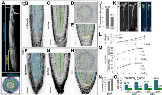
Through a forward genetic screen we isolated the recessivenomad (nom) mutant (see supplementary Materials and Methods). In the epidermis of WT roots, 98.84% of the symmetric divisions are in a transverse orientation relative to the proximodistal axis of the root, whereas innom, only 51.13% are transverse and the other 48.87% are oblique (Fig. 1B,F,J). In the cortical tissue, cells divide again in a transverse orientation in the wild type (WT) and this is unchanged in nom(Fig. 1C,G). The concentric organisation of root tissues and the organisation of the stem cells niche reflect the ability of the stem cells to divide asymmetrically and to give rise to the different tissue types (Dolan et al., 1993), and they are the same innomand WT (Fig. 1D,H,E,I). Innom, some defects in division orientation can been seen in the endodermis, the tissue subtending the cortex (Fig. 1D,H); however, division patterns along the root-hypocotyl axis during embryonic development are unaltered (Fig. S1). This indicates that the nom mutation alters the orientation of the symmetric divisions, but does not affect the root asymmetric divisions in the seedlings or the regular division patterns during embryogenesis.
Mutantnomseedlings can be discriminated from WT seedlings at 4 days post germination (dpg) by a small reduction in root length, which becomes more pronounced at 8 dpg (Fig. 1K,L), but along the proximodistal axis of the root, the meristem size of nom is unchanged compared with that of the WT (Fig. 1M). Within the radial dimension, 8 dpgnomroot meristems were 20% wider than the WT (Fig. 1D,H,N). Although tissues were also mono-layered in thenomepidermis, they had 58.5% more cells than in the WT; by contrast, the increase in cell numbers was not as great in thenom cortex compared with the WT (+18%; Fig. 1,O). As division orientation determines to which growth axis of the organ the new cell will contribute, such a difference in epidermal cell number can be correlated with the oblique orientation of nom epidermal divisions.
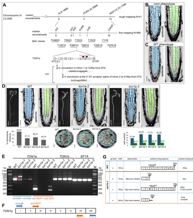
The nom mutation was mapped to the TON1a (At3g55000) locus (Fig. 2A). Complementation with a genomic fragment that restores the nom phenotype to WT (Fig. 2B,C) and the identification of two recessive, T-DNA insertion alleles, ton1a-2
Received 18 August 2015; Accepted 8 February 2016
Department of Cell and Developmental Biology, John Innes Centre, Norwich NR4 7UH, UK.
*Present address: Department of Biology, University of North Carolina, Chapel Hill, NC, USA.
‡Author for correspondence ([email protected])
This is an Open Access article distributed under the terms of the Creative Commons Attribution License (http://creativecommons.org/licenses/by/3.0), which permits unrestricted use, distribution and reproduction in any medium provided that the original work is properly attributed.
DEVEL
O
(GK-016D04) and ton1a-3 (GK-727H06), which display epidermal-specific division defects like nom, confirmed its identity (Fig. 2D). Thenom allele was renamedton1a-1. TON1a lies in tandem to TON1b; they encode proteins that are 85% identical at the amino acid level and the two genes have been proposed to function redundantly (Azimzadeh et al., 2008). However, more recently, a unique function for the TON1agene was hypothesised from biochemical studies (Spinner et al., 2013) and from a genetic interaction found between afass/ton2-15allele and the ton1a-te500 allele that has a WT root phenotype (Kirik et al., 2012). Our RT-PCR analysis shows that in the roots of the ton1a-1,ton1a-2andton1a-3alleles there is a severe reduction in the TON1a transcript compared with that in WT roots and the TON1bgene is expressed as normal (Fig. 2E-G). This suggests that the consistent mutant phenotype we observed in the three ton1a alleles is caused by a reduction in theTON1atranscript and that the threeton1aalleles are hypomorphic alleles of TON1a. Thus, our data show the first direct, genetic evidence of a requirement for a functionalTON1agene alone in the control of symmetric division orientation within the root epidermis, but not in the underlying cortical tissue.
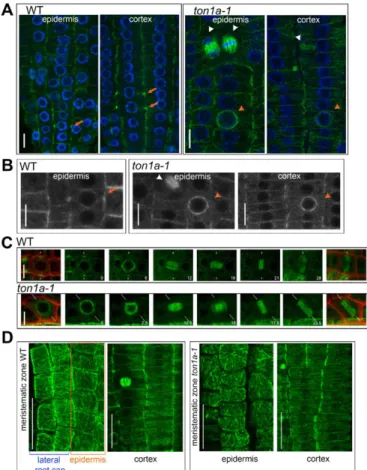
To test whether the root epidermal and cortical cells inton1a-1 form PPBs, we used anti-α-tubulin immunolocalisation. The narrow PPB ring of microtubules that forms at the cellular periphery can be seen as bright foci on each side of the cell in median confocal
sections within WT cells (Fig. 3A). Instead, in median confocal sections withinton1a-1cells of the epidermal and cortical tissues, or of the inner tissues, we did not detect any bright foci, or structures resembling it and only detected more pronounced labelling around the nuclear periphery (Fig. 3A; Fig. S2), like in the studies of the loss-of-function ton1 and fass mutants (Azimzadeh et al., 2008; Camilleri et al., 2002; Traas et al., 1995). Later division structures (spindle and cytokinetic phragmoplast) were normal in ton1a-1 compared with the WT (Fig. 3A). Only occasionally were PPBs seen in lateral root cap cells, which surround the epidermis (data not shown). The absence of PPB formation in the epidermis and cortex was confirmed by examining in vivo the expression of the microtubule marker RFP-TUA5 in theton1a-1mutant (Fig. 3B). Also, we were unable to detect PPBs inton1a-1epidermal cells by in vivotime-course imaging using GFP-β-tubulin (Fig. 3C). Thus, TON1aappears consistently necessary to form the narrow PPBs in both the epidermis and the cortex, yet division orientation is correctly in place in the cortex, but not in the epidermis. Such ability of cells in the cortex to divide correctly is consistent with studies on cell cultures that have shown division plane can also be correctly placed without the formation of narrow PPBs (Chan et al., 2005; Marcus et al., 2005) and with the observations that not all plant species or all cell types form PPBs (Pickett-Heaps et al., 1999; Rasmussen et al., 2013). This suggests that PPB-independent mechanisms can guide division plane positioning in organised tissues.
Fig. 1. Thenommutation affects the patterns of symmetric cell divisions in theArabidopsisroot meristem epidermis but not in the underlying cortical tissue.(A) Top, SEM image of anArabidopsisroot with a superimposed confocal image of the root epidermis in the meristem; bottom, radial organisation of the root in a transverse section in the meristem: the epidermis (in blue) overlays the cortical tissue (in green) and on the outside is surrounded by the lateral root cap tissue (in dark blue). (B-I) Organisation of the root meristem in WT andnommutant seedlings at 8 dpg. Note that in plants, the orientation of the cell walls, highlighted in black by propidium iodide (PI) staining, reflects the orientation of the division planes. Epidermal cells are pseudocoloured in blue, cortical cells in green. (B,C,F,G) Cellular organisation and division plane orientation in the epidermis and underlying cortical layer, images are single, longitudinal confocal sections of Schiff-PI stained meristems. Scale bars: 50μm. (D,H) Transverse sections in the root meristem of resin-embedded seedlings; each black dot marks a single epidermal cell. Scale bars: 25μm. (E,I) Single, median-longitudinal confocal sections in Schiff-PI-stained root meristems; stem cell niche are
pseudocoloured in yellow. Scale bars: 25μm. (J) percentage of transverse divisions in the root, meristematic epidermis of 8 dpg seedlings; WTn=19,nom n=20. (K) Whole seedlings at 4 and 8 dpg. Scale bars: 2 mm and 7.5 mm, respectively. (L-N) Data quantification. Root length (L) and meristem size (M) measured over time; for each time point, WTn=58,nom n=58 (L), WTn=40,nom n=40 (M); (N) diameter of the meristem; WTn=23,nom n=26; (O) number of epidermal and cortical cells scored in transverse sections within the root meristem at 4 and 8 dpg; at 4 dpg, WTn=319 epidermal and 113 cortical cells in 14 roots,nom n=786 epidermal and 206 cortical cells in 23 roots; at 8 dpg, WTn=629 epidermal and 188 cortical cells in 23 roots,nom n=1127 epidermal and 251 cortical cells in 26 roots. All data are means±s.d.; mean values are shown above bars for J,N,O.
DEVEL
O
Fig. 2. Thenommutation maps to theTON1agene and the threeton1aalleles identified are hypomorphic alleles ofTON1a.(A) Genetic mapping delimited thenommutation to BAC clone F28P10. Schematic representation of the organisation of the gene, position of the point mutations relative to the start codon and position of the T-DNA insertions in theton1a-2andton1a-3alleles;nomwas later renamed as theton1a-1allele. (B,C) Confocal images of 4 dpg T2 seedlings segregatingnomand WT phenotypes from T1 BASTA-resistantnomplants carrying the complementing construct. Epidermis is pseudocoloured in blue, cortex in green. Scale bars: 50μm. (D) Phenotypic characterisation of theton1a-2andton1a-3alleles. Stereomicroscope images of whole seedlings and confocal images of the epidermis and cortex in the root meristem of WT andton1a-2andton1a-3alleles at 4 dpg, quantification of epidermal transverse divisions at 8 dpg; WTn=19,ton1a-2 n=18,ton1a-3 n=21; reconstructed transverse sections and respective quantification of epidermal and cortical cell numbers at 8 dpg, including those calculated for theton1a-1allele; WTn=629 epidermal and 188 cortical cells in 23 roots,nom n=1127 epidermal and 251 cortical cells in 26 roots,ton1a-2 n=751 epidermal and 183 cortical cells in 18 roots,ton1a-3 n=883 epidermal and 204 cortical cells in 21 roots. Data are means±s.d. Scale bars: 2 mm for all seedlings, 50μm for epidermis and cortex and 25μm for transverse sections. (E-G) Molecular characterisation of the threeton1aalleles. (E) RT-PCR analysis with primer pairs specific forTON1aex1to8f/r (in blue) produces in theton1a-1allele three main transcripts (indicated with red b,c,d letters), and in the ton1a-2andton1a-3alleles produces a severe reduction in theTON1atranscript compared with the WT. The transcripts inton1a-1were cloned and sequenced to confirm they resulted from mis-splicing. Primer pairs ex2-3to7f/r (in orange) amplify only the correctly spliced WT transcript ofTON1aand produce consistently barely detectable WT transcript for theton1a-1allele. As a loading control, primer pairs that amplify the elongation factor 1A (EF1A) were used. (F) Schematic representation of primer locations on theTON1acDNA; exons are indicated by roman numerals. (G) Schematic summary of RT-PCR and cloning results with the predicted amino acid sequences resulting from the mis-splicing ofTON1atranscript in theton1a-1allele and the location of the LisH
dimerisation motif.
DEVEL
O
The restriction of the defects in symmetric division orientation and patterning to the epidermal tissue in the three ton1a mutant alleles we have characterised raises the question of how such a tissue-specific phenotype might emerge.TON1ais part of the large TTP (TON1/TRM/PP2A) protein complex (Spinner et al., 2013). The possibility that mutations in TON1a affect the interactions between TON1a and the other components of the TTP complex in a tissue-specific manner seems unlikely becauseton1aepidermal and cortical cells are equally unable to form narrow PPBs. An alternative possibility is that in the two tissues, cells have different requirements for a functionalTON1agene and for the formation of the PPB in the
control of division plane orientation. We speculated whether the organisation of the interphase microtubules might underlie such different requirements. We found that, similar to what was also observed in WT meristems by Bichet et al. (2001), in both WT and ton1a-1meristems the interphase microtubules are not organised in epidermal cells and instead have a prevalent transverse organisation in cortical cells, which is parallel to the correct orientation of the division plane (Fig. 3D; Fig. S3). This suggests that in root meristematic cells, theTON1Agene is required for the formation of PPBs and not for the overall organisation of interphase microtubules. In addition, this result raises the possibility that the PPB might function to accurately fix division plane orientation in epidermal cells, where interphase microtubules and the cues responsible for division plane orientation are not aligned. Otherwise, where interphase microtubules are already aligned with such cues, PPB might be redundant, as seen in cortical cells. Such a possibility will need to be experimentally tested in further studies. Our results point to the existence of tissue-specific responses to the signals guiding the orientation of symmetric cell division during tissue patterning. This could be key to ensuring organised growth within proliferating meristems.
MATERIALS AND METHODS Plant lines and growth conditions
TheArabidopsis thalianaT-DNA insertion lines GK-727H06 (N469786), GK-016D04 (N401480) and theGFP-TUB6line (N6550) were obtained from the NottinghamArabidopsisstock centre (NASC). Descriptions of the following lines have been published:GL2::GUS(Masucci and Schiefelbein, 1996), GFP-MBD (Granger and Cyr, 2001) and RFP-TUA5 (Gutierrez et al., 2009). Phenotypic analysis was undertaken on ton1a-1 mutant seedlings backcrossed three times into the parentalGL2::GUSline. The
ton1a-1 phenotype was always compared against the phenotype of the parental lineGL2::GUS(referred to here as WT). GK-727H06 (N469786) and GK-016D04 (N401480) T-DNA insertion lines were confirmed by PCR genotyping using primers listed in Table S1 as described in the supplementary Materials and Methods.
Seeds were surface sterilised in 10% sodium hypochlorite and sown on plates prepared with Murashige and Skoog (MS) salts (Duchefa), 1% sucrose and 0.5% phytagel (Sigma) medium ( pH 5.8). Seeds were stratified in the dark at 4°C for 3 days and grown in a vertical position under continuous light at 28°C. Thenommutant was generated and characterised using standard techniques as detailed in the supplementary Materials and Methods. All experiments represent at least two independent replicates.
Cell division orientation
In plants, cell wall orientation reflects cell division orientation and those cell walls oriented at a 90° angle with the proximodistal axis were scored as transverse divisions; cell walls whose orientation differed more than a 10° angle from the transverse orientation were scored as oblique. Using ImageJ (NIH), cell walls were scored within a rectangular frame of 100 μm (length)×50μm (height) drawn over confocalz-series and centred at 100μm from the quiescent centre of 8 dpg root tips stained with Schiff and propidium iodide (PI).
Cell counting and meristem measurements
Epidermal and cortical cell numbers were counted on confocal, reconstructed transverse sections centred at 100μm from the quiescent centre of 8 dpg root tips stained with Schiff-PI. The diameters of the meristems were measured on the same sections, excluding the lateral root cap tissue from the measurements. Root meristem size was measured on confocal, median, longitudinal sections of PI-stained roots using ImageJ software (NIH) as described previously (Dello Ioio et al., 2007).
Immunocytochemistry
α-tubulin immunostaining of 4 dpg seedlings was carried out using established techniques (Collings and Wateneys, 2005; Sauer et al., 2006) with some modifications as in supplementary Materials and Methods. Fig. 3. In the root meristem,TON1ais required to form PPBs in both
the epidermis and the cortex but does not control the organisation of interphase microtubules, which differs between the two tissues. (A,B) Localisation of cytoskeletal structures in the meristematic epidermis of WT andton1a-1roots, PPB (orange arrows), abundant microtubules surrounding the nucleus (orange arrowheads), spindle and cytokinetic phragmoplast (white arrowheads); all images are single, confocal sections. (A) Roots immunostained withα-tubulin (green), nuclei are stained blue with DAPI; WTn=6 roots observed 13 PPBs in the epidermis and 23 PPBs in the cortex,ton1a-1 n=6 roots. (B) Representative close-up images of meristematic epidermal and cortical cells expressingin vivothe tubulin marker RFP-TUA5 (grey); WTn=12 roots observed 37 PPBs in the epidermis and 48 PPBs in the cortex,ton1a-1 n=10 seedlings. (C) Stills from a confocal time-lapse movie of epidermal cells of WT andton1a-1seedlings expressing GFP-β-tubulin from G2 phase to the end of M-phase (left to right); time is indicated in minutes, maximum intensity projections of confocalz-series, in the first and last stills the GFP images have been merged with PI images (red) to outline the cell walls, the white lines indicate the position of the division plane. (D) Representative images of maximum intensity projections of confocal optical sections in the epidermis and cortex of WT andtona1-1roots immunostained withα-tubulin (green); WTn=6,ton1a-1 n=6. Note that in the first panels, epidermal cells (framed within an orange outline) are present in the same projections as lateral root cap cells. Scale bars: 10μm (A-C) and 25μm (D).
DEVEL
O
Microscopy and image processing
Confocal laser microscopy was performed with a Leica LCS SP5II microscope equipped with HyD detectors. The following wavelengths were used for fluorescence detection. Schiff-PI staining (Truernit et al., 2006): excitation, 488 nm and detection, 600-700 nm; GFP: excitation, 488 nm and detection, 493-550 nm; RFP: excitation, 561 nm and detection, 550-700 nm; DAPI: excitation, 405 nm and detection, 430-500 nm, as described in the supplementary Materials and Methods. Seedlings expressing the marker GFP-TUB6 were mounted in 50% MS liquid medium on slides and meristematic epidermal cells were imaged immediately.
Confocal images were processed with Image J64 software; reconstructed transverse sections were obtained by orthogonal projection of z-series collected at 0.4-0.5μm intervals. Maximum intensity projections were done onz-series collected at 0.5μm intervals for GFP-TUB6. Photoshop CS6 was used to prepare and pseudocolour the images for the figures. Images of longitudinal confocal sections and reconstructed transverse sections of PI-stained seedlings illustrating the organisation of tissues were inverted and their levels adjusted so that the PI staining, which outlines the profile of the cells, is in black.
Acknowledgements
We thank L. Dolan, C. Lloyd, J. Langdale, C. Dean, P. Shaw, H. Buschmann and J. Chan for discussions and advice; G. Calder for advice and assistance with confocal microscopy; and N. Stacey for assistance with historesin sectioning. We are indebted to D. Bradley for support and comments on the manuscript.
Competing interests
The authors declare no competing or financial interests.
Author contributions
Y.Z. and M.I. provided technical support with the genetic mapping; S.C. conceptualised the work, designed and executed the experiments, analysed the data, processed the images and wrote the paper. All authors commented on the manuscript.
Funding
This work was funded by a Royal Society University Research Fellowship to S.C. and by support from the John Innes Centre. Deposited in PMC for immediate release.
Supplementary information
Supplementary information available online at
http://dev.biologists.org/lookup/suppl/doi:10.1242/dev.129502/-/DC1
References
Azimzadeh, J., Nacry, P., Christodoulidou, A., Drevensek, S., Camilleri, C., Amiour, N., Parcy, F., Pastuglia, M. and Bouchez, D. (2008). Arabidopsis TONNEAU1 proteins are essential for preprophase band formation and interact with centrin.Plant Cell20, 2146-2159.
Bichet, A., Desnos, T., Turner, S., Grandjean, O. and Hofte, H.(2001). BOTERO1 is required for normal orientation of cortical microtubules and anisotropic cell expansion in Arabidopsis.Plant J.25, 137-148.
Camilleri, C., Azimzadeh, J., Pastuglia, M., Bellini, C., Grandjean, O. and Bouchez, D.(2002). The Arabidopsis TONNEAU2 gene encodes a putative novel protein phosphatase 2A regulatory subunit essential for the control of the cortical cytoskeleton.Plant Cell14, 833-845.
Chan, J., Calder, G., Fox, S. and Lloyd, C.(2005). Localization of the microtubule end binding protein EB1 reveals alternative pathways of spindle development in Arabidopsis suspension cells.Plant Cell17, 1737-1748.
Dello Ioio, R., Linhares, F. S., Scacchi, E., Casamitjana-Martinez, E., Heidstra, R., Costantino, P. and Sabatini, S.(2007). Cytokinins determine Arabidopsis root-meristem size by controlling cell differentiation.Curr. Biol.17, 678-682.
Dolan, L., Janmaat, K., Willemsen, V., Linstead, P., Poethig, S., Roberts, K. and Scheres, B. (1993). Cellular organisation of the Arabidopsis thaliana root.
Development119, 71-84.
Granger, C. L. and Cyr, R. J.(2001). Spatiotemporal relationships between growth and microtubule orientation as revealed in living root cells ofArabidopsis thaliana
transformed with green-fluorescent-protein gene construct GFP-MBD.
Protoplasma216, 201-214.
Gutierrez, R., Lindeboom, J. J., Paredez, A. R., Emons, A. M. and Ehrhardt, D. W.(2009). Arabidopsis cortical microtubules position cellulose synthase delivery to the plasma membrane and interact with cellulose synthase trafficking compartments.Nat. Cell Biol.11, 797-806.
Kirik, A., Ehrhardt, D. W. and Kirik, V.(2012). TONNEAU2/FASS regulates the geometry of microtubule nucleation and cortical array organization in interphase Arabidopsis cells.Plant Cell24, 1158-1170.
Marcus, A. I., Dixit, R. and Cyr, R. J.(2005). Narrowing of the preprophase microtubule band is not required for cell division plane determination in cultured plant cells.Protoplasma226, 169-174.
Masucci, J. D. and Schiefelbein, J. W.(1996). Hormones act downstream of TTG and GL2 to promote root hair outgrowth during epidermis development in the Arabidopsis root.Plant Cell8, 1505-1517.
Pickett-Heaps, J. D., Gunning, B. E. S., Brown, R. C., Lemmon, B. E. and Cleary, A. L.(1999). The cytoplast concept in dividing plant cells: Cytoplasmic domains and the evolution of spatially organized cell division.Am. J. Bot.86, 153-172.
Rasmussen, C. G., Humphries, J. A. and Smith, L. G.(2011). Determination of symmetric and asymmetric division planes in plant cells.Annu. Rev. Plant Biol.62, 387-409.
Rasmussen, C. G., Wright, A. J. and Müller, S.(2013). The role of the cytoskeleton and associated proteins in determination of the plant cell division plane.Plant J.
75, 258-269.
Spinner, L., Gadeyne, A., Belcram, K., Goussot, M., Moison, M., Duroc, Y., Eeckhout, D., De Winne, N., Schaefer, E., Van De Slijke, E. et al.(2013). A protein phosphatase 2A complex spatially controls plant cell division. Nat. Commun.4, 1863.
Torres-Ruiz, R. A. and Jurgens, G.(1994). Mutations in the FASS gene uncouple pattern formation and morphogenesis in Arabidopsis development.Development
120, 2967-2978.
Traas, J., Bellini, C., Nacry, P., Kronenberger, J., Bouchez, D. and Caboche, M.
(1995). Normal differentiation patterns in plants lacking microtubular preprophase bands.Nature375, 676-677.
Truernit, E., Siemering, K. R., Hodge, S., Grbic, V. and Haseloff, J.(2006). A map of KNAT gene expression in the Arabidopsis root.Plant Mol. Biol.60, 1-20.