Genetic Regulation of Cell Death and Disease Resistance in Arabidopsis
Melinda Margaret Roberts
A dissertation submitted to the faculty of the University of North Carolina at Chapel Hill in partial fulfillment of the requirements for the degree of Doctor of Philosophy in the Department of Biology.
Chapel Hill 2012
ii
ABSTRACT
MELINDA MARGARET ROBERTS: Genetic Regulation of Cell Death and Disease Resistance in Arabidopsis
(Under the direction of Jeff Dangl)
Plants are constantly identifying and responding to cues and threats from their surroundings, such as changes in light, temperature, and humidity, mechanical damage from herbivores and insect, and pathogen attack. Resistance to plant pathogens involves both passive barriers and active, inducible disease resistance responses. Induction of immune responses in plants leads to, for example, cellular redox changes, activation of MAP kinase cascades, massive transcriptional reprogramming, and frequently culminates in a form of programmed cell death known as the hypersensitive response. In my dissertation work, I characterized proteins involved in the regulation of cell death and disease resistance in the model plant Arabidopsis thaliana.
iii
This activity could be partially controlled by LSD1 sequestering NF-YC3 in the cytosol, thereby preventing its nuclear relocalization and subsequent disease resistance function.
The latter half of my work involved the characterization of a positive regulator of lsd1 rcd, ADR1-L2. ADR1-L2 belongs to a small family of NB-LRRs, the main class of
iv
v
ACKNOWLEDGEMENTS
My first thanks must go, of course, to Jeff. Throughout my years in his lab he has challenged me, taught me, supported me, and overall made me a much better scientist, and much, much more of a critical thinker, than I ever thought I could be. He also gave me the best gift of all: a great scientific home in which I was able to learn from a huge variety of individuals, about science, about life, and about the type of person I want to be. The Dangl lab is truly a unique place, and a fitting tribute to its leader.
My gratitude also goes to the other members of my committee: Joe, Jason, Greg, and Mark. Each of you has supported me in many ways during my time here, both through great scientific ideas and insights, and by being excellent members of a caring department. When, as a naive first year, I chose my committee, I had no idea how interesting it would be to have Joe, Mark, and Jeff all together at the same meetings, frequently talking over each other to make their points. This combination of friendship and scientific curiosity is really one of the things that makes the Biology department at UNC a wonderful place to study. Plus, Joe always let me crash his lab’s happy hours, which was a welcome release on several Fridays after some very long weeks.
vi
Emily, Marc, and Baltrus, made for excellent pit-going friends and I still miss sitting around there on warm days discussing anything and everything. Emily, you are one of my most favorite people in the world, and to quote another one of your fans “when I grow up, I want to be like Emily Fisher”. Vera and Anna made my last project, and last two years, so much better than they otherwise would have been. And to Terry and Erica, for helping me all along but being especially supportive at the end: I don’t think I would’ve made it through the last 18 months in lab without you guys. Thank you!
Outside of our lab, there were so many people who made my time here great. The upper-level grad students who were in the department when I arrived made my move to the East Coast so easy, and my fellow cohorts (“the first years”, as some people still insist on calling us) gave me one of the most fun years of my life when we first arrived here, and then have remained good friends all along. And special thanks go to the Kieber lab girls (Cris, Blaire, and Tracy) and to Erin Friedman for helping to make Coker Hall more tolerable, and to the Chemistry Boys, for letting me crash your parties and being great friends (who also happened to introduce me to Michael). I also have to thank all of the people involved with Central Carolina Synchro-getting away from lab and to the rink really helped me stay sane! Kim and Dan of course get a very special thank you. Our friendship means so much to me, and I will miss you most of all. Good thing we have Yosemite, Harry Potter World, and an epic road trip in the future so that there will be lots more time to hang out (and yes Kim, Bridal Mart too-we’ll figure out a way to make it all work).
vii
though the hard times, or what exactly I spend all of my time doing, or if I was ever going to finish, but you all have been wonderful and understanding, and I can’t wait to talk to you and see you more now that it’s over. Special thanks go to Erin, who knew what I was getting myself into and let me do it anyway. Thank you for leading me down this path and doing everything first so I could see what it was all like. And to Cristina, whose life has changed more during the time I’ve been here than ever before, thank you for all of your help and support and listening and understanding. I’m so proud of you in everything that you do, and so thankful to have you in my life. Now it’s your turn to go back to school!
viii
through in a way that nobody else could. Bryan gives me the best advice, no matter the topic, and is the older brother I never had. He always had a fantastic outsider’s prospective, and there were many, many times where talking to him allowed me to see things for what they were, rather than what I was making them out to be. And to Michelle, who is the best of sisters and the best of friends, I am thankful for all of your support and love, and so so so happy to have the two (almost three!) new people you have blessed us with in my life. There’s not much better when I am tired or frustrated than to video chat with Owen and Emma. They make everything better.
ix
Table of Contents
List of Tables ... xi
List of Figures ... xii
List of Terms ... xiii
Chapter 1 ... 1
PRRs and MAMP Triggered Immunity ... 4
NB-LRRs and Effector Triggered Immunity ... 7
Salicylic Acid, a Central Molecule in Plant Defense Responses ... 14
Programmed Cell Death in Plant Pathogenesis ... 17
Conclusions ... 19
References ... 23
Chapter 2 ... 32
Preface ... 32
Abstract ... 33
Introduction ... 33
Results ... 38
LSD1 interacts with the transcription factor NF-YC3. ... 38
NF-YC3 localization is dependent on GxP-mediated LSD1 interaction. ... 39
NF-YC3 is a positive regulator of disease resistance. ... 41
NF-YC3 function requires proper heterotrimeric NF-Y formation. ... 44
Discussion ... 45
Materials and Methods ... 49
References ... 61
Chapter 3 ... 66
Preface ... 66
Abstract ... 68
x
Results ... 73
Members of the ADR1 family of NB-LRRs are required for runaway cell death in lsd1. ... 73
ADR1-L2 is required at the specific site undergoing cell death. ... 74
The function of ADR1-L2 in lsd1 rcd is P-loop dependent. ... 75
An autoactive version of ADR1-L2 displays P-loop dependent, ectopically activated immune responses. ... 75
ADR1-L2D484V autoactivity is regulated by lsd1 suppressors. ... 78
lsd1 ADR1-L2D484V lethality requires EDS1. ... 81
RAR1 is dispensable for accumulation of ADR1-L2. ... 83
Discussion ... 84
Materials and Methods ... 90
References ... 111
xi
List of Tables
xii
List of Figures
Figure 1.1. The zig-zag model of plant defense... 21
Figure 2.1: LSD1, a negative regulator of cell death, interacts with members of the NF-Y transcription factor family ... 53
Figure 2.2: NF-YC Transcription factor subunits are conserved across eukaryotes ... 54
Figure 2.3: LSD1 interacts with the NF-Y subunit NF-YC3 ... 56
Figure 2.4: Semi-in vivo pulldown ... 57
Figure 2.5: NF-YC3 is required for full pathogen resistance ... 58
Figure 2.6: NF-Y assembly and DNA interactions are required for induced pathogen resistance ... 60
Figure 3.1. A family of CC-NB-LRR proteins is required for lsd1 runaway cell death. .. 96
Figure 3.2. ADR1-L2 is required at the site undergoing cell death. ... 97
Figure 3.3. ADR1-L2AAA is not sufficient to trigger lsd1 rcd following BTH treatment ... 98
Figure 3.4. ADR1-L2D484V ectopically activates basal defense ... 99
Figure 3.5. An intact P-loop catalytic domain is required for the ADR1-L2D484V morphological phenotype ... 100
Figure 3.6. lsd1 suppressors are regulators of ADR1-L2D484V autoactivity ... 102
Figure 3.7. eds1 lsd1 ADR1-L2D484V plants lose ectopic activation phenotypes... 104
Figure 3.8. eds1 D484V plants segregating LSD1 show both wild-type and extreme cpr phenotypes ... 106
Figure 3.9. RAR1 is not required for either steady state ADR1-L2 accumulation or BTH-mediated induction... 107
Figure 3.10. A model for the regulation of ADR1-L2D484V activity ... 108
xiii
List of Terms
ADR1 Activated Disease Resistance 1
BiFC Bimolecular fluorescence complementation BTH Benzothiadiazole
CC Coiled-coil
Dex Dexamethasone
EDS1 Enhanced Disease Susceptibility 1 ETI Effector-triggered immunity ETS Effector-triggered susceptibility Hpa Hyaloperonospora arabidopsidis
HR Hypersensitive response ICS1 Isochorismate Synthase 1 LSD1 Lesion Simulating Disease 1
MAMP Microbe-associated molecular patterns MTI MAMP-triggered immunity
NB-LRR Nucleotide-binding leucine-rich repeat NPR1 Nonexpressor of PR Genes 1
NLR Nucleotide-binding domain and leucine-rich repeat-containing NF-Y Nuclear factor Y
PCD Programmed cell death PR Pathogenesis related
PRR Pattern recognition receptors
Pto Pseudomonas syringae pathovar tomato
xiv RCD Runaway cell death
RLK Receptor-like kinase RLP Receptor-like protein
ROI Reactive oxygen intermediates SA Salicylic acid
SAR Systemic acquired resistance
STAND Signal transduction ATPases with numerous domains TF Transcription factor
TIR Toll/interleukin-1
Chapter 1
Introduction
Plants, like all other organisms, must properly respond to changes in their surroundings. Examples of these responses include finely-tuned tropism reactions to water gradients, light, and gravity (Eapen et al., 2005; Holland et al., 2009; Moulia and Fournier, 2009); proper timing of seed germination (Penfield and King, 2009); and correct responses to attacks from herbivores, phytophagous insects, and pathogens. As sessile organisms without adaptive or circulatory immune systems, plants have had to evolve a set of cell-autonomous defense responses. Many times, these endogenous disease resistance mechanisms are not successful: plant pathogens alone contribute up to $30 billion in annual losses to the US agriculture industry (Pimentel et al., 2000). However, despite the inherent limitation of not having an adaptive or circulatory immune system, most plants are resistant to most pathogens (McDowell and Simon, 2008).
2
locally and systemically, and can be divided into two parts. The first of these branches includes recognition of microbe-associated molecular patterns (MAMPs) by transmembrane pattern recognition receptors (PRRs) in the host plant. PRRs frequently bind proteins and other molecules that are particularly important to the pathogen’s function (Zipfel, 2009). While MAMP-triggered immunity, or MTI, is an effective and robust defense strategy, pathogens have, by definition, evolved methods of evading it and are thus able to colonize their hosts. Pathogens, such as the model bacteria Pseudomonas syringae pathovar tomato (Pto), secrete effector proteins into the host plant. These
specialized proteins antagonize MTI responses by, for example, blocking cell wall callose deposition, interrupting plant hormone signaling important for a proper defense response, and interfering with cell death responses triggered by other effectors (Grant et al., 2006). Effector proteins that are able to suppress MTI help in a successful colonization of the host plant; this process is known as effector-triggered susceptibility (ETS). Plants have, in turn, developed a system for responding to ETS. This response depends on disease resistance, or R, genes.
effector-3
triggered immunity (ETI), resulting in a disease resistance response that is both faster and stronger than MTI (Jones and Dangl, 2006). ETI responses include calcium influx, protein kinase activation, production of reactive oxygen intermediates, transcriptional reprogramming, and, frequently, the hypersensitive response (HR), a type of programmed cell death (Dangl and Jones, 2001).
Basal defense, or the responses triggered by virulent pathogens on susceptible hosts, and ETI are easily thought of as different magnitudes of the same defense responses. This is best visualized by the zigzag model put forth by Jones and Dangl in 2006 (Figure 1). Their model presents MTI as the primary, low-level amplitude reaction to pathogens. Successful pathogens utilize effector proteins to overcome this first response, and resistant plants employ NB-LRRs to recognize these intruder proteins and mount a stronger defense, including localized cell death (HR). Inherent in this model is the resulting evolutionary “arms race” between pathogens and their potential hosts. When plants begin to recognize existing effector proteins, the pathogen will evolve a new array of effectors that cannot be recognized or which can counteract the plant’s original ETI. Plants, in turn, evolve new NB-LRRs, capable of recognizing the new effectors, and the cycle will begin again.
4
PRRs and MAMP Triggered Immunity
The first layer of inducible defense responses involves direct perception of non-host elicitors, or MAMPs, by PRRs. To avoid confusion between MAMPs and effectors, MAMPs are defined as being “conserved among a large group or class of microbes”, whereas effectors evolve within a single or small group of microbial species (Zipfel, 2009). Continuous addition to the body of knowledge about elicitors and effectors makes categorizing these molecules an ongoing effort. Interactions between MAMPs and their receptors occur at the plant cell’s plasma membrane, and all currently identified PRRs are transmembrane receptor-like proteins (RLPs) or kinases (RLKs). RLKs and RLPs have similar extracellular structures with multiple LRR domains and similar transmembrane helices, but RLKs posses a cytoplasmic kinase domain (Tor et al., 2009). The two best-studied examples of MAMP receptors are FLS2 and EFR, RLKs which bind flagellin and elongation factor-Tu (EF-Tu), respectively. Additional PRRs include XA21, a rice protein whose ligand Ax21 was recently discovered (Lee et al., 2009) and CERK1, a LysM-RLK which recognizes chitin (Petutschnig et al., 2010). Activation of any of these receptors leads to a common set of downstream defense responses, and efr and fls2 mutants are more susceptible to a range of pathogens (Zipfel, 2009).
5
natural plant-pathogen interaction, but do offer an overall picture of MAMP-initiated defense effects (Segonzac and Zipfel, 2011). The most frequently studied elicitor-receptor interactions are flagellin-FLS2 and EF-Tu-EFR; the former will be used as an example here. Flagellin, or the minimal signaling epitope known as flg22 which is derived from the N-terminus of flagellin, is recognized at the cell surface by FLS2, a glycosylated, transmembrane RLK (Gomez-Gomez and Boller, 2000; Chinchilla et al., 2006). This extracellular detection leads to a heteromerization between FLS2 and BAK1, a short LRR RLK which is a member of the Somatic Embryogenesis Receptor Kinase (SERK) family (Chinchilla et al., 2007). This PRR/RLK complex also binds other SERKs (Roux et al., 2011). In addition to the formation of the receptor/kinase complex, phosphorylation of both FLS2 and BAK1 quickly follows elicitor recognition (Schulze et al., 2010), though the relevant residues are currently unknown.
6
NADPH oxidase RbohD in a cell autonomous manner, and is a way for the initial defense signal to be propagated across the leaf (Torres et al., 2002; Miller et al., 2009).
Classification of pathogen molecules as MAMPs versus effectors, and, in a related manner, PRRs versus R proteins, is an important and constantly evolving process. Initial categorization can be used as preliminary insight into the role of a new pathogenesis-related protein, but if incorrect can lead to faulty assumptions about that protein. For instance, classifying a protein as an effector leads to the conclusion that it will act within the plant cell, whereas a MAMP functions at the extracellular membrane. The experimental approaches used to test the functions of these two proteins are inherently different. Therefore, incorrectly identifying a protein makes it difficult to properly dissect the genetics and biochemistry of the defense processes in which it is involved. As more MAMP/PRR pairs are identified, there will be a better understanding of the common signaling components involved in MTI. Proper classification of MAMPs and effectors also allows for robust evolutionary studies, which will further inform the overall picture of plant-pathogen interactions.
7
tomato, a species that does not normally carry this PRR, leads to broad-spectrum bacterial resistance in these plants (Lacombe et al., 2010). Examples such as this prove that MTI-related research has developed rapidly over the course of the last 10 years. Continuing efforts should uncover a much more complete view of the path from ligand perception to disease resistance.
NB-LRRs and Effector Triggered Immunity
Pathogens, in an evolutionary response to MTI, evolved effector proteins to combat the defense responses triggered by their MAMPs. Bacteria utilize the type three secretion system (TTSS), a syringe-like apparatus that sends 15-30 such effector proteins into the host plant (Cornelis and Van Gijsegem, 2000; Alfano and Collmer, 2004). Effectors target the function of proteins important for MTI, thereby increasing pathogen virulence and causing ETS. For instance, the Pto effector AvrPto suppresses basal defenses in tomato, Arabidopsis, and the model plant Nicotiana benthamiana. AvrPto binds EFR and FLS2 (Xiang et al., 2008), and targets BAK1 (Shan et al., 2008), disrupting FLS2-BAK1 interactions and suppressing flg22-induced MPK3 and MPK6 activation, cell death, and callose deposition (Hann and Rathjen, 2007).
8
trafficking, and MIN7 is required for full bacterial resistance in Arabidopsis. HopM1 uses the proteaosome of the host plant to degrade MIN7, thereby increasing bacterial virulence (Nomura et al., 2006). Left unchecked, effectors can overcome MTI and lead to host plant susceptibility. Thus, plants have evolved a way to recognize and respond to pathogen effector proteins.
Recognition of pathogen effectors by the host requires the proper detection by and function of NB-LRRs, and proper recognition leads to ETI. Interaction between NB-LRR and effector proteins is hypothesized to occur in one of two ways: directly or indirectly. Direct interactions occur when an effector and an NB-LRR bind to each other. Examples of this include the Arabidopsis protein RRS1-R directly interacting with the Ralstonia Avr protein PopP2 (Deslandes, PNAS 2003), the rice Pi-ta NBS-LRR directly associating with Avr-Pita from rice blast (Jia et al., 2000), and L5, L6, and L7 proteins from flax, which directly recognize the products of the rust flax AvrL567 genes (Dodds et al., 2006).
9
One well-studied example of the guard hypothesis involves the P. syringae effectors AvrB and AvrRpm1, along with the Arabidopsis proteins RIN4 and RPM1. RPM1 encodes a CC-NB-LRR and guards RIN4, a small, membrane-bound protein that
is a negative regulator of basal defense (Mackey et al., 2002). RIN4 is modified when either of the two sequence-unrelated effector proteins, AvrB or AvrRpm1, is introduced to the system via delivery by the TTSS of Pto DC3000. Neither effector is a kinase, but their interaction with RIN4 leads to phosphorylation of RIN4 at threonine 166 (Chung et al., 2011). This phosphorylation of RIN4 is perceived by RPM1, which then triggers a series of pathogen defense responses, including HR (Boyes et al., 1998; Chung et al., 2011). In an rpm1 mutant, the lack of RPM1 protein allows AvrRpm1 or AvrB to enter the cell undetected. From there at least AvrRpm1 acts as a virulence factor, promoting bacterial growth and disease (Ritter and Dangl, 1995). RIN4 is also guarded by a second, independent NB-LRR, RPS2. RPS2 is triggered when AvrRpt2, a third P.syringae effector, cleaves RIN4 at two sites. This cleavage is detected by RPS2, triggering a similar series of defense responses to those activated by RPM1. A fourth effector, HopF2, also targets RIN4 (Wilton et al., 2010). These interactions involve four different effector proteins that are all found to trigger defense responses through the same protein, in fact, by their action on the same ~30 amino acid domain of RIN4. RIN4 is guarded by at least two different NB-LRRs. The RIN4 example provides proof that Arabidopsis is able to maximize its pathogen recognition specificity utilizing a small, non-adaptive set of NB-LRRs.
10
triggered when necessary, and thus they must be under finely-tuned control. One model of disease resistance shows NB-LRRs functioning as molecular switches, with multiple subdomains responsible for keeping the protein in the resting, or “off”, state, thereby preventing spurious NB-LRR activation (Takken et al., 2006). NB-LRR proteins, and NLR homologs in animals, are members of the NTPase superfamily and belong to the signal transduction ATPases with numerous domains (STAND) subclade (Leipe et al., 2004). In the “off” conformation, STAND proteins bind ADP, which must be exchanged for ATP in order to trigger defense responses (Takken et al., 2006).
11
nucleotide exchange. After pathogen recognition, where specificity is typically conferred by the LRR, this autoinhibition is released, allowing ADP to be exchanged for ATP and initiation of defense signaling events.
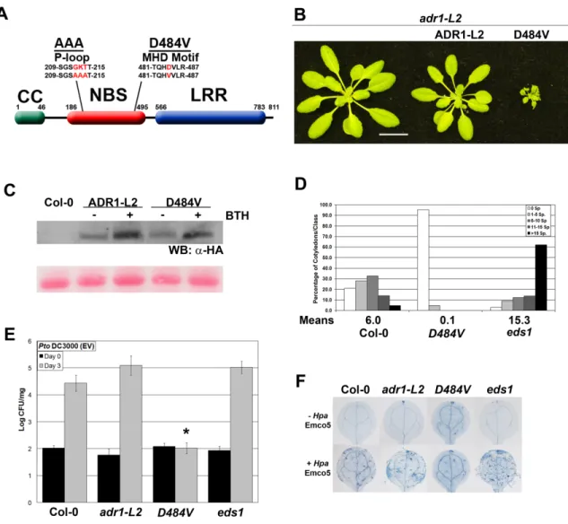
Attempts to study autoactive mutants have been made in Arabidopsis, flax, and tobacco (Table 1). While the majority of NB-LRR autoactive mutations recovered have been in the MHD domain (Bendahmane et al., 2002; Howles et al., 2005; Tameling et al., 2006; Gao et al., 2011; Williams et al., 2011; Zhang et al., 2012), there are also mutations that lead to autoactivity which occur outside of this domain (Zhang et al., 2003; Igari et al., 2008; Huang et al., 2010). Much of the work done with these autoactive alleles has been carried out in transient over-expression assays in flax or tobacco systems, making it difficult to test their biological relevance. However, some key work in Arabidopsis and flax has shown that these autoactive mutations lead to lethality or dwarfed morphology (Howles et al., 2005; Gao et al., 2011; Zhang et al., 2012). Additionally, there is evidence that NB-LRR autoactivity directly affects the immune system signaling pathway, as some of these mutants exhibit hallmarks of defense activation, including high steady-state SA levels (Zhang et al., 2003; Huang et al., 2010) and increased resistance to infection with virulent pathogens (enhanced basal defense) (Gao et al., 2011). Overall, these autoactive mutants clearly show that correctly controlled function of NB-LRRs is necessary for both plant fitness and defense activation.
12
typical ATPase activities. In these cases, NB-LRRs may not work as canonical ‘sensors’, but might instead act as ‘helper’ proteins. These ‘helpers’ potentially function as scaffolding proteins, perhaps working with other immune-related proteins, including canonical NB-LRRs, to trigger defense responses (Bonardi et al., 2012). These examples show us that there is still much to be learned about the overall role that NB-LRR proteins play in defense. Chapter 3 addresses the characterization of a unique NB-LRR with both ‘helper’ and P-loop dependent functions in disease resistance.
13
SGT1 is also required for proper NB-LRR stability. RAR1 and SGT1 interact via the C-terminal CHORDII domain of RAR1 and the CS domain of SGT1. Arabidopsis contains two orthologues of this gene; mutations in SGT1b, but not STG1a, can alter the functions of some NB-LRR proteins. SGT1 double mutants are lethal (Azevedo et al., 2006).
In planta, both RAR1 and SGT1 associate independently with the cytosolic
protein HSP90. HSP90 is a chaperone protein that is responsible for the proper folding of its “client” proteins, and it is known to regulate accumulation of wild-type amounts of protein for all tested NB-LRRs. The ATPase domain of cytosolic HSP90 associates with both CHORDI of RAR1 and the CS domain of SGT1b. This association is clearly important to NB-LRR function, as point mutations in the ATPase domain of one isoform of HSP90, hsp90.2, cause a large reduction in the accumulation of the NB-LRR RPM1 (Hubert et al., 2003). This data lead to a model where NB-LRR proteins are clients of HSP90, and are held in proper conformation and therefore maintain proper protein levels with the co-chaperones RAR1 and SGT1b. Additional hsp90.2 alleles were identified that suppressed rar1 phenotypes, allowing accumulation of functional levels of NB-LRRs in this background (Hubert et al., 2009). These alleles furthered the model of HSP90-regulated protein accumulation, showing that RAR1 normally functions to physically regulate HSP90-dependent dynamic protein turnover. Overall, disruption to RAR1, HSP90, or SGT1 can lead to an alteration in NB-LRR accumulation, and potentially affects disease resistance.
14
Proper signal transduction from TIR-NB-LRRs is dependent on EDS1 (Enhanced Disease Susceptibility 1), PAD4 (Phytoalexin Deficient 4) and SAG101 (Senescence-Associated Gene 101), while CC-NB-LRRs require NDR1 (Non-race-specific Disease Resistance 1) for proper function (Glazebrook, 2001). Together, these NB-LRR regulatory proteins and domains attempt to balance plant cell damage caused by virulent pathogens with fitness costs stemming from disease responses whose amplitude is too high.
Salicylic Acid, a Central Molecule in Plant Defense Responses
15
Dewdney et al., 2000). An additional isochorismate synthase, ICS2, also exists in Arabidopsis, and it is responsible for generating the SA measured in sid2 mutants (Garcion et al., 2008). There is also an ICS-independent pathway for SA synthesis, as ics1 ics2 double mutants still display very low levels of SA (Garcion et al., 2008).
In addition to those genes encoding the proteins required for the biosynthesis of SA, several other genes are positive regulators of SA. The best characterized of these include EDS1, PAD4, and NDR1, though a handful of additional positive regulators have been recently characterized (Lu, 2009). Both EDS1 and PAD4, as well as several other positive regulators of SA, are also SA-inducible, and the loss of resistance phenotypes seen in eds1, pad4, and ndr1 plants can be reversed by exogenous application of BTH (Zhou et al., 1998; Falk et al., 1999; Shapiro and Zhang, 2001). This suggests that SA regulation occurs in a feedback loop: many positive regulators of SA are induced by SA, leading to dramatic increases in this molecule after disease resistance pathways are triggered.
Much of the signaling downstream of SA requires NONEXPRESSOR OF PR GENES 1 (NPR1) (Cao et al., 1997). NPR1 is found in both the cytosol and the nucleus,
16
responses can be of high fitness cost to a plant, and thus, inducers of defense must be tightly regulated. In the case of NPR1, this regulation comes in the form of proteasome-mediated degradation, which uniquely both prevents spurious gene activation in plants not undergoing pathogen attack and stimulates defense-related gene expression when plant defense responses are turned on (Spoel et al., 2009). Very recently, a potential mechanism for SA perception and monitoring was proposed (Fu et al., 2012). The data in this paper demonstrates that NPR3 and NPR4, paralogues of NPR1, are SA receptors with different binding affinities for the molecule. These two proteins function in the SA-mediated degradation of NPR1, and the authors propose that their different affinities for NPR1 sets up the proper regulation of NPR1 protein levels mentioned above.
17
Much work has gone into trying to identify the molecule responsible for the spread of SAR. Early studies of SA showed that large amounts of the compound accumulate in and around the lesions that form at the site of pathogen infection (Enyedi et al., 1992). SA levels are also known to increase throughout the plant after pathogen recognition, including in the phloem (Yalpani et al., 1991). Given this data, it was originally thought that SA might be the SAR potentiation signal. However, grafting studies showed that SA is not necessary for development of the signal at the site of infection, though it is required for SAR at distal sites in the plant (Vernooij et al., 1994). SA can be reversibly turned into methylsalicylic acid (MeSA), and studies in tobacco found that this compound fit all the requirements to be the SAR signaling compound (Park et al., 2007). However, in Arabidopsis, studies showed that MeSA accumulation mutants still could induce SAR (Attaran et al., 2009). Thus, the search for the SAR systemic signal continues.
Programmed Cell Death in Plant Pathogenesis
Programmed cell death (pcd) in plants can be induced by a variety of abiotic and biotic stressors, including high light, heat shock or chilling, and the chemical inducers H2O2 and paraquat. One of the hallmarks of pathogen recognition is the HR, a type of pcd
18
(Levine et al., 1994; Delledonne et al., 1998). Increases in ROS occur in and around the infected cell, and they are important signaling molecules for HR propagation across the leaf (Nanda et al., 2010). AtRbohD is required for the oxidative burst, and in wild-type plants this burst signals the cells proximal to sites of infection to induce transcription of defense genes and suppress cell death (Jabs et al., 1996; Torres et al., 2005). In distal cells, ROS and SA function as signal transduction molecules, potentiating cell death throughout the leaf. This cell death must be kept in check to prevent unnecessary death of parts of or the whole plant.
The Arabidopsis lesions simulating disease1 (lsd1) mutant provides an excellent background to study the roll of cell death in disease resistance. LSD1 is a cytosolic zinc finger protein, and in wild-type plants it functions as a negative regulator of cell death. lsd1 mutant plants exhibit inappropriately regulated cell death, also known as runaway
cell death (rcd), and increased resistance to multiple pathogens (Dietrich et al., 1997). Triggers of rcd in an lsd1 mutant include pathogen infection, changes in day length, and exogenous application of SA or BTH; lsd1 plants are unable to stop the propagation of cell death from any trigger. As a cytosolic zinc finger protein, LSD1 functions as a potential interacting platform for other proteins involved in pcd (Kaminaka et al., 2006). Yeast two-hybrid and phage display screens identified several other LSD1 interactors, including NF-YC3 and NF-YC2, both encoding CAAT Box-binding Factor CBF-C subunits of heterotrimeric CAAT-binding TFs. These proteins and their roles in plant defense are further explored in chapter 2.
19
Runaway cell death in lsd1 also requires both SA and NPR1 (Aviv et al., 2002). In lsd1, but not wild-type plants, SA is able to trigger rcd, indicating that LSD1 is normally working as a negative regulator of SA-dependent cell death (Dietrich et al., 1994). These results position LSD1 and SA in a feedback loop, where the presence of LSD1 is necessary and sufficient to stop SA-potentiated rcd. NPR1 is also necessary for lsd1-mediated rcd, making it clear that SA is at least partially required as a signal initiator in rcd. It is also important to note that these SA requirements are not the same for the lsd1-related basal defense phenotypes: SA-depleted lsd1 plants still show increased resistance (Aviv et al., 2002).
Experiments looking for positive regulators of lsd1 rcd also uncovered the ADR1 family of NB-LRRs, members of which function as ‘helper’ NB-LRRs in basal defense (Bonardi et al., 2011). As previously stated, at least one member of this family also has canonical, P-loop dependent immune functions which are discussed chapter 3.
Conclusions
20
gene families (Tsuda et al., 2008; Miller et al., 2009). Another example of overlap, published recently, shows that there is an in planta association between the PRR FLS2 and the R proteins RPM1, RPS2, and RPS5 (Qi et al., 2011), though the functional consequences of this, if any, remain to be defined.
21
23
References
Ade, J., DeYoung, B.J., Golstein, C., and Innes, R.W. (2007). Indirect activation of a plant nucleotide binding site-leucine-rich repeat protein by a bacterial protease. PNAS 104, 2531-2536.
Alfano, J.R., and Collmer, A. (2004). Type III secretion system effector proteins: double agents in bacterial disease and plant defense. Annu Rev Phytopathol 42, 385-414.
Attaran, E., Zeier, T.E., Griebel, T., and Zeier, J. (2009). Methyl salicylate production and jasmonate signaling are not essential for systemic acquired resistance in Arabidopsis. Plant Cell 21, 954-971.
Aviv, D.H., Rusterucci, C., Holt, B.F., 3rd, Dietrich, R.A., Parker, J.E., and Dangl, J.L. (2002). Runaway cell death, but not basal disease resistance, in lsd1 is SA- and NIM1/NPR1-dependent. Plant J 29, 381-391.
Azevedo, C., Betsuyaku, S., Peart, J., Takahashi, A., Noel, L., Sadanandom, A., Casais, C., Parker, J., and Shirasu, K. (2006). Role of SGT1 in resistance protein accumulation in plant immunity. EMBO J 25, 2007-2016.
Bendahmane, A., Farnham, G., Moffett, P., and Baulcombe, D.C. (2002). Constitutive gain-of-function mutants in a nucleotide binding site-leucine rich repeat protein encoded at the Rx locus of potato. Plant J 32, 195-204.
Bieri, S., Mauch, S., Shen, Q.H., Peart, J., Devoto, A., Casais, C., Ceron, F., Schulze, S., Steinbiss, H.H., Shirasu, K., et al. (2004). RAR1 positively controls steady state levels of barley MLA resistance proteins and enables sufficient MLA6 accumulation for effective resistance. Plant Cell 16, 3480-3495.
Bonardi, V., Cherkis, K., Nishimura, M.T., and Dangl, J.L. (2012). A new eye on NLR proteins: focused on clarity or diffused by complexity? Curr Opin Immunol 24, 41-50. Bonardi, V., Tang, S., Stallmann, A., Roberts, M., Cherkis, K., and Dangl, J.L. (2011). Expanded functions for a family of plant intracellular immune receptors beyond specific recognition of pathogen effectors. Proc Natl Acad Sci U S A 108, 16463-16468.
Boyes, D.C., Nam, J., and Dangl, J.L. (1998). The Arabidopsis thaliana RPM1 disease resistance gene product is a peripheral plasma membrane protein that is degraded coincident with the hypersensitive response. PNAS 95, 15849-15854.
Cao, H., Glazebrook, J., Clarke, J.D., Volko, S., and Dong, X. (1997). The Arabidopsis NPR1 gene that controls systemic acquired resistance encodes a novel protein containing ankyrin repeats. Cell 88, 57-63.
24
Chinchilla, D., Zipfel, C., Robatzek, S., Kemmerling, B., Nurnberger, T., Jones, J.D., Felix, G., and Boller, T. (2007). A flagellin-induced complex of the receptor FLS2 and BAK1 initiates plant defence. Nature 448, 497-500.
Chung, E.H., da Cunha, L., Wu, A.J., Gao, Z., Cherkis, K., Afzal, A.J., Mackey, D., and Dangl, J.L. (2011). Specific threonine phosphorylation of a host target by two unrelated type III effectors activates a host innate immune receptor in plants. Cell host & microbe 9, 125-136.
Coll, N.S., Vercammen, D., Smidler, A., Clover, C., Van Breusegem, F., Dangl, J.L., and Epple, P. (2010). Arabidopsis type I metacaspases control cell death. Science 330, 1393-1397.
Cornelis, G.R., and Van Gijsegem, F. (2000). Assembly and function of type III secretory systems. Annu Rev Microbiol 54, 735-774.
Dangl, J.L., and Jones, J.D. (2001). Plant pathogens and integrated defence responses to infection. Nature 411, 826-833.
DebRoy, S., Thilmony, R., Kwack, Y.B., Nomura, K., and He, S.Y. (2004). A family of conserved bacterial effectors inhibits salicylic acid-mediated basal immunity and promotes disease necrosis in plants. PNAS 101, 9927-9932.
Delledonne, M., Xia, Y., Dixon, R.A., and Lamb, C. (1998). Nitric oxide functions as a signal in plant disease resistance. Nature 394, 585-588.
Despres, C., DeLong, C., Glaze, S., Liu, E., and Fobert, P.R. (2000). The Arabidopsis NPR1/NIM1 protein enhances the DNA binding activity of a subgroup of the TGA family of bZIP transcription factors. Plant Cell 12, 279-290.
Dewdney, J., Reuber, T.L., Wildermuth, M.C., Devoto, A., Cui, J., Stutius, L.M., Drummond, E.P., and Ausubel, F.M. (2000). Three unique mutants of Arabidopsis identify eds loci required for limiting growth of a biotrophic fungal pathogen. Plant J 24, 205-218.
Dietrich, R.A., Delaney, T.P., Uknes, S.J., Ward, E.R., Ryals, J.A., and Dangl, J.L. (1994). Arabidopsis mutants simulating disease resistance response. Cell 77, 565-577. Dietrich, R.A., Richberg, M.H., Schmidt, R., Dean, C., and Dangl, J.L. (1997). A novel zinc finger protein is encoded by the Arabidopsis LSD1 gene and functions as a negative regulator of plant cell death. Cell 88, 685-694.
Dodds, P.N., Lawrence, G.J., Catanzariti, A.M., Teh, T., Wang, C.I., Ayliffe, M.A., Kobe, B., and Ellis, J.G. (2006). Direct protein interaction underlies gene-for-gene specificity and coevolution of the flax resistance genes and flax rust avirulence genes. Proc Natl Acad Sci U S A 103, 8888-8893.
25
Eapen, D., Barroso, M.L., Ponce, G., Campos, M.E., and Cassab, G.I. (2005). Hydrotropism: root growth responses to water. Trends in plant science 10, 44-50.
Enyedi, A.J., Yalpani, N., Silverman, P., and Raskin, I. (1992). Localization, conjugation, and function of salicylic acid in tobacco during the hypersensitive reaction to tobacco mosaic virus. Proc Natl Acad Sci U S A 89, 2480-2484.
Falk, A., Feys, B.J., Frost, L.N., Jones, J.D., Daniels, M.J., and Parker, J.E. (1999). EDS1, an essential component of R gene-mediated disease resistance in Arabidopsis has homology to eukaryotic lipases. PNAS 96, 3292-3297.
Fu, Z.Q., Yan, S., Saleh, A., Wang, W., Ruble, J., Oka, N., Mohan, R., Spoel, S.H., Tada, Y., Zheng, N., et al. (2012). NPR3 and NPR4 are receptors for the immune signal salicylic acid in plants. Nature advance online publication.
Gao, Z., Chung, E.H., Eitas, T.K., and Dangl, J.L. (2011). Plant intracellular innate immune receptor Resistance to Pseudomonas syringae pv. maculicola 1 (RPM1) is activated at, and functions on, the plasma membrane. Proc Natl Acad Sci U S A 108, 7619-7624.
Garcion, C., Lohmann, A., Lamodiere, E., Catinot, J., Buchala, A., Doermann, P., and Metraux, J.P. (2008). Characterization and biological function of the ISOCHORISMATE SYNTHASE2 gene of Arabidopsis. Plant Physiol 147, 1279-1287.
Glazebrook, J. (2001). Genes controlling expression of defense responses in Arabidopsis--2001 status. Curr Opin Plant Biol 4, 301-308.
Glazebrook, J. (2005). Contrasting mechanisms of defense against biotrophic and necrotrophic pathogens. Annu Rev Phytopathol 43, 205-227.
Gomez-Gomez, L., and Boller, T. (2000). FLS2: an LRR receptor-like kinase involved in the perception of the bacterial elicitor flagellin in Arabidopsis. Mol Cell 5, 1003-1011. Grant, S.R., Fisher, E.J., Chang, J.H., Mole, B.M., and Dangl, J.L. (2006). Subterfuge and manipulation: type III effector proteins of phytopathogenic bacteria. Annu Rev Microbiol 60, 425-449.
Hann, D.R., and Rathjen, J.P. (2007). Early events in the pathogenicity of Pseudomonas syringae on Nicotiana benthamiana. Plant J 49, 607-618.
Hanson, P.I., and Whiteheart, S.W. (2005). AAA+ proteins: have engine, will work. Nat Rev Mol Cell Biol 6, 519-529.
Holland, J.J., Roberts, D., and Liscum, E. (2009). Understanding phototropism: from Darwin to today. Journal of experimental botany 60, 1969-1978.
26
Holt, B.F., 3rd, Hubert, D.A., and Dangl, J.L. (2003). Resistance gene signaling in plants--complex similarities to animal innate immunity. Curr Opin Immunol 15, 20-25.
Howles, P., Lawrence, G., Finnegan, J., McFadden, H., Ayliffe, M., Dodds, P., and Ellis, J. (2005). Autoactive alleles of the flax L6 rust resistance gene induce non-race-specific rust resistance associated with the hypersensitive response. Mol Plant Microbe Interact 18, 570-582.
Huang, X., Li, J., Bao, F., Zhang, X., and Yang, S. (2010). A gain-of-function mutation in the Arabidopsis disease resistance gene RPP4 confers sensitivity to low temperature. Plant Physiol 154, 796-809.
Hubert, D.A., He, Y., McNulty, B.C., Tornero, P., and Dangl, J.L. (2009). Specific Arabidopsis HSP90.2 alleles recapitulate RAR1 cochaperone function in plant NB-LRR disease resistance protein regulation. Proc Natl Acad Sci U S A 106, 9556-9563.
Hubert, D.A., Tornero, P., Belkhadir, Y., Krishna, P., Takahashi, A., Shirasu, K., and Dangl, J.L. (2003). Cytosolic HSP90 associates with and modulates the Arabidopsis RPM1 disease resistance protein. The EMBO journal 22, 5679-5689.
Igari, K., Endo, S., Hibara, K., Aida, M., Sakakibara, H., Kawasaki, T., and Tasaka, M. (2008). Constitutive activation of a CC-NB-LRR protein alters morphogenesis through the cytokinin pathway in Arabidopsis. Plant J 55, 14-27.
Jabs, T., Dietrich, R.A., and Dangl, J.L. (1996). Initiation of runaway cell death in an Arabidopsis mutant by extracellular superoxide. Science 273, 1853-1856.
Jia, Y., McAdams, S.A., Bryan, G.T., Hershey, H.P., and Valent, B. (2000). Direct interaction of resistance gene and avirulence gene products confers rice blast resistance. The EMBO journal 19, 4004-4014.
Jones, J.D., and Dangl, J.L. (2006). The plant immune system. Nature 444, 323-329. Kaminaka, H., Nake, C., Epple, P., Dittgen, J., Schutze, K., Chaban, C., Holt, B.F., 3rd, Merkle, T., Schafer, E., Harter, K., et al. (2006). bZIP10-LSD1 antagonism modulates basal defense and cell death in Arabidopsis following infection. The EMBO journal 25, 4400-4411.
Kofoed, E.M., and Vance, R.E. (2011). Innate immune recognition of bacterial ligands by NAIPs determines inflammasome specificity. Nature 477, 592-595.
Lacombe, S., Rougon-Cardoso, A., Sherwood, E., Peeters, N., Dahlbeck, D., van Esse, H.P., Smoker, M., Rallapalli, G., Thomma, B.P., Staskawicz, B., et al. (2010). Interfamily transfer of a plant pattern-recognition receptor confers broad-spectrum bacterial resistance. Nat Biotechnol 28, 365-369.
27
Leipe, D.D., Koonin, E.V., and Aravind, L. (2004). STAND, a class of P-loop NTPases including animal and plant regulators of programmed cell death: multiple, complex domain architectures, unusual phyletic patterns, and evolution by horizontal gene transfer. J Mol Biol 343, 1-28.
Levine, A., Tenhaken, R., Dixon, R., and Lamb, C. (1994). H2O2 from the oxidative burst orchestrates the plant hypersensitive disease resistance response. Cell 79, 583-593. Lu, H. (2009). Dissection of salicylic acid-mediated defense signaling networks. Plant signaling & behavior 4, 713-717.
Mackey, D., Holt, B.F., 3rd, Wiig, A., and Dangl, J.L. (2002). RIN4 interacts with Pseudomonas syringae type III effector molecules and is required for RPM1-mediated resistance in Arabidopsis. Cell 108, 743-754.
McDowell, J.M., and Simon, S.A. (2008). Molecular diversity at the plant-pathogen interface. Dev Comp Immunol 32, 736-744.
Miller, G., Schlauch, K., Tam, R., Cortes, D., Torres, M.A., Shulaev, V., Dangl, J.L., and Mittler, R. (2009). The plant NADPH oxidase RBOHD mediates rapid systemic signaling in response to diverse stimuli. Science signaling 2, ra45.
Mou, Z., Fan, W., and Dong, X. (2003). Inducers of plant systemic acquired resistance regulate NPR1 function through redox changes. Cell 113, 935-944.
Moulia, B., and Fournier, M. (2009). The power and control of gravitropic movements in plants: a biomechanical and systems biology view. Journal of experimental botany 60, 461-486.
Naito, K., Taguchi, F., Suzuki, T., Inagaki, Y., Toyoda, K., Shiraishi, T., and Ichinose, Y. (2008). Amino acid sequence of bacterial microbe-associated molecular pattern flg22 is required for virulence. Mol Plant Microbe Interact 21, 1165-1174.
Nanda, A.K., Andrio, E., Marino, D., Pauly, N., and Dunand, C. (2010). Reactive oxygen species during plant-microorganism early interactions. Journal of integrative plant biology 52, 195-204.
Nawrath, C., and Metraux, J.P. (1999). Salicylic acid induction-deficient mutants of Arabidopsis express PR-2 and PR-5 and accumulate high levels of camalexin after pathogen inoculation. Plant Cell 11, 1393-1404.
Nomura, K., Debroy, S., Lee, Y.H., Pumplin, N., Jones, J., and He, S.Y. (2006). A bacterial virulence protein suppresses host innate immunity to cause plant disease. Science 313, 220-223.
28
Penfield, S., and King, J. (2009). Towards a systems biology approach to understanding seed dormancy and germination. Proceedings Biological sciences / The Royal Society 276, 3561-3569.
Petutschnig, E.K., Jones, A.M., Serazetdinova, L., Lipka, U., and Lipka, V. (2010). The lysin motif receptor-like kinase (LysM-RLK) CERK1 is a major chitin-binding protein in Arabidopsis thaliana and subject to chitin-induced phosphorylation. J Biol Chem 285, 28902-28911.
Pimentel, D., Lach, L., Zuniga, R., and Morrison, D. (2000). Environmental and economic costs of nonindigenous species in the United States. Bioscience 50, 53-65. Qi, Y., Tsuda, K., Glazebrook, J., and Katagiri, F. (2011). Physical association of pattern-triggered immunity (PTI) and effector-pattern-triggered immunity (ETI) immune receptors in Arabidopsis. Molecular plant pathology 12, 702-708.
Rairdan, G.J., and Moffett, P. (2006). Distinct domains in the ARC region of the potato resistance protein Rx mediate LRR binding and inhibition of activation. Plant Cell 18, 2082-2093.
Ritter, C., and Dangl, J.L. (1995). The avrRpm1 gene of Pseudomonas syringae pv. maculicola is required for virulence on Arabidopsis. Mol Plant Microbe Interact 8, 444-453.
Roux, M., Schwessinger, B., Albrecht, C., Chinchilla, D., Jones, A., Holton, N., Malinovsky, F.G., Tor, M., de Vries, S., and Zipfel, C. (2011). The arabidopsis leucine-rich repeat receptor-like kinases BAK1/SERK3 and BKK1/SERK4 are required for innate immunity to Hemibiotrophic and Biotrophic pathogens. Plant Cell 23, 2440-2455. Rusterucci, C., Aviv, D.H., Holt, B.F., 3rd, Dangl, J.L., and Parker, J.E. (2001). The disease resistance signaling components EDS1 and PAD4 are essential regulators of the cell death pathway controlled by LSD1 in Arabidopsis. Plant Cell 13, 2211-2224.
Schulze, B., Mentzel, T., Jehle, A.K., Mueller, K., Beeler, S., Boller, T., Felix, G., and Chinchilla, D. (2010). Rapid heteromerization and phosphorylation of ligand-activated plant transmembrane receptors and their associated kinase BAK1. The Journal of biological chemistry 285, 9444-9451.
Segonzac, C., and Zipfel, C. (2011). Activation of plant pattern-recognition receptors by bacteria. Curr Opin Microbiol 14, 54-61.
Sels, J., Mathys, J., De Coninck, B.M., Cammue, B.P., and De Bolle, M.F. (2008). Plant pathogenesis-related (PR) proteins: a focus on PR peptides. Plant physiology and biochemistry : PPB / Societe francaise de physiologie vegetale 46, 941-950.
Shah, J. (2009). Plants under attack: systemic signals in defence. Curr Opin Plant Biol 12, 459-464.
29
MAMP receptor-signaling complexes and impede plant immunity. Cell host & microbe 4, 17-27.
Shapiro, A.D., and Zhang, C. (2001). The role of NDR1 in avirulence gene-directed signaling and control of programmed cell death in Arabidopsis. Plant Physiol 127, 1089-1101.
Shirasu, K., Lahaye, T., Tan, M.W., Zhou, F., Azevedo, C., and Schulze-Lefert, P. (1999). A novel class of eukaryotic zinc-binding proteins is required for disease resistance signaling in barley and development in C. elegans. Cell 99, 355-366.
Silverstein, K.A., Graham, M.A., Paape, T.D., and VandenBosch, K.A. (2005). Genome organization of more than 300 defensin-like genes in Arabidopsis. Plant Physiol 138, 600-610.
Spoel, S.H., Mou, Z., Tada, Y., Spivey, N.W., Genschik, P., and Dong, X. (2009). Proteasome-mediated turnover of the transcription coactivator NPR1 plays dual roles in regulating plant immunity. Cell 137, 860-872.
Taiz, L., and Zeiger, E. (2002). Plant physiology, 3rd edn (Sunderland, Mass., Sinauer Associates).
Takken, F.L., Albrecht, M., and Tameling, W.I. (2006). Resistance proteins: molecular switches of plant defence. Curr Opin Plant Biol 9, 383-390.
Tameling, W.I., Elzinga, S.D., Darmin, P.S., Vossen, J.H., Takken, F.L., Haring, M.A., and Cornelissen, B.J. (2002). The tomato R gene products I-2 and MI-1 are functional ATP binding proteins with ATPase activity. Plant Cell 14, 2929-2939.
Tameling, W.I., Vossen, J.H., Albrecht, M., Lengauer, T., Berden, J.A., Haring, M.A., Cornelissen, B.J., and Takken, F.L. (2006). Mutations in the NB-ARC domain of I-2 that impair ATP hydrolysis cause autoactivation. Plant Physiol 140, 1233-1245.
Tian, D., Traw, M.B., Chen, J.Q., Kreitman, M., and Bergelson, J. (2003). Fitness costs of R-gene-mediated resistance in Arabidopsis thaliana. Nature 423, 74-77.
Tor, M., Lotze, M.T., and Holton, N. (2009). Receptor-mediated signalling in plants: molecular patterns and programmes. J Exp Bot 60, 3645-3654.
Torres, M.A., Dangl, J.L., and Jones, J.D. (2002). Arabidopsis gp91phox homologues AtrbohD and AtrbohF are required for accumulation of reactive oxygen intermediates in the plant defense response. PNAS 99, 517-522.
Torres, M.A., Jones, J.D., and Dangl, J.L. (2005). Pathogen-induced, NADPH oxidase-derived reactive oxygen intermediates suppress spread of cell death in Arabidopsis thaliana. Nat Genet 37, 1130-1134.
30
Van der Biezen, E.A., and Jones, J.D. (1998). Plant disease-resistance proteins and the gene-for-gene concept. Trends Biochem Sci 23, 454-456.
van Loon, L.C., Rep, M., and Pieterse, C.M. (2006). Significance of inducible defense-related proteins in infected plants. Annu Rev Phytopathol 44, 135-162.
van Loon, L.C., and van Strien, E.A. (1991). The families of pathogenesis-related proteins, their activities, and comparative analysis of PR-1 type proteins. Physiological and Molecular Plant Pathology 55, 85-97.
Vernooij, B., Friedrich, L., Morse, A., Reist, R., Kolditz-Jawhar, R., Ward, E., Uknes, S., Kessmann, H., and Ryals, J. (1994). Salicylic Acid Is Not the Translocated Signal Responsible for Inducing Systemic Acquired Resistance but Is Required in Signal Transduction. Plant Cell 6, 959-965.
Vlot, A.C., Dempsey, D.A., and Klessig, D.F. (2009). Salicylic Acid, a multifaceted hormone to combat disease. Annual review of phytopathology 47, 177-206.
Wildermuth, M.C., Dewdney, J., Wu, G., and Ausubel, F.M. (2001). Isochorismate synthase is required to synthesize salicylic acid for plant defence. Nature 414, 562-565. Williams, S.J., Sornaraj, P., deCourcy-Ireland, E., Menz, R.I., Kobe, B., Ellis, J.G., Dodds, P.N., and Anderson, P.A. (2011). An autoactive mutant of the M flax rust resistance protein has a preference for binding ATP, whereas wild-type M protein binds ADP. Mol Plant Microbe Interact 24, 897-906.
Wilton, M., Subramaniam, R., Elmore, J., Felsensteiner, C., Coaker, G., and Desveaux, D. (2010). The type III effector HopF2Pto targets Arabidopsis RIN4 protein to promote Pseudomonas syringae virulence. Proc Natl Acad Sci U S A 107, 2349-2354.
Xiang, T., Zong, N., Zou, Y., Wu, Y., Zhang, J., Xing, W., Li, Y., Tang, X., Zhu, L., Chai, J., et al. (2008). Pseudomonas syringae effector AvrPto blocks innate immunity by targeting receptor kinases. Current biology : CB 18, 74-80.
Yalpani, N., Silverman, P., Wilson, T.M., Kleier, D.A., and Raskin, I. (1991). Salicylic acid is a systemic signal and an inducer of pathogenesis-related proteins in virus-infected tobacco. Plant Cell 3, 809-818.
Zhang, Y., Goritschnig, S., Dong, X., and Li, X. (2003). A gain-of-function mutation in a plant disease resistance gene leads to constitutive activation of downstream signal transduction pathways in suppressor of npr1-1, constitutive 1. Plant Cell 15, 2636-2646. Zhang, Z., Wu, Y., Gao, M., Zhang, J., Kong, Q., Liu, Y., Ba, H., Zhou, J., and Zhang, Y. (2012). Disruption of PAMP-induced MAP kinase cascade by a Pseudomonas syringae effector activates plant immunity mediated by the NB-LRR protein SUMM2. Cell Host Microbe 11, 253-263.
31
Zhou, N., Tootle, T.L., Tsui, F., Klessig, D.F., and Glazebrook, J. (1998). PAD4 functions upstream from salicylic acid to control defense responses in Arabidopsis. Plant Cell 10, 1021-1030.
Chapter 2
NF-YC3 is a positive regulator of plant disease resistance to
Hyaloperonospora arabidopsidis that is negatively regulated by LSD1
Preface
33
Abstract
Plants induce a variety of defense responses upon pathogen recognition. A hallmark of disease resistance in plants is the hypersensitive response (HR), a type of programmed cell death. Genetic regulators of cell death have been identified and include the cytosolic zinc finger protein LESION SIMULATING DISEASE 1 (LSD1), a negative regulator of cell death and disease resistance. Here we demonstrate that LSD1 can interact with NF-YC3, a NUCLEAR FACTOR Y, subunit C protein. NF-YC proteins are components of NF-Y transcription factor complexes that regulate many genes in diverse eukaryotic lineages. The LSD1 interaction could sequester NF-YC3 in the cytosol, which would prevent the formation of active NF-Y complexes. Using the combined techniques of yeast two-hybrid, phage display, and site directed mutagenesis, we define a single GxP motif in NF-YC3 as necessary for the LSD1 interaction. nf-yc3 mutants display moderately increased susceptibility to the oomycete pathogen Hyaloperonospora arabidopsidis (Hpa). Alternatively, plants conditionally over-expressing NF-YC3 exhibit
increased nuclear accumulation of NF-YC3 and corresponding enhancement of resistance to Hpa. Therefore, NF-YC3 is a positive regulator of disease resistance.
Introduction
34
initiates a defense signaling cascade that leads to disease resistance (Pitzschke et al., 2009). Successful disease resistance requires transcriptional re-programming and consequently the production of myriad proteins and cell wall re-enforcements to stop pathogen growth and colonization (Dangl and Jones, 2001). Signal transduction subsequent to recognition thus is likely to culminate in activation of latent transcription factors to up- or down-regulate the transcription of disease resistance-related genes.
The Arabidopsis thaliana (Arabidopsis) genome encodes more than 1500 transcription factors (Riechmann et al., 2000). Of these, five families of transcription factors are known to play roles in defense responses: AP2/ERF (APETALA2
/Ethylene-response factors), bHLH (basic helix-loop-helix), bZIP (basic leucine zipper), MYB
(myeloblast), and WRKY (characterized by the amino acids tryptophan (W), arginine
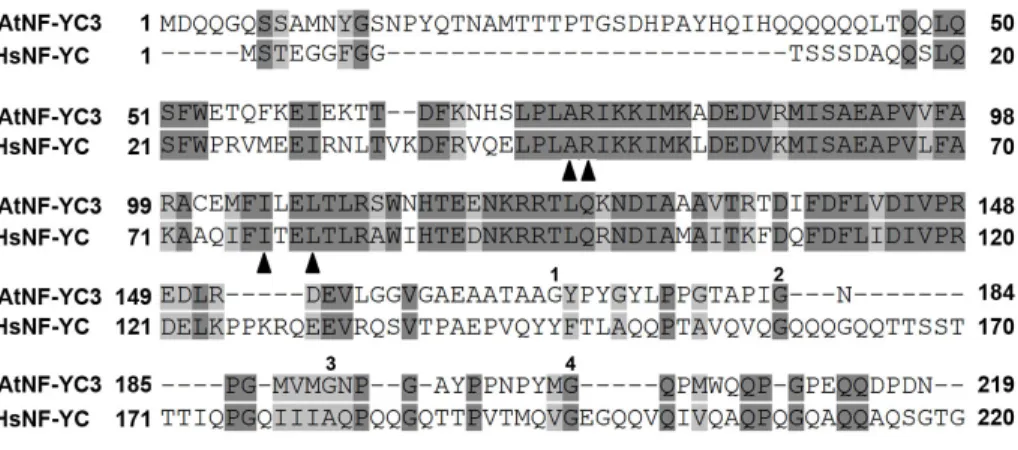
(R), lysine (K), and tyrosine (Y)) (van Verk et al., 2009). Another transcription factor found in Arabidopsis is the heterotrimeric Nuclear Factor Y (NF-Y), also referred to as the heme-activated protein (HAP) or CCAAT binding factor (CBF). This transcription factor is found in all eukaryotes and regulates a diverse set of genes. In most organisms, each of the three unique NF-Y subunits (NF-YA, NF-YB, and NF-YC) is encoded by one or two genes (Riechmann et al., 2000). However, in Arabidopsis there are 10 NF-YA, 13 NF-YB, and 13 NF-YC subunits (Siefers et al., 2009b). Brachypodium distachyon and Triticum aestivum also have 35 or more NF-Ys in each of their genomes (Cao et al.,
2011; Stephenson et al., 2007), indicating that there has been a generalized NF-Y expansion in the plant lineage.
35
cytosol, where they initially form a dimer (Frontini et al., 2004; Goda et al., 2005; Tuncher et al., 2005). NF-YB/C heterodimerization is required for translocation into the nucleus; once there the heterodimer binds the third subunit of the NF-Y family (NF-YA). The mature NF-Y complex binds DNA at the nucleotide sequence CCAAT (the “CCAAT box”) (Ceribelli et al., 2008). The CCAAT box is a frequent and widespread promoter element, with functional sites minimally occurring in ~7-8% of mammalian promoters (FitzGerald et al., 2004; Testa et al., 2005). There is no accurate estimate for the number of functional CCAAT sites in plants, but Arabidopsis promoters have a higher frequency of this simple pentamer sequence than what is found in humans (Siefers et al., 2009a). NF-Y transcription factors are able to both up- and down-regulate the transcription of CCAAT box containing genes (Mantovani, 1999).
36
transcriptional activation, it follows that transcription factors such as NF-Ys must themselves be under some form of control.
Localization can regulate transcription factor activity (Whiteside and Goodbourn, 1993); cytoplasmic retention prevents transcription factors from entering the nucleus, thereby thwarting transcriptional activation. In some cases, transcription factors are retained in the cytosol until an appropriate signal causes them to move into the nucleus (Whiteside and Goodbourn, 1993). Such retention can result from the binding of transcription factors to cytosolic proteins that function as interaction modules. One known group of cytosolic interaction modules are zinc finger proteins (Krishna et al., 2003). These molecules use zinc ions to stabilize their protein folds and can bind DNA, RNA and small proteins (Krishna et al., 2003). In Arabidopsis, one such cytosolic zinc finger protein is LSD1, a proposed interaction module and a negative regulator of cell death (Dietrich et al., 1997).
lsd1 mutant plants exhibit inappropriately activated and uncontrolled cell death
37
module, LSD1 is known to interact with both transcription factors (Kaminaka et al., 2006) and positive regulators of cell death (Coll et al., 2010; Epple et al., 2003). These interactions take place in the cytosol. The transcription factor bZIP10 is a positive mediator of rcd. LSD1 functions to sequester bZIP10 in the cytosol, thereby preventing its function in transcription of a pro-cell death regulon (Kaminaka et al., 2006). LOL1 and AtMC1, two proteins with LSD1-like zinc-finger motifs, also interact with LSD1 (Coll et al., 2010; Epple et al., 2003). These proteins are also positive regulators of cell death, and in the absence of LSD1 each protein is required for rcd. Taken together, these data indicate that LSD1 may act as a cytoplasmic scaffolding protein, sequestering proteins necessary to appropriately balance cell death and defense responses. As such, other proteins which interact with LSD1 could be important for rcd and/or disease resistance.
NF-38
YC3 could be partially controlled by LSD1 sequestering it in the cytosol, thereby preventing NF-YC3 movement into the nucleus and its subsequent disease resistance function.
Results
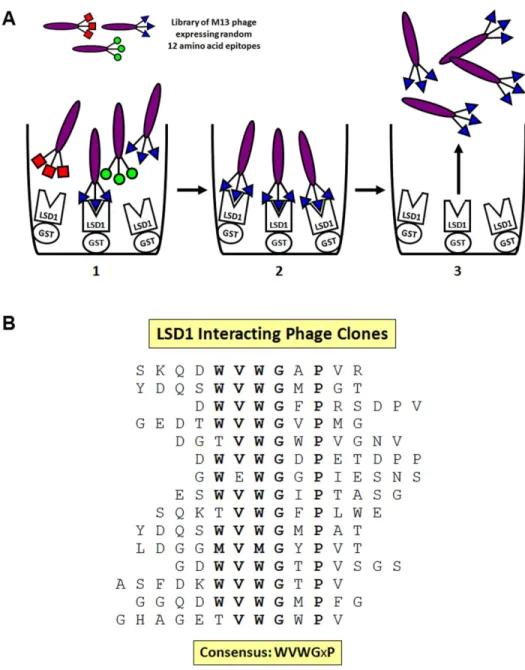
LSD1 interacts with the transcription factor NF-YC3. LSD1 is necessary for proper regulation of defense responses and interacts with proteins important in disease resistance (Coll et al., 2010; Dietrich et al., 1994; Kaminaka et al., 2006). To identify additional LSD1-interacting peptides, we performed a phage display using a library of random 12aa epitopes (Kay et al., 1996). GST:LSD1 fusion proteins were purified on glutathione sepharose beads and incubated with the phage library. Phage that bound to LSD1 were isolated and independent phage plaques were sequenced, yielding fifteen unique LSD1-interacting peptides (Figure 2.1A). The consensus sequence WVWGxP was found in 11 of the sequenced epitopes, and the G and P positions were invariant in all 15 LSD1 interacting peptides (Figure 2.1B). One of the sequenced variants was a near exact match to a peptide in NF-YC3, which had previously been isolated as an LSD1 interacting protein in Y2H assays. Arabidopsis NF-YC3 has homology to mammalian NF-YC, including the residues required for proper NF-Y formation (Figure 2.2).
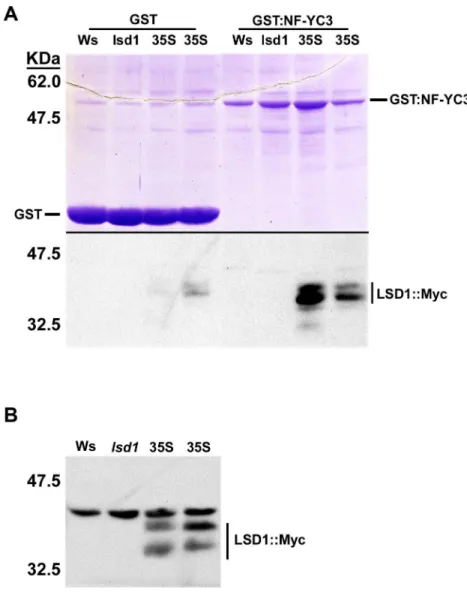
To confirm the interaction between LSD1 and NF-YC3, we used a combination of in vitro and semi-in vivo methods. We first confirmed the Y2H interaction between LSD1
39
(Figure 2.4). Purified GST-NF-YC3 was incubated with total protein extracts from Arabidopsis expressing LSD1-Myc under control of the 35S promoter. Excess protein was washed off and proteins bound to GST-NF-YC3 were eluted and separated on an SDS-PAGE gel. GST-NF-YC3 pulled down myc-tagged LSD1 protein, whereas a GST control did not (Figure 2.4A). Protein blots of input proteins showed that these two bands were specific to LSD1-Myc (Figure 2.4B).
NF-YC3 localization is dependent on GxP-mediated LSD1 interaction. As an additional test of whether LSD1 interacts with NF-YC3, we utilized the plant-specific GxP motif found in the phage display. This sequence was found in all phage display clones that bound LSD1 (Figure 2.1B), leading us to hypothesize that it would be necessary for the interaction between LSD1 and NF-YC3. There are 4 sequential GxP motifs in a Q-rich region at the C-terminus of NF-YC3. A truncation containing only this region retained interaction with LSD1 in Y2H assays (Figure 2.3A). Further, using a series of truncation mutations and a point mutation in the second GxP motif (GP2, labeled in the Figure 2.2 alignment), we found that this motif is necessary and likely sufficient for the interaction with LSD1. We note that this particular GxP motif is in a region divergent from human NF-YC.
(p35S:NF-40
YC3∆GP2-GFP, expressing a G182A/P184A mutation). Interestingly, NF-YC3∆
GP2-GFP was only present in the nucleus (Figure 2.3B, right panel), indicating that the GxP motif was required for accumulation in the cytosol. As LSD1 is a known cytosolic protein and previous studies have shown that it works to sequester other transcription factors out of the nucleus (Kaminaka et al., 2006), these results are consistent with the suggestion that LSD1 could retain NF-YC3 in the cytosol.
If LSD1 interacts with NF-YC3, there must be direct interaction between these two proteins in plant cells. To test this hypothesis, we used a bimolecular fluorescence complementation (BiFC) assay to check for direct interaction between the two proteins, albeit under conditions of transient over-expression. LSD1 fused to N-terminal YFP (YFPN-LSD1) and empty vector C-terminal fragments of YFP (YFPC) did not produce YFP fluorescence (Figure 2.3C, top). However, strong YFP fluorescence was observed in protoplasts expressing both YFPN-LSD1 and YFPC-NF-YC3, indicating that these two proteins are interacting (Figure 2.3C, middle). When the GP2 mutant construct YFPC -NF-YC3∆GP2 was expressed in the same cells as YFPN-LSD1, there was no fluorescent signal (Figure 2.3C, bottom), further indicating that the GxP motif is necessary for interaction between LSD1 and NF-YC3.
41
not be as effectively retained in the cytoplasm. Five week old Col-0 (wild-type) and lsd1-2 plants were sprayed with benzothiadiazole (BTH), a synthetic SA functional analog that
induces rcd in lsd1 (Lawton et al., 1996), and leaf tissue was collected at regular intervals. Protein blots with an NF-YC3-specific antibody (Kumimoto et al., 2010b) demonstrated that NF-YC3 was detectable in the nuclear-enriched fraction of both wild-type and lsd1-2 plants, and that the amount of protein increased after BTH activation (Figure 2.3D). However, the lsd1-2 plants showed an overall increased level of nuclear-localized YC3 compared to Col-0, indicating that LSD1 can function to keep NF-YC3 in the cytosol. The lack of hyper-accumulation of NF-NF-YC3 in the nucleus of non-induced (0 time) lsd1-2 plants suggests that there are likely additional factors besides LSD1 involved in the cytoplasmic retention of NF-YC3.
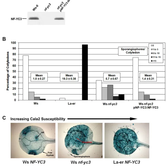
NF-YC3 is a positive regulator of disease resistance. LSD1 interactors can regulate pathogen responses (Coll et al., 2010; Kaminaka et al., 2006); therefore, NF-YC3 may also play a role in disease resistance. To test this hypothesis, we looked at the effects of NF-YC3 on disease resistance using the obligate biotrophic oomycete Hpa. We used the Hpa isolate Cala2, which is virulent on the Arabidopsis La-er ecotype (Holub et al., 1995). On the Ws ecotype, relatively weak resistance to Hpa Cala2 is conferred by RPP1A (Botella et al., 1998). We isolated nf-yc3 homozygous mutant plants in the Ws