THE QUALITY OF FIXED PROSTHODONTIC IMPRESSIONS: AN ASSESSMENT OF CROWN AND BRIDGE IMPRESSIONS RECEIVED AT COMMERCIAL
LABORATORIES
Clayton T. Rau
A thesis submitted to the Faculty of The University of North Carolina at Chapel Hill in partial fulfillment of the requirements for the degree of Master on Science in the Department of
Operative Dentistry of the School of Dentistry
Chapel Hill 2015
ii
The views expressed in this presentation are those of the author and do not necessarily reflect the official policy or position of the Department of the Navy, Department of Defense, nor the U.S.
Government. I am a military service member. This work was prepared as part of my official duties. Title 17, USC, §105 provides that ‘Copyright protection under this title is not available for any work of the U.S. Government.’ Title 17, USC, §101 defines a U.S. Government work as
a work prepared by a military service member or employee of the U.S. Government as part of that person’s official duties.
iii
ABSTRACT
Clayton T. Rau: The Quality of Fixed Prosthodontic Impressions: An Assessment of Crown and Bridge Impressions Received at Commercial Laboratories
(Under the direction of Terence E. Donovan)
Purpose: The objective of this study was two-fold. First, to evaluate and quantify clinically detectable errors commonly seen in impressions sent to commercial laboratories. Second, to determine if impressions from students at the University of North Carolina school of Dentistry are comparable to those made by private practitioners. Materials and Methods: Three large dental laboratories and one small dental laboratory were visited over a 12 month period. Impressions were evaluated by one of three calibrated examiners. All impressions were
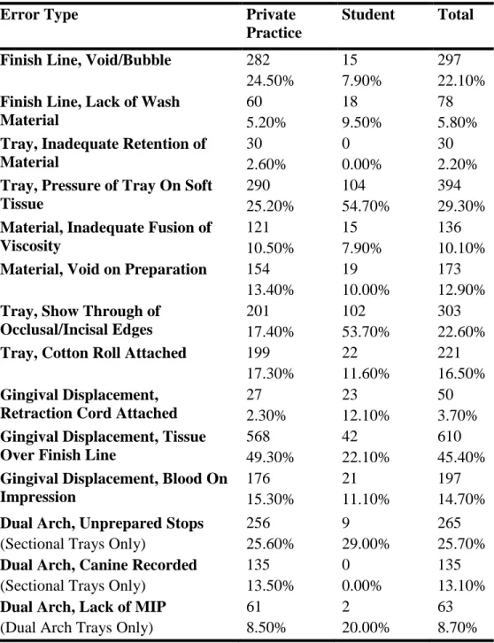
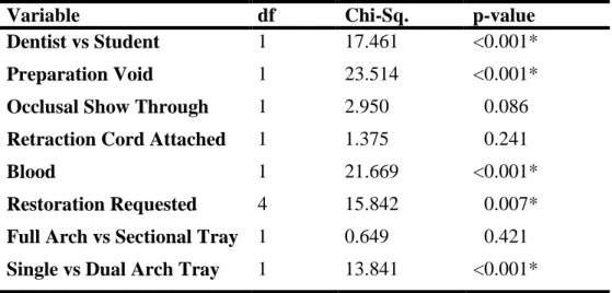
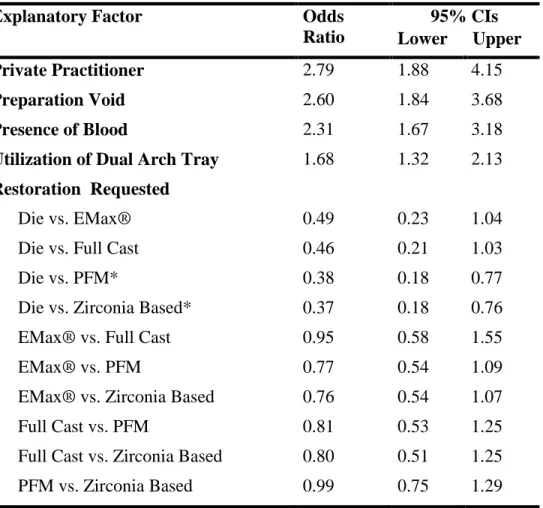
evaluated for errors using 2.5x magnification loupes under ambient room lighting without the aid of additional illumination. Result: A total of 1,347 impressions were evaluated. The largest error category evaluated, with a rate of 49.3%, was tissue impinging on the finish line. Multiple logistic regression analysis for factors influencing finish line error was statistically significant for the following variables: provider type (OR 1.68, p<0.001), blood (OR 2.31, p<0.001), tray type (OR 1.68, p<0.001) and restoration requested (p=0.007). Conclusion: Within the limitations of this study, marginal discrepancies made up the largest category of error noted in impressions evaluated. Impressions made by private practice dentists were significantly worse than those made by students. Simplified impression techniques such as dual arch impression trays increase the risk of obtaining critical errors. Although students made the same errors as private
iv
To my fiancé Meridith Pumphrey, it is your understanding, support and love that kept me going throughout this project.
To my brother and parents, thank you for your guidance over the years and supporting me in life’s difficult times.
To my mentor Terry Donovan, you are an inspiration to all who practice dentistry and a voice of evidence based reason in an opinionated world. Your contributions to your previous, current, and
v
ACKNOWLEDGEMENTS
I would like to express my sincere appreciation to my mentor Dr. Terry Donovan and the committee members, Dr. Lee Boushell, Dr. Alex Delgado and Dr. André V. Ritter, for their invaluable guidance, patience, time and effort. I would also like to thank Dr. Ceib Phillips and her staff for their extensive help in the data analysis.
Special thanks to Dr. Ed Swift, Dr. Harold Heymann, Dr. John Sturdevant, Dr. Andrea Zandona, Dr. Ken May, Dr. Rick Walter and Dr. Scott Eidson. It has been an honor and life changing experience to be part of the Operative Dentistry Family.
I thank the staff of the Department of Operative Dentistry, Mrs. Shannon Tate, Mrs. Dayna McNaught, Mrs. Barbara Walton, Mrs. Cynthia Lambert and Mrs. Rosanna Arrington, for all your devotion and commitment.
Also to my co-residents that made this quite an experience, Fernando Astorga, Kristy Erickson, Hiroko Nagaoka, Sumitha Ahmed, Silvia Amaya-Pajares, Upoma Guha, Vilhelm Olafsson, Mohammad Atieh, Taiseer Sulaiman, Edward Epure, Caroline Nguyen, and Leslie Trippe.
vi
TABLE OF CONTENTS
LIST OF TABLES ... viii
LIST OF FIGURES ... ix
LIST OF ABREVIATIONS ...x
1. CHAPTER 1: LITERATURE REVIEW ...1
1.1 A History Of Impression Quality ...2
1.2 Impression Materials ...4
1.2.1 General Properties ...4
1.2.2 Effects Of Moisture...6
1.2.3 Interactions With Other Materials ...9
1.3 Impression Technique ...11
1.3.1 Tray Flexure ...11
1.3.2 Custom Trays ...13
1.3.3 Dual Arch Trays ...14
1.4 Margin Design and Placement ...16
1.4.1 Subgingival Margins ...17
1.4.2 Biologic Width ...18
1.4.3 Bacterial Accumulation ...21
1.5 Gingival Displacement ...23
1.5.1 Gingival Retraction Cords and Medicaments ...24
vii
1.5.3 Alternative Methods...28
1.6 Conclusion ...29
REFERENCES ...30
2. CHAPTER 2: The Quality of Fixed Prosthodontic Impressions: An Assessment of Crown and Bridge Impressions Received at Commercial Laboratories ...41
2.1 Introduction...41
2.2 Material and Methods ...43
2.3 Statistical Analysis...45
2.4 Results ...45
2.4.1 Descriptive Statistics ...45
2.4.2 Factors Effecting Finish Line Errors...46
2.5 Discussion ...47
2.6 Limitations ...52
2.7 Conclusions ...52
3. TABLES ...54
4. FIGURES ...60 42
viii
LIST OF TABLES
Table 1. Unacceptable Criteria Description and Examples ...54 Table 2. Frequency of Observed Errors, Private Practice
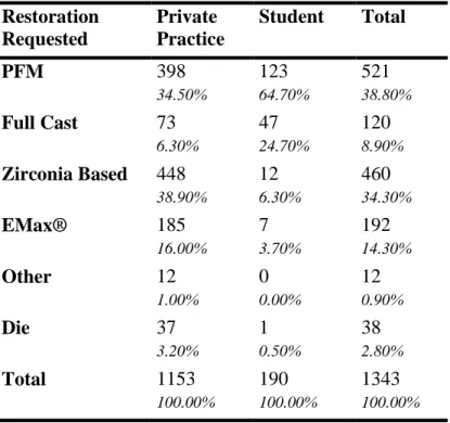
and Student Breakouts ...55 Table 3. Frequency of Restorations Requested, Private
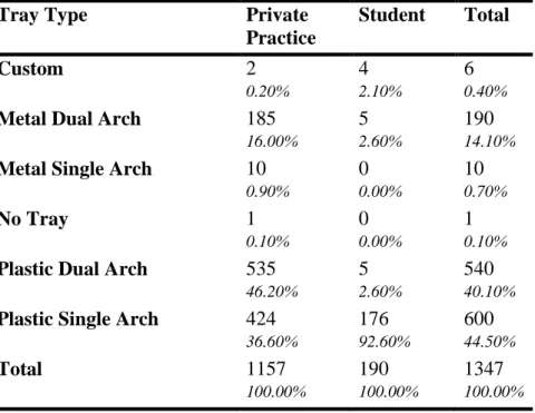
Practice and Student Breakout ...56 Table 4. Frequency of Type of Impression Tray Utilized,
Private Practice and Student Breakout ...57 Table 5. Factors Leading to Critical Error, Multiple
Logistic Regression Data ...58 Table 6. Odds Ratios for Significant Variables from
ix
LIST OF FIGURES
Figure 1. Impression Evaluation Form ...60
Figure 2. Finish Line, Void/Bubble ...61
Figure 3. Finish Line, Lack of Wash Material ...62
Figure 4. Tray, Inadequate Retention ...63
Figure 5. Tray, Pressure of Tray on Soft Tissue ...64
Figure 6. Tray, Show Through of Occlusal/Incisal Edges ...65
Figure 7. Material, Inadequate Fusion of Viscosity ...66
Figure 8. Material, Void on Preparation ...67
Figure 9. Material, Lack of Polymerization ...68
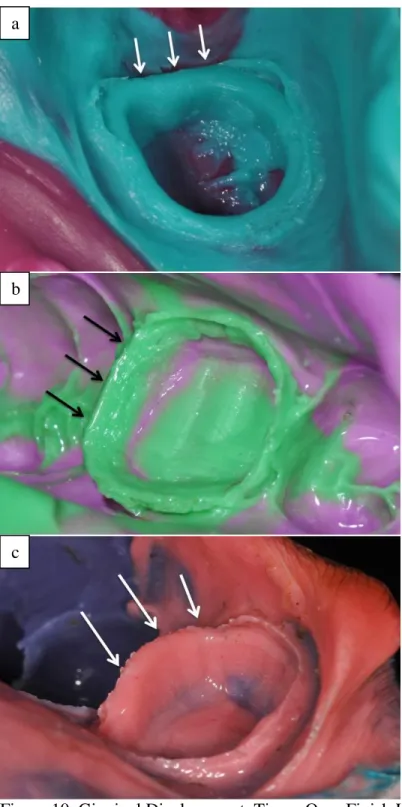
Figure 10. Gingival Displacement, Tissue Over Finish Line ...69
Figure 11. Gingival Displacement, Blood on Impression...70
Figure 12. Dual Arch, Lack of MIP ...71
Figure 13. Dual Arch, Unprepared Stops...72
x
LIST OF ABREVIATION
PE Polyether
1
CHAPTER 1: LITERATURE REVIEW
The transfer of accurate records to the dental laboratory is an important part of prosthesis fabrication in fixed prosthodontics. Obtaining an optimal impression for fixed dental prostheses is still one of the most challenging procedures in dentistry.1,2 While there are many steps that must be taken to fabricate an indirect restoration where an error can occur, the technician can only be expected to produce a quality restoration if the impression itself is of adequate quality. All dentists must possess the ability and willingness to analyze the quality of impressions, as this will ultimately determine success or failure of the restoration.3
2
1.1. A History Of Impression Quality
Published in vivo studies show a disturbing history in the quality of impressions. This may be a reason the dentist/laboratory relationships tend to be less than ideal and often are relatively short term.12 Aquilino and Taylor recognized there was a disconnect between what was happening in the institutional setting and private practice practitioners.10 Their 1984 study suggests that even though most schools require dental students to perform certain fixed prosthodontic laboratory procedures, educators were becoming concerned with the ability of recent graduates to perform these procedures with limited amounts exposure and experience. Concerns were also expressed at how quickly recent graduates abandoned the sound principles they were taught in school. Evaluation of the survey, which the study was focused upon, reveals that dental laboratory technicians were also concerned about this trend as well.
Winstanely et al.8 later performed a survey of 4 commercial dental laboratories in England to assess the quality of impressions received for crowns and bridges. This research study analyzed a total of 290 impressions and evaluated several factors including the type of tray utilized, presence of contamination, and adequacy of the impressions. A satisfactory restoration could be made for only 57% of the impressions, and making an adequate restoration was doubtful or impossible for 20% of the impressions. The single largest cause of defects in the working impressions was indistinct recording of the preparation margins. Irreversible hydrocolloid was the medium of choice in nearly all of the impressions evaluated at the time of the study.
3
being unusable, unsatisfactory, acceptable, or satisfactory. They found the quality of 50% of impressions and dies to be unsatisfactory or unusable. One interesting finding was the discrepancy in the source of the impression and the satisfactory ratings. Although compromising only 20 impressions in the study, those from providers employed at government or educational institutions were all rated as acceptable or satisfactory. When these impressions were removed from the database, the percentage of acceptable impressions was only 47% for non-institutionalized dentists. Sixty five percent of the impressions utilized irreversible hydrocolloid as the impression medium, but it is not known whether this had any bearing on the quality of the impressions reviewed.
In 2005 Samet et al.3 evaluated 193 impressions from 11 different laboratories in Israel. Evaluation criteria in this study were more sophisticated and not only evaluated preparation margins but also included several other variables such as pressure of the tray on the soft tissue, exposure of the heavy body material through the wash, and adhesive usage. Polyether and silicone materials were primarily used; irreversible hydrocolloid was not used for any impression. Eighty-nine percent of all impressions evaluated had at least one detectable error. The authors discussed that these findings were in agreement with previous studies8,10,13 despite the differences in criteria. This is mainly based on the fact that 51% of the defects involved the cervical finish line.
4
conducted. The impressions were made by “experienced dental clinicians” from the departments from which the patients were recruited. These clinicians followed a careful protocol in which retraction cord was used in all patients and left in place for 10 to 15 minutes. Strict attention to detail continued as the impressions were not made until all evidence of active bleeding had been stopped. Finally, all preparations were thoroughly dried prior to insertion of the impression material. This careful attention to detail was likely the reason for the low failure rate. Unfortunately, based on the findings of other research studies, similar levels of detail may not occur in most practices. This research study is an excellent example of how excellent soft tissue management and impression technique will lead to successful impressions.
The most recent evaluation of the quality of impressions took place in 2009 by Mitchell
et al.14 Although designed to evaluate the impression tray type and manner in which the trays
were used, the authors also noted the overall impression quality. The general overview of success rates for impressions varied widely from 44% to 83% depending on the type of tray and preparation location.
1.2. Impression Materials
Extensive impression material research and development has been accomplished in dentistry. Due largely to differences of material improvement, polysulfide rubber and reversible hydrocolloid are rarely used today in fixed prosthodontics. Most impressions are made with polyether (PE) and polyvinyl siloxane (PVS) based impression materials. Therefore the rest of this literature review will focus on the properties and limitations of PE and PVS materials.
1.2.1. General Properties
5
paste that is composed of long-chain polyether copolymer with alternating oxygen atoms, methylene groups, and reactive terminal groups. The ends of these macromolecular chains are converted into reactive rings, which transform into cross-linked final reaction products. The ether-dominated polymer backbone makes this group of materials the most hydrophilic of all elastomeric impression materials.15 PE material is fairly rigid upon completion of polymerization. Newer formulations of “soft” PE are less stiff and, as such, are easier to remove from the impressed teeth than earlier formulations. However, these materials still have an increased tendency to lock into undercuts not properly blocked out and have the potential to fracture delicate gypsum dies. 16
Although PEs are the most hydrophilic elastomeric material, they are only moderately hydrophilic and have limited ability to displace fluids in the process of impression making. Therefore the preparation unequivocally must be dry. Due to their hydrophilic nature, these materials also must be handled with strict criteria after setting. Kanehira et al17 have shown distortion of PE materials over time. Specifically, Impregum® has the ability to absorb water and deform at relative humidity levels about 50%. Because all PE materials deform when in constant contact with moisture from disinfection fluids, it is recommended that PE impressions should be rinsed, dried, and poured after 10 minutes of disinfectant contact time. 9,16,17
6
byproduct, which is then scavenged by the platinum in the catalyst. The consistency of the material is controlled by the amount of silica filler. More silica filler increases material viscosity and rigidity.9,15,16 Unlike PE, PVS materials are dimensionally stable over time and can be stored for several weeks without loss of accuracy.6 However, PVS materials are very moisture sensitive in their unset phase and detail loss from contact with blood or saliva can affect their accuracy.6,9,15,16
According to Chai et al.18 there are three mechanical properties that are clinically relevant when discussing impression materials. First, yield strength determines the ability of a material to withstand stress without permanent deformation. Second, strain at yield point indicates the amount of undercut that the impression material can overcome without permanent elastic deformation. Finally, tear energy indicates the resistance to tear of impression material. The ideal impression material is one which absorbs the most energy not to the point of tearing, but rather just prior to the point of critical, permanent deformation.19 No differences in the relative amount of distortion have been detected between PE and PVS when these materials are used with impression techniques that provide adequate bulk of material in the area of the preparation margins.20
1.2.2. Effects Of Moisture
7
their hydrophilic properties. PVS materials are principally hydrophobic because of their chemical structure. They contain hydrophobic aliphatic hydrocarbon groups around the siloxane bond.9,15,21,22 PE materials are more hydrophilic because they contain carbonyl (C=O) and ether (C-O-C) groups that chemically attract and interact with water via hydrogen bonding.9,22
Peutzfeldt and Asmussen22 evaluated how the properties of hydrophilicity and viscosity affected the ability of hydrophilic and hydrophobic impression materials to displace water and replicate surface characteristics that had been placed onto a ground dentin surface. The materials were injected in such a way that they had to flow across the test surface to displace the water from the periphery, trying to mimic clinical conditions. Their results split the materials into two distinct groups with a 70 degree contact angle as the dividing line between the groups. In the hydrophilic group, those which presented contact angles below 70 degrees, it was found that as materials increased in hydrophilicity, they performed better at displacing the water. The hydrophobic materials, with contact angles over 70 degrees, showed a propensity to displace water more readily with increases in viscosity. The authors stressed that when water was omitted from the dentin surface and testing was repeated, all PE and PVS materials tested achieved 100% reproduction of the groove pattern being evaluated.
8
Given this, all PE and PVS samples except one, a dual-phase PVS, would have produced acceptable detail to meet the current ISO standards for fine detail reproduction.
During the same year Johnson et al.23 published their findings, Petrie et al.24 published findings of a very similar study. Rather than evaluating dry and wet conditions only, they made an effort to create what dry, wet, and “moist” surfaces to test the detail reproduction of two PVS materials. Steel dies, similar to those used in ADA Specification 19, were utilized during the experiment. For making impressions in “moist” conditions, a fine mist of water was applied to the surface of the die just before the impression material was added to the surface. For wet conditions, the steel die was immersed in a water bath and the impression was made with the die and impression syringe under water. The research study revealed that, as the moisture level increased from dry to wet, the ability of the PVS materials to reproduce the surface details was significantly affected. This finding was similar to the Johnson et al study.23 It was also found that both PVS materials were able to reproduce the steel die without error in dry conditions. Two years later Walker et al.25 repeated the study with the addition of two PE materials. It was found that the PVS materials still were unable to reproduce the die details under moist conditions; however the PE materials were able to achieve complete reproduction of the surface under dry and moist conditions. The “wet” test condition with a submerged die was not repeated in the Walker study.
9
60 minutes of set time, only 2 of the 6 PVS materials became more hydrophilic than the PE.27 Therefore, the only added benefits to addition of surfactants to PVS impression materials relates to the process of pouring the impression and not in the impression making process itself.
It can be concluded that PEs are hydrophilic in that they will absorb some moisture in the process of impression making, but still require a relatively dry field. PVS materials, despite the addition of surfactants to make them perform in a more hydrophilic nature, still do not readily interact with moist surfaces. For the present time, it does not seem to matter if the PVS is termed “improved,” “hydrophilic,” or “smart wetting;” as none will compensate for poor control of moisture. Therefore, factors such as those described by Chai et al.,18 moisture control, and rheologic properties of the impression material have the most direct impact on the final quality of the impression.
1.2.3. Interactions With Other Materials
While PEs seem to be largely unaffected by any other materials, it has been shown that PVS materials can have interactions with many items commonly used during restorative procedures.6,7,9,16,29-31 Since PVS requires a small amount of catalyst to initiate the setting reaction, anything that interferes with the catalyst may prevent cross-linking and thereby cause the surface to remain tacky after the bulk of the material has set. Contamination is commonly a result of interaction with sulfur or sulfur containing compounds.9 This contamination may occur by either direct contact with the unset PVS materials or by indirect contact with compounds that remain adhered to the teeth and soft tissues.
10
method of inhibition is not clearly known; however, it is thought that the chloroplatinic acid catalyst reacts with unreacted sulfur in the latex products.32 While many believe that the interaction is not the same with latex-free, vinyl products,6,16 others warn that these products still have the potential to inhibit polymerization because of the sulfur containing stabilizers used in the manufacturing process.9 A recent research study demonstrated that two light body PVS materials can be inhibited by direct contact with several latex and latex-free products; however, no indirect contact inhibition was seen between any latex or latex-free product and the PVS materials tested.33
Metal salts, which are found in many hemostatic and retraction solutions, have been thought to inhibit the set of PVS. The result of such an interaction would result in lack of polymerization is the critical area of the preparation margin. The research study of de Camargo
et al.34 evaluated if such an interaction does exist. In the evaluation 3 latex samples, 5 retraction
cords and 4 medicaments were allowed to contact PVS during the setting reaction. Neither the retraction cord nor the medicaments inhibited the PVS setting reaction, as opposed to latex control samples. It was concluded that the medicaments and retractions cord tested are safe for use, and that the previous reports of polymerization inhibition were due to handling of the cords with latex gloves. A more recent study by Machado and Guedes35 identified that there was no inhibitory affect with any combination of gloves or hemostatic agents tested. This may be the result of improvements in materials to make them less reactive or non-reactive to excess sulfur in dental products.
11
set of oxygen.15 The oxygen inhibited layer has an inhibitory effect on the polymerization of PVS and will result in unset material around any preparation that has been immediately sealed or restored with freshly placed composite resins.9,36 To avoid this interaction, the inhibition layer must be removed by curing through a glycerin gel or DeOx (Ultradent, Utah, USA), preparation of the resin coating by fine diamond instrumentation at low speed, airborne particle abrasion, or flour of pumice.6,9,16,19,36
1.3. Impression Technique
The influence of tray selection on successful impression taking is often overlooked. Numerous factors must be taken into consideration when selecting the correct impression tray including size, shape, and rigidity. The importance of correct tray selection cannot be overstated as it can make the difference between success and failure. Gordon et al.37 stated that many dentists consistently use less expensive prefabricated plastic trays because of the time and cost associated with the fabrication of custom impression trays. However many dentists are not aware of the shortcomings related to the usage of these trays.
A trend in tray selection can be seen when looking at a history of research studies dating from 1980-2009.3,8,10,11,14,38 Since 1980, the usage of stock trays has increased from 75%38 to nearly 100%,11,14 and the use of quadrant trays has greatly increased from 35%38 to 88%.14 It is unknown if the use of these alternative trays results in a clinical outcome equivalent to traditional procedures?
1.3.1. Tray Flexure
12
potential to reduce its overall lifespan. The rigidity of commercially available disposable plastic trays, especially with higher viscosity materials, has been questioned. Many clinicians choose higher viscosity materials under the assumption that these will compensate for the added volume needed when using stock plastic trays and that more rigid materials will resist distortion. Cho and Chee39 determined that the mean cross arch and cross section change respectively for metal stock trays were 0.001 mm and 0.002 mm, for plastic trays these values were 0.099 mm and 0.120 mm. The difference was found to be statistically significant and the authors raised concerns that use of plastic impression trays with high viscosity materials may lead to marginal and occlusal discrepancies when seating final restorations.
Carrotte et al.40 evaluated several different tray systems and classified them as rigid, semi-rigid, or flexible mainly based on the approximate thickness of the plastic tray material and presence of a reinforcing rolled peripheral border. After each putty wash impression was made, a casting was fabricated and the marginal discrepancy was measured. Rigid, 3mm thick, plastic trays and metal controls were virtually identical with approximately 50 µm openings. In contrast with rigid trays, the marginal opening increased to 151 µm and 208 µm for the semi-rigid and flexible trays, respectively. Use of softer putty decreased these numbers to 90 µm and 178 µm, respectively, but all results were significantly poorer than with rigid plastic and metals trays were used. This data, like that of Cho and Chee,39 indicated that more rigid putty may actually be worse for the overall restoration than using a heavy body, syringeable material.
13
trays, they are more susceptible to error both in the clinical and laboratory setting and their routine usage should be questioned.14
1.3.2. Custom Trays
It is clear that some stock trays may not provide adequate rigidity and flex during the impression making process, but these trays also do not provide a uniform thickness of the impression materials.42 The varying thickness of the material is most commonly a cause of the stock tray not being correctly oriented, yielding inadequate bulk in some areas and too much bulk in others. While bulk may not have been relevant for hydrocolloid impression materials, with non-aqueous impression materials a uniform bulk of material of 2 mm is optimal.15,43,44 Eames et al.43 evaluated the amount of dimensional change that occurred with varying thicknesses of impression materials of a master die and found that not only was distortion increased as the material thickness increased, but over a 24 hour period the initial distortions were magnified. With few clinicians performing their own lab work, it may be that increased distortion resulting from increased delays are likely more representative of reality.
14
between stock and custom trays,37,46,47 many researchers and clinicians still recommend their routine usage. 2,6,7,18,30,40,43-45,48,49
1.3.3. Dual Arch Trays
Dual arch, or “closed bite”, impressions have been in use in dentistry since they were first mentioned by Wilson and Werrin in the early 1980’s.50
They are designed to simultaneously obtain an impression of the prepared teeth, the opposing dentition, and the intercuspal relationship while using less material than traditional full arch impressions.51 They are, however, not without limitations for use. The indications and requirements for their accurate utilization are as follows: 1) a maximum of two prepared teeth, 2) unprepared stops both anterior and posterior to the preparations, 3) stable, reproducible intercuspal position, 4) the patient must be able to close into maximum intercuspal position with the tray in place, 5) existing anterior guidance, 6) the canine must be recorded in the impression, 7) the tray must not impinge on any teeth or soft tissue, and 8) the provider must be familiar with the procedures being performed.6,7,52-54 Contraindications for the utilization of dual arch trays are 1) group function occlusal pattern, 2) unstable maximum intercuspal position, and 3) a planned alteration of the vertical dimension of occlusion.55 Some studies have shown that preparations with detailed intra-coronal preparation aspects, such as inlays, are also not reproduced well by the dual arch impression technique.56
15
important. Cox et al.60 and Wostmann56 both showed the dimensional accuracy of metal dual arch trays were superior to plastic varieties. Wostmann argues that it is not the primary stiffness of a tray but its tendency to reset after deformation that is the crucial factor that affects impression accuracy. Therefore, it is not the initial deformation, but the elastic recovery of the tray which causes the actual distortion of the preparation.56 To avoid the possible effects of soft tissue impingement and the risk of inducing tray distortion, many manufacturers have little or no sidewall on the impression trays. Several potential problems could arise from the lack of sidewall including creep of the material away from the preparation and pressure from the tongue thinning away or removing material from the preparation. Therefore, it is recommended that trays have sidewalls that extend to at least the gingival margin of the preparation.54
Johnson et al.63 in a study involving 116 dual arch impressions showed that 64% of impressions were successful in capturing all relevant aspects of the preparation. However, PVS produced significantly more successful impressions compared with PE, 70% and 58% respectively. de Lima et al.64 were also able to show greater distortion obtained when using PE material. The most common error in the Johnson et al.63 study pertained to the finish line and was attributed to inadequate gingival displacement. The success rate of this study is in general agreement with that of previous studies which evaluated full arch impressions by private practitioners.3,8,10,11,14
16
1.4. Margin Design and Placement
Clinicians must determine what type of margin is best for a given situation and the location of that margin. Although many factors such as material, esthetics, and access influence the selection, most dentists probably have a “preferred” design they feel comfortable preparing.65 A discussion in gingival margin design must include a clarification of terminology, as different sources often refer to a similarly termed margin as being prepared in several different ways. For simplification of terminology throughout the remainder of this section, finish line configurations will be discussed as described by Hunter and Hunter65,66 and Shillingburg.67
The chamfer is the preferred gingival margin design for restorations having a metal
margin. This variant of finish line has been shown to exhibit the least stress so the underlying cement will have less likelihood of failure. Preparation of this variety of finish line is most often accomplished with a round-end or torpedo shaped diamond bur. The final preparation should have the geometry of rounded internal angles with an approximately 90 degree cavosurface angle.65-67
Although they are more destructive to the remaining tooth structure than chamfer preparations, shoulder margins have been the finish line of choice for ceramic restorations. The margin geometry consists of an approximately 90 degree cavosurface margin with a wide, butt joint ledge to provide resistance to occlusal forces and allow bulk of porcelain to minimize the risk of fracture. Proper width of the butt joint is critical for restoration contour, strength, and esthetics. Previously a sharp, 90 degree internal angle was advocated, but it is now recommended that the pulpogingival line angle be rounded.65-68
Knife edge finish lines and otherwise beveled margins have been advocated to improve
17
Unless these margins are cut as designed, the axial reduction may “fade out” instead of terminating in a definite finish line. The thin marginal design is harder for the technician to read, wax, and cast compared to other varieties. The thinner area of restorative material is also more susceptible to distortion for metals or fracture for ceramics. Although intraoral finishing is advocated as a benefit to this margin design, it is often difficult, impractical, or simply not attempted. 65-67 Others point out the metals used in metal-ceramic restorations are not suitable for burnishing and attempting to do so may fracture the overlying porcelain.69
No matter what margin is chosen, the advantages of improved control of contours, esthetics, structural rigidity, ease of evaluating preparations, and clearer impressions allowed by wider margins must be considered.1,65 Overcontour of restorations, so as to provide adequate bulk of materials, often result in a compromised gingival health by impeding plaque removal.65,70 Donovan and Chee68 state that the following criteria for margin selection should be considered: 1) the selected margin must provide a predictable level of integrity, 2) to minimize plaque accumulation, the selected margin must present smooth materials to the gingival sulcus, and 3) in some situations, the margin also must provide acceptable esthetics.
1.4.1. Subgingival Margins
18
subgingivally include caries, extension over previously existing restorations, trauma, root sensitivity, severe non-carious cervical defects and esthetics.67,72,73
Placement of margins subgingivally for esthetics is a matter of debate. A classic study by Crispin and Watson74 evaluated the amount of gingival display among 425 subjects during normal and exaggerated smiles. What they found was that during normal smiling, the gingival margin remained hidden for 44% of canines, 34% of lateral incisors, and 50% of central incisors. For an exaggerated smile, these numbers decreased to 26% of canines, 16% of lateral incisors, and 24% of central incisors. Therefore, it must not automatically be assumed that the patient will show all of the anterior gingival margins when treatment planning for the type of restorations to be placed and margin location.
When subgingival margins are indicated there are numerous theories about where they should be placed. Historically, the location of the margin has been suggested to be 1) the base of the gingival crevice, 2) half the distance between the base of the gingival crevice and the gingival margin, 3) the crest of the gingival margin, and 4) supragingivally.70 Today, recommendations have become more limited to universally placing margins 0.5 mm apical to the free gingival margin, or, when situations dictate, sounding of the alveolar crest so as to make sure the biologic width is not violated.68,75,76 The latter recommendation comes from Kois76 who makes mention of the fact that the relationship of the margin location to the bone is more critical than the distance below the free gingival margin.
1.4.2. Biologic Width
The concept of biologic width was first described by Gargiulo et al.77 in 1961 when he measured the average length of the gingival attachment to the root, the junctional epithelium, and the sulcus depth in human cadavers. However, it wasn’t until Loe78
19
reaction of gingival tissues to restorative procedures that the profession started looking at the iatrogenic biological damage done to the periodontium when this area was violated.75 Generally it is thought that there is a need for 1mm of gingival attachment to the root, 1 mm of junctional epithelium and 1 mm of sulcus depth to maintain normal gingival and osseous health immediately adjacent to each tooth. Most consider the total biologic width to be approximately 2-3 mm but this depends on the reference point and if the depth is considered as originating from the depth of the sulcus or the free gingival margin. The average is not a true measure though, as junctional epithelium measurements vary widely in length.75,77 Sounding the osseous crest has been recommended as the most accurate determinant of how far subgingivally, if at all, margins can be placed without violating the biologic width, and still allow the space needed for a healthy gingival attachment, junctional epithelium and gingival.76
Numerous studies exist that show how subgingival margins can negatively affect the periodontium when the aspects of biologic width are not considered. Newcomb79 demonstrated increasing levels of inflammation in anterior teeth with direct correlation to the distance remaining between the crown margin and the base of the sulcus. Interestingly in this research study, the crown margins had lower plaque indexes compared to the control teeth. This is not saying that bacteria are not a potential cause of the inflammation, because they can exist in the niche between the restoration and tooth, but simply stating that the margin location can directly affect inflammation.
20
amount marginal discrepancy and measured pocket depths in the same locations. The authors’ stated that the current methods of judging subgingival margin discrepancies are inadequate, which is in agreement with Christensen81 who showed that providers generally do not detect subgingival margin discrepancies until they are greater than 120 µm. When looking at the Felton80 and Newcomb79 research studies together, it may be concluded that the margin location or the bacterial accumulation may increase gingival inflammation.
Richter and Ueno82 placed mandibular first molar crowns with each crown containing a facial margin which was located half supragingivally and half subgingivally. They were unable to show any difference in the gingival index scores for the areas that were supra- or sub-gingival regardless of plaque levels. The authors’ of the research study mentioned that meticulous detail was given to make sure margins were sealed and that the restorations were not over contoured. Ultimately Reeves83 summarized the findings on subgingival margin placement by stating that the degree of inflammation is influenced by a confluence of four factors: 1) failure to maintain proper emergence profile, 2) inability to adequately finish subgingival margins, 3) placement of the margin in an area with minimum to no attached gingiva, and 4) violation of biologic width. Although metal allergies and reactions to dental cements due occur, the inflammation resulting from subgingival margin is generally not a reaction to the materials themselves.
21
premolars.72,73 Inexperienced clinicians have a tendency to extend the tooth preparation to one circumferential depth, disregarding this disparity in height, and will likely violate the interproximal biologic width.68
1.4.3. Bacterial Accumulation
Perfect restoration marginal adaptation is nearly impossible, especially in the subgingival area. Placement of preparation margins subgingivally compromises the ability to record margins during the impression making process, which ultimately limits the accuracy of the marginal fit of the restoration. Compromised subgingival marginal fit will adversely affect the health of the periodontium. Beier et al.1 showed that increasing the depth of a margin subgingivally significant increased the risk of obtaining an unacceptable impression. Therefore, all subgingival margins present a potential point of bacterial colonization.84
Lang et al.,85 in a classic article, showed an increase in bleeding, inflammation, and an increase in bacterial flora in conjunction with overhangs of 1 mm. The predominant species of bacteria were noted as Gram-negative, anaerobic bacteria and black pigmented bacteria. It was also noted that the anaerobic:facultative ratio was increased, indicating a shift away from periodontally healthy bacteria. The most interesting outcome of the study was that when the overhang restoration was replaced with one having margins that restored normal anatomic contours, the bacteria in the subgingival area returned to that seen healthy periodontal tissues.
22
resulting in restoration margins short of the original preparation margins. Beier et al.1 showed in their study that, in addition to the subgingival depth having an effect, beveled margins significantly increased the risk of obtaining an unacceptable impression as compared with preparations having a definitive finish line. The clinical ramifications of these findings is that subgingival knife-edge/beveled marginal design may result in an areas of tooth structure that has been roughened during the process of preparation that not will not be covered with the final restoration. These roughened (damaged) areas thereby predispose the patient to constant, localized gingival inflammation problems.80
23
bacterial component in disease progression, are among the most common complications experienced with prosthodontic work that successfully restores normal anatomic contours. 92 Increased bacterial colonization that occurs secondary to the failure to properly prepare and record the knife-edge/beveled margin even further predisposes the patient to caries and periodontitis in the area.
Subgingival margins are an often wanted and sometimes a needed part of clinical dentistry. Great care must be taken in placing and properly recording these margins to ensure long term health of the restoration and periodontium. When done correctly, ill effects will result; however, when poorly done, even patients with excellent home care and receiving regular, professional preventive care, will experience perpetual problems.93-97
1.5. Gingival Displacement
According to The Glossary of Prosthodontic Terms98 gingival displacement is defined as “the deflection of the marginal gingiva away from the tooth.” The basic criteria for what would be considered acceptable gingival displacement material have been defined by Nemetz et al.99 as: 1) the creation of sufficient lateral and vertical space between the finish line and gingival tissues to allow the preparation margin to be recorded in an impression medium, 2) provide absolute control of gingival fluid seepage and hemorrhage, 3) no significant, irreversible soft or hard tissue damage resulting from the procedure, and 4) not produce any potentially dangerous side effects. To accomplish this task, the provider may choose a technique classified as mechanical, chemical, surgical, or utilize a combination of the methods.4,99,100
24
procedures. They recommend twice daily rinses 2 weeks prior to crown preparation, during the provisionalization period, and 2 weeks after final prosthesis delivery. Compared to the study’s control group, which required remaking 6 of 15 (40%) of impressions due to inadequate quality, none of the experimental CHX treatment patients required a remake. Additionally, the plaque and gingival index levels in the experimental CHX group were significantly better than the control group.
1.5.1. Gingival Retraction Cords and Medicaments
A variety of different gingival retraction cords have been advocated over the years. Currently braided and knitted retraction cords have become the primary ones reported as being used by clinicians.4,38,104,105 Braided retraction cords are characterized by a consistently tight weave pattern which makes them resistant to separation or fray during placement. This feature makes them easy to manipulate with smooth or serrated edge packing instruments.106 Knitted retraction cords require the use of non-serrated instruments to prevent fraying or dislodgement of the cord, but have been shown to increase in size after placement in the sulcus leading to an increase in their popularity.101 There is an overall lack of standardization in cord size and efficacy between manufacturers and no scientific evidence to suggest a difference in performance; therefore the selection of cord type is mainly a matter of handling characteristics preferred by the provider.4,100
25
polymerization PVS or PE materials.6,34,35 A trend in decreased usage of epinephrine as a medicament has been noted over the years.38,104,105,107,108 Epinephrine has been linked to adverse clinical side effect such as anxiety, tachycardia, and increased respiratory rate.38,102,107,109,110 In a recent survey of prosthodontists, one third reported that their patients experienced symptoms consistent with reaction to epinephrine.105 The severity of these adverse reactions seems to be increased when the epithelial lining of the sulcus is damaged. Researchers have noted spikes in blood levels of epinephrine upon initial placement and removal of retraction cords containing epinephrine.108 There is clinical data to suggest no difference in the gingival displacement efficacy between safer materials, like aluminum chloride, and epinephrine.103,111 Given the evidence at hand, the routine use of epinephrine in conjunction with gingival displacement procedures is not recommended.
1.5.2. Classical Displacement Methods
The most traditional form of gingival displacement taught in dental institutions is the chemicomechanical as described by Schillingburg67 in his text “Fundamentals of Fixed
Prosthodontics.” By convention this involves the utilization of 1 or 2 cords placed in the gingival
sulcus with the addition of a hemostatic medicament. These are known as the single- or
double-cord techniques and they are the method utilized by 98% of prosthodontists.105 No clinical study
has demonstrated superiority of one technique over another as long as the techniques were used correctly for the given clinical situation.100
26
used and is removed just prior to impression making. Christensen5 believes this method to be the most often used, and one of the least successful methods, owing to the frequent presence of blood and lack of proper management. Many providers choose to leave the single cord in place during impression making. If the clinician’s choice is to leave the single cord in place, secondary evaluation of the margins is required to ensure proper exposure.
In the double cord technique an initial, smaller cord is placed into the sulcus prior to placement of the second, larger diameter cord. This technique is indicated for all situations, but is specifically recommended for situations which involve multiple abutment teeth, compromised soft tissue health, margins that extend beyond 1 mm subgingivally and when a single cord does not provide sufficient lateral tissue displacement.4,99,100 Many dentists and laboratories consider this technique to be the standard by which all other gingival retraction methods should be compared and is the method of choice for 43% of prosthodontists surveyed.5,102 Just prior to making the impression the second, larger diameter cord is removed leaving the smaller cord in place. The advantage of the small cord remaining in place is that it can absorb moisture from the gingival crevice, reduce the collapse of tissues against the preparation and provide continued control hemorrhage via the presence of medicaments in the cord..4,99
27
than monophase impression techniques. Some researchers advocate that if a sulcus width of 0.2 mm is required, then the clinician should aim to open the crevice by at least 0.3-0.4 mm.7
In order to obtain the critical sulcular width of 0.2 mm that will remain present for up to 20 seconds after cord removal, Baharav et al.114 showed the retraction cord needs to be left in place for a minimum of 4 minutes. In this same research study a significant difference was not seen when leaving the cords in beyond 4 minutes, however the sulcular width remained above the 0.2 mm width for nearly twice as long in cords left in place for 8 minutes. Laufer et al.115 showed at the transitional line angle the sulcus was closed an average of 35% at 20 seconds and 53% at 40 seconds after cord removal when using the double cord technique for 6 minutes,. In the area of the mid-buccal sulcus the closure was measured at 11% and 19% for these same time periods.
28
reactions to retraction cord, noted the cord was often packed into the supra-alveolar connective tissue attachment, indicating excessive pressure when placing cords.
1.5.3. Alternative Methods
Although retraction cord is the most utilized method of gingival displacement, the discussion would not be complete with mentioning the alternative treatments currently available. Historically, electrosurgery has been used to reduce hyperplastic tissue, expose gingival margins and control hemorrhage.. According to the review by Baba et al.4 electrosurgery accomplishes gingival retraction by the removal of several layers of cells to expose the gingival margin and, when used appropriately, has no adverse effects on would healing. Electrosurgery is strongly contraindicated in patients with pacemakers and/or implanted cardioverter defibrillators, and should be used with caution around metallic restorative materials and implants. Electrosurgery removes tissue and therefore could potentially have permanent effects on the soft tissue contours.5,7,119
Soft tissue lasers can be used as another alternative method with less inflammation, less hemorrhage, and faster, painless healing when compared to mechanochemical methods.119,120 However, the amount of time taken to complete the procedure with lasers has been reported to be much longer.5 Soft tissue lasers, similar to electrosurgery, also removes gingival tissue and therefore may potentially have permanent effects on the soft tissue contours.5,7,119
The most recent alternative to traditional methods are the so called “cordless” impression systems. These are meant to be injected into the gingival sulcus where they expand to apply lateral pressure on the tissue and provide hemostasis through incorporated medicaments. 4,100,121-124
29
anesthesia.4,125 Compared to mechanochemical methods there is no difference in measured crevicular fluid flow124 or achieved lateral gingival displacement,121 but there is a compromised ability of these materials to move vertically in the sulcus and displace deeper gingival margins.123,125 Comparatively, these materials produce less hemorrhaging than treated cords,122,124 however, the studies mentioning this difference removed the retraction cord in a dry state and may have induced bleeding that would not have occurred if the cord was wet.117
1.6. Conclusion
Achieving excellent results with an indirect restoration absolutely depends on obtaining an accurate representation of the preparation and soft tissue. Modern impression materials, when used properly, are very accurate. Correct impression technique includes proper tray selection, adequate moisture control, effective gingival displacement, and appropriate choice of impression material. Each clinical situation will require changes in one or multiple factors in order to ensure successful transfer of preparation specifics to the dental laboratory technician. One broad method will not work for all situations.
30
REFERENCES
1. Beier US, Grunert I, Kulmer S, Dumfahrt H. Quality of impressions using hydrophilic polyvinyl siloxane in a clinical study of 249 patients. Int J Prosthodont 2007;20(3):270-4.
2. Christensen GJ. Have fixed-prosthodontic impressions become easier? J Am Dent Assoc 2003;134(8):1121-3.
3. Samet N, Shohat M, Livny A, Weiss EI. A clinical evaluation of fixed partial denture impressions. J Prosthet Dent 2005;94(2):112-7.
4. Baba NZ, Goodacre CJ, Jekki R, Won J. Gingival displacement for impression making in fixed prosthodontics: contemporary principles, materials, and techniques. Dent Clin North Am 2014;58(1):45-68.
5. Christensen GJ. Simplifying and improving soft-tissue management for fixed-prosthodontic impressions. J Am Dent Assoc 2013;144(2):198-200.
6. Donovan TE, Chee WW. A review of contemporary impression materials and techniques. Dent Clin North Am 2004;48(2):vi-vii, 445-70.
7. Stewardson DA. Trends in indirect dentistry: 5. Impression materials and techniques. Dent Update 2005;32(7):374-6, 79-80, 82-4 passim.
8. Winstanley RB, Carrotte PV, Johnson A. The quality of impressions for crowns and bridges received at commercial dental laboratories. Br Dent J 1997;183(6):209-13.
9. Rubel BS. Impression materials: a comparative review of impression materials most commonly used in restorative dentistry. Dent Clin North Am 2007;51(3):629-42, vi.
10. Aquilino SA, Taylor TD. Prosthodontic laboratory and curriculum survey. Part III: Fixed prosthodontic laboratory survey. J Prosthet Dent 1984;52(6):879-85.
31
12. Leeper SH. Dentist and laboratory: a "love-hate" relationship. Dent Clin North Am 1979;23(1):87-99.
13. Albashaireh ZS, Alnegrish AS. Assessing the quality of clinical procedures and technical standards of dental laboratories in fixed partial denture therapy. Int J Prosthodont 1999;12(3):236-41.
14. Mitchell ST, Ramp MH, Ramp LC, Liu PR. A preliminary survey of impression trays used in the fabrication of fixed indirect restorations. J Prosthodont 2009;18(7):582-8.
15. Anusavice KJ, Phillips RW, Shen C, Rawls HR. Phillips' science of dental materials. 12th ed. St. Louis, Mo.: Elsevier/Saunders; 2013.
16. Hamalian TA, Nasr E, Chidiac JJ. Impression materials in fixed prosthodontics: influence of choice on clinical procedure. J Prosthodont 2011;20(2):153-60.
17. Kanehira M, Finger WJ, Endo T. Volatilization of components from and water absorption of polyether impressions. J Dent 2006;34(2):134-8.
18. Chai J, Takahashi Y, Lautenschlager EP. Clinically relevant mechanical properties of elastomeric impression materials. Int J Prosthodont 1998;11(3):219-23.
19. Perakis N, Belser UC, Magne P. Final impressions: a review of material properties and description of a current technique. Int J Periodontics Restorative Dent 2004;24(2):109-17.
20. Laufer BZ, Baharav H, Ganor Y, Cardash HS. The effect of marginal thickness on the distortion of different impression materials. J Prosthet Dent 1996;76(5):466-71.
21. Giordano R, 2nd. Impression materials: basic properties. Gen Dent 2000;48(5):510-2, 14, 16.
22. Peutzfeldt A, Asmussen E. Impression materials: effect of hydrophilicity and viscosity on ability to displace water from dentin surfaces. Scand J Dent Res 1988;96(3):253-9.
32
24. Petrie CS, Walker MP, O'Mahony A M, Spencer P. Dimensional accuracy and surface detail reproduction of two hydrophilic vinyl polysiloxane impression materials tested under dry, moist, and wet conditions. J Prosthet Dent 2003;90(4):365-72.
25. Walker MP, Petrie CS, Haj-Ali R, et al. Moisture effect on polyether and polyvinylsiloxane dimensional accuracy and detail reproduction. J Prosthodont 2005;14(3):158-63.
26. Rupp F, Axmann D, Jacobi A, Groten M, Geis-Gerstorfer J. Hydrophilicity of elastomeric non-aqueous impression materials during setting. Dent Mater 2005;21(2):94-102.
27. Rupp F, Geis-Gerstorfer J. Hydrophilicity of unset and set elastomeric impression materials. Int J Prosthodont 2010;23(6):552-4.
28. Balkenhol M, Haunschild S, Lochnit G, Wostmann B. Surfactant release from hydrophilized vinylpolysiloxanes. J Dent Res 2009;88(7):668-72.
29. Baumann MA. The influence of dental gloves on the setting of impression materials. Br Dent J 1995;179(4):130-5.
30. Chee WW, Donovan TE. Polyvinyl siloxane impression materials: a review of properties and techniques. J Prosthet Dent 1992;68(5):728-32.
31. Kahn RL, Donovan TE, Chee WW. Interaction of gloves and rubber dam with a poly(vinyl siloxane) impression material: a screening test. Int J Prosthodont 1989;2(4):342-6.
32. Cook WD, Thomasz F. Rubber gloves and addition silicone materials. Current note no. 64. Aust Dent J 1986;31(2):140.
33. Amaya-Pajares S.P. Delgado A.J., and Donovan T.E. Inhibition of Polymerization of Contemporary Polyvinyl Siloxane Impression Materials by Latex-Free Products FORUM for Dental Student Research and Innovation 2014;2(2):12-19.
33
35. Machado CE, Guedes CG. Effects of sulfur-based hemostatic agents and gingival retraction cords handled with latex gloves on the polymerization of polyvinyl siloxane impression materials. J Appl Oral Sci 2011;19(6):628-33.
36. Magne P, Nielsen B. Interactions between impression materials and immediate dentin sealing. J Prosthet Dent 2009;102(5):298-305.
37. Gordon GE, Johnson GH, Drennon DG. The effect of tray selection on the accuracy of elastomeric impression materials. J Prosthet Dent 1990;63(1):12-5.
38. Shillingburg HT, Jr., Hatch RA, Keenan MP, Hemphill MW. Impression materials and techniques used for cast restorations in eight states. J Am Dent Assoc 1980;100(5):696-9.
39. Cho GC, Chee WW. Distortion of disposable plastic stock trays when used with putty vinyl polysiloxane impression materials. J Prosthet Dent 2004;92(4):354-8.
40. Carrotte PV, Johnson A, Winstanley RB. The influence of the impression tray on the accuracy of impressions for crown and bridge work--an investigation and review. Br Dent J 1998;185(11-12):580-5.
41. Larson TD, Nielsen MA, Brackett WW. The accuracy of dual-arch impressions: a pilot study. J Prosthet Dent 2002;87(6):625-7.
42. Bomberg TJ, Hatch RA, Hoffman W, Jr. Impression material thickness in stock and custom trays. J Prosthet Dent 1985;54(2):170-2.
43. Eames WB, Sieweke JC, Wallace SW, Rogers LB. Elastomeric impression materials: effect of bulk on accuracy. J Prosthet Dent 1979;41(3):304-7.
44. Millstein P, Maya A, Segura C. Determining the accuracy of stock and custom tray impression/casts. J Oral Rehabil 1998;25(8):645-8.
45. Christensen GJ. Now is the time to change to custom impression trays. J Am Dent Assoc 1994;125(5):619-20.
34
47. Rueda LJ, Sy-Munoz JT, Naylor WP, Goodacre CJ, Swartz ML. The effect of using custom or stock trays on the accuracy of gypsum casts. Int J Prosthodont 1996;9(4):367-73.
48. Johnson GH, Craig RG. Accuracy of addition silicones as a function of technique. J Prosthet Dent 1986;55(2):197-203.
49. Singh K, Sahoo S, Prasad KD, Goel M, Singh A. Effect of different impression techniques on the dimensional accuracy of impressions using various elastomeric impression materials: an in vitro study. J Contemp Dent Pract 2012;13(1):98-106.
50. Wilson EG, Werrin SR. Double arch impressions for simplified restorative dentistry. J Prosthet Dent 1983;49(2):198-202.
51. Lane DA, Randall RC, Lane NS, Wilson NH. A clinical trial to compare double-arch and complete-arch impression techniques in the provision of indirect restorations. J Prosthet Dent 2003;89(2):141-5.
52. Kaplowitz GJ. Trouble-shooting dual arch impressions. J Am Dent Assoc 1996;127(2):234-40.
53. Kaplowitz GJ. Trouble-shooting dual arch impressions II. J Am Dent Assoc 1997;128(9):1277-81.
54. Small BW. Revisiting impressions using dual-arch trays. Gen Dent 2012;60(5):379-81.
55. Parker MH, Cameron SM, Hughbanks JC, Reid DE. Comparison of occlusal contacts in maximum intercuspation for two impression techniques. J Prosthet Dent 1997;78(3):255-9.
56. Wostmann B, Rehmann P, Balkenhol M. Accuracy of impressions obtained with dual-arch trays. Int J Prosthodont 2009;22(2):158-60.
35
58. Ceyhan JA, Johnson GH, Lepe X, Phillips KM. A clinical study comparing the three-dimensional accuracy of a working die generated from two dual-arch trays and a complete-arch custom tray. J Prosthet Dent 2003;90(3):228-34.
59. Cox JR. A clinical study comparing marginal and occlusal accuracy of crowns fabricated from double-arch and complete-arch impressions. Aust Dent J 2005;50(2):90-4.
60. Cox JR, Brandt RL, Hughes HJ. A clinical pilot study of the dimensional accuracy of double-arch and complete-arch impressions. J Prosthet Dent 2002;87(5):510-5.
61. Kang AH, Johnson GH, Lepe X, Wataha JC. Accuracy of a reformulated fast-set vinyl polysiloxane impression material using dual-arch trays. J Prosthet Dent 2009;101(5):332-41.
62. Gates GN, Nicholls JI. Evaluation of mandibular arch width change. J Prosthet Dent 1981;46(4):385-92.
63. Johnson GH, Mancl LA, Schwedhelm ER, Verhoef DR, Lepe X. Clinical trial investigating success rates for polyether and vinyl polysiloxane impressions made with full-arch and dual-arch plastic trays. J Prosthet Dent 2010;103(1):13-22.
64. de Lima LM, Borges GA, Junior LH, Spohr AM. In vivo Study of the Accuracy of Dual-arch Impressions. J Int Oral Health 2014;6(3):50-5.
65. Hunter AJ, Hunter AR. Gingival crown margin configurations: a review and discussion. Part I: Terminology and widths. J Prosthet Dent 1990;64(5):548-52.
66. Hunter AJ, Hunter AR. Gingival margins for crowns: a review and discussion. Part II: Discrepancies and configurations. J Prosthet Dent 1990;64(6):636-42.
67. Shillingburg HT. Fundamentals of fixed prosthodontics. 3rd ed. Chicago: Quintessence Pub. Co.; 1997.
68. Donovan TE, Chee WW. Cervical margin design with contemporary esthetic restorations. Dent Clin North Am 2004;48(2):vi, 417-31.
36
70. Becker CM, Kaldahl WB. Current theories of crown contour, margin placement, and pontic design. J Prosthet Dent 1981;45(3):268-77.
71. Goldberg PV, Higginbottom FL, Wilson TG. Periodontal considerations in restorative and implant therapy. Periodontol 2000 2001;25:100-9.
72. Nevins M. Periodontal considerations in prosthodontic treatment. Curr Opin Periodontol 1993:151-6.
73. Nevins M, Skurow HM. The intracrevicular restorative margin, the biologic width, and the maintenance of the gingival margin. Int J Periodontics Restorative Dent 1984;4(3):30-49.
74. Crispin BJ, Watson JF. Margin placement of esthetic veneer crowns. Part I: Anterior tooth visibility. J Prosthet Dent 1981;45(3):278-82.
75. Block PL. Restorative margins and periodontal health: a new look at an old perspective. J Prosthet Dent 1987;57(6):683-9.
76. Kois JC. Altering gingival levels: The restorative connection Part I: Biologic variables. J Esthet Dent 1994;6:3-9.
77. Gargiulo AW, Wentz, F.M., and Orban, B. Dimensions and relations of the dentogingival junction in man. J Periodontol 1961;32:261-7.
78. Loe H. Reactions to marginal periodontal tissues to restorative procedures. Int Dent J 1968;18(4):759-78.
79. Newcomb GM. The relationship between the location of subgingival crown margins and gingival inflammation. J Periodontol 1974;45(3):151-4.
80. Felton DA, Kanoy BE, Bayne SC, Wirthman GP. Effect of in vivo crown margin discrepancies on periodontal health. J Prosthet Dent 1991;65(3):357-64.
81. Christensen GJ. Marginal fit of gold inlay castings. J Prosthet Dent 1966;16(2):297-305.
37
83. Reeves WG. Restorative margin placement and periodontal health. J Prosthet Dent 1991;66(6):733-6.
84. Sorensen JA. A rationale for comparison of plaque-retaining properties of crown systems. J Prosthet Dent 1989;62(3):264-9.
85. Lang NP, Kiel RA, Anderhalden K. Clinical and microbiological effects of subgingival restorations with overhanging or clinically perfect margins. J Clin Periodontol 1983;10(6):563-78.
86. Quirynen M, Bollen CM. The influence of surface roughness and surface-free energy on supra- and subgingival plaque formation in man. A review of the literature. J Clin Periodontol 1995;22(1):1-14.
87. Quirynen M, Marechal M, Busscher HJ, et al. The influence of surface free energy and surface roughness on early plaque formation. An in vivo study in man. J Clin Periodontol 1990;17(3):138-44.
88. Bollen CM, Lambrechts P, Quirynen M. Comparison of surface roughness of oral hard materials to the threshold surface roughness for bacterial plaque retention: a review of the literature. Dent Mater 1997;13(4):258-69.
89. Teughels W, Van Assche N, Sliepen I, Quirynen M. Effect of material characteristics and/or surface topography on biofilm development. Clin Oral Implants Res 2006;17 Suppl 2:68-81.
90. Al-Omari WM, Mitchell CA, Cunningham JL. Surface roughness and wettability of enamel and dentine surfaces prepared with different dental burs. J Oral Rehabil 2001;28(7):645-50.
91. Ayad MF, Rosenstiel SF, Hassan MM. Surface roughness of dentin after tooth preparation with different rotary instrumentation. J Prosthet Dent 1996;75(2):122-8.
92. Goodacre CJ, Bernal G, Rungcharassaeng K, Kan JY. Clinical complications in fixed prosthodontics. J Prosthet Dent 2003;90(1):31-41.
38
94. Sorensen JA, Doherty FM, Newman MG, Flemmig TF. Gingival enhancement in fixed prosthodontics. Part I: Clinical findings. J Prosthet Dent 1991;65(1):100-7.
95. Valderhaug J. Periodontal conditions and carious lesions following the insertion of fixed prostheses: a 10-year follow-up study. Int Dent J 1980;30(4):296-304.
96. Valderhaug J, Birkeland JM. Periodontal conditions in patients 5 years following insertion of fixed prostheses. Pocket depth and loss of attachment. J Oral Rehabil 1976;3(3):237-43.
97. Valderhaug J, Ellingsen JE, Jokstad A. Oral hygiene, periodontal conditions and carious lesions in patients treated with dental bridges. A 15-year clinical and radiographic follow-up study. J Clin Periodontol 1993;20(7):482-9.
98. The glossary of prosthodontic terms. J Prosthet Dent 2005;94(1):10-92.
99. Nemetz H, Donovan T, Landesman H. Exposing the gingival margin: a systematic approach for the control of hemorrhage. J Prosthet Dent 1984;51(5):647-51.
100. Donovan TE, Chee WW. Current concepts in gingival displacement. Dent Clin North Am 2004;48(2):vi, 433-44.
101. Morgano SM, Malone WF, Gregoire SE, Goldenberg BS. Tissue management with dental impression materials. Am J Dent 1989;2(5):279-84.
102. Benson BW, Bomberg TJ, Hatch RA, Hoffman W, Jr. Tissue displacement methods in fixed prosthodontics. J Prosthet Dent 1986;55(2):175-81.
103. Weir DJ, Williams BH. Clinical effectiveness of mechanical-chemical tissue displacement methods. J Prosthet Dent 1984;51(3):326-9.
104. Ahmed SaD, TE. A survey of dentists as to gingival displacement procedures used in their practice. J Prosthet Dent 2015;In Press.
39
106. Kumbuloglu O, User A, Toksavul S, Boyacioglu H. Clinical evaluation of different gingival retraction cords. Quintessence Int 2007;38(2):e92-8.
107. Donovan TE, Gandara BK, Nemetz H. Review and survey of medicaments used with gingival retraction cords. J Prosthet Dent 1985;53(4):525-31.
108. Shaw DH, Krejci RF, Todd GL, 3rd, Reinhardt RA. Determination of plasma catecholamines in dogs after experimental gingival retraction with epinephrine-impregnated cord. Arch Oral Biol 1987;32(3):217-9.
109. Bader JD, Bonito AJ, Shugars DA. A systematic review of cardiovascular effects of epinephrine on hypertensive dental patients. Oral Surg Oral Med Oral Pathol Oral Radiol Endod 2002;93(6):647-53.
110. Buchanan WT, Thayer KE. Systemic effects of epinephrine-impregnated retraction cord in fixed partial denture prosthodontics. J Am Dent Assoc 1982;104(4):482-4.
111. Jokstad A. Clinical trial of gingival retraction cords. J Prosthet Dent 1999;81(3):258-61.
112. Laufer BZ, Baharav H, Cardash HS. The linear accuracy of impressions and stone dies as affected by the thickness of the impression margin. Int J Prosthodont 1994;7(3):247-52.
113. Finger WJ, Kurokawa R, Takahashi H, Komatsu M. Sulcus reproduction with elastomeric impression materials: a new in vitro testing method. Dent Mater 2008;24(12):1655-60.
114. Baharav H, Laufer BZ, Langer Y, Cardash HS. The effect of displacement time on gingival crevice width. Int J Prosthodont 1997;10(3):248-53.
115. Laufer BZ, Baharav H, Langer Y, Cardash HS. The closure of the gingival crevice following gingival retraction for impression making. J Oral Rehabil 1997;24(9):629-35.
116. Csempesz F, Vag J, Fazekas A. In vitro kinetic study of absorbency of retraction cords. J Prosthet Dent 2003;89(1):45-9.