DIFFERENCES IN POSTURAL CONTROL RESPONSES TO LEVELS OF VISUAL OCCLUSION IN INDIVIDUALS WITH CHRONIC ANKLE INSTABILITY
Lillian H. VanDeMark
A thesis submitted to the faculty at the University of North Carolina at Chapel Hill in partial fulfillment of the requirements for the degree of Master of Arts in the Department of Exercise and Sport Science (Athletic Training) in the College of Arts &
Sciences
Chapel Hill 2019
Approved by:
Erik Wikstrom
Jason Mihalik
ii
©2019
iii ABSTRACT
Lillian H. VanDeMark: Differences in Postural Control Responses to Levels of Visual Occlusion in Individuals with Chronic Ankle Instability
(Under the direction of Erik Wikstrom)
Chronic ankle instability (CAI) is a condition characterized by recurrent ankle
sprains. Those with CAI are described as visually reliant due to postural control deficits
observed under fully occluded visual conditions. Little is known about the influence of
partially occluded vision on postural control in those with CAI. The purpose of this
study was to examine the effect of CAI on postural control under progressive levels of
visual occlusion and relate postural control under these conditions to visual function
and sensory integration in those with CAI.
Thirty-five participants with CAI and sixteen controls completed postural
control assessments under four visual conditions:1) eyes-open, 2) low-occlusion, 3)
high-occlusion, and 4) eyes-closed. Participants also completed visual performance
(Senaptec Sensory Station) and sensory integration (Sensory Organization Test)
assessments.
Both groups demonstrated worse postural control under limited-vision
conditions compared to eyes-open. Some measures of visual performance predicted
iv
TABLE OF CONTENTS
LIST OF TABLES ... vi
LIST OF ABBREVIATIONS ... vii
CHAPTER I: INTRODUCTION ... 1
Specific Aims ... 3
CHAPTER II: REVIEW OF LITERATURE ... 4
Chronic Ankle Instability ... 4
Vision ... 16
CHAPTER III: METHODS ... 21
Study Design ... 21
Participants ... 21
Visual Performance ... 22
Sensory Integration ... 22
Postural Control ... 24
Statistical Analysis ... 25
CHAPTER IV ... 36
Overview ... 36
v
Methods ... 40
Results... 42
Discussion ... 43
Conclusions ... 46
Conflict of Interest Statement ... 46
CHAPTER V ... 47
Overview ... 47
Introduction ... 49
Methods ... 51
Results... 56
Discussion ... 57
Conclusion ... 59
vi
LIST OF TABLES
Table 1 – Demographic information by group. Mean (SD)………....26 Table 2 – Average postural control outcomes for healthy (n=16)
and CAI (n=35) groups by visual condition. Mean (SD)……….27 Table 3 – Average time-to-boundary under various levels of
visual occlusion with effect size, collapsed across group. Mean (SD)….………..28 Table 4 – Senaptec Sensory System Test detailed
outcomes and procedures………..29
Table 5 – Average Senaptec Sensory System and
Sensory Organization Test outcomes. Mean (SD)……….………..31
Table 6 – Univariable analyses of Senaptec variables included
in the time-to-boundary anterior-to-posterior stepwise model………....32 Table 7 – Univariable analyses of Senaptec variables included in
the the time-to-boundary medial-to-lateral stepwise model…...…….………...33 Table 8 – Stepwise multivariable regression analysis for
time-to-boundary anterior-to-posterior outcomes under
both eyes-open and limited-vision conditions………..34 Table 9 – Stepwise multivariable regression analysis for
time-to-boundary medial-to-lateral outcomes under
vii
LIST OF ABBREVIATIONS
CAI Chronic Ankle Instability
DLS Double-Limb Support
COP Center of Pressure
SOT Sensory Organization Test
1
CHAPTER I: INTRODUCTION
Chronic ankle instability (CAI) is a costly and disabling condition characterized
by recurrent sprains, episodes of giving way, pain, and/or weakness that linger after an
injury to the ankle, typically a lateral ankle sprain.1,2 Ankle sprains are one of the most
common injuries treated by health care professionals, with an estimated 23,000 ankle
sprains occurring per day in the United States.3 Approximately 40% of these individuals
develop CAI as a result4 which may lead to further disability including post-traumatic
osteoarthritis in the ankle joint5,6 and decreases in physical activity7,8.
Individuals with CAI have sensorimotor adaptations that are a result of the
initial ligamentous injury9 and lead to deficits in postural control. These adaptations
include altered somatosensory input and neuromuscular response. A cascade of events
is thought to follow damaged mechanoreceptors upon initial injury leading to long
lasting altered cortical level somatosensory integration.9–11 First, the initial ligamentous
injury interrupts continuous somatosensory input from the joint regarding joint
position sense, joint stress, and joint velocity.9,11 Without this information, the motor
component of the sensorimotor system must adopt a new strategy of obtaining
sufficient information to maintain postural control via dynamic joint stability.9,11 It
appears that in the absence of sufficient somatosensory input, the motor control system
reweights the level of visual feedback used to maintain postural control.9 Over time, the
2
responses. A recent meta-analysis found that individuals with CAI have significantly
greater postural control declines during single-limb balance tasks under eyes-closed
conditions compared to eyes-open.12 This indicates that the sensorimotor system
cannot compensate for the removal of vision and therefore suggests a reliance on visual
information to maintain postural control.12 It is unclear whether long-term visual
reliance in CAI patients is a positive or negative adaptation. Reliance on information
from the visual system to maintain postural control—due to dysfunctional
somatosensory system integration—could leave the individual vulnerable to reinjury
when the visual system is overloaded with tasks, such as in sport, when one has to
identify and respond to multiple stationary and dynamic objects.11 However,
incorporating faulty somatosensory information to determine correct motor response
may result in an incorrect response and, predispose an individual to injury.11
Whether or not the adaptation of visual reliance is necessary or detrimental, the
topic is worth exploring in future research. Current balance training methods often
involve exercises in static posture with eyes-closed,13,14 but these do not alter visual
reliance in CAI15. Incorporating dynamic postural exercises with limited visual
information should be explored as a method of altering visual reliance. A study by Kim
et al.16 found interrupting visual information with stroboscopic goggles caused
significant postural instability during single-leg stance compared to eyes open
conditions in healthy subjects. However, the current literature surrounding the postural
response of the CAI population to interrupted visual information is limited. Therefore,
the overall purpose of this study is to examine the difference in postural control
3
controls. We will also relate postural control response under these conditions to visual
and somatosensory function, and the sensory integration strategies in those with CAI.
Specific Aims
We will approach this topic with the following aims:
Aim 1: To specify changes in postural control during balance conditions with varying
levels of visual occlusion in those with CAI and healthy controls.
Hypothesis 1: Both groups will demonstrate worse spatiotemporal postural control
outcomes (shorter time to boundary) during balance conditions with any limitation in
visual information (low occlusion, high occlusion, and eyes closed) compared to eyes
open with no occlusion. Furthermore, those with CAI will have significantly worse
postural control outcomes compared to healthy controls during all limited vision
conditions.
Aim 2: To test the influence of visual function and performance measures on changes in
postural control under varying levels of visual occlusion in those with CAI.
Hypothesis 2: Worse spatiotemporal postural control outcomes (shorter TTB) will be
associated with poor visual performance scores on the Senaptec Sensory Station
battery of vision and sensory performace tests, specifically the Perception Span,
Eye-Hand Coordination, Go/No-Go, .
Aim 3: To test the influence of sensory integration measures on changes in postural
control under varying levels of visual occlusion in those with CAI.
Hypothesis 3: Worse spatiotemporal postural control outcomes (shorter TTB) will be
4
score) and decreased ability to utilize visual input (higher VIS ratio score) on the
4
CHAPTER II: REVIEW OF LITERATURE
Chronic Ankle Instability
Epidemiology
Chronic ankle instability (CAI) is a debilitating condition of the ankle joint
characterized by recurrent sprains, episodes of “giving way”, pain, or weakness in the ankle.1,2,4 CAI arises from an acute sprain of the ankle. Acute ankle sprains are one of the
most commonly occurring injuries seen in active populations17,18 regardless of
competition level.1 It has been estimated that over 11, 000 ankle sprains occur in
collegiate athletics per year, accounting for 15% of all injuries.1 Out of all ankle sprains,
the most common type is the lateral ankle sprain, occurring due to overstretching of the
lateral ligaments during extreme inversion.1,18 The majority of individuals who
experience an acute ankle sprain go on to sustain one or more recurrent sprains.1,13
Yeung et. al. showed that the rate of re-injury across multiple competition levels was as
high as 74%.1It has been estimated that 40% of individuals who sustain an ankle sprain
develop CAI.4
Substantial costs are associated with ankle sprains including initial medical
expenses, lost earnings, and reduced quality of life costs.5 In 2010, United States
emergency departments saw over 1 million ankle sprains, charging an average of
$1,211.15 for initial treatment.18 These costs do not consider long-term effects of ankle
5
ankle.6,7 An arthroscopic study done on individuals with CAI prior to surgical
reconstruction of the ankle ligaments showed that 55% of participants had either
lateral or medial talar cartilage lesions.5 Taga et al. found the incidence of talar cartilage
damage in those with CAI to be as high as 95%.19 Increased stress distribution on the
medial aspect of the ankle joint has also been shown in this population and is
hypothesized to contribute to cartilage damage.5 Cartilage damage as a result of ankle
sprains/CAI has been related to the development of post-traumatic osteoarthritis
regardless of initial injury severity.6 Given these long-term consequences that effect
quality of life, addressing CAI should be a priority for clinicians. In order to manage CAI,
an understanding of the mechanism driving the instability is crucial. One of the major
contributing factors to CAI is adaptation to the sensorimotor system.
Sensorimotor Adaptations
Mechanical and/or functional instability contribute to CAI development;20 where
mechanical instability refers to physical changes to static stabilizers of the ankle joint
which include ligaments, joint capsule, cartilage, and bony geometry.20 Long after the
initial injury, functional deficits remain that cannot be explained by increased laxity or
dysfunction of static ankle joint stabilizers.9 This is referred to as functional instability
and is often attributed to sensorimotor system dysfunction.21 The sensorimotor system
is a complex subcomponent of the motor control system in the body.22 This system is
responsible for maintaining dynamic joint stability through central integration and
processing of the sensory and motor systems.22 The sensory contribution involves
integration and processing of visual, vestibular, and somatosensory inputs.22 The visual
6
discussed in further detail later on in this chapter. The vestibular component
contributes information regarding the position of the head in respect to gravity.23 The
somatosensory system is composed of a network of peripheral sensory receptors.22
Broadly defined as mechanoreceptors, they are subcategorized by their location and
thus include tenomuscular (tendon and muscle), articular(ligaments and joint capsule),
cutaneous receptors (skin).22 Tenomuscular receptors are sensitive to changes in
muscle length and tension.22 Articular receptors are sensitive to joint position,
displacement, stress, and velocity.22 Cutaneous receptors are sensitive to touch,
pressure, pain, and temperature.22 Accurate information from all three systems is
crucial in anticipating and formulating the correct motor response to internal or
external perturbations in order to maintain stability.22
The motor contribution to the sensorimotor system includes the motor output
dictated by sensory input22 in order to maintain joint equilibrium. Motor output can be
subcategorized into feedforward and feedback responses.22 Feedforward indicating
anticipatory muscle activation stemming from previous experience and sensory
information.22 Feedback indicating reactive or reflexive muscle activity to internal or
external perturbations, determined by input from sensory receptors.22 Appropriate
motor response is essential in producing movements that prevent unfavorable joint
positions that could result in tissue damage. Alterations in either sensory input or
motor output can decrease joint stability and predispose an individual to injury.
Function of the sensorimotor system has been throroughly examined in the CAI.24–29
Those with CAI have been shown to have alterations in the sensorimotor system that
7
Function of the somatosensory system has been assessed in the CAI
population.10,25,27,30,31 Docherty and Arnold found that CAI patients had a decreased
ability to replicate a given eversion force, indicating dysfunction of articular receptors
responsible for sensing joint force.31 Other studies have shown more evidence of
articular receptor dysfunction in CAI patients through a decreased ability to reproduce
a given joint position.32,33 Cutaneous receptor function has been assessed using Semmes
Weinstein Monofilaments to measure cutaneous sensation threshold in the ankle and
foot complex.27 The CAI population has been shown to have higher light-touch
cutaneous sensation thresholds in the foot and ankle complex compared to healthy
controls.27,34 Vibratory perception threshold, measured with a handheld
biothesiometer, has also been used to assess the function of cutaneous receptors in CAI
patients.25,35 In the CAI population, plantar cutaneous vibratory perception threshold
has been shown to be higher compared to controls, including at 1st and 5th metatarsal
sites, similar to monofilament findings above.25,36 Deficits in joint force and position
sense along with higher light touch and vibratory thresholds indicate poor
somatosenstion25,35 and therefore indicate an altered sensorimotor system, leading to
potential for injury via inadequate joint stability.
Adaptations to the motor response component of the sensorimotor system have
also been reported in the CAI population.37 Commonly, muscle weakness, muscle
8
motorneuron pool excitability.28,38 Those with CAI have been found to have a depressed
H-reflex in the soleus and peroneal muscles of the involved limb, indicating decreased
muscle activation capacity.37 This spinal-level adaptation, often referred to as
arthrogenic muscle inhibition, is thought to be a consequence of the initial injury.37
Upon initial injury, inflammation, pain, and altered sensory output inhibit muscles
around the affected joint.22 Arthrogenic muscle inhibition of the soleus and peroneal
muscles is thought to result in muscle weakness and motor activation alteration in the
CAI population.37 A study by Bowker et al. agreed with previous evidence37 and found
decreased H-reflex excitability in the soleus of CAI patients compared to copers, but no
difference in mechanical laxity between the two groups.39 This finding indicates that
mechanical laxity may not play a major role in the sensorimotor adaptations seen in CAI
patients.39 The soleus and peroneal muscles play an important role in maintaining
dynamic joint stability at the ankle.29 Therefore, motor dysfunction in these muscles
may increase risk of reinjury.
In healthy individuals, it has been shown that the H-reflex is reduced or
down-modulated with increasingly complex postural tasks.28,29 It is hypothesized that the
H-reflex is down-modulated during these tasks in order to shift motor-control from spinal
to supra-spinal levels.28 This shift to supra-spinal centers allows for finer control of
increasingly complex postural tasks.28 In the CAI population however, altered
modulation of the H-reflex has been found in the soleus and peroneal muscles of the
involved limb during increasingly complex postural tasks, indicating decreased cortical
9
injury. Measuring the magnitude of motor-related cortical potentials (MRCP) with EEG
is one way to determine the extent to which cortical resources are required to maintain
stability. A study by Burcal et. al. saw increased MRCPs in the CAI population during
leaning tasks, especially when leaning toward the involved limb, indicating increased
cortical resources necessary and less automatic ability to maintain postural control.40
Other post injury cortical level adaptations include altered working memory
capacity—short-term memory associated with immediate perceptual processing. This was observed in those with ligamentous injury to the ACL; though evidence in the CAI
population is lacking.41 Alterations in working memory may affect the feedback
mechanism used to maintain postural control9 due to its role in determining
appropriate action based on perception. The combination of reduced H-reflex
excitability, altered modulation of the H-reflex, decreased working memory capacity
predisposes the individual to postural control deficits and, therefore, risk of reinjury.
Postural Control Deficits
The sensorimotor system is responsible for maintaining postural control, as
described above. Postural control, or balance, broadly refers to the ability to maintain
stability of the body and its segments through anticipatory and reactive motor
responses.23,42 Postural control is essential in both static and dynamic circumstances to
prevent movement that may cause injury.23 Impaired postural control, therefore,
presents a concern. Deficits in postural control have been exhibited in the CAI
population.43 These deficits have been shown as worse postural control outcomes in
10
In the laboratory, time-to-boundary (TTB) —a force plate measurement that determines the time it would take for the center of pressure (COP) to leave the
boundaries of the base of support if it continued at the same velocity and direction—is
one way of evaluating postural control.45A shorter TTB indicates that an individual’s
sensorimotor system would have less time to make a postural adjustment before the
COP leaves the boundaries of the base of support.46 McKeon et. al. showed that
individuals with CAI, regardless of sex, have significantly shorter TTB under eyes-closed
conditions than healthy controls, indicating decreased postural stability.43
The Balance Error Scoring System is a simple, clinically-based tool used to assess
balance deficits and has been used in the CAI population.26 During various stance
conditions, balance errors are subjectively measured, more balance errors indicate
worse postural control.26 Powell et al. found those with CAI had higher balance error
scores under both firm and foam conditions.27 The Star Excursion Balance Test is
another clinically-based assessment tool that requires the patient to stand on one limb
and reach as far as possible in 1 of 8 directions with the other limb.26 The reach distance
is measured and farther distances indicate better postural control.26 Those with CAI
have been shown to have lower SEBT scores indicating worse postural controls
compared to healthy individuals.44,46
Impaired postural control could be explained by the multifaceted dysfunction of
the sensorimotor system described above. Formerly mentioned neuromuscular
adaptations as well as somatosensory and motor cortex alterations may play a role in
11
Sensory Reweighting Compensation
The exact cause of the sensorimotor system adaptations mentioned above that
result in postual control deficits in CAI patients is not fully understood. One theory,
derived mainly from ACL research, involves evidence of reorganization, or
neuroplasticity, of higher level integration centers.9,11 It is hypothesized that initial
ligamentous injury leads to damaged mechanoreceptors in the tendinous and
capsuloligamentous structures in the area, which would subsequently alter the sensory
information that is being transmitted to the spinal cord/brain about joint position and
joint movement.9–11 This consequence, often referred to as peripheral deafferentation,9
along with pain and inflammation associated with the initial injury, disrupt sensory
feedback to the brain.9 Neuroplasticity of the somatosensory cortex is a possible
consequence of the interruption of once continuous sensory input.9 Neuroplasticity of
the somatosensory cortex has been shown by measuring somatosensory-evoked
potentials (SEPs) using electroencephalography (EEG).9 Studies have provided evidence
that while the somatosensory-evoked potentials from the involved region may reach
the spinal cord, they may not reach the somatosensory cortex in those with ligamentous
injury.9 Neuroplasticity of the motor cortex, evidenced by changes in excitability of
descending cortical pathways,24 could be a potential result of changes to the
somatosensory cortex.9 To review, appropriate selection of motor output requires the
integration of sensory inputs from the visual, vestibular, and somatosensory in order to
maintain postural control. However, the use of these three systems by the brain is not
uniform and is reweighted depending on the the health of the individual, demands of
12
somatosensory input in ligamentous injuries, the highly adaptive central nervous
system increases the use of visual feeback for motor processing.9 With altered input
from the somatosensory cortex, the motor cortex would be forced to adopt a new
strategy of obtaining sufficient information to produce appropriate motor
response.9,11,23 Over time, it is thought that the motor control system relies on the visual
component of the sensorimotor system in order to maintain postural control.11
Information is lacking regarding the sensory organization strategies of the CAI
population, but it appears in emerging literature to involve this compensation of
sensory reweighting to the visual system.12,43,48 A recent systematic review with
meta-analysis15 evaluated studies that compared postural control outcomes during eyes-open
and eyes-closed conditions to evaluate the ability to reweight somatosensory
information in the absence of visual information in those with CAI and healthy controls.
Results of this study showed that compared to healthy controls, the magnitude of
postural control declines were significantly worse in eyes-closed conditions relative to
the eyes-open condition, indicating a reduced ability to reweight sensory information
and therefore an increased reliance on visual information to maintain postural control
in single limb stance.12 This provides evidence that the initial compensation of sensory
reweighting to visual information remains after healing and leads to visual reliance in
those with CAI.12
Lasting alteration of the somatosensory cortex from the initial injury may be an
underlying issue. Those with CAI have been shown to have alterations to the
somatosensory cortex after injury healing.49 Needle et al. investigated activation of the
13
copers, and CAI.49 They found that while the somatosensory and motor cortices were
active in all groups during joint loading, these measures in the CAI group increased in
the initial phase of loading but did not increase with joint load.49 This finding indicates
that while the somatosensory and motor cortices may receive information about the
presence of joint loading, information about the magnitude of the load is not
perceived.49 The continued disruption of accurate somatosensory information to the
motor control system may be the cause of the continued visual reliance, even after joint
mechanoreceptors are healed. In essence, the message sent from somatosensory
receptors may not be received by the brain (absence of SEPs), or may only be received
in part (increase in activity of somatosensory cortex during initial phase of loading
only) causing changes to the system.9,49
Determination of whether visual reliance is a positive or negative adaptation has
not been reached. On the one hand, reliance on visual information could have negative
effects. The inability to reweight the sensory system to integrate somatosensory
information when visual information is absent could predispose the individual to injury
due to impaired postural control.23,47The individual’s sensorimotor system fails to
benefit from unintegrated sensory information, unless this information is faulty in
nature. Assuming that somatosensory receptors, themselves, are capable of
transmitting accurate sensory information once they are healed, this information would
serve to benefit the sensorimotor system, especially when visual input is disrupted. The
question remains if it is possible to reverse the neuroplasticity of the somatosensory
cortex that potentially lead to the reliance of visual information after initial ligamentous
14
maintain posture could place a burden on visual processing power during complex
motor and cognitive tasks such as participating in sport. Poor visual processing speed
and reaction time have been shown to be risk factors in ACL injury, indicating that
decreased visual processing ability already predisposes the individual to injury without
the added demand resulting from visual reliance.11 This may leave the individual predisposed to injury as there may not be “enough visual resources to go around”. This way of thinking would lead to the conclusion that developing methods of training
sensory reweighting to incorporate somatosensory input and decrease visual reliance
are important and necessary.
Conversely, visual reliance may not be a negative adaptation. Recovery of normal
somatosensory cortex function and information integration may not be possible
(evidenced by failure of balance training to alter visual reliance)15. In this case, reliance
on visual information may be a necessary adaptation to maintain postural control.
Without the adaptation of visual reliance, sensory information sufficient enough to
produce accurate motor resonse may not be available. The individual may positively
adapt to a higher demand placed on visual resources. If somatosensory information is
not to be trusted—if somatosensory receptors themselves are dysfunctional—
incorporating this information may be more detrimental than ignoring it. In this case, it
could be concluded that improving visual processing capacity is necessary for CAI
patients to allow the affordance of higher demand placed on this system.
Visual reliance, good or bad, is an important factor for the clinician to take into
account as it is prevalent in the CAI population and is not addressed through traditional
15
Current Treatment Methods
Functional rehabilitation has been shown to be one of the most important
aspects of managing an ankle sprain and reducing postural control deficits.50 These
programs typically follow a period of immobilization and activity restriction51
complimented with rest, ice, compression, and elevation and non-steroidal
anti-inflammatory drugs.52 Balance training is a central component of CAI rehabilitation
with goals of strengthening surrounding musculature and improve postural control.46,53
Balance training usually consists of a progression from wide to narrow base of support,
firm to unstable surface, and eyes-open to eyes-closed tasks.13,14 These balance
programs have been shown to improve patient-reported and postural control outcomes
in those with CAI.46,54 One study reported a decrease of 60% in patient reported
episodes of “giving way” one year after completing a six week balance and coordination
program.55 McKeon et al. found increased (ie improved) TTB in eyes-closed static
single-limb stance in those with CAI following a 4 week balance training program.46
Improvement in balance tasks is often attributed to improvement of proprioception,14
however, it has yet to be supported that true measures of proprioception, such as joint
position sense and passive joint movement detection threshold, are improved through
balance training.14 Improvements following balance training in those with CAI are often
attributed to better organization of the sensorimotor system and better
proprioception.14,46 However, recent meta-analysis has found that even after balance
training, those with CAI remain visually reliant15 indicating a lack of sensory
reorganization. The underlying reason for these improvements remains unclear. Given
16
those with CAI rely more heavily on visual information to maintain postural control,12
determining if there is a relationship between visual and somatosensory performance,
the integration of these systems, and postural response to alteredvisual information is
key to understanding the role of visual reliance in those with CAI.
Vision
Visual Contribution to Postural Control
In order to understand the role of vision in those with CAI, it is important to first
understand the function of vision in healthy human postural control and locomotion.
Vision is an essential source of information used in planning and producing stable,
purposeful movement.57 Before it can be processed, visual information must be
received from various receptors (i.e. the retina).58 From this information, only the most
relevant—largely determined by past experience—is selected for further processing.58
Two modes of visual processing have been identified: focal and ambient. The focal
mode of vision answers the question of “what?” about an object and registers events primarily in the central retina.59 Ambient vision answers the question of “where?” about both an object’s location relative to the observer and the observer’s location relative to
the environment.59 It functions primarily in the periphery and registers low spatial
frequencies in a large area of the visual field59. Ambient vision, therefore, is essential in
the maintenance of postural stability and works in conjunction with the somatosensory
and vestibular systems to provide accurate sensory information to the motor control
system.60 After pertinent information is selected, the decision mechanism decides what
action is required and selects a response based on prior experience and expectations.58
17
the effector mechanism organizes the aspects of the desired movement and produces a
neural command which travels to the specified muscle groups and produces a
contraction with the magnitude of force and time indictated by the effector
mechanism.58 Improving visual processing would therefore benefit the sensorimotor
system by providing accurate and timely information about the environment. Steps to
improving the visual processing system begin with assessing its current function and
performance.
Assessing Visual Function and Performance
Visual function refers to the capacity of the visual sensory organs to receive
information58 and influences both focal and ambient modes of visual processing.58,59
Measures of visual acuity, depth perception, occular muscle balance, color vision,
contrast sensitivy, and peripheral visual range are examples of visual function that are
commonly assessed.58 Assessments of visual acuity, or sharpness of vision, such as the
Snellen Eye Chart and the Early Treatment of Diabetic Retinopathy Chart are frequently
administered because they are simple and cost effective. Other charts and instruments
have been developed to assess contrast sensitivy (Pelli-Robson Chart), color vision
(Ishihara 38 Plates CVD Test), and peripheral visual range (kinetic perimetry). More
comprehensive tools of assessing many aspects of visual function have been developed
such as the Senaptec Sensory Station, which assesses many aspects of visual function
using an interactive computerized system.
Visual performance often refers to how well the individual is able to process and
respond to visual information.58 This involves proper extraction of relevant
18
appropriate attentional focus to avoid processing of irrelevant visual information.58
Assessing visual performance is often done by measuring visual-motor reaction time
with tasks that require the reception and processing of visual information dictating a
motor response. An example of this might be a task such as spotting a visual queue in
the periphery and quickly reaching to touch it, which would evaluate
hand-eye-coordination as well as peripheral field view. It is unclear whether visual function can
be improved through training programs, however there is evidence that visual
processing can be improved.58
Improving Visual Performanceand Stroboscopic Goggles
There is great interest in the ability to improve visual performance as it has
broad applications as a range of populations could seek to benefit from improvement.
For example, deterioration in visual performance is thought to be a contributing factor
to automobile crashes in elderly population.61 Higher level athletes have been found to
have better visual processing abilities.58 For the purpose of this study, ability to
improve visual performance could function to improve postural stability in those with
CAI who are visually reliant.
There is evidence that visual training improves visual performance.58 Recent
interest in this area has developed with the use of stroboscopic goggles to improve
visual processing performance. Strobe glasses intermittently flash between transparent
and opaque lenses with the option to adjust the frequency of transitions in one or both
lenses.62 They have been shown to improve anticipatory timing63 and short-term
19
processing performance, the use of these goggles has been used in recent research to
evaluate the sensorimotor system.66
One recent study showed stroboscopic visual conditions had the same disruptive
effect on postural control measures as eyes-closed conditions in healthy individuals in
single-limb stance on a foam pad.16 This indicates that during a disruption to the
somatosensory system (i.e. foam pad), even a partial obstruction of visual information
may lead to an impairment of postural stability such as that under eyes-closed
conditions.16 This has important implications for individuals with CAI as they have
greater reliance on visual information and downregulate the use of somatosensory
information to maintain postural control compared to healthy controls.12 Balance
training does not improve visual reliance.15 This could leave the CAI population
vulnerable to injury during activities that require high levels of visual attention on the
environment, potentially leaving less cognitive resources available to neuromuscular
control.11 However, if using stroboscopic eyewear is as effective in disrupting visual
input as eyes-closed conditions, this tool has potential to train sensory reweighting with
dynamic exercises, previously infeasible due to potential hazards. Dynamic exercises
completed under eyes-closed conditions present the danger of being unaware of one’s
surroundings; stroboscopic eyewear would allow enough visual information to avoid
obstacles while stressing the sensorimotor system as effectively as eyes-closed
conditions. This type of training may improve the efficiency of the sensorimotor system
when visual information is less available. If training sensory reweighting is not
effective, stroboscopic vision training may still benefit those with CAI as it could
20
Limited evidence exists surrounding the effect of varying levels of visual
occlusion on postural control measures in either healthy or CAI populations. Determing
the postural control response to levels of visual occlusion can help determine if using
limited vision conditions (strobe goggles) in place of no vision conditions (eyes-closed)
in balance training provides the same stress to the sensorimotor system with the added
capability of performing functional tasks. Also limited is our understanding of the
influence of visual and function and performance and the sensory integration strategy
in CAI patients on postural control measures during varying levels of visual occlusion.
This information could provide a broader understanding of the role of these sensory
inputs in postural control under varying visual conditions. This study aims to expand
21
CHAPTER III: METHODS Study Design
This quasi-experimental cross-sectional study was approved by the university’s
institutional review board and performed in a clinical research center. Participants
completed one 2-hour long testing session including visual performance, sensory
integration, and balance testing as part of a larger study. Visual performance was
assessed using the Senaptec Sensory Station battery. Sensory integration was assessed
using the Sensory Organization Test. Finally, an assessment of postural control was
completed involving double-limb balance on a triaxial forceplate under varying levels of
visual input produced by stroboscopic eyewear.
Participants
Fifty-one participants (15 males (29.4%); age=21.1±2.0 years; mass=66.2±10.7
kg; height=1.7±0.1 m) participating in a larger study provided written informed consent
and were included presently. All participants qualified as having CAI according to the
International Ankle Consortium inclusion criteria recommendations2. All participants
were physically active, defined as completing moderate to vigorous activity at least 3
times per week for at least 30 minutes during the past 3 months. Table 1 outlines participant demographic information. Recruitment of participants within the university
22 Visual Performance
The Senaptec Sensory Station (Senaptec, Beaverton, Oregon) was used to assess
visual function, processing, and performance. The system consists of two
touch-sensitive, high resolution liquid crystal monitors (22-inch and 42-inch) controlled by a
single computer and a Motorola Moto G3 smartphone (Motorola Mobility, LLC,
Schaumburg, IL) used remotely to register participant responses to the Senaptec
system assessments. The Senaptec Sensory Station assessment takes approximately 25
minutes to complete and includes a battery of 10 tests: visual clarity, contrast
sensitivity, depth perception, near-far quickness, perception span, multiple object
tracking, hand-reaction time, target capture, hand-eye-coordination, and go-no-go.
Participants were provided verbal instruction on, a demonstration of, and allowed to
practice prior to completing each test. Table 4 provides a detailed explanation for each
test and describes the outcomes of interest that served as our independent variables.
The Senaptec Sensory Station is the successer to a comparable computer-based system
(Nike Sensory Station) of which these series of visual function and performance tests
have been found reliable.67
Sensory Integration
Sensory integration was evaluated using the Sensory Organization Test (SOT) of
the SMART Balance Master (NeuroCom International, Clackamas, OR). The SOT
measures dynamic posturography using two 9 X 18 inch force plates under 6 conditions
designed to alter the visual, vestibular, and visual sensory systems: 1) eyes open,
stationary support surface, 2) eyes closed, stationary support surface, 3) eyes open,
sway-23
referenced support surface, 5) eyes closed with sway-referenced support surface, and
6) eyes open, with sway-referenced visual and support surface. Each condition is
repeated 3 times for a total of eighteen, 20 second trials. Participants stood without
shoes on the system’s forceplates in double-limb support (DLS). Foot placement was adjusted per system requirements, and was readjusted if movement during the trial
occurred. Participants were instructed to stand as still as possible with their arms at
their sides and remain quiet throughout the trials. The first 6 trials were completed in
order of conditions 1-6 to acclimate the participant to the test. The next 6 trials began
with condition 1, followed by condition 2, followed by conditions 3-6 in an
operator-randomized order. The last 6 trials were operator-randomized by the operator. SOT procedures
have been thoroughly detailed in previous work.68
Equilibrium scores for each trial were computed based on an algorithm
developed for the SMART Balance Master and used to calculate an overall composite
score and three ratio scores (vestibular, visual, and somatosensory) for each
participant. The composite score represents the weighted average sway across all
testing conditions. Better postural control is indicated by a higher composite score.68 The ratio scores represent the individual’s ability to use the specified sensory system to
maintain balance when the other two systems are unavailable or altered, and also
served as independent variables.68 Vestibular, visual, and somatosensory ratio scores
are calculated by comparing select conditions (5, 4, and 2 respectively) to the reference
condition (1) in which all sensory systems are available and unaltered. Higher ratio
scores represent better ability to integrate information for the specific sensory
24 Postural Control
A triaxial forceplate (AMTI, Watertown, MA) was used to examine postural
control by recording center-of-pressure (COP) measurements at a sampling frequency
of 100Hz via Balance Clinic software (version 2.02.01). A Matlab software program
(MathWorks, Inc., Natick, MA, USA) was used to compute the postural control outcomes.
Low (level 2) and high (level 6) visual occlusion conditions were produced using
Senaptec Strobe Training Goggles (Senaptec, Beaverton, OR). Participants completed a
3-minute trial in DLS under four visual conditions: 1) eyes-open, 2) low-occlusion, 3)
high-occlusion, and 4) eyes-closed. Participants were instructed to stand still with arms
at side and focus on a self-selected point on the wall in front of them. Foot placement
was preserved between trials to ensure consistency.69,70
Time-to-boundary mean of minima in the mediolateral (TTB-ML) and
anteroposterior (TTB-AP) directions served as dependent variables in this study.
Previously described by Hertel et al70, TTB ML and TTB AP are calculated in seconds using COP excursion velocity and moments as well as the dimensions of the individual’s
base-of-support. TTB is an estimate of the time a person has before their COP would
extend past the base of support, resulting in balance loss, if a postural correction is not
made.70 Therefore, a shorter TTB indicate worse postural control.71 TTB mean of
minima has been shown to be a reliable measure of postural control (ICC=0.62-0.87).70
This measure is commonly used in this field of study because it has greater sensitivity
to postural control differences between the CAI population and healthy controls
compared to other center-of-pressure measures that do not consider boundaries of
25
on a personal laptop with data acquisition and data-analyses software. Original
forceplate COP data were processed with a fourth order, zero-lag, low-pass Butterworth
filter with a cutoff frequency of 5 Hz.43,69,72 Data from the 1st minute of each trial acted
as four 15-second trials to allow better congruence with previous methodology in this
field.12,44 Due to a limited sample size, TTB AP and TTB ML outliers—defined as falling
outside 2 standard deviations above or below the mean—were adjusted by averaging the preceding two extreme values.
Statistical Analysis
For all aims, continuous clinical variables are summarized as mean standard
deviation and categorical variables are presented as frequencies and corresponding
percentages.
Aim 1: Two-way repeated measures ANOVAs were used to assess differences
between groups (CAI vs. healthy) and visual conditions (eyes-open, low-occlusion,
high-occlusion, and eyes-closed) on TTB AP/ML minima means using statistical software
(SAS 9.4).
Aim 2: Initial univariable regressions were computed to evaluate individual
relationships between the 10 Senaptec subtest’s and postural control outcomes (TTB
AP/ML mean of minimas) under the eyes-open and limited conditions. Univariable
relationships that demonstrated a p-value of p ≤ 0.1 were considered for inclusion in the stepwise models. Two stepwise multivariable regressions (eyes-open and limited
conditions) were calculated to examine the influence of visual system performance
measures on postural control outcomes under varying levels of visual occlusion in those
26
Senaptec variables that demonstrated the greatest relationship with each dependent
variable separately.
Aim 3: To test the influence of individual sensory system integration capability
on postural control under eyes-open and limited-visual conditions in those with CAI,
four multivariable regressions were calculated (two conditions, two dependent
variables). Visual, vestibular, and somatosensory ratio scores were put into the models
27
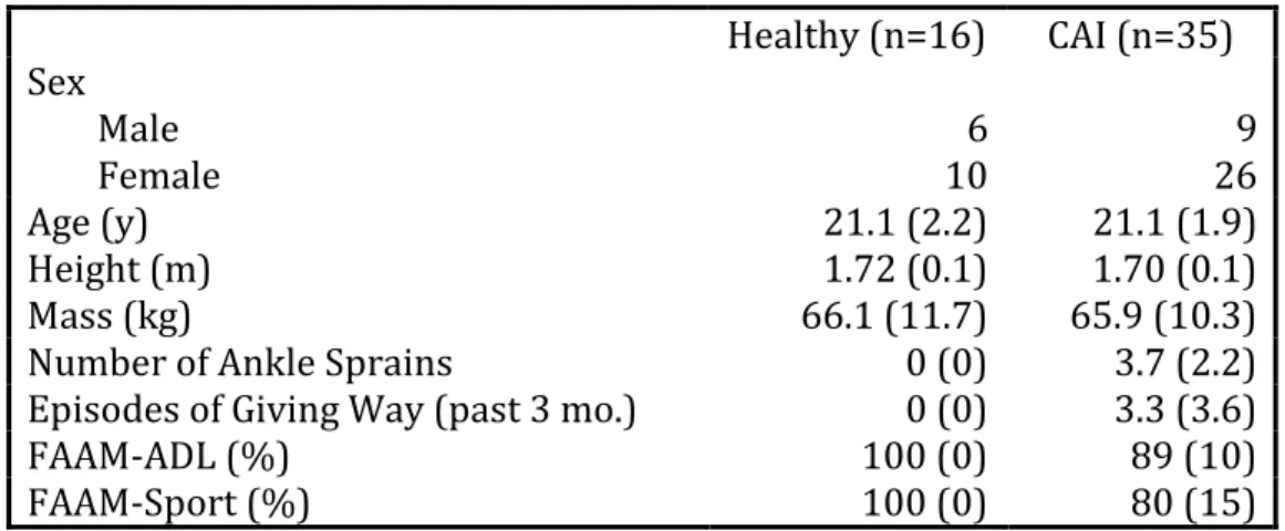
Table 1.Demographic information by group. Mean (SD)
Healthy (n=16) CAI (n=35) Sex
Male
Female 10 6 26 9
Age (y) 21.1 (2.2) 21.1 (1.9)
Height (m) 1.72 (0.1) 1.70 (0.1)
Mass (kg) 66.1 (11.7) 65.9 (10.3)
Number of Ankle Sprains 0 (0) 3.7 (2.2)
Episodes of Giving Way (past 3 mo.) 0 (0) 3.3 (3.6)
FAAM-ADL (%) 100 (0) 89 (10)
FAAM-Sport (%) 100 (0) 80 (15)
28
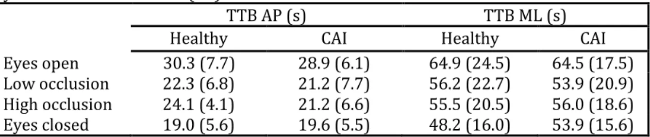
Table 2. Average postural control outcomes for healthy (n=16) and CAI (n=35) groups by visual condition. Mean (SD)
TTB AP (s) TTB ML (s)
Healthy CAI Healthy CAI
Eyes open 30.3 (7.7) 28.9 (6.1) 64.9 (24.5) 64.5 (17.5)
Low occlusion 22.3 (6.8) 21.2 (7.7) 56.2 (22.7) 53.9 (20.9)
High occlusion 24.1 (4.1) 21.2 (6.6) 55.5 (20.5) 56.0 (18.6)
Eyes closed 19.0 (5.6) 19.6 (5.5) 48.2 (16.0) 53.9 (15.6)
29
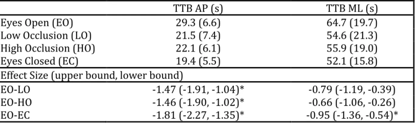
Table 3. Average time-to-boundary under various levels of visual occlusion with effect size, collapsed across group. Mean (SD)
TTB AP (s) TTB ML (s)
Eyes Open (EO) 29.3 (6.6) 64.7 (19.7)
Low Occlusion (LO) 21.5 (7.4) 54.6 (21.3)
High Occlusion (HO) 22.1 (6.1) 55.9 (19.0)
Eyes Closed (EC) 19.4 (5.5) 52.1 (15.8)
Effect Size (upper bound, lower bound)
EO-LO -1.47 (-1.91, -1.04)* -0.79 (-1.19, -0.39)
EO-HO -1.46 (-1.90, -1.02)* -0.66 (-1.06, -0.26)
EO-EC -1.81 (-2.27, -1.35)* -0.95 (-1.36, -0.54)*
*Represents a significant (p ≤ 0.05) effect size. TTB AP=time-to-boundary in the anterior-to-posterior
29
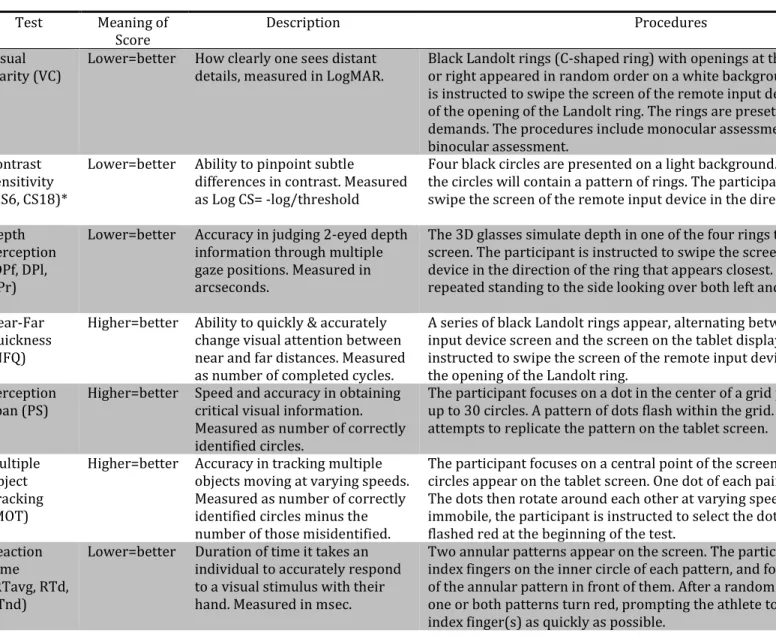
Table 4. Senaptec Sensory System Test detailed outcomes and procedures.
Test Meaning of
Score Description Procedures
Visual
Clarity (VC) Lower=better How clearly one sees distant details, measured in LogMAR. Black Landolt rings (C-shaped ring) with openings at the top, bottom, left, or right appeared in random order on a white background. The participant is instructed to swipe the screen of the remote input device in the direction of the opening of the Landolt ring. The rings are preset at varying acuity demands. The procedures include monocular assessments followed by a binocular assessment.
Contrast Sensitivity (CS6, CS18)*
Lower=better Ability to pinpoint subtle
differences in contrast. Measured as Log CS= -log/threshold
Four black circles are presented on a light background. At random, one of the circles will contain a pattern of rings. The participant is instructed to swipe the screen of the remote input device in the direction of this circle.
Depth Perception (DPf, DPl, DPr)
Lower=better Accuracy in judging 2-eyed depth information through multiple gaze positions. Measured in arcseconds.
The 3D glasses simulate depth in one of the four rings that appear on the screen. The participant is instructed to swipe the screen of the remote input device in the direction of the ring that appears closest. Test procedures are repeated standing to the side looking over both left and right shoulders.
Near-Far Quickness (NFQ)
Higher=better Ability to quickly & accurately change visual attention between near and far distances. Measured as number of completed cycles.
A series of black Landolt rings appear, alternating between the remote input device screen and the screen on the tablet display. The participant is instructed to swipe the screen of the remote input device in the direction of the opening of the Landolt ring.
Perception
Span (PS) Higher=better Speed and accuracy in obtaining critical visual information. Measured as number of correctly identified circles.
The participant focuses on a dot in the center of a grid pattern composed of up to 30 circles. A pattern of dots flash within the grid. The participant then attempts to replicate the pattern on the tablet screen.
Multiple Object Tracking (MOT)
Higher=better Accuracy in tracking multiple objects moving at varying speeds. Measured as number of correctly identified circles minus the number of those misidentified.
The participant focuses on a central point of the screen. Two to five sets of circles appear on the tablet screen. One dot of each pair briefly flashes red. The dots then rotate around each other at varying speeds. Once the dots are immobile, the participant is instructed to select the dot in each pair that flashed red at the beginning of the test.
Reaction Time (RTavg, RTd, RTnd)
Lower=better Duration of time it takes an individual to accurately respond to a visual stimulus with their hand. Measured in msec.
30
Target
Capture (TC) Lower=better Ability to shift visual attention and recognize peripheral targets. Threshold reached measured in msec.
The participant focuses on a central black dot until a Landolt ring appears briefly in one of the corners on Senaptec Sensory Station display. The participant is instructed to swipe in the direction of the opening of the Landolt ring.
Eye-Hand Coordination (EHC)
Lower=better Ability to make quick and accurate visually-guided hand responses to rapidly changing targets. Total time to complete test, measured in msec.
A grid is presented with ten columns and eight rows of equally sized and spaced circles. A green dot appears within one circle of the grid. Participant is instructed to touch the dot as quickly as possible with either hand. As soon as they touch the dot, another dot will be presented. 80 dots will appear.
Go/No-Go
(GNG) Higher=better Ability to make quick and accurate decision responses to rapidly changing targets.
Measured as (total green dots hit plus 0.25*total green dots hit within 0.5 seconds of
disappearing) minus (total red dots hit plus 0.25*red dots within 0.5 seconds of disappearing).
An identical grid as Eye-Hand Coordination test appears. A green or red dot will appear. If the dot is green, the participant is instructed to touch it. If the dot is red, the participant is instructed not to touch it. Eighty dots will appear in a pseudorandomized sequence.
For VC, CS, DP, NFQ, and TC subtests, participants stood 10 ft from tablet, holding remote input device. For PS, MOT, and RT subtests participants stood 2 ft from and responded on tablet. For EHC and GNG subtests, participants stood 2 ft from and responded on 42-in display.
31
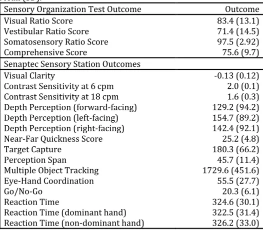
Table 5. Average Senaptec Sensory System and Sensory Organization Test outcomes. Mean (SD).
Sensory Organization Test Outcome Outcome
Visual Ratio Score 83.4 (13.1)
Vestibular Ratio Score 71.4 (14.5)
Somatosensory Ratio Score 97.5 (2.92)
Comprehensive Score 75.6 (9.7)
Senaptec Sensory Station Outcomes
Visual Clarity -0.13 (0.12)
Contrast Sensitivity at 6 cpm 2.0 (0.1)
Contrast Sensitivity at 18 cpm 1.6 (0.3)
Depth Perception (forward-facing) 129.2 (94.2)
Depth Perception (left-facing) 154.7 (89.2)
Depth Perception (right-facing) 142.4 (92.1)
Near-Far Quickness Score 25.2 (4.8)
Target Capture 180.3 (66.2)
Perception Span 45.7 (11.4)
Multiple Object Tracking 1729.6 (451.6)
Eye-Hand Coordination 55.5 (27.7)
Go/No-Go 20.3 (6.1)
Reaction Time 324.6 (30.1)
Reaction Time (dominant hand) 322.5 (31.4)
32
Table 6. Univariable analyses of Senaptec variables included in the time-to-boundary anterior-to-posterior stepwise model.
Condition Senaptec Variable Estimate 95% CI P
Eyes-open VC B 15.25 -2.85 33.35 0.096
PS 0.23 0.05 0.42 0.013
RT D -0.06 -0.13 0.004 0.065
Limited-vision VC B 15.94 -2.21 34.10 0.083
TC -0.03 -0.06 0.005 0.091
PS 0.23 0.05 0.41 0.016
EHC <0.01 -0.00001 0.0001 0.096
33
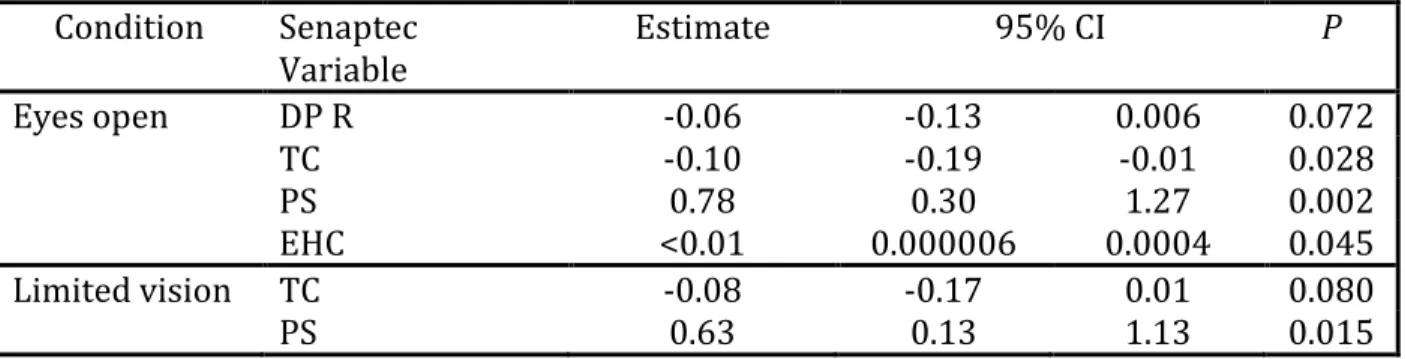
Table 7.Univariable analyses of Senaptec variables included in the time-to-boundary medial-to-lateral stepwise model.
Only Senaptec outcomes that demonstrated adequate significance (p > 0.10) are included in this table. Condition Senaptec
Variable Estimate 95% CI P
Eyes open DP R -0.06 -0.13 0.006 0.072
TC -0.10 -0.19 -0.01 0.028
PS 0.78 0.30 1.27 0.002
EHC <0.01 0.000006 0.0004 0.045
Limited vision TC -0.08 -0.17 0.01 0.080
34
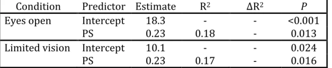
Table 8. Stepwise multivariable regression analysis for time-to-boundary anterior-to-posterior outcomes under both eyes-open and limited-vision conditions
Condition Predictor Estimate R2 ∆R2 P
Eyes open Intercept 18.3 - - <0.001
PS 0.23 0.18 - 0.013
Limited vision Intercept 10.1 - - 0.024
35
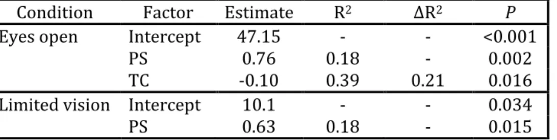
Table 9. Stepwise multivariable regression analysis for time-to-boundary medial-to-lateral outcomes under both eyes-open and limited-vision conditions
Condition Factor Estimate R2 ∆R2 P
Eyes open Intercept 47.15 - - <0.001
PS 0.76 0.18 - 0.002
TC -0.10 0.39 0.21 0.016
Limited vision Intercept 10.1 - - 0.034
36 CHAPTER IV
Overview
Those with chronic ankle instability (CAI) are often described as visually reliant
due to postural control deficits observed under fully occluded visual conditions. Little is
known about the influence of partially occluded vision on postural control in those with
CAI.
To examine differences in postural control in those with and without CAI under
increasing levels of visual occlusion during static stance compared to eyes-open.
Thirty-five participants with CAI and sixteen participants with no history of
lower extremity injury completed four 3-minute postural control assessments in
double-limb stance on a triaxial forceplate under the following four visual conditions: 1)
eyes-open, 2) low-occlusion, 3) high-occlusion, and 4) eyes-closed. Low (level 2) and
high (level 6) occlusion conditions were produced using stroboscopic eyewear. Postural
control outcomes included time-to-boundary minima means in the anteroposterior
(TTB-AP) and mediolateral (TTB-ML) directions, calculated from forceplate data.
Two-way repeated measures ANOVAs assessed differences between groups and the 4 visual
conditions. Alpha level was set at p=0.05.
A condition main effect for both TTB-ML (F3,138=22.9; p<0.001) and TTB-AP
(F3,138=93.7; p<0.001) was observed. Specifically, our observed main effects were
37
for TTB ML (p=0.006) and TTB AP (p<0.001). Additionally, significant differences in
TTB-AP were found between eyes-open and both low- (p<0.001) and high-occlusion
(p<0.001).
Those with and without CAI have impaired postural control under limited and
no vision conditions. Both occlusion conditions produced similar postural control
outcomes as eyes-closed, suggesting a disruption to the sensorimotor system. Future
research should examine the effect of stroboscopic eyewear on postural control in
single-limb support and functional activities as well as the effect of stroboscopic
38 Introduction
Chronic ankle instability (CAI) is a debilitating condition of the ankle joint
characterized by recurrent sprains, episodes of “giving way”, pain, or weakness in the
ankle.1,2,4 CAI arises from an acute sprain of the ankle, one of the most commonly
occurring injuries seen in active populations.17,18 It has been estimated that 40% of
individuals who sustain an ankle sprain develop CAI which decreases in physical
activity and facilitates post-traumatic ankle osteoarthritis development. 4–6
Individuals with CAI have sensorimotor adaptations that are a result of the
initial ligamentous injury and lead to postural control deficits.9 These adaptations
include altered somatosensory input and neuromuscular responses that leave the
individual vulnerable to re-injury.25,26,29 A cascade of events is thought to follow
damaged mechanoreceptors upon initial injury leading to long-lasting alterations in
cortical level somatosensory integration and resultant reliance on visual information to
maintain postural control.9–11
First, initial ligamentous injury interrupts once continuous somatosensory input
from the joint to higher level integration centers regarding joint position, stress, and
velocity.9,11 Without this information, the central nervous system must adopt a new
strategy of obtaining sufficient information for dictation of motor strategies to maintain
postural control.9,11 It appears that in the absence of sufficient somatosensory input, the
motor control system reweights the level of visual feedback used to prepare for and
respond to external perturbation.9 Over time, the motor control system begins to rely
on visual feedback to produce appropriate motor responses9,11 and long-term changes
39
individuals with CAI have significantly worse postural control measures during
single-limb balance tasks under eyes-closed conditions compared to eyes-open.12 While it is
true that healthy controls also have worse postural control under eyes-closed
conditions compared to open, the magnitude of change from open to
eyes-closed conditions are greater in the CAI population.12,43 This indicates that those with
CAI lack appropriate sensorimotor compensation when visual information is disturbed
and, therefore, suggests a reliance on visual information to maintain postural control.12
It is unclear whether long-term visual reliance in CAI patients is a positive or negative
adaptation.
Whether or not a reliance on visual information is a positive or negative
adaptation, the topic is worth exploring in future research. Current balance training
methods often involve exercises in static posture with eyes closed13,14 that do not alter
visual reliance in CAI.15 Incorporating dynamic postural exercises with limited visual
information to stress the sensory reweighting mechanism should be explored as a
method of examining visual reliance. Research has shown that interrupting visual
information with stroboscopic eyewear caused significant postural instability during
single-limb stance (SLS) compared to eyes open conditions in healthy subjects.16
Current literature surrounding the postural response of the CAI population to
interrupted visual information is non-existent. Therefore, the purpose of this study was
to examine changes in spatiotemporal measures of postural control during balance
conditions with varying levels of visual occlusion in those with CAI and healthy
controls. We hypothesized that worse spatiotemporal postural control outcomes (i.e.
40
balance conditions with any limitation in visual information compared to eyes open.
Furthermore, we hypothesize that those with CAI would have significantly worse
postural control outcomes compared to healthy controls during limited vision
conditions.
Methods
Study Design
This quasi-experimental cross-sectional study was approved by the university’s
institutional review board and performed in a clinical research center. Participants
completed a postural control assessment in double-limb support under varying levels of
visual input as the final assessment within the larger study’s assessment battery that
included various visual and balance performance measures.
Participants
Fifty-one participants (15 males (29.4%); age=21.1(2.0 years; mass=66.2 (10.7
kg; height=1.7(0.1 m)) participating in a larger study were included in the present
investigation. Participants who met the International Ankle Consortium inclusion
criteria recommendations2 (n=35) were included in the CAI group while healthy
participants (n=16) had no history of lower extremity injury. All participants were
physically active, defined as participating in moderate to vigorous activity at least 3
days per week for at least 30 minutes, over the past 3 months. All participants provided
written informed consent prior to study participation. Table 1 outlines participant demographic information.
Instrumentation
41
control by recording center-of-pressure (COP) measurements at a sampling frequency
of 100Hz via Balance Clinic software (version 2.02.01). A Matlab software program
(MathWorks, Inc., Natick, MA, USA) was used to compute the postural control outcomes.
Low (level 2) and high (level 6) visual occlusion conditions were produced using
Senaptec Strobe Training Goggles (Senaptec, Beaverton, OR).
Procedures
The postural control balance protocol required participants to complete a
3-minute trial in double-limb support (DLS) under four visual conditions: 1) eyes-open, 2)
low-occlusion, 3) high-occlusion, and 4) eyes-closed. Participants were instructed to
stand as still as possible with arms at side and focus on a self-selected point on the wall
in front of them. Placement of the feet was marked to capture foot width and length and
ensure consistency between all trials.69,70
Outcomes
Time-to-boundary minima means in the anteroposterior (TTB-AP) and
mediolateral (TTB-ML) directions were calculated in seconds, as previously described
by Hertel et al.70 TTB uses COP excursion velocity and position in reference to the
boundaries of the base-of-support to estimate the time a person has to make a postural
correction before the COP reaches a base-of-support boundary assuming that COP
excursion direction and velocity remain constant70. A shorter TTB indicates worse
postural control.71 Time-to-boundary minima mean has been shown to be a reliable
measure of postural control (ICC=0.62-0.87).70 In addition, this measure has been found
to be more sensitive to postural control differences between those with CAI and healthy
42
Original forceplate COP data were processed with a fourth order, zero-lag,
low-pass Butterworth filter with a cutoff frequency of 5 Hz.43,69,72 Data from the 1st minute of
each trial acted as four 15-second trials to allow better congruence with previous
methodology in this space.12,44 Due to a limited sample size, TTB AP and TTB ML
outliers—defined as falling above or below 2 standard deviations of the mean—were adjusted by averaging the preceding two extreme values.
Statistical Analysis
Separate two-way repeated measures ANOVAs were used to assess differences
between groups (CAI vs. healthy) and visual conditions (eyes-open, low-occlusion,
high-occlusion, and eyes-closed) on TTB AP/ML minima means using statistical software
(SAS 94). Hedge’s g effect sizes and 95% confidence intervals were calculated between conditions. A-priori alpha level was set to p=0.05.
Results
All subjects completed the full 3-minute trial under each visual condition, with
the exception of one who felt dizzy under the first stroboscopic condition, discontinued
the testing session, and was removed from the study. We did not observe any Group x
Condition interaction for TTB-AP (F3,196=0.55; p=0.652) or TTB-ML (F3,196=0.35;
p=0.788). No group main effects were observed for TTB-AP (F1,46=1.57; p=0.211) or
TTB-ML (F1,46=0.10; p=0.757). We did observe a condition main effect for both TTB-AP
(F3,138=93.7; p<0.001) and TTB-ML (F3,138=22.9; p<0.001). Specifically, our observed
main effects were driven by differences between the open condition and the
eyes-closed condition for TTB AP (p<0.001) TTB ML (p=0.006). Additionally, significant