ORIGINAL ARTICLE
Myeloid but not epithelial tissue factor exerts protective
anti-inflammatory effects in acid aspiration-induced acute
lung injury
J . B . K R A L - P O I N T N E R , * W . C . S C H R O T T M A I E R , * V . H O R V A T H , * H . D A T L E R , * L . H E L L ,†C . A Y ,† B . N I E D E R R E I T E R ,‡B . J I L M A ,§J . A . S C H M I D ,¶A . A S S I N G E R , * N . M A C K M A N , * * S . K N A P P† † ‡ ‡ and G . S C H A B B A U E R *
*Institute for Physiology, Center for Physiology and Pharmacology, Medical University of Vienna;†Clinical Division of Hematology and Hemostaseology, Department of Medicine I, Medical University of Vienna;‡Division of Rheumatology, Internal Medicine III, Medical University of Vienna;§Departments of Clinical Pharmacology, Department of Medicine I, Medical University of Vienna;¶Department for Vascular Biology and Thrombosis Research, Center for Physiology and Pharmacology, Medical University of Vienna, Vienna, Austria;
**Division of Hematology/Oncology, Thrombosis and Hemostasis Program, UNC McAllister Heart Institute, University of North Carolina, Chapel Hill, NC, USA;††CEMM, Research Center for Molecular Medicine of the Austrian Academy of Sciences; and‡‡Laboratory of Infection Biology, Department of Medicine I, Medical University of Vienna, Vienna, Austria
To cite this article:Kral-Pointner JB, Schrottmaier WC, Horvath V, Datler H, Hell L, Ay C, Niederreiter B, Jilma B, Schmid JA, Assinger A, Mackman N, Knapp S, Schabbauer G. Myeloid but not epithelial tissue factor exerts protective anti-inflammatory effects in acid
aspiration-induced acute lung injury.J Thromb Haemost2017;15: 1625–39.
Essentials
• Tissue factor (TF) represents a central link between hemostasis and inflammation.
• We studied the roles of myeloid and airway epithelial TF in acid-caused acute lung injury (ALI).
• TF on myeloid cells displays a non-coagulatory role regulating the inflammatory response in ALI.
• Airway epithelial TF contributes to hemostatic func-tions, but is dispensable in ALI pathogenesis.
Summary.Introduction:Acute lung injury (ALI) is a
life-threatening condition characterized by damaged alveolar– capillary structures and activation of inflammatory and hemostatic processes. Tissue factor (TF) represents a cru-cial link between inflammation and coagulation, as inflammatory mediators induce myeloid TF expression, and TF initiates extrinsic coagulation. Objective:As pul-monary inflammation stimulates TF expression and TF modulates immune responses, we aimed to elucidate its impact on ALI. In particular, we wanted to distinguish
the contributions of TF expressed on airway epithelial cells and TF expressed on myeloid cells. Methods:Mice with different cell type-specific TF deficiency and wild-type littermates were intratracheally treated with hydrochloric acid, and leukocyte recruitment, cytokine levels, thrombin–antithrombin (TAT) complexes and pul-monary protein-rich infiltrates were analyzed. Results: Our data demonstrate that a lack of epithelial TF did not influence acute responses, as bronchoalveolar neutrophil accumulation 8 h after ALI induction was unaltered. However, it led to mild, prolonged inflammation, as pul-monary leukocyte and erythrocyte numbers were still increased after 24 h, whereas those in wild-type mice had returned to basal levels. In contrast, myeloid TF was pri-marily involved in regulating the acute phase of ALI without affecting local coagulation, as indicated by increased bronchoalveolar neutrophil infiltration, pul-monary interleukin-6 levels, and edema formation, but equal TAT complex formation, 8 h after ALI induction. This augmented inflammatory response associated with myeloid TF deficiency was confirmed in vitro, as lipopolysaccharide-stimulated TF-deficient alveolar macrophages released increased levels of chemokine (C-X-C motif) ligand 1 and tumor necrosis factor-a as compared with wild-type macrophages. Conclusion:We conclude that myeloid TF dampens inflammation in acid-induced ALI.
Keywords: acute lung injury; blood coagulation;
inflammation; macrophages; tissue factor.
Correspondence: Gernot Schabbauer, Center for Physiology and Pathophysiology, Medical University of Vienna, Schwarzspanier-strasse 17, A-1090 Vienna, Austria.
Tel: +43 1 40160 31427.
E-mail: [email protected]
Received 6 July 2016
Manuscript handled by: W. Ruf
Final decision: P. H. Reitsma, 5 May 2017
Introduction
Tissue factor (TF), also referred to as factor III, thrombo-plastin, or CD142, is a glycosylated integral transmem-brane protein that initiates the extrinsic coagulation cascade. TF is constitutively expressed on the surface by various extravascular tissues, such as pulmonary epithe-lium. Upon injury, e.g. to the alveolar–endothelial barrier, extravascular TF is exposed to plasmatic coagulation fac-tors, e.g. FVII, and initiates clot formation. The generated thrombin triggers fibrin formation, which stabilizes the primary platelet plug. Hence, TF is important for protect-ing vascular integrity, and thus preventprotect-ing uncontrolled and life-threatening blood loss [1–3]. Upon infection or inflammation, TF expression is readily induced on leuko-cytes that are usually devoid of surface TF. These TF-expressing leukocytes comprise members of the first line of defense, such as macrophages, but also circulating mono-cytes or neutrophils. Furthermore, blood vessel-lining endothelial cells are able to synthesize TF [4–6]. Stimuli triggering bloodborne TF expression during infection include pathogen-associated molecular patterns, e.g. lipopolysaccharide (LPS) or lipoteichoic acid, and endoge-nous inflammatory cytokines, e.g. tumor necrosis factor (TNF)-a and interleukin (IL)-1b [7,8]. This process involves several transcription factors that are essential for immune functions, such as nuclear factor-jB (NF-jB), AP-1, nuclear factor of activated T cells, and early growth response-1 [9–12]. Hence, the properties of TF are not strictly confined to hemostatic functions, but also promote host defense and inflammation [13]. Therefore, reduced TF activity in mice leads to more severe bacteremia and increased mortality [14]. However, uncontrolled coagula-tion cascade activacoagula-tion markedly contributes to dissemi-nated intravascular coagulation. Correspondingly, reduced TF expression protects mice from systemic endotoxemia [15], indicating that finely balanced TF expression is essen-tial for adequate immune responses during sepsis. Further-more, TF inhibition prevents secondary lung injury in a baboon sepsis model [16]. However, it remains incom-pletely understood how TF contributes to local infection and inflammation in organs such as the lung.
Acute lung injury (ALI) represents a multifactorial dis-ease that can be induced by aspiration of non-infectious acidic gastric contents as well as by pathogenic lung infec-tions. The role of TFs in different pathologies of lung injury are still undefined. A recent study investigating low-TF mice subjected to LPS-induced ALI suggested that reduced TF expression exacerbates disease pathogen-esis [17]. Cell type-specific TF deficiency in type I and type II lung epithelial cells worsens LPS-induced ALI by increasing alveolar permeability and hemorrhage. In con-trast, mice devoid of myeloid TF show no overt differ-ences in the severity of lung damage [18]. In the present study, we focused on cell type-specific roles of TF in acid-induced ALI. This clinically relevant mouse model mimics
aspiration of gastric contents leading to lung complica-tions (particularly in patients admitted to intensive care units), which accounts for 11% of all ALI cases in clinics [19], and is characterized by the release of endogenous alarm signals, damage-associated molecular patterns (DAMPs), and simultaneous Toll-like receptor activation [20,21]. The resulting inflammation is associated with endothelial cell activation, leukocyte and platelet influx, activation of coagulation, fibrin deposition, and, eventu-ally, damage to the alveolar–capillary barrier [22]. Nota-bly, activation of the coagulation cascade plays a relevant role in disease progression [23].
As coagulatory and inflammatory processes affect the pathogenesis of ALI, we wanted to elucidate the impact of TF on these events during ALI. Thus, we endeavored to elucidate whether the presence of TF is required to protect the integrity of alveolar–capillary structures. Moreover, we aimed to define cell type-specific functions of TF, and therefore performed acid-induced ALI in mice with TF deficiency in either myeloid cells (monocytes, macrophages, and granulocytes) or alveolar type I and type II epithelial cells. Our data indicate that TF acts in a cell type-specific manner, and that myeloid TF, in con-trast to airway epithelial TF, dampens inflammatory responses during acid-induced ALI.
Materials and methods
Mice
All experiments and animal studies were conducted according to institutional guidelines and were approved by the Animal Care and Use Committee of the Medical University of Vienna (BMWF-66.009/0320-II/3b/2013; BMWF-66.009/0254-ll/3b/2013).
All animal experimentation was littermate-controlled. We used previously described LysM cre+/TFflox/flox (referred to as TFΔmye) [24,25] or SPC cre+/TFflox/flox (referred to as TFΔepi) [18] mice on a C57BL/6J genetic background (backcrossed for six generations), and 8–12-week-old, sex-matched littermates (control LysM cre/TFflox/flox and SpcCre/TFflox/flox, referred as TF+/+or wild-type).
Direct PCR of lysed tissue was performed for genotyp-ing. (Primers: TF forward, 50-ATGAGGAGCTGTGTTA
AAGGGTCGCAGA-30; TF reverse, 50-TGCAGTAA
ATGCACGTGTCTGCCAT-30; Cre forward, 50-TCGC
GATTATCTTCTATATCTTCAG-30; Cre reverse, 50-GC
TCGACCAGTTTAGTTACCC-30.)
Anesthesia
ALI and administration of anti-TF antibody
Sedated mice were intratracheally treated with 50lL of 0.1Mhydrochloric acid (HCl) to induce ALI.
Wild-type mice (C57BL/6J genetic background) were intraperitoneally injected once with monoclonal inhibitory rat anti-mouse TF (1H1) (20lg g1) prior to ALI induc-tion.
Bronchoalveolar lavage fluid (BALF) and alveolar macrophage isolation and stimulation
For quantification of cytokines and leukocytes in the bron-choalveolar space, the trachea was exposed and punctured with a vein catheter, and the bronchoalveolar space was lavaged with 1 mL of phosphate-buffered saline (PBS). Cells were pelleted for flow cytometry (600 9g, 10 min), and supernatants were used for ELISA.
Alveolar macrophages were isolated by repeating lavaging (10 times), and seeded into non-tissue culture-treated plates (2 h, 37°C, and 5% CO2; RPMI medium (Sigma-Aldrich,
Vienna, Austria) dosed with 10% fetal bovine serum, 1% penicillin, streptomycin, and fungizone, and 1%L-glutamine)
before non-adherent lymphocytes were discarded.
Alveolar macrophages were stimulated with 10 ng mL1 or 100 ng mL1LPS (LPS-EB ultrapure fromEscherichia coli O111:B4; Invivogen, Toulouse, France) for the indi-cated times, and supernatant or cells with peqGOLD Tri-Fast (VWR, Darmstadt, Germany) were harvested.
Bone marrow-derived macrophage (BMDM) isolation
Murine bone marrow was isolated and incubated with granulocyte–macrophage colony-stimulating factor (GM-CSF) (10 ng mL1) for 1 week to generate BMDMs, which were then stimulated with LPS (10 ng mL1) for the indicated times.
Lung homogenization for ELISA and western blotting
Lungs were homogenized in 4lL of PBS mg–1 with 5-mm beads in a Precellys24 homogenizer (Bertin Technolo-gies, Aix-en-Provence, France). Supernatants were mixed with Greenberger Lysis buffer and protease inhibitor (Roche Life Sciences, Basel, Switzerland), incubated (20 min on ice), and centrifuged (135 9g, 15 min, 4 °C), and the supernatant was used for ELISA.
For immunoblotting, lungs were homogenized in 10lL of RIPA buffer mg–1, and supernatants were mixed with Laemmli buffer.
RNA isolation and quantitative PCR (qPCR)
RNA was isolated from TriFast-homogenized lungs or macrophages, transcribed into cDNA (High-Capacity cDNA Reverse transcription Kit; Applied Biosystems,
Carlsbad, CA, USA), and analyzed by qPCR with Fast SYBR Green Master Mix (Applied Biosystems, Carlsbad, CA, USA) on a StepOne Real-Time PCR System (Applied Biosystems, Foster City, CA, USA). The results were eval-uated according to Pfaffl [26] and expressed as fold con-trol values. Primer sequences are supplied in Data S1.
Western blotting
Proteins were separated by SDS-PAGE (10% acrylamide gels), and semi-dry-blotted onto a poly(vinylidene difluo-ride) membrane. The fibrin b-monomer 59D8 and anti-b -actin (Sigma-Aldrich) and horseradish peroxidase-linked secondary antibodies were used. Immunoblots were devel-oped with SuperSignal West Femto Maximum Sensitivity Substrate (Thermo Fisher Scientific, Waltham, MA, USA), imaged with FluorChem HD2 Chemiluminesence Imager (Alpha Innotech, San Leandro, CA, USA), and densitometrically analyzed with IMAGEJ 1.47v. Bands were
normalized tob-actin.
Flow cytometry
BALF cells were incubated (20 min) with anti-CD45– PerCP, anti-F4/80–fluorescein isothiocyanate, anti-Ly6G/ Ly6C–phycoerythrin, and anti-Ter119–allophycocyanin
(1 : 80; BioLegend, London, UK), fixed with 1%
formaldehyde, and measured by flow cytometry (BD Accuri flow cytometer with BD ACCURIC6, and FLOWJO9
[Treestar, Ashland, OR, USA]). The following gating strat-egy was applied. CD45+ cells (leukocytes) were analyzed for F4/80 positivity (macrophages). Ly6C+ macrophages were termed inflammatory macrophages. CD45+F4/80 cells were analyzed for Ly6G expression (neutrophils).
Total cell counts of BALF were calculated by multiply-ing the events by
Final dilution for flowcytometer
Measured volume
Resupensionvolume Stained volume
Injected PBS
Recollected PBS
Histologic analysis
The left lung was extracted, fixed in 4% formaldehyde, and embedded in paraffin. Three-micrometer lung sec-tions were stained with anti-mouse GR-1 or TF antibody, and counterstained with hematoxylin or with hematoxylin and eosin (Carl Roth, Karlsruhe, Germany). Images were obtained with an Axio Imager.Z1 microscope (Carl Zeiss, Vienna, Austria).
TF activity assay
salt solution with 0.1% bovine serum albumin (HBSA), and microvesicles were pelleted (18 0009 g, 20 min, 4°C), washed, and incubated with anti-mouse TF (1H1) (100lg mL1) or control IgG (15 min) before addition of FVII (4.88 nM), FX (150 nM), and CaCl2(10 mM). The
reaction was stopped by addition of EDTA HBSA buffer before addition of chromogenic substrate and absorbance measurement. TF-dependent activated FX (FXa) genera-tion was determined by subtracting FXa levels generated in the presence of TF antibody from the levels in IgG control samples.
ELISA
ELISAs were performed according to manufacturer’s pro-tocols. Myeloperoxidase (MPO), IL-33 and IL-6, and
che-mokine (C-X-C-motif) ligand-1 (KC), TNF-a and
thrombin–antithrombin (TAT) ELISAs were purchased from eBioscience (San Diego, USA), R&D Systems (Min-neapolis, MN, USA) and Siemens Healthcare Diagnostics (Vienna, Austria), respectively. Absorbance was measured at 450 nm with an EL808 Ultra Microplate Reader (Bio-Tek Instruments, Winooski, VT, USA).
Bicinchoninic acid (BCA) assay
The protein concentration in BALF was determined with a Pierce BCA Protein Assay Kit (Thermo Fisher Scientific, Wal-tham, MA, USA) according to the manufacturer’s guidelines.
Edema formation
Murine lungs were extracted, weighed, dried at 55°C for 48 h, and weighed again. The ratio between wet weight and dry weight was calculated.
Statistical analysis
Data were depicted as bar diagrams showing the
meanstandard error of the mean, as box plots indicat-ing the median, the first and third quartiles, and the mini-mum and maximini-mum, or as scatter plots with indicated means. The results were evaluated with GRAPHPAD PRISM5
(GraphPad, San Diego, CA, USA) by one-way-ANOVAwith
Dunn’s multiple comparisons test, two-way ANOVA with
Sidak’s multiple comparisons test, unpaired, two-tailed t-tests, or the Mann–Whitney test, if the samples did not pass the Kolmogorov–Smirnov test for normal distribu-tion. AP-value of<0.05 was considered to be significant.
Results
TF expression during acid-induced ALI
Activation of coagulation is an integral step in the
pathol-ogy of ALI. We established a mouse model of
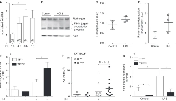
acid-induced ALI by intratracheal administration of HCl. Lung TF mRNA expression was increased within the first 8 h of ALI, which represents the most critical time in the pathology (Fig. 1A), whereas we could not detect simulta-neous accumulation of fibrin b-monomers in lungs (Fig. 1B–D). This indicates rather modest activation of coagulation during acid-induced ALI. To investigate the role of TF in acid-induced ALI, we used mice with a cell type-specific TF deficiency (Fig. S1) either in myeloid cells (TFDmye) or in lung epithelial cells (TFDepi) in littermate-controlled experiments. We found a significant increase in TF mRNA expression in TFDmyemice during ALI, which was significantly more pronounced than in wild-type mice (Fig. 1E). This indicates that non-myeloid cells upregulate TF during ALI. As fibrin generation was only moderately affected during ALI, we used thrombin, the protease-cleaving fibrinogen, as a functional readout for coagula-tion activacoagula-tion. Formed thrombin becomes rapidly inacti-vated via binding of antithrombin III, leading to the formation of TAT complexes. However, we found only modest TAT complex upregulation in BALF 8 h after ALI, perhaps because of low FVII and FX BALF levels. TAT complex formation only tends to be increased in TFDmyemice (Fig. 1F).In vitro, we confirmed TF deletion at the mRNA level in alveolar macrophages with and without LPS stimulation (Fig. 1G).
TF on myeloid cells negatively regulates leukocyte recruitment during acid-induced ALI
and wild-type littermates. Only inflammatory macrophages (Fig. 2F), characterized by F4/80+ and Ly6C+ [28,29], were marginally, but not significantly (P =0.09), increased in number. To analyze the specificity of this phenomenon, we applied a leukocyte recruitment model to TFDmyeand wild-type mice. To this end, we induced thioglycollate-eli-cited sterile inflammation, and analyzed leukocyte influx in an early phase (4 h) and a late phase (72 h). However, no differences in infiltrating leukocytes, macrophages or neu-trophils were observed, perhaps because of the strong inflammatory stimulus (Fig. S3A–D). In vitro analysis of alveolar macrophages revealed that, in response to LPS, TF deficiency increased the release of the chemokine KC, which primarily attracts neutrophils (Fig. 2G). These data may indicate that myeloid TF plays a lung-specific role in regulating leukocyte alveolar trafficking.
TF dampens the inflammatory response in acid-induced ALI
Another hallmark of acid-induced ALI is the induction of an inflammatory response, which is usually triggered by
endogenous alarmins or DAMPs, owing to cellular dam-age. DAMPs include nuclear components such as histones or high-mobility group box 1, but also IL-33 [30]. We observed a strong increase in the release of IL-33 into the bronchoalveolar compartment in response to ALI, but no differences between TFDmyeand wild-type littermate mice (Fig. 3A). Although granulocyte numbers in TFDmyemice (Fig. 2B) were markedly increased, we found only a slight trend for increased MPO levels in BALF and lung homo-genates in TFDmye mice (Fig. 3B). However, analysis of total protein in BALF revealed an increased protein con-tent in TFDmye mice subjected to acid-induced ALI as compared with wild-type mice (Fig. 3C). This finding points towards increased vascular leakage and loss of alveolar–capillary barrier function in the lungs of TFDmye mice. Next, we measured the level of pulmonary IL-6 of acid-treated mice, which we considered to be the most robust marker for inflammation in this rather mild inflammatory model. We observed significantly increased IL-6 production and release in BALF and total lung after acid-induced ALI in TFDmye mice as compared with 0
0 0 0
2 4 6 8
5 10
P = 0.15 TAT BALF
TAT (ng mL
–1
)
Fold change normalized
to HPRT
15
HCI – – + + HCI – – + +
1 2 3 4 5
6 *
tf tf
TF+/+
TFΔmye
TF+/+ TFΔmye
TF+/+
TFΔmye 0 h
HCI 4 h 6 h 8 h
Control HCI 8 h
Fibrinogen
Actin
0.0 0
1 2 3 4
Control HCI Control HCI
Control LPS 0.5
1.0 1.5 2.0
Fibrinogen/actin (a.u.)
Fibrin (ogen) degradation products
Fibrin (-ogen) degradation
product/actin (a.u.)
1 2 3 4 5
A
E F G
B C D
*
* *
(8) (10) (9) (8)
Fold change TF
normalized to HPRT
Fold change normalized
to HPRT
Fig. 1.Tissue factor (TF) expression and activation of coagulation during acute lung injury (ALI). (A) TF mRNA expression in lungs of wild-type mice after HCl-induced ALI was determined by qPCR at the indicated time points. Data show fold control (0 h) values normalized to
hypoxanthine guanine phosphoribosyl transferase (HPRT); numbers are shown in parentheses. (B) Western blot analysis for fibrin-bof
homoge-nized lung samples 8 h after HCl treatment (n=3) and of controls (n=2); actin was used as a loading control. (C, D) Western blot
densitomet-ric analysis of (C) fibrinogen and (D) the major fibrin(ogen) degradation product. (E) TF mRNA expression in total lung tissue of control (nTF+/
+=2;nTFDmye=1) and HCl-treated (nTF+/+=9;nTFDmye=4) myeloid TF-deficient (TFD
mye
) or wild-type (TF+/+) mice was analyzed by
q-PCR 8 h after treatment. (F) Thrombin–antithrombin (TAT) complexes in the bronchoalveolar lavage fluid (BALF) of control (n=4) and
HCl-treated (nTF+/+=10;nTFDmye=12) wild-type or myeloid TF-deficient mice 8 h after treatment. (G) TF mRNA expression of alveolar
macrophages, isolated from lungs of myeloid TF knockout mice and wild-type littermates and stimulated with lipopolysaccharide (LPS)
(100 ng mL1)in vitrofor 3 h;n=5. For statistical analysis, one-way-ANOVAwith Dunn’s multiple comparisons test (A), unpaired Student’s
0
SSC
SSC
SSC-A :: SSC-A
FL3-A :: FL3_CD45-A
HCI CD45
2967 4431
– – + +
HCI – – + +
HCI – – + + HCI – – + +
*
* 2
3399
100
100 102 104 106
FL2-A :: FL2_Ly6G-A 100 102 104 106
FL2-A :: FL2_Ly6G-A 100 102 104 106
FL3-A :: FL3_CD45-A 100 102 104 106
101
102
103
104
105
106
107
SSC-A :: SSC-A
100
101
102
103
104
105
106
107
SSC-A :: SSC-A
100
101
102
103
104
105
106
107
SSC-A :: SSC-A
100
101
102
103
104
105
106
107
4605 4
6
A
B
C
D
E F G
0 2 4 6
TF+/+
TF+/+
TF+/+ TF+/+
TF+/+ HCI CD45+ leukocytes
Log CD45
+ (count per mouse)
Log Ly6G
+ (count per mouse)
Log F4/80
+ (count per mouse)
F4/80+ macrophages Inflammatory macrophages Alveolar macrophages
Log Ly6C
+ F4/80 + (count per mouse)
Ly6G+ neutrophils
Ly6G
Control
0 2 4 6
0 0
PBS 3 h 6 h 24 h PBS 3 h 6 h 24 h
LPS LPS
500 1000
KC (pg mL
–1
)
2000
1500
* *
2 4
P = 0.09 P = 0.07
6 TFΔmye
TFΔmye
TFΔmye TFΔmye
TF+/+ TFΔmye
TF+/+ TFΔmye
wild-type mice (Fig. 3D,E). However, pulmonary IL-1b and IL-12 p40 levels were unaltered (Fig. S4A,B).
TF dampens the inflammatory response of macrophages
in vitro
To determine the possible source of increased IL-6 levels in HCl-treated TFDmye mice and the impact of myeloid TF on inflammatory responses of macrophages, we iso-lated alveolar macrophages from naive TFDmye mice and
wild-type littermates. Analysis of IL-6 at the mRNA and protein levels revealed a significant increase in IL-6 mRNA 3 h after LPS stimulation (Fig. 4A,B). Moreover, TNF-a mRNA and protein levels were significantly increased 3 h after LPS treatment (Fig. 4C,D). As alveo-lar macrophages are very limited in number, and their development depends on GM-CSF [31], we also used GM-CSF-induced BMDMs. LPS-stimulated TF-deficient BMDMs expressed similar levels of IL-6 mRNA and pro-tein (Fig. 4E,F); however, TNF-a secretion was
0
0 0 0
100 200 300 400 500
200 400 600 800 1000 1200
100 200 300 400
0 0
5000 10 000 15 000
500 000 1 000 000 1 500 000
HCI – – + + HCI – – + + HCI – – + +
HCI – – + + HCI – – + + HCI – – + +
200 400
IL-33 (pg mL
–1)
Protein content (µg mL
–1
)
MPO (pg mL
–1)
IL-6 (pg mL
–1
)
IL-6 (pg mL
–1
)
MPO (pg mL
–1)
600
A
C D E
B
IL-33
Protein IL-6 IL-6
BALF:
BALF: BALF:
* * *
Lung:
BALF: Lung:
MPO MPO
TF+/+ TFΔmye
TF+/+
TFΔmye
TF+/+
TFΔmye
TF+/+
TFΔmye TF+/+
TFΔmye TF+/+
TFΔmye
Fig. 3.Increased inflammation in myeloid tissue factor (TF)-deficient mice in response to acid-induced acute lung injury (ALI). (A–D)
Inter-leukin (IL)-33 (A) (ncontrol TF+/+=6,ncontrol TFDmye=5,nHCl TF+/+=7,nHCl TFDmye=8), myeloperoxidase (MPO) (B) (bronchoalveolar
lavage fluid [BALF]–ncontrol TF+/+=6,ncontrol TFDmye=4,nHCl TF+/+=11,nHCl TFDmye=12; lung–ncontrol TF+/+=5,ncontrol TFDmye=5,
nHCl TF+/+=14,nHCl TFDmye=14) and IL-6 (D) (ncontrol TF+/+=6,ncontrol TFDmye=5,nHCl TF+/+=10,nHCl TFDmye=12) were measured in
BALF by ELISA. (C) The protein content in BALF was measured with a bicinchoninic acid protein assay (ncontrol TF+/+=3,
ncontrol TFDmye=3,nHCl TF+/+=7,nHCl TFDmye=10). (E) IL-6 levels in total lung tissue were determined by ELISA (ncontrol TF+/+=7, ncontrol TFDmye=8;nHCl TF+/+=11,nHCl TFDmye=13). Parameters were analyzed 8 h after HCl treatment. For statistical analysis, unpaired
Student’st-tests (A–E) were performed;*P<0.05. Littermate-controlled experiments were performed.
Fig. 2.The effect of myeloid tissue factor (TF) on pulmonary leukocyte recruitment during acute lung injury (ALI). (A, B, E, F) Flow
cyto-metric analysis of leukocytes in the bronchoalveolar lavage fluid of control mice (nTF+/+=10;nTFDmye=11) and HCl-treated (nTF+/+=22;
nTFDmye=27) myeloid TF-deficient mice or wild-type mice 8 h after ALI. (A) CD45+leukocytes. (B) CD45+Ly6G+F4/80neutrophils. (E)
CD45+F4/80+macrophages. (F) CD45+Ly6C+F4/80+inflammatory macrophages (n
control TF+/+=9;ncontrol TFDmye=11;
nHCl TF+/+=21;nHCl TFDmye=26). (A, B) Representative blots of HCl-treated mice. (C,D) Lung sections of myeloid TF-deficient mice or
wild-type mice 8 h after ALI induction stained for GR-1 and hematoxylin (C) or hematoxylin and eosin (D). Scale bar: 25lm. Magnification:
9200. Arrows indicate neutrophils. (G) Alveolar macrophages were stimulated with lipopolysaccharide (LPS) for the indicated time points
in vitro, and released chemokine (C-X-C-motif) ligand-1 (KC) was measured by ELISA;n=3. For statistical analysis (A–F), unpaired
Student’st-tests and (G) two-wayANOVAwith Sidak’s multiple comparison were performed;*P<0.05. Littermate-controlled experiments were
significantly increased (Fig. 4G). To identify potential sig-naling pathways affected by TF, we analyzed the expres-sion of EGR1, IjBa, and STAT1. We found that TF significantly reduced the expression of EGR1 1 h after
LPS stimulation, whereas IjBaand STAT1 were similarly expressed (Fig. 4H–J), indicating that the mitogen-acti-vated kinase pathway was primarily affected, rather than the interferon and NF-jB pathways.
0
A B
C
E
H I J
F G D 0.0 0 500 GM-CSF BMDM 0 0 0 10 20 30 10 20 30 40 50 0 0 10 000 20 000 30 000 40 000 50 000 10 000 20 000 30 000 40 000 2 4 4000 8000 12 000 16 000 20 000 1000 TNF-α (pg mL –1 ) TNF-α (pg mL –1 ) 1500 2.5 5.0 0.06 7.5 10.0
Fold change normalized
to HPRT
Fold change normalized
to HPRT
Fold change normalized
to HPRT
Fold change normalized
to HPRT
0 5 10 15
Fold change normalized
to HPRT 75 150 225 300 0 100 200 300 5000
IL-6 (pg mL
–1)
IL-6 (pg mL
–1 ) 10 000 15 000 20 000 25 000 10
Fold change normalized
to HPRT 20 3000 Control LPS Control LPS Control LPS ns **** * **** *** TNF-α tnf-α
TNF-α
LPS PBS 3 h 6 h 24 h
LPS PBS 3 h 6 h 24 h
LPS PBS 3 h 6 h 24 h
LPS PBS 3 h 6 h24 h
LPS PBS 3 h 6 h 24 h
LPS PBS 3 h 6 h24 h LPS
PBS 3 h 6 h
24 h
LPS PBS 3 h 6 h 24 h LPS
PBS 3 h 6 h 24 h
LPS PBS 3
h 6 h 24 h LPS
PBS 3 h 6 h 24 h
LPS PBS 3 h 6 h 24 h LPS
PBS 3 h 6 h 24 h
LPS PBS 3 h 6 h 24 h 6000 9000 12 000 **** ** *** * * * il-6 il-6
egr1 ikba stat1
IL-6
IL-6 Alveolar macrophages
TF+/+ TFΔmye
TF+/+ TFΔmye
TF+/+ TFΔmye
TF+/+ TFΔmye
TF+/+
TFΔmye TF
+/+ TFΔmye TF+/+
TFΔmye
TF+/+ TFΔmye TF+/+
As TF activity leads to activated FVII (FVIIa) and thrombin generation, we examined the effects of 3 h of human FVIIa and thrombin stimulation on wild-type BMDMs. We found that concomitant stimulation with LPS and thrombin significantly reduced the expression of IL-6 and tended to decrease the expression of TNF-aand IjBa (Fig. S5A–C). Similarly, FVIIa showed a tendency to reduce IL-6 levels and, to a lesser extent, TNF-a and IjBamRNA levels (Fig. S5D–F). However,in vivo, other mechanisms, such as the intrinsic coagulation cascade, can also contribute to thrombin generation. Moreover, TF can directly signal and thereby modulate macrophage functions.
These results indicate that the differential expression of proinflammatory cytokines during ALI is probably depen-dent on TF surface expression on innate immune cells.
TF on epithelial cells does not contribute to tissue inflammation 8 h after acid-induced ALI
To determine the contribution of lung epithelial TF to the pathophysiology of acid-induced ALI, we used TFDepi mice. We used immunostaining to confirm that there was appropriate TF deficiency in the lungs of TFDepimice [18]
(Fig. S1B). Analysis of TF mRNA expression in whole lung tissue during ALI revealed a trend for increased TF expression 8 h after acid instillation in wild-type mice. In contrast, TF mRNA levels were strongly reduced in TFDepi mice (Fig. 5A), indicating that epithelial cells express the majority of lung TF mRNA during ALI. The SPC cre efficiently targets TF expression in most of the lung epithelial cells [32]. Moreover, upon LPS-induced ALI, lung epithelial cells represent the major TF-expres-sing cell type responsible for protection of alveolar– capil-lary integrity [18]. Hence, we wanted to determine whether this is also the case during acid-induced ALI. Measure-ment of TF activity revealed slightly increased FXa gener-ation and TAT complex formgener-ation 8 h after disease induction in wild-type littermates. In comparison, TFDepi
mice showed a trend for reduced FXa generation but no differences in TAT complex formation (Fig. 5B,C). Next, we tested endothelial and epithelial barrier function by measuring BALF protein levels. Surprisingly, we did not observe significant differences in BALF protein levels and
edema formation between acid-treated TFDepi mice and wild-type mice (Fig. 5D,E). Similarly, we did not observe alterations in erythrocyte levels in BALF (Fig. 5J). To test the inflammatory response, we measured pulmonary IL-6 mRNA expression and IL-6 protein levels in BALF and lung tissue, and pulmonary neutrophil transmigration, but did not detect any differences between acid-treated TFDepi mice and wild-type littermates (Fig. 5F–I).
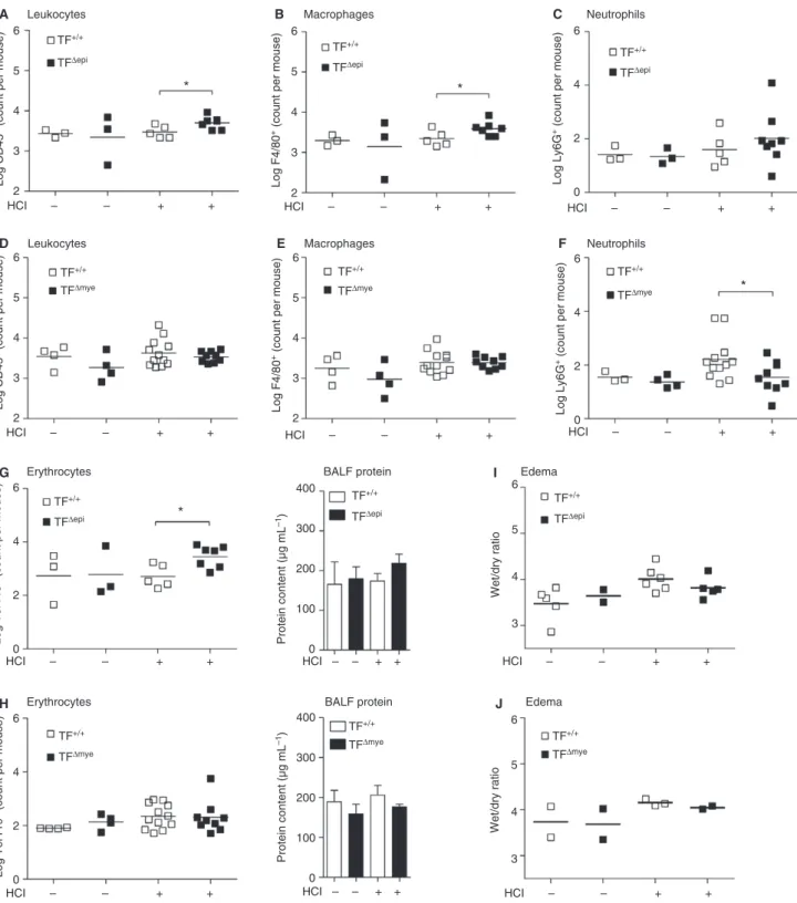
Restoration of tissue integrity is disturbed at late stages of acid-induced ALI in TFDepimice but not in TFDmyemice
The current literature on TF in ALI pathogenesis is focused on early responses in disease progression. There-fore, we aimed to elucidate the effects of TF deletion at late stages of ALI. Twenty-four hours after acid treatment, the initial injury and inflammation in wild-type mice had resolved. Extravasation of leukocytes, including neu-trophils and macrophages, as well as erythrocytes and pro-tein infiltrates, returned to baseline levels (Fig. 6A–H). Surprisingly, we observed increased leukocyte counts in the bronchoalveolar compartment in TFDepimice as compared with wild-type littermates at this time point, suggesting that TFDepi mice were unable to completely resolve the
mild acid-induced injury (Fig. 6A). Notably, most of this increase could be attributed to macrophages, and not to neutrophils (Fig. 6B,C). In contrast, TFDmyemice did not show any overt pulmonary inflammation 24 h after ALI induction, as indicated by baseline levels of leukocytes, macrophages and neutrophils in BALF (Fig. 6D–F). Simi-larly, we found significantly more erythrocytes in HCl-trea-ted TFDepimice and a trend for more protein in the BALF (Fig. 6G), whereas TFDmye mice and wild-type mice did not show increased levels in comparison with untreated mice (Fig. 6H). Edema formation was still slightly increased in all four HCl-treated groups, but no differences between genotypes were observed (Fig. 6I,J).
Systemic TF targeting by antibody does not affect
inflammation but increases the risk of bleeding during acid-induced ALI
To elucidate a possible systemic role of TF in the pro-gression of acid-induced ALI, we administered the TF-Fig. 4.Myeloid tissue factor (TF) deficiency promotes a proinflammatory phenotype of alveolar and bone marrow-derived macrophages
(BMDMs). (A–D) Alveolar macrophages of TFDmyeand TF+/+mice were isolated and stimulated with lipopolysaccharide (LPS) (10 ng mL1)
or vehicle for the indicated time points. Interleukin (IL)-6 (A) (n=5) and tumor necrosis factor (TNF)-a(C) (n=3) mRNA expression in
iso-lated alveolar macrophages, stimuiso-lated with LPS or vehicle for 3 h were determined by qPCR. Data are depicted as fold TF+/+control. (B, D)
IL-6 (B) and TNF-a(D) protein levels in supernatants of alveolar macrophages were measured by ELISA at the indicated time points after LPS
stimulation;n=3. (E–J) Bone marrow cells of TFDmyeand TF+/+mice were differentiated towards macrophages by granulocyte–macrophage
colony-stimulating factor (GM-CSF) (10 ng mL1) and stimulated with LPS (10 ng mL1) or vehicle at the indicated time points. (E) IL-6
mRNA (ncontrol TF+/+=5; all othersn=6) expression 3 h after stimulation was quantified by qPCR, and the data are depicted as fold TF
+/+
control. (F) IL-6 (n6 h=4; all othersn=6). (G) TNF-a(n=6) levels in supernatants were determined by ELISA. (H–J) EGR1 (H) (n=5–6),
IKBa(I) (n=5–6) and STAT1 (J) (n=5–6) were measured by qPCR, and are depicted as fold TF+/+control. For statistical analysis, two-way
0
0
0
0 3.0
3.5 4.0 4.5 5.0 5.5
2 4 6
Log Ly6G
+ (count per mouse)
Log Ter119
+ (count per mouse)
0 0
200 400 600 800
100 200 300 400
Neutrophils
2015 1475
90 110
Erythrocytes
Ter119
IL-6 (pg mL
–1
)
IL-6 (pg mL
–1
)
IL-6 mRNA normalized to HPRT
SSC
Ly6G
SSC
1 2 3
2 3 4
Wet/dry ratio
5 6
100 200
Protein content (µg mL
–1) 300 400
BALF protein Edema
Lung IL-6 BALF IL-6
0 0
2 4 6 8 10 12
20 40
FXa (pg mL
–1)
TAT complex (ng mL
–1
)
60 80 100
HCI
WT
– – + + HCI – – + +
HCI – – + +
HCI – – + +
HCI – – + + HCI – – + +
ND ND
HCI – – + +
HCI – – + + ***
1 2
P = 0.07
P = 0.16
P = 0.06
TF mRNA nnormalized to HPRT
3 A
D E
F
I J
G H
B C
TF+/+
tf
il-6
Xa BALF TAT
TFΔepi
TF+/+
TFΔepi
TF+/+
TF+/+ TF+/+
TFΔepi
TF+/+
TFΔepi
TF+/+
TFΔepi
TFΔepi TFΔepi
TF+/+
TFΔepi
TFΔepi
WT TFΔepi
TF+/+
TFΔepi
TF+/+
TFΔepi
SSC-A :: SSC-A
100 101 102 103 104 105 106 107
SSC-A :: SSC-A
100 101 102 103 104 105 106 107
SSC-A :: SSC-A
100 101 102 103 104 105 106 107
SSC-A :: SSC-A
100 101 102 103 104 105 106 107
100 102 104 106 FL2-A :: FL2-A
100 102 104 106 FL2-A :: FL2-A
100 102 104 106 FL4-A :: FL4-A
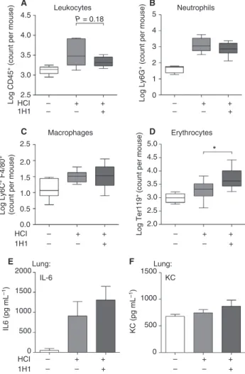
blocking antibody 1H1 intraperitoneally in C57BL/6J lit-termates [33]. We observed no adverse events such as fatal bleeding during the 8-h observation period after intratracheal acid instillation. When we analyzed ALI-induced leukocyte influx in BALF, we found no differ-ences for CD45+leukocytes, Ly6G+neutrophils, or F4/ 80+ macrophages (Fig. 7A–C). In contrast to what was seen in cell type-specific TF-deficient mice, systemic blockade of TF led to a significant increase in erythrocyte numbers in BALF (Fig. 7D), indicating that TF is required to maintain bronchoalveolar barrier function. Accordingly, we did not observe gross changes in IL-6 (Fig. 7E) or KC (Fig. 7F) levels.
Discussion
TF and the extrinsic coagulation cascade are intricately involved in the disease mechanisms underlying ALI. For instance, exacerbated fibrin deposition is detrimental to the function of the alveolar–capillary system by causing barrier dysfunction and hampering gas exchange [34]. We found that myeloid TF deficiency exacerbated the pathol-ogy of acid-induced ALI. Surprisingly, TF deficiency of lung epithelial cells resulted in an unusually long-lasting (24 h) disease pathology of acid-induced ALI, accompa-nied by modest intra-alveolar hemorrhage. This is in line with previous findings showing that epithelial TF exerts protective effects, e.g. supporting the alveolar–capillary barrier, in various infectious and non-infectious ALI models [18,35,36]. For instance, in an LPS-induced lung inflammation model, the presence of epithelial cell TF determines the severity of the lung pathology [18,33]. Whereas, in LPS-induced ALI, myeloid TF is of minor importance [36], we provide evidence that, in acid-induced ALI, deficiency of myeloid TF promoted the inflamma-tory response and thereby significantly exacerbated dis-ease progression. Incrdis-eased inflammation may be the reason for increased TF expression in whole lung tissue, as observed in TFDmye mice as compared with wild-type mice during ALI. Previous studies have indicated that the effect of myeloid TF depends significantly on the disease model. Whereas, in low-TF mice, bacterial burden and cytokine release were unaltered as compared with
wild-type mice during tuberculosis [37], myeloid TF defi-ciency augmented the pulmonary growth of Mycobac-terium tuberculosis, most likely because of reduced fibrin generation and shifting macrophage polarization [38].
Such disease model-dependent differences could explain our observations regarding the role of myeloid TF in ALI. In contrast to LPS-induced ALI, which induces apoptosis in epithelial and endothelial cells [39,40], acid-induced ALI creates a direct cell-damaging disease pat-tern. Intratracheal instillation of HCl mainly affects epithelial cells, leading to impaired epithelial cell function and fluid transport, and indirect damage to the capillaries [41]. Epithelial cell injury was also exemplified by dam-age-associated release of IL-33 into the bronchoalveolar space. IL-33 is abundantly expressed in lung epithelial cells [42]. However, we did not observe any differences in IL-33 levels in BALF between TFDmyemice and wild-type mice, indicating comparable initial insults by acid aspira-tion in both groups. However, during acid-induced ALI, TFDmyemice showed increased pulmonary TF expression, indicating enhanced activation of non-hematopoietic cells, e.g. type I and type II lung epithelial cells, upon acid injury. The most apparent differences in inflammation were significant increases in proinflammatory IL-6 levels and leukocyte/neutrophil influx. These occurred indepen-dently of coagulation activation or the presence of TF on epithelial cells, and was solely dependent on myeloid TF. Although there was a tendency for there to be increased cytokine expression in TFDmye mice during LPS-induced ALI, no significant differences were observed [36].
In line with elevated cytokine levels, we also observed increased recruitment of leukocytes into the bronchoalve-olar space of TFDmye mice, comprising inflammatory macrophages and, particularly, neutrophils; however, total MPO levels were only slightly increased. These find-ings indicate that, although coagulation is not affected by the lack of TF on myeloid cells and, in particular, on alveolar macrophages, the proinflammatory environment created by acid-induced ALI further substantiates the damage. This idea is supported by the significant upregu-lation of proinflammatory cytokines, e.g. IL-6, by isolated TF-deficient alveolar macrophages upon LPS stimulation in vitro.
Fig. 5.Epithelial tissue factor (TF) plays a minor role in acid-induced acute lung injury (ALI). Epithelial TF-deficient and wild-type (WT) mice
were treated with HCl for 8 h. (A) TF mRNA expression of lung tissue was determined by qPCR, and is depicted as fold wild-type control (n
con-trol TF+/+=5;ncontrol TFDmye=3;nHCl TF+/+=9;nHCl TFDmye=10). (B, C) Activated factor X (FXa) generation (B) (ncontrol TF+/+=4;n con-trol TFDmye=2;nHCl TF+/+=6;nHCl TFDmye=4) and thrombin–antithrombin (TAT) complex (C) were measured in bronchoalveolar lavage fluid
(BALF) (ncontrol TF+/+=4;ncontrol TFDmye=1;nHCl TF+/+=9;nHCl TFDmye=7). (D) Protein content in BALF samples
(ncontrol TF+/+=4;ncontrol TFDmye=2;nHCl TF+/+=10;nHCl TFDmye=6). (E) Lung edema was determined by measuring the weight ratio of wet
and dried whole lung tissue (ncontrol TFDmye=2; all othersn=5) (F–H) IL-6 mRNA expression (F) (ncontrol=4;nHCl TF+/+=5;nHCl TFDmye=8)
and IL-6 protein levels in (G) total lung tissue (ncontrol TFDmye=1; all othersn=4) and (H) BALF (ncontrol TF+/+=4;
ncontrol TFDmye=1;nHCl TF+/+=6;nHCl TFDmye=3). (I, J) Flow cytometric analysis of (I) neutrophils (CD45 +
Ly6G+F4/80) and (J)
erythrocytes (Ter119+) in the BALF of epithelial TF-deficient and WT mice (ncontrol TF+/+=4;ncontrol TFDmye=2;nHCl TF+/+=11;
However, neutrophils can also upregulate TF expres-sion during inflammation [5,43]. Infection with influenza virus A/H1N1 induces TF expression on various cell
types, including neutrophils [44]. Thus, we cannot exclude the possibility that lack of TF on neutrophils contributes to the observed phenotype during ALI.
2
HCI – – + +
HCI – – + +
HCI – – + + HCI – –
HCI – – + + HCI – – + +
HCI – – + +
+ +
HCI – – + +
HCI – – + +
HCI – – + + HCI – – + +
HCI – – + +
3
Log CD45
+ (count per mouse)
Log F4/80
+ (count per mouse)
Log Ly6G
+ (count per mouse)
4 5 6
A
D
G
H J
I
E F
B C
2 3
Log CD45
+ (count per mouse)
Log Ter119
+ (count per mouse)
Log Ter119
+ (count per mouse)
Protein content (µg mL
–1
)
Wet/dry ratio
4 5 6
2 3 4 5 6
Log F4/80
+ (count per mouse)
2 3 4 5 6
0 2 4 6
0 0
3 4 5 6
Wet/dry ratio
3 4 5 6 100
200 300 400
Protein content (µg mL
–1)
0 100 200 300 400 2
4 6
0 2 4 6
Log Ly6G
+ (count per mouse)
0 2 4 6 Leukocytes
Leukocytes
Erythrocytes
Erythrocytes BALF protein
BALF protein Edema
Edema Macrophages
Macrophages
Neutrophils
Neutrophils *
*
* *
TF+/+
TFΔepi
TF+/+ TFΔmye
TF+/+
TFΔepi
TF+/+
TFΔmye
TF+/+ TFΔmye TF+/+
TFΔmye TF+/+
TFΔepi
TF+/+
TFΔepi TF+/+
TFΔmye
TF+/+
TFΔmye TF+/+
TFΔepi
TF+/+
TFΔepi
Fig. 6.Leukocyte extravasation into the lung 24 h after HCl-induced acute lung injury (ALI) in epithelial and myeloid tissue factor
(TF)-defi-cient mice. The presence of (A, D) leukocytes (CD45+), (B, E) macrophages (CD45+F4/80+), (C, F) neutrophils (CD45+Ly6G+F4/80)
and (G, H) erythrocytes (Ter119+) in the bronchoalveolar lavage fluid (BALF) was determined by flow cytometry. (I, J) BALF protein was
measured with a bicinchoninic acid assay. (A–C, G)ncontrol=2–3;nHCl TF+/+=5;nHCl TFDepi=7–8. (D–F, H)ncontrol=3–4;
nHCl TF+/+=12;nHCl TFDmye=9. (I)ncontrol TF+/+=5;ncontrol TF+/+=2;nHCl TF+/+=6;nHCl TFDepi=5. (J)n=2–3. For statistical
TF upregulation during ALI results not only in a mild hypercoagulable state but also in an inflammatory state, as FVIIa, FXa, and activated FII can activate protease-activated receptors, which induce the expression of vari-ous cytokines [45]. However, we provide evidence that myeloid TF itself dampens inflammatory reactions via downregulation of macrophage IL-6 expression. Our data indicate that there is TF cell type-specific and disease-spe-cific regulation of inflammatory responses, suggesting that TF acts as a double-edged sword in the cross-talk between inflammation and coagulation.
Taking our findings together, we conclude that myeloid TF has roles in addition to activating the extrinsic coagu-lation cascade, as TF clearly shows immunomodulatory potential. Thus, myeloid TF may potently regulate the intensity of inflammatory responses to exogenous lung insults such as that caused by gastric contents.
Addendum
J. B. Kral-Pointner designed and performed experiments, analyzed the results, and wrote the manuscript. W. C. Schrottmaier and V. Horvath planned and performed research, and analyzed the data. H. Datler was involved in data collection and animal experiments. C. Ay, B. Niederreiter, and L. Hell were involved in data collec-tion. N. Mackman provided conditional knockout mice and supervised the study. B. Jilma and J. A. Schmid were involved in interpreting and discussing the results. A. Assinger and S. Knapp participated in study design and contributed to the discussion and interpretation of the data. G. Schabbauer designed the study, was involved in result interpretation and discussion of the research pro-gress, and wrote the manuscript. All authors confirm that they had full access to the data and the manuscript; they revised its intellectual content and approved the final version.
Acknowledgements
We thank H. Paar, B. Birnecker, and A. Kn€obl for their excellent technical assistance. Floxed TF mice crossbred with LysMcre and SPC cre mice were a kind gift from N. Mackman. We wish to thank C. T. Esmon for kindly providing the fibrin b-monomer-specific antibody 59D8, C. Binder and M. Schwameis for fruitful discussions, and J. S. Brunner, L. Hanzl, and T. Afonyushkin for excellent technical advice. The study was supported by the Aus-trian Science Fund FWF with the projects FWF-SFB 54 and FWF-P24978.
Disclosure of Conflict of Interests
The authors state that they have no conflict of interest.
Supporting Information
Additional Supporting Information may be found in the online version of this article:
Data S1.Methods.
Fig. S1. Genotyping data of myeloid and airway
epithe-lial TF-deficient mice.
Fig. S2. Macrophage recruitment into the lung 8 h after
acid-induced acute lung injury (ALI).
Fig. S3. Myeloid TF does not influence leukocyte
recruit-ment during sterile peritonitis. 2.5
0.0
0 500 1000
IL6 (pg mL
–1)
KC (pg mL
–1)
1500
0 500 1000 1500 2000Lung:
IL-6 KC
Lung: 2.0 2.5 3.0 3.5 4.0 4.5 5.0
*
0.5 1.0 1.5 2.0 2.5
0 1 2 3 4 5
HCI 1H1
– –
+ –
+ +
HCI 1H1
– –
+ –
+ +
– –
+ –
+ +
HCI 1H1
– –
+ –
+ +
– –
+ –
+ + –
– + –
+ + 3.0
Log CD45
+ (count per mouse)
Log Ly6G
+ (count per mouse)
Log Ly6C
+ F4/80
+
(count per mouse)
Log Ter119
+ (count per mouse)
3.5 4.0 4.5
A B
C
E F
D
Leukocytes
Macrophages Erythrocytes P = 0.18
Neutrophils
Fig. 7.Systemic tissue factor (TF) inhibition in acid-induced acute lung injury (ALI). Wild-type mice were intraperitoneally injected with anti-TF blocking antibody (1H1) or vehicle prior to HCl
treat-ment, and parameters were analyzed 8 h after HCl treatment. (A–D)
Extravasation of (A) leukocytes (CD45+), (B) neutrophils
(CD45+Ly6G+F4/80), (C) macrophages (CD45+F4/80+) and
(D) erythrocytes (Ter119+) in the bronchoalveolar lavage fluid
(BALF) was determined by flow cytometry. (E, F) Interleukin (IL)-6 (E) and chemokine (C-X-C-motif) ligand-1 (KC) (F) levels in BALF
were determined by ELISA. (A)ncontrol WT=5;nHCl WT=12;
nHCl 1H1=9. (B)ncontrol WT=5;nHCl WT=8;nHCl 1H1=8. (C) ncontrol WT=7;nHCl WT=8;nHCl 1H1=8. (D)ncontrol WT=7; nHCl WT=10;nHCl 1H1=10. (E, F)ncontrol WT=3;nHCl WT=6; nHCl 1H1=6. For statistical analysis, unpaired Student’st-tests were
performed;*P<0.05. Littermate-controlled experiments were
Fig. S4. No significant changes in IL-12 and IL-1b levels between myeloid TF and wild-type littermates 8 h after acid-induced ALI.
Fig. S5. Effect of thrombin and FVIIa on the
inflamma-tory response of macrophages.
References
1 Mackman N, Taubman M. Tissue factor: past, present, and
future.Arterioscler Thromb Vasc Biol2009;29: 1986–8.
2 Williams JC, Mackman N. Tissue factor in health and disease. Front Biosci (Elite Ed)2012;4: 358–72.
3 Wilcox JN, Smith KM, Schwartz SM, Gordon D. Localization of tissue factor in the normal vessel wall and in the
atheroscle-rotic plaque.Proc Natl Acad Sci USA1989;86: 2839–43.
4 Moosbauer C, Morgenstern E, Cuvelier SL, Manukyan D, Bidz-hekov K, Albrecht S. Eosinophils are a major intravascular
loca-tion for tissue factor storage and exposure. Blood 2007; 109:
995–1002.
5 Maugeri N, Brambilla M, Camera M, Carbone A, Tremoli E, Donati MB, de Gaetano G, Cerletti C. Human polymorphonu-clear leukocytes produce and express functional tissue factor
upon stimulation.J Thromb Haemost2006;4: 1323–30.
6 Steinemann S, Ulevitch RJ, Mackman N. Role of the
lipopolysaccharide (LPS)-binding protein/CD14 pathway in LPS
induction of tissue factor expression in monocytic cells.
Arte-rioscler Thromb1994;14: 1202–9.
7 Drake TA, Cheng J, Chang A, Taylor FB Jr. Expression of tis-sue factor, thrombomodulin, and E-selectin in baboons with
lethal Escherichia coli sepsis.Am J Pathol1993;142: 1458–70.
8 Erlich J, Fearns C, Mathison J, Ulevitch RJ, Mackman N. Lipopolysaccharide induction of tissue factor expression in
rab-bits.Infect Immun1999;67: 2540–6.
9 Cui MZ, Parry GC, Oeth P, Larson H, Smith M, Huang RP, Adamson ED, Mackman N. Transcriptional regulation of the tissue factor gene in human epithelial cells is mediated by Sp1
and EGR-1.J Biol Chem1996;271: 2731–9.
10 Mackman N, Brand K, Edgington TS. Lipopolysaccharide-mediated transcriptional activation of the human tissue factor gene in THP-1 monocytic cells requires both activator protein 1
and nuclear factor kappa B binding sites.J Exp Med1991;174:
1517–26.
11 Oeth P, Parry GC, Mackman N. Regulation of the tissue factor gene in human monocytic cells. Role of AP-1, NF-kappa B/Rel, and Sp1 proteins in uninduced and lipopolysaccharide-induced
expression.Arterioscler Thromb Vasc Biol1997;17: 365–74.
12 Schabbauer G, Schweighofer B, Mechtcheriakova D, Lucerna M, Binder BR, Hofer E. Nuclear factor of activated T cells and early growth response-1 cooperate to mediate tissue factor gene induction by vascular endothelial growth factor in endothelial
cells.Thromb Haemost2007;97: 988–97.
13 Engelmann B, Massberg S. Thrombosis as an intravascular
effec-tor of innate immunity.Nat Rev Immunol2013;13: 34–45.
14 Luo D, Szaba FM, Kummer LW, Plow EF, Mackman N, Gai-lani D, Smiley ST. Protective roles for fibrin, tissue factor,
plas-minogen activator inhibitor-1, and thrombin activatable
fibrinolysis inhibitor, but not factor XI, during defense against
the gram-negative bacterium Yersinia enterocolitica. J Immunol
2011;187: 1866–76.
15 Pawlinski R, Pedersen B, Schabbauer G, Tencati M, Holscher T, Boisvert W, Andrade-Gordon P, Frank RD, Mackman N. Role of tissue factor and protease-activated receptors in a mouse
model of endotoxemia.Blood2004;103: 1342–7.
16 Welty-Wolf KE, Carraway MS, Ortel TL, Ghio AJ, Idell S, Egan J, Zhu X, Jiao JA, Wong HC, Piantadosi CA. Blockade of
tissue factor–factor X binding attenuates sepsis-induced
respira-tory and renal failure.Am J Physiol Lung Cell Mol Physiol2006;
290: L21–31.
17 Bastarache JA, Sebag SC, Clune JK, Grove BS, Lawson WE, Janz DR, Roberts LJ, Dworski R, Mackman N, Ware LB. Low levels of tissue factor lead to alveolar haemorrhage, potentiating murine
acute lung injury and oxidative stress.Thorax2012;67: 1032–9.
18 Shaver CM, Grove BS, Putz ND, Clune JK, Lawson WE, Car-nahan RH, Mackman N, Ware LB, Bastarache JA. Regulation of alveolar procoagulant activity and permeability in direct acute
lung injury by lung epithelial tissue factor.Am J Respir Cell Mol
Biol2015;53: 719–27.
19 Rubenfeld GD, Caldwell E, Peabody E, Weaver J, Martin DP, Neff M, Stern EJ, Hudson LD. Incidence and outcomes of acute
lung injury.N Engl J Med2005;353: 1685–93.
20 Bianchi ME. DAMPs, PAMPs and alarmins: all we need to
know about danger.J Leukoc Biol2007;81: 1–5.
21 Imai Y, Kuba K, Neely GG, Yaghubian-Malhami R, Perkmann T, van Loo G, Ermolaeva M, Veldhuizen R, Leung YH, Wang H, Liu H, Sun Y. Identification of oxidative stress and Toll-like
receptor 4 signaling as a key pathway of acute lung injury.Cell
2008;133: 235–49.
22 Reiss LK, Uhlig U, Uhlig S. Models and mechanisms of acute
lung injury caused by direct insults.Eur J Cell Biol 2012; 91:
590–601.
23 Glas GJ, Van Der Sluijs KF, Schultz MJ, Hofstra JJ, Van Der Poll T, Levi M. Bronchoalveolar hemostasis in lung injury and acute
respiratory distress syndrome.J Thromb Haemost2013;11: 17–25.
24 Faust N, Varas F, Kelly LM, Heck S, Graf T. Insertion of enhanced green fluorescent protein into the lysozyme gene cre-ates mice with green fluorescent granulocytes and macrophages. Blood2000;96: 719–26.
25 Rautou PE, Tatsumi K, Antoniak S, Owens AP III, Sparken-baugh E, Holle LA, Wolberg AS, Kopec AK, Pawlinski R, Luyendyk JP, Mackman N. Hepatocyte tissue factor contributes to the hypercoagulable state in a mouse model of chronic liver
injury.J Hepatol2016;64: 53–9.
26 Pfaffl MW. A new mathematical model for relative
quantifica-tion in real-time RT-PCR.Nucleic Acids Res2001;29: e45.
27 Stojkovic S, Kaun C, Basilio J, Rauscher S, Hell L, Krychtiuk
KA, Bonstingl C, de Martin R, Groger M, Ay C, Holnthoner€
W, Eppel W, Neumayer C, Huk I, Huber K, Demyanets S, Wojta J. Tissue factor is induced by interleukin-33 in human endothelial cells: a new link between coagulation and
inflamma-tion.Sci Rep2016;6: 25171.
28 Geissmann F, Jung S, Littman DR. Blood monocytes consist of
two principal subsets with distinct migratory properties.
Immu-nity2003;19: 71–82.
29 Garcia-Bonilla L, Faraco G, Moore J, Murphy M, Racchumi G, Srinivasan J, Brea D, Iadecola C, Anrather J. Spatio-temporal profile, phenotypic diversity, and fate of recruited monocytes
into the post-ischemic brain.J Neuroinflammation2016;13: 285.
30 Martin NT, Martin MU. Interleukin 33 is a guardian of barriers
and a local alarmin.Nat Immunol2016;17: 122–31.
31 Wicks IP, Roberts AW. Targeting GM-CSF in inflammatory
dis-eases.Nat Rev Rheumatol2016;12: 37–48.
32 Tanjore H, Xu XC, Polosukhin VV, Degryse AL, Li B, Han W, Sherrill TP, Plieth D, Neilson EG, Blackwell TS, Lawson WE. Contribution of epithelial-derived fibroblasts to
bleomycin-induced lung fibrosis. Am J Respir Crit Care Med 2009; 180:
657–65.
33 Kirchhofer D, Moran P, Bullens S, Peale F, Bunting S. A mono-clonal antibody that inhibits mouse tissue factor function. J Thromb Haemost2005;3: 1098–9.
lung injury. Am J Physiol Lung Cell Mol Physiol 2006; 291:
L307–11.
35 Antoniak S, Tatsumi K, Hisada Y, Milner JJ, Neidich SD, Shaver CM, Pawlinski R, Beck MA, Bastarache JA, Mackman N. Tissue factor deficiency increases alveolar hemorrhage and death in
influenza A infected mice.J Thromb Haemost2016;14: 1238–48.
36 Shaver CM, Grove BS, Clune JK, Mackman N, Ware LB, Bas-tarache JA. Myeloid tissue factor does not modulate lung inflam-mation or permeability during experimental acute lung injury. Sci Rep2016;6: 22249.
37 Kothari H, Rao LV, Vankayalapati R, Pendurthi UR. Mycobac-terium tuberculosis infection and tissue factor expression in
macrophages.PLoS ONE2012;7: e45700.
38 Venkatasubramanian S, Tripathi D, Tucker T, Paidipally P, Cheekatla S, Welch E, Raghunath A, Jeffers A, Tvinnereim AR, Schechter ME, Andrade BB, Mackman N, Idell S, Vankayalap-ati R. Tissue factor expression by myeloid cells contributes to protective immune response against Mycobacterium tuberculosis
infection.Eur J Immunol2016;46: 464–79.
39 Kawasaki M, Kuwano K, Hagimoto N, Matsuba T, Kunitake R, Tanaka T, Maeyama T, Hara N. Protection from lethal apop-tosis in lipopolysaccharide-induced acute lung injury in mice by
a caspase inhibitor.Am J Pathol2000;157: 597–603.
40 Wang HL, Akinci IO, Baker CM, Urich D, Bellmeyer A, Jain M, Chandel NS, Mutlu GM, Budinger GR. The intrinsic apop-totic pathway is required for lipopolysaccharide-induced lung
endothelial cell death.J Immunol2007;179: 1834–41.
41 McAuley DF, Frank JA, Fang X, Matthay MA. Clinically rele-vant concentrations of beta2-adrenergic agonists stimulate maxi-mal cyclic adenosine monophosphate-dependent airspace fluid clearance and decrease pulmonary edema in experimental
acid-induced lung injury.Crit Care Med2004;32: 1470–6.
42 Prefontaine D, Nadigel J, Chouiali F, Audusseau S, Semlali A, Chakir J. Increased IL-33 expression by epithelial cells in
bron-chial asthma.J Allergy Clin Immunol2010;125: 752–4.
43 Higure A, Okamoto K, Hirata K, Todoroki H, Nagafuchi Y, Takeda S, Martin JG, Hamid Q. Macrophages and neutrophils infiltrating into the liver are responsible for tissue factor
expres-sion in a rabbit model of acute obstructive cholangitis.Thromb
Haemost1996;75: 791–5.
44 Prutkina EV, Sepp AV, Tsybikov NN. Lung tissue factor expres-sion changes in relation to the stage of respiratory distress
syn-drome in the presence of pneumonia in A/H1N1 influenza.Arkh
Patol2013;75: 16–21.
45 Mackman N. The many faces of tissue factor.J Thromb Haemost