PERIAPICAL MICROSURGERY: THE EFFECTS OF LOCALLY INJECTED DEXAMETHASONE ON POST-OPERATIVE HEALING
Elena V. Kan
A thesis submitted to the faculty at the University of North Carolina at Chapel Hill in partial fulfillment of the requirements for the degree of Masters of Science in the School of Dentistry
(Endodontics).
Chapel Hill 2015
© 2015 Elena V. Kan
ABSTRACT
Elena V. Kan: Periapical Microsurgery: The Effects of Locally Injected Dexamethasone on post-operative Healing
(Under the direction of Peter Z. Tawil)
Substantial inflammation, bruising and pain have been an inevitable consequence of oral surgery. Objectives: To study a protocol to reduce these complications after periapical microsurgery. Hypothesis is that a single local submucosal injection of 4.0mg of dexamethasone at the time of periapical microsurgery can reduce the postoperative
complications. Mate
ACKNOWLEGEMENTS
I wish to thank and acknowledge my fellow endodontic residents: Dr. Melita Islambasic, Dr. Bryan Mitchell, Dr. Tangit Taggar, Dr. Jeffrey Parker, and Dr. Alison St. Paul for their clinical performance in surgeries. I wish to recognize my committee and advisors Dr. Peter Z. Tawil, Dr. Jonathan M. Reside, Dr. Asma A. Kan for their expertise and patience. I wish to make special recognition to Dr. Derek Duggan whose guidance was invaluable.
I wish to thank and acknowledge the American Endodontic Association for their financial support and the Angelus Company for their product support. Also, I wish to thank Dr. Veeral Saraiya for providing statistical analysis.
TABLE OF CONTENTS
LIST OF TABLES ………..………...………… vii
LIST OF FIGURES ………... viii
LIST OF ABBREVIATIONS …..……….………... ix
CHAPTER 1: INTRODUCTION, STUDY DEVELOPMENT AND ANALYSIS ……. 1
Introduction ………..………..….………...………. 1
Review of Inflammation ……….………...………..……….. 3
Review of Steroids ………...………...………....………..… 6
Indications and contraindications of steroids …...…...…… 7
Study Development ………...………..………..…. 11
Sample size ……….……… 11
Patient selection ……… 12
Criteria and restrictions ……….……… 13
Surgical procedure ……….……… 14
Calibration ……….………... 17
Required Information and Measurement ……….………...…… 18
Survey ……….……… 19
Pain ………..……….……… 19
Swelling ……….………..……… 20
Development of the Survey ……….………...…… 22
Double-Blind Procedure ……….……….………...…… 24
Collection of Data …………..……….……….………...…… 25
Analysis ………...………..………..………..…. 26
Overview of General Statistics ………...………...…… 26
Statistical Methodology .………...………...………...…… 27
Results .………...………..………...………...…… 29
Discussion ……...………..………...………...…… 36
References ………..…..……...………..…….……… 40
CHAPTER 2: FORMATED SECTIONS FOR THE JOURNAL OF ENDODONTICS.. 49
Abstract ………..…………..………...…….………...…… 49
Introduction ………..………...…….………...…… 50
Materials and Methods ……...……….……….………...…… 51
Results ………...……..……….……….………...…… 54
Discussion ……….………...………...…… 55
Conclusion ………...…….………...………...…… 58
LIST OF TABLES
Table 1: Case Distribution ………...….. 62 Table 2: Summary of Pain, Swelling, Bruising and Intra-oral healing
by high and low ratings ………….……….………...….. 63 Table 3: Patient Survey Ratings for Pain – Raw Data …….………...… 64 Table 4: General Statistics of the Dexamethasone and Placebo Group’s Pain Ratings.. 66 Table 5: Patient Survey Ratings for Swelling – Raw Data …………...……..……...…. 67 Table 6: General Statistics of the Dexamethasone and Placebo Group’s
Swelling Rating ………. 69
Table 7: Patient Survey Ratings for Bruising – Raw Data ………....……. 70 Table 8: General Statistics of the Dexamethasone and Placebo Group’s
Bruising Ratings ……….……….…...…... 72 Table 9: Patient Survey Ratings for Intra-Oral Healing – Raw Data ……...………..… 73 Table 10: General Statistics of the Dexamethasone and Placebo Group’s
LIST OF FIGURES
Figure 1: Inflammatory Cascade ………...… 76
Figure 2: Inflammatory Cascade, NSAIDs and Corticosteroids ..………..… 77
Figure 3: Patient Survey ………….………...……… 78
Figure 4: Chi-square P values ……… 79
Figure 5: Dexamethasone and Placebo Swelling Rating Frequency Distribution at 24 hours ………...……… 80
LIST OF ABBREVIATIONS AND SYMBOLS
AAE American Association of Endodontics ACTH Adrenocorticotropic hormone
CRP C-reactive protein
ESR Erythrocyte Sedimentation Rate
HIPAA Health Insurance Portability and Accountability Act IDS Investigational Drug Services
IRB Internal Review Board MAP Micro apical placement MG Milligrams
MTA Mineral trioxide aggregate
NSAID Nonsteroidal anti-inflammatory drug
CHAPTER 1: INTRODUCTION, STUDY DEVELOPMENT AND ANALYSIS
Post-treatment complications in endodontics continue to be a significant problem in
dentistry. Initial endodontic therapy does not produce an acceptable treatment outcome in
approximately one-third of all cases and, therefore, endodontic retreatment is required (
Introduction
1).
Analysis of surgical and non-surgical retreatment therapies (2) have found that healing rates
are not substantially different between the two approaches (3).
Traditionally, the cost of retreatment is higher since treatment typically takes multiple
appointments. This is in contrast to a surgical retreatment which can be performed in a single
appointment. Furthermore, a traditional retreatment approach requires access through the
crown and can involve replacing the crown. This is in contrast to periapical surgery that only
involves resection of soft tissue, bone and root structure without penetrating the crown
structure. In addition, there is a high frequency of flare-ups experienced by patients who are
treated with the traditional orthograde retreatment of root canal therapy (4). Periapical
surgery is a more efficient and less expensive method for retreatment relative to the
orthograde method which involves removing posts, root canal filling material, and often
replacing a crown (3, 5).
Periapical surgery is regarded as an integral part of modern endodontics (6, 7).
Endodontic surgeries account for approximately 6 to 10% of the typical endodontic practice
and long term evaluations, periapical surgeries have a 91 to 97% healing success rate (10, 11)
making periapical microsurgery a good treatment option.
Research indicates that inflammation, pain, swelling, and bruising have long been an
inevitable consequence of oral surgery (5). The same research indicates that post-endodontic
surgical discomfort can cause 23% of patients to miss work (5). Some studies have found that
patients with pre-operative endodontic pain will continue to have post-treatment pain in up to
80% of the cases (12-14). The greatest concern among endodontic surgery patients is
experiencing pain (10). According to one study, patients experience their peak pain by the
end of the day of their endodontic surgery (5). Although all patients endure some level of
pain, approximately two-thirds of the patients that undergo endodontic surgery require
analgesics to lessen their pain (15).
Pain is a complex process that involves sensory, emotional, and conceptual aspects
(16). Post-treatment analgesic intervention is necessary in many endodontic cases to manage
pain. There are various classes of drugs that have been proposed in the management of
post-treatment endodontic pain and discomfort. These classes include NSAIDs, Acetaminophen,
opioids, and corticosteroids (12, 14, 15, 17). Research has proposed that corticosteroids are
effective in treating pain (12, 14).
The majority of evidence evaluating post-surgical discomfort and complications
utilizes the oral surgery third molar extraction model and views the aspect of pain as a
consequence of the acute inflammatory reaction. However, pain in endodontic origin differs
from this model in that it is associated with chronic inflammation (18), the presence of
Wound healing or repair after surgical procedures is an important component of inflammation
(20) and is a necessary physiological response of the body to the injury (21).
Review of Inflammation
Inflammation has been defined as “the local reaction of vascularized tissue to injury”
(22). Manipulation of the soft and hard tissues during endodontic surgery leads to an
inflammatory cascade via biological mediators (e.g. prostaglandins, leukotrienes, bradykinins,
histamine, serotonin, and others) that are released from blood vessels and cells in the injured
area (22) and serve a specific role at each stage of the inflammatory process (20). This
inflammatory cascade results in pain and vasodilation which leads later to edema and
hematoma in the injured area (21).
Inflammation can be divided into three stages: acute, chronic, and repair with no clear
dividing line among them (20, 23). Periapical microsurgery involves all three stages: chronic
inflammation due to the periapical lesion that is present at the time of treatment, acute
inflammation at the time of hard and soft tissue manipulation, and repair after conclusion of
the surgical intervention.
Acute inflammation is characterized by the transudation of leukocytes into the tissue,
whereas chronic inflammation features the presence of leukocytes in the tissues (23). Acute
inflammation is an exudative process where small vessels become permeable. This allows
plasma proteins and fluid to leave the bloodstream and enter the tissue to form a loose
network of fluid, fibrin, and white blood cells (20). Chronic inflammation, on the other hand,
is a proliferative process with the presence of fibroblasts and angioblasts (20) and nerve
inflammation involves mononuclear inflammatory cells, such as macrophages, lymphocytes,
and plasma cells (23, 24).
The first event in any inflammatory reaction is tissue injury. After the initial injury, a
transient vasoconstriction occurs and is followed by vasodilation of the tissue. The injured
tissue becomes painful, hot, erythematous, and edematous (20, 21). There are many
biochemical mediators that take place to contribute to the symptoms and signs of
inflammation.
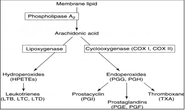
Arachidonic acid is a major precursor of inflammatory reaction and its metabolites are
important mediators in the inflammatory cascade (Figure 1). Major arachidonic acid
derivatives include prostaglandins and leukotrienes. Both prostaglandins and leukotrienes are
responsible for delaying and prolonging stages in vascular permeability. They are long-chain,
lipid-soluble fatty acids that are present in all tissues and are formed within seconds after
various stimuli. In inflammation, macrophages and neutrophiles are responsible for the
production of both prostaglandins and leukotrienes. Arachidonic acid serves as the precursor
to both prostaglandins and leukotrienes and is produced by the action of phospholipase A2.
Phospholipase A2 is an enzyme that is found in all human cells.
The oxidation of phospholipase A2 leads to the formation of arachidonic acid from
cell membrane phospholipids. Once arachidonic acid is formed, it is metabolized by two
major enzymes: cyclooxygenase or lipoxygenase. The cyclooxygenase pathway involves
prostaglandins, which produce vasodilation of tissues and increase of vascular permeability.
The lipoxygenase pathway involves leukotrienes (21).
All of the components of inflammatory cascade cause patient discomfort.
One method to decrease these critical signs of inflammation is through the use of
anti-inflammatory agents. These agents aid in the reduction of unpleasant side-effects of the
inflammatory cascade through the inhibition of the steps in the formation of arachidonic acid
and its metabolites. The potential for anti-inflammatory agents to prevent pain, swelling, and
bruising depends on the suppression of the release of the inflammatory mediators. A decrease
in the amount of inflammatory mediators present leads to a reduction in vascular permeability.
This in turn decreases fluid accumulation within tissues, resulting in a decreased tissue
pressure that translates to less pain, swelling and potential bruising (14). Various medications
have been used to interfere with inflammation to prevent or stop pain, swelling and/or
bruising. Two commonly used classes of medications for this are NSAIDs and
corticosteroids.
NSAIDs are cyclooxygenase inhibitors and prevent formation of prostaglandins and
thromboxanes from arachidonic acid (25), but do not affect the lipoxygenase pathways
(Figure 2). In contrast, corticosteroids prevent the release of arachidonic acid which inhibits
both inflammatory pathways and effectively prevent inflammation (26-28). As a result, the
anti-inflammatory efficacy of corticosteroids is more pronounced than NSAIDs, which has led
to their use after surgical procedures (29-32).
Many different steroids are currently available on the market. One of the first clinical
applications of corticosteroids was the use of compound E, cortisone, compound F, and
hydrocortisone, in the treatment of rheumatic fever was in the late 1940’s (33). Past studies
have primarily focused on the use of glucocorticosteroids (cortisone and hydrocortisone) in
dental applications. Dexamethasone is one of the most recent corticosteroids to become
sequelae (10, 29-32, 34-40) that typically accompanies oral surgery. The anti-inflammatory
effect is a result of the suppression of the migration of neutrophils, leukocytes, and
macrophages through the inhibition of the formation of arachidonic acid, thus blocking the
cyclo-oxygenase and lipoxygenase pathways and respective synthesis of prostaglandins and
leukotrienes (41, 42). Corticosteroids can be delivered through a variety of methods,
including orally, intravenously (37, 43), intramuscularly (12, 14, 31), submucosally (36),
intraligamentary (29, 30, 38, 40, 41, 44), supraperiostealy (45) and intraosseously (46, 47).
The submucosal injection enables the application of anti-inflammatory agents at a precise site
to effect a pharmacological action in sufficient quantities. More importantly, dentists are
familiar with submucosal injections over other techniques.
Review of Steroids
The adrenal cortex synthesizes corticosteroids from cholesterol. Corticosteroids
contain 21 carbon atoms in a 4 member hydrocarbon ring. Adrenal corticosteroids are
necessary regulators of homeostasis (48). These corticosteroids are produced naturally and
include different classes, mineralocorticoid (aldosterone), the sex hormones (testosterone,
estrogen, progesterone), and glucocorticosteroids (cortisol). Aldosterone affects the human
body’s water and electrolyte balance. It is primarily secreted by stimulation of the kidney’s
renin-angiotensin system. Therefore, water and electrolyte balance are not affected by
suppression of adrenal glands (48). Sex hormones are produced by gonads and adrenal glands
(48).
Glucocorticosteroids act on multiple sites to inhibit immune and inflammatory
reactions. Cortisol is the primary glucocorticosteroid that is synthesized by the body and is
pituitary glands. Along with the adrenal cortex, these structures make up the
hypothalamic-pituatatry-adrenal axis. This system regulates glucorticosteroid synthesis. The hypothalamus
produces corticotropin-releasing hormone, which travels to the anterior pituitary gland via the
hyphothalamic-hypohyseal portal system. This corticotropin-releasing hormone stimulates
release of the adrenocorticotropic hormone (ACTH) by the pituitary gland (49). ACTH is the
main regulator of cortisol. The metabolism of corticosteroids occurs in the liver, and later,
through excretion in the urine (41, 42, 48).
The chemical modification of cortisol produces a number of synthetic corticosteroids.
Synthetic corticosteroids, as well as cortisol, are 90% bound to plasma proteins (albumin and
corticosteroid-binding globulin). Only a small unbound portion of corticosteroids are free to
enter the cell and mediate the anti-inflammatory effect at any time. Although cortisol
normally has a half-life of 90 minutes, chemical modifications to its composition can cause it
to have a greater anti-inflammatory effect and an increased duration of action.
Indications and Contraindications of Steroids
There are many applications of corticosteroids in dentistry. Oral surgery studies have
used dexamethasone for reducing edema, pain, and trismus after extraction of third molars
utilizing various injection methods (31, 36, 37) and oral formulations (32, 35, 38). Research
has demonstrated a pain reduction of 50% when dexamethasone was used pre- and
post-operatively after extraction of 3rd molars (32). More than 1/3 of patients traditionally require post-operative analgesics after surgical extraction of third molars. Evidence suggests that 8.0
mg of dexamethasone administered orally significantly reduced post-operative pain (35).
Early case reports indicated the use of corticosteroids in dentistry for alleviating
Pre-operative administration of oral dexamethasone in a periodontal surgery model
finds that it is effective in reducing pain after periodontal surgeries for the initial eight hours
following treatment (30). The same author found in another study that it is better to use a
single oral dose of 8.0 mg of dexamethasone than two doses of 4.0 mg of dexamethasone in
reducing pain one-hour prior to periodontal surgery (29). Patient compliance and the need for
repeated doses to sustain adequate steroid concentration can become problematic with oral
administration. It is more predictable and effective to inject the dexamethasone at the time of
the surgery rather than using the oral route.
An animal study showed that the supraperiosteal infiltration of dexamethasone into the
submucosal tissue had a significant anti-inflammatory effect on the injured periapical tissues
(61). The study also stressed the importance of patient compliance and use of an infiltration
method instead of oral administration. Submucosal deposition of dexamethasone is the
preferred method of delivery, as an application at the site of inflammation provides the
maximum anti-inflammatory effect on tissues. Intramuscular administration has been shown
to have the same effect as oral infiltration (14). However, a practitioner should be
experienced in giving an intramuscular injection as accuracy is paramount. The oral
infiltration injection is a familiar procedure in dentistry. Another animal study found similar
effects of dexamethasone that was deposited and absorbed in the maxilla and mandible (62).
This finding can be used to prevent flare-ups after endodontic treatment and periapical
microsurgery using similar doses for the maxilla and/or mandible.
Studies have demonstrated that corticosteroids can prolong pulpal anesthesia with
inferior alveolar nerve blocks in patients that experience the painful condition of irreversible
painful and not recommended as a supplemental treatment in patients with irreversible pulpitis
(54).
Corticosteroids have been an effective treatment for the injured and/or compressed
inferior alveolar nerve after the extrusion of root canal filling materials (55).
There are documented cases of corticosteroids being used in instances of sodium
hypochlorite incidents with or without neurological deficit during endodontic procedures (56,
57), thus, promoting recovery.
In some instances, placing corticosteroids locally into the root canal space prevents
and possibly treats flare-ups after endodontic treatments (17, 52, 58-60).
Supra-periosteal infiltration with a single dose of 4.0 mg of dexamethasone has been
found to be effective in reducing acute pain after endodontic treatment if administered within
the first 24 hours, but not more than 48 hours (45). Most endodontic patients with acute pain
experience pain even after endodontic treatment had been performed (5, 58).
Some endodontic microsurgery protocols incorporate oral dexamethasone to be
administered preoperatively and postoperatively (34, 39, 40). One endodontic surgical model
suggested that the routine use of oral dexamethasone is a safe method to reduce pain and
swelling after endodontic surgery (10).
Corticosteroids at higher levels with multiple dosages have been found to cause
adrenal fatigue and can mask symptoms of bacterial infection (48, 49). One study showed
that a one-week course of corticosteroids is not harmful, but instead, very effective in
reducing post-surgical dental pain and swelling (15). The important aspect of this study was
that patients required less NSAIDs and opioids to control pain. A single dose of
(39). One of the earlier studies concluded that a single large intravenous dose of
dexamethasone (2.0 milligrams per kilogram of body weight) does not have any harmful side
effects (43).
Some studies raised concerns regarding expanded uses of corticosteroids in dentistry
due to their adverse reactions. These studies concluded that corticosteroids can cause
psychosis, memory disturbances, and hallucinations (63-65). These case documented reports
are based upon small doses that patients were taking for prolonged periods of time. Patients
who suffer from mental illnesses, pregnant woman, immune-compromised patients (i.e.
Cushing’s syndrome, tuberculosis, systemic fungal infection, uncontrolled diabetes), chronic
pain patients, or patients with hyperthyroidism should not take systemic corticosteroids for a
prolonged period of time (48, 66-69). Based on the above, this study’s small single dose of
corticosteroids at the conclusion of the periapical microsurgery is safe, effective and efficient
Determining the sample size for a research survey is the task of choosing the number of observations or patients to include in the statistical sample. The goal of this study was to make statistical inferences about the population from the sample. As with any empirical study, the sample size was of paramount importance. In som
Study Development
Sample Size
be divided into different In this case, for the sake of statistical tests and administrative ease, the same number of patients were placed in each group.
The statistical power of a sample size relies on many subjective estimates. There has been little endodontic research in this specific area. Therefore, there was no specific proxy as to expected variance or sample size. The only guidance was based upon anecdotal evidence from oral surgeons’ experience with an injection-form of dexamethasone.
After balancing the statistical methods to be employed, costs, and timeline, it was estimated that the sample size of sixty patients (n = 60) would provide the necessary statistical significance to make valid conclusions. Patients were selected from those that were currently or previously recommended for endodontic periapical microsurgery by the UNC Dental School Endodontic Department. Participants were randomly placed into two groups by UNC
Investigational Drug Services (IDS). The first group of participants, which totaled thirty (x = 30), received an injection of 4.0 mg of dexamethasone solution. The second group of
n = x + y Where,
x = Number of dexamethasone group patients y = Number of placebo group patients
n = Total population of the sample
Patient Selection
This study involved sixty (n = 60) patients that were prequalified and selected for endodontic periapical microsurgery from the UNC Dental School Endodontic Department’s patient files. This study included the first sixty qualified patients on a first-come-first-served basis that agreed to participate. Current patients that would likely qualify were considered immediately. Endodontic residents in the department were aware of the research and alerted the patient and the principal investigator of a potential case. Clinical and radiographic exams were used to determine patient qualifications. The selection process sought males or females of at least eighteen years of age. Patients were required to be in relatively good health with no significant medical conditions.
All root canal treated cases with apical periodontitis were eligible for inclusion where previously endodontically treated teeth had non-healing lesions, endodontic retreatment was impossible (post, anatomy), or cases with a high possibility of failure after a traditional root canal treatment. All teeth groups (anterior, premolars, molars, maxillary or mandibular teeth) were eligible for the study.
or III mobility, horizontal/vertical root fractures, combined endodontic-periodontal lesions, compromised crown-to-root ratio or patients with systemic conditions were removed from consideration.
Written and verbal informed consents were obtained from qualified participants. Among much other information, patients were informed that they may or may not actually receive the drug dexamethasone. Patients were informed that they might actually receive a placebo. Given the nature and importance of these communications and the survey, those not literate in English were removed from consideration.
Criteria and Restrictions
Patients were subject to certain study criteria and restrictions with the aim of upholding the validity of the study while maintaining a safe procedure for patients. As with most clinical studies, one goal of the criteria and restrictions is to create a uniform environment. The study boundaries can be used to isolate the event to be studied from the influence of other factors. This provides the truest cause-and-effect result.
Clinical and radiographic examinations as well as a medical history review were used to determine whether a potential candidate’s qualifications adhered to the criteria and restrictions.
Another goal of the study conditions is to maintain patient health and safety. The study submitted to the stringent conditions and restrictions place upon it by the IDS and IRB. Patient candidates whose physical qualities might overshadow the study results were removed from consideration. Disqualification was also done in some instances out of consideration of the candidate’s health and safety.
unforeseen risks of the study.
Candidates were questioned pre-operatively about their health and medications that they were taking. As all candidates were current patients at the UNC Dental School, merely
updating their records was the primary task. As current patients, this provided a preliminary screening of that patient’s vital readings (blood pressure and pulse) to ensure they were within safety standards as set in the IDS and IRB as well as the school’s standards. Candidates that were pregnant were removed from consideration. Although the drug dosage was small and local, pregnant candidates were removed from the selection out of an abundance of caution for the fetus. Females of child-bearing age were offered a pregnancy test to determine if they were pregnant. Every female patient was required to give verbal confirmation that she was not pregnant or planning to become pregnant prior to inclusion in the study.
Surgical Procedure
All research and surgical consents were filled out by the participants prior to the surgical procedures. With the exception of the incisions and suturing, all microsurgical procedures were performed using a Surgical Operating Microscope (Global G6 Microscope, Global Surgical Corporation, St. Louis, MO). All surgeries were performed using modern microsurgical techniques (70, 71). All participants were required to rinse their mouth with 0.12 % chlorhexidinegluconate rinse (Peridex 3M ESPE) for one minute immediately prior to the periapical microsurgery. Chlorhexidine mouth rinse plays an important role in
pre-disinfection of the surgical area. About 30% of chlorhexidine may be retained in the mouth after rinsing for one minute (72) and once bound to the oral tissues, chlorhexidine can be released for up to 12 hours for a prolonged bacteriocidal effect (72, 73).
the area of the surgery via local infiltration. In addition, a 2% lidocaine solution with 1:50,000 of epinephrine was administered in the planned incision area in order to achieve improved hemostasis. Local anesthesia serves two purposes: to prevent pain during surgery and to minimize surgical hemorrhage due to vasoconstriction. A 2% lidocaine solution with epinephrine is the anesthetic of choice since it activates alpha receptors that are present in the muscles of the arterioles, periodontium, and submucosa causing vasoconstriction (74, 75).
After anesthesia, a #15C surgical blade was used to make papilla-base incisions and vertical releasing incisions to allow adequate access to the surgical area (76, 77). A coronal split-thickness and apical full-thickness mucoperiosteal flap was used to ensure
standardization and to allow for the most esthetic outcome (78-80). Participants were made aware of the risk that gingival recession might occur after the periapical microsurgery (81, 82).
If necessary, osteotomies were performed using a #6 round carbide bur in a high-speed impact air hand piece (Sybron Endo, USA) under copious water irrigation. The modified surgical hand piece used a 45-degree angulated head for visibility and no air ejection to prevent emphysema. Carbide burs were used during the osteotomy to ensure safe and clean cutting. Water irrigation was necessary to minimize thermal injury to the adjacent bone (83, 84).
The apicoectomy was performed with a 0-30% bevel (87) by sectioning 3.0 mm of the root tip (88) with a high-speed surgical hand piece using a multi-purpose bur (Dentsply Maillefer, Milford, DE) under copious water irrigation. A resection of at least 3.0 mm of the root tip was completed to ensure the best healing potential due to the potential for accessory anatomy in the apical portion of the root (88). A minimal bevel was desired because it required a smaller osteotomy, minimal loss to a buccal cortical plate, and eliminates missed anatomy (87, 89).
The resected surface of the root was stained with methylene blue dye (Vista BLUE, VISTA Dental Products, USA) and inspected under a microscope using a micro-mirror to detect any cracks, fractures, dentinal defects or missed anatomy. The microscope’s trans-illumination and the methylene blue aided in detecting the etiology of the non-healing endodontic lesions (90).
The root-end preparation was performed at least 3.0 mm into the canal space following the long axis of the tooth using ultrasonic surgical tips (Obtura, Spartan) under sterile saline irrigation. Using ultrasonic surgical tips for the end-root preparation ensured a smooth surface (91) and appropriate depth/diameter for placement of the retro-filling material. Cleaning deep isthmuses (92) with any other means does not guarantee a favorable result (93, 94).
created a seal to prevent the ingress of microorganisms or their byproducts into the canal. There are many acceptable retro-filling materials on the market (96, 97). Evidence suggests that MTA provides the best seal (98). In addition, MTA is biocompatible and has a high pH balance upon setting (99-101). This study used a white MTA-Angulus (Angelus Dental Solutions, Londrina, Parana, Brazil) due to its excellent handling property and predictable setting time (98, 100, 101). A post-operative radiograph was taken to confirm the adaptation of the root-end filling to root-end preparation. Following completion of apicoectomy, the mucoperiosteal flap was irrigated with copious sterile saline solution.
The soft-tissues were then repositioned with 5.0 and 4.0 chromic gut sutures to obtain primary closure. The sutures maintain the position of the flap during the initial healing via primary intention. The approximation of the papilla with a smaller suture size gave the best esthetic outcome (80, 102). A local administered submucosally injection of dexamethasone 4.0 mg or placebo saline solution was then placed at the conclusion of the surgery.
All participants, regardless of the group in which they were assigned, received the same standard post-operative care instructions in verbal and written form. All participants were also given a standardized post-operative surgery prescription for pain control for the following three days to be taken if needed. The prescription included twelve 500 mg tablets of Tylenol and twelve tablets of Vicodin 3/500. Participants were instructed to take both tablets every 6 hours if they experienced pain. Participants were informed not to take more than 4 dosages of either medication in a 24 hour period.
Calibration
of the periapical microsurgeries. Prior to participation in this study, each resident had performed at least forty endodontic surgeries. Besides having been trained in the same endodontic program, all participating endodontic residents attended a 6-hour lecture and watched a video on the standardized surgical protocol prior to the study. In addition to this calibration and standardization, the principal investigator performed or assisted in all of the research surgeries. In order to avoid variances/errors during each procedure, the principal investigator, when not performing research surgery, was assisting in order to verify accurate papilla-base flap design and flap reflection, standard osteotomy, curettage, root-end resection, root end-preparation and the placement of MTA (confirmed with a radiograph), and suturing. At the conclusion of every surgery, the primary investigator administered the study protocol injections at the mucobuccal fold, adjacent to the target tooth with advancement of the needle to approximate the osteotomy site. As a last safety measure, an attending endodontic faculty closely monitored all surgical procedures to ensure quality of care.
All of the residents and the patients that were participating in the study were informed that the principal investigator could intervene at any time during the surgery in order to be consistent with the study protocol. Only the principal investigator performed the follow-up evaluations for uniformity of the results. Given the above, the standardization and oversight within an endodontic resident program minimized the chance of error or variance in the procedure.
Required Information and Measurement
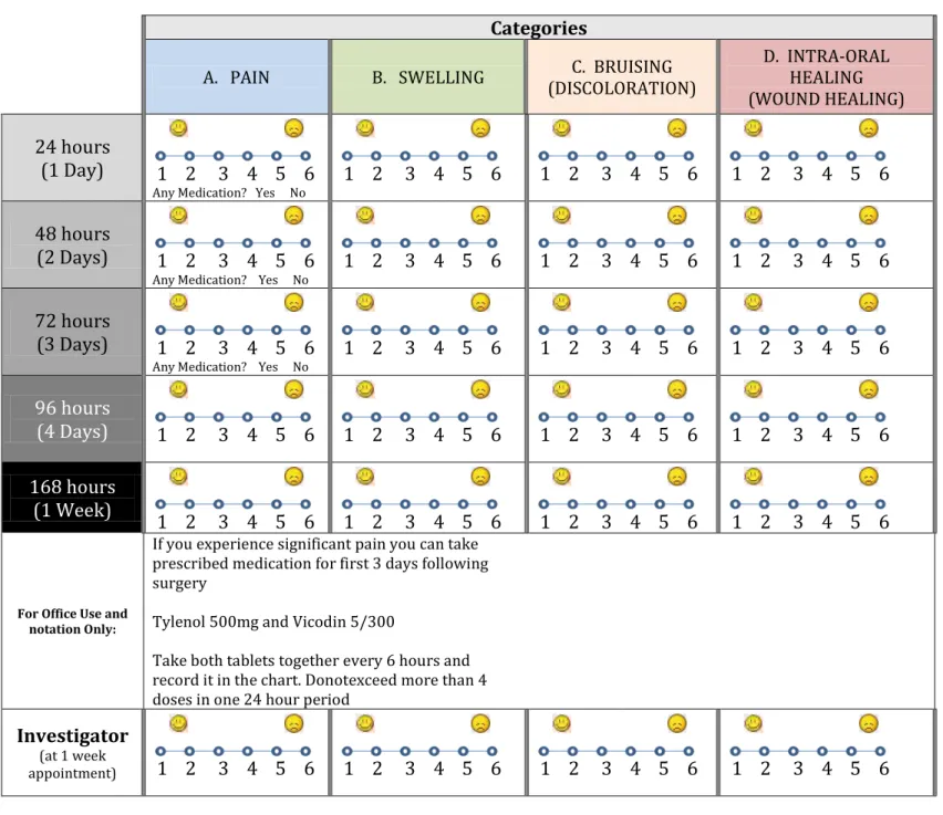
Pain, swelling, bruising and intra-oral healing were the four general areas of recovery that the study assessed (Figure 3). It was necessary that the study measure these areas from the standpoint of the patient experience as well as the perspective of the investigator for consistency and confirmation. Photographic and survey documentation at pre and post-operative
examinations provided data for analytical comparison.
The patients were instructed to complete the self-evaluation surveys on a 6-point likert-like visual analog scale for each of the post-operative categories of pain, swelling, bruising, and intra-oral healing (Figure 3). The visual analogue scale is
Survey
in subjective post-operatively. Patients were informed of the characteristic pain, swelling, bruising, and intra-oral healing that one would anticipate after such a procedure. When responding to an item in this survey, patients were to indicate their rating by indicating a position along a continuous line between two end-points. Patients made observations at four consecutive 24-hour intervals and recorded their experience on the survey. One week after the surgery, patients met with the investigator to submit the subject’s survey responses and complete a clinical evaluation. This method allow for the study of absolute levels of healing as well as the rates of recovery.
The pain scale’s purpose was to measure the Pain
the different dimensions of pain they experienced to develop consistency among patients in the investigator’s rating. The questions included the site of the pain, such as: “Where specifically is the pain?” The patient was questioned about the type of pain, such as: “What does the pain feel like?”, or, “Did the pain impact your everyday life?” Patients were also asked questions regarding exacerbating or relieving factors, such as: “Was there anything that made the pain worse or better?”, or, “Was it necessary to take pain relief medication?” Patients were instructed to take 500 mg of Tylenol and Vicodin 5/300 in order not to affect the observations inflammation. The relative pain level assessment at each interval was regarded as an important element in judging recovery. Because pain is a subjective and an internal experience, the evaluation of pain in this study was best performed by using patient self-reports. This appears to be the most frequently used technique in other pain studies (12, 15, 25, 29, 30, 38).
Measuring the change in acute phase reactants is one method of detecting
inflammation. The body reacts to inflammation by changing the manufacturing of protein in the liver and other protein creating organs. Acute phase reactants are proteins whose blood levels are altered by inflammation. Two techniques for measuring the change in acute phase reactants are the Erythrocyte Sedimentation Rate (ESR) and the C - reactive protein (CRP) (112, 113). These techniques measure the rate at which red blood cells settle over a small interval of time. The rate is directly proportionate to the amount of reactant proteins that are Swelling
Swelling of tissue creates a tight barrier which keeps bacteria out. The increased blood supply provides a defense mechanism. Inflammation’s basic role of isolating injury,
present. The presence of inflammation increases the amount of proteins in the blood,
accordingly, the rate of production increases. Unfortunately, the rates are not specific and can be altered by other circumstances such as anemia. The levels for both ESR and CRP can be influenced by both gender and age. Finally, and most importantly, inflammation due to other causes is not distinguishable from swelling caused by the surgical procedure. The size of the surgical wound and swelling would be relatively small and short in duration in contrast to other whole body causes.
Like pain, inflammation is almost always present with a surgical procedure. The swelling scale’s purpose was to measure the degree and persistence of inflammation in and around the site. In this study, the swelling score was based on self-report by the patient’s daily survey observations as well as the investigator’s perspective. This was deemed to be a better approach in contrast to ESR testing given the specific and local nature of the wound, duration of recovery and expense and time of ESR testing.
Innovative measures of bruising, such as Electrical Impedance Measurement, were not a practical application in this study (114). Many methods for measuring bruising are
destructive in nature as they require penetration, piercing and cutting the tissue. Infrared Bruising
spectroscopy, hyperspectral imaging, thermal imaging and nuclear magnetic resonance imaging are cutting edge technologies that are non-destructive (115-118). However, after consideration, they are unproven technologies in dentistry, impractical for the oral cavity, exceeded study time constraints and/or cost prohibitive for this study.
Unlike pain and inflammation, significant bruising does not always accompany an oral surgical procedure. The bruising scale’s purpose was to measure the degree and persistence of bruising in and around the site. In this study, the bruising score was based on self-report by the patient’s daily survey observations as well as the investigator’s perspective.
The goal of the survey was to provide the patient with a document to record and collect data on their recovery experience. Pain, swelling, bruising (discoloration) and intra-oral healing (wound healing) are the four areas of recovery that the patient assessed (Figure 3). Factual information was included on the form such as date, coded patient identifier, investigator’s name, Intra-Oral Healing
Periapical surgery is an invasive procedure. There is no alternative to the process of gingival incisions and root resection. This process of removing unhealthy tissues and requires damaging the surrounding healthy tissue to access the root. The surgical wound can be a source of general discomfort during the recovery period (34).
Part of surgical wound assessment is wound measurement. Due to the variation in access points and severity of incisions, it was difficult to use a consistent technique among cases. Some patients were unable to retract their cheek and/or lips to evaluate the surgical site. In these instances, patients were asked not to evaluate their intraoral healing to avoid disruption of the sutured site.
tooth type and the diagnosis.
The survey was created to be simple and easy to use. Although English literacy was required of patients for participation, minimal instructions with simple words were used in the document’s instructions. Qualitative description of categories can impact how patients use the rating scale. For example, if only the points 1-6 are given without description, some might rarely select 6, whereas others may select the category often. If, instead, "6" is described as "near the maximum," the category is more likely to mean the same thing to different people. This could apply to all categories and not just the extreme points. Smiling and frowning emoticons were used instead of descriptive words to qualitatively characterize the scale and minimize the different interpretations. Although instructions were to be given to patients post-operatively when delivering the survey to the patient, it was created to be self-explanatory.
The featured in surveys. It is often used to measure attitudes and other factors. The original scale featured five points. Over time, there have been many discussions and disagreements focused on what works best with the likert scale to give the most accurate responses. Most agree that more than seven points on a scale are too much. Studies show that people are not able to distinguish a scale greater than seven. Studies are not conclusive on which number scale is best.
made observations at four consecutive 24-hour intervals and recorded their experience on the survey. One week after the surgery, patients met with the investigator to submit their survey responses and complete a clinical evaluation.
At the one-week meeting, the principal investigators rated the patient’s different healing categories. This allowed for verification of the magnitude of the patient’s ratings. Ratings at the various time intervals allowed for the study of absolute levels of healing as well as the rates of recovery in the category over time.
Double-Blind Procedure
A blind experiment is a or service that might lead t after the test. An open trial where such information is not concealed is vulnerable to such intentional or unconscious biases. The blind technique is used to eliminate human bias or influence of the study results.
A basic blind study would be an experiment where only the subject was unaware of information of the product or service being received. The person or investigator conducting the study would know beforehand which subjects were receiving which product or service. Researchers suggest that the person or investigator administering the experiment could influence the results of such studies by picking and choosing which subjects received which product or service. This would degrade the randomness of the study and could introduce bias.
against both experimenter bias and placebo effects. This study was performed under double-blind conditions.
The IDS at UNC provided control to ensure the double-blind conditions.
Randomization of who received the drug or placebo injection was performed by the IDS. IDS also maintained custody of the syringes and were the only ones privy to who received which injections. Once notified of upcoming surgeries, IDS provided the identically prepared syringes the morning of the surgery. The identity of which patients received which injection was unknown to all operators, examiners, and resident surgeons and patients. Only after the completion of the treatments and examinations were the identities of the injections revealed.
Collection of Data
The collection of data in this study began with the initial consultation and concluded with the post-surgery evaluation.
Patients were scheduled to return one week after their surgical procedure with their completed survey. Photographs, clinical assessment and notations were made at the post-operative appointments by the principal investigator. There were two instances where the patients forgot to bring the survey document to their follow-up appointment. One patient sent the survey form to the investigator electronically and the other dictated survey form ratings verbally via telephone shortly after their appointment. Completed survey documents were stored in individual envelopes until the data analysis was initiated.
The distribution of race that were treated in the dexamethasone group included one of Asian descent, seven of African-American descent, three of Hispanic descent and nineteen of Caucasian descent. The distribution of race that were treated in the placebo group included one
Analysis
Overview of General Statistics
All sixty patients completed their surgical procedures, completed their surveys and attended their post-operative evaluations. Upon conclusion of the study, the information held by the Investigational Drug Services was then released to the study’s investigator. General statistics were compiled about the total sample and groups before the statistical methodology was applied and hypothesis testing began.
The total sample of the patients consisted of twenty-seven males and thirty-three
females. The patients’ ages were categorized as forty years old or under or forty-one and older. Fourteen were in the younger age group and forty-six were in the older age group. The race of the total sample included two of Asian descent, thirteen of African-American descent, six of Hispanic descent and thirty-nine of Caucasian descent (Table 4). The distribution of teeth that were treated included eight mandibular/anterior, thirteen mandibular/posterior, twenty-five maxillary/anterior and fourteen maxillary/posterior.
of Asian descent, six of African-American descent, three of Hispanic descent and twenty of Caucasian descent. Seven of the total sample patients were regular smokers of tobacco. Five of the thirty in the Dexamethasone Group were regular smokers of tobacco. Two of the thirty in the placebo group was regular smokers of tobacco.
The distribution of teeth that were treated in dexamethasone group included four mandibular/anterior, six mandibular/posterior, thirteen maxillary/anterior and seven
maxillary/posterior. The distribution of teeth that were treated in placebo group included four mandibular/anterior, seven mandibular/posterior, twelve maxillary/anterior and seven
maxillary/posterior.
Supportive Chi-square and Fisher’s exact tests were performed to determine if the distribution of patients between the two groups was significantly different. The distribution of patients based on age, race, gender, tobacco users and teeth did not appear to be significantly different between the two groups. All p-values for the tests were p>0.05 which resulted in failure to reject the null hypothesis of equal proportions of patient personal characteristics and traits across treatment groups. This implies that IDS’s randomization of patients provided a balanced and normal distribution of patients and their personal traits between the treatment groups.
Statistical Methodology
the course of the survey. This casts doubt on the assumption that the patients view the scale as discrete, in which case a parametric test and assumption of continuous data may be more appropriate.
The statistical methodology employed to analyze the survey results and test the null hypothesis was the Pearson Chi-Squared test (χ2
). It is a goodness-of-fit statistical model that is often used and well known in the scientific research community. In order to avoid any
perceived subtleties and fractions in the rating system, the rating from the surveys were grouped into two categories. The “Low” rating category was comprised of ratings 3 and below. The “High” rating category is comprised of ratings above 3.
The Pearson Chi-square test estimates or approximates how likely it is that an observed sampling distribution is due to chance. A Pearson Chi-square test is designed to analyze
categorical data. In this study, analysis is of the High versus Low ratings of the patients that received dexamethasone versus the placebo. This test compares the observed data matrix to a matrix that distributes the data according to the expectation that the variables are independent. When observed data does not match expected data, the likelihood that the variables are
dependent increases. With greater differences, the statistical significance increases. This increased significance can disprove the null hypothesis.
The Pearson Chi-square tests the(H0) that the variables (ratings) are
independent of the injection that the patient received. The Null Hypothesis in this Study is: There is no statistically significant difference in postoperative complications with a single injection of dexamethasone versus a placebo at the time of periapical microsurgery.
H0: μ1 = μ2
H0 = the null hypothesis,
μ1 = the distribution of observed data, and
μ2 = the distribution of expected data
If the null hypothesis is rejected, the alternative hypothesis must be accepted. The Alternative Hypothesis for this study is:
There is a reduction of postoperative complications with a single injection of dexamethasone at the time of periapical microsurgery compared to placebo injection.
Fisher's Exact Test is a scientific research community in the analysis of
significance of the association (contingency) between two kinds of classification. It is most often used for the sample sizes are small. It derives its name as the P-value significance of the deviation from probabilities based on the hyper-geometric probability distribution.
Results
The survey ratings for the recovery categories of pain, swelling, bruising, intra-oral healing were analyzed individually. The following reviews the results of the individual categorical analysis.
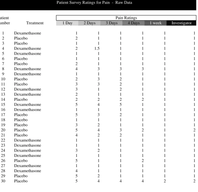
days, 3 days, 4 days and 1 week after the procedure. The higher the pain level experienced, the higher the rating (Tables 2, 3).
Thirty-two of the sixty patients were symptomatic and were experiencing pain the day of the surgical procedure (Table 1). Those that received an injection of dexamethasone rated pain from 1 to 6 with an average rating of 2.5 and a standard deviation of 1.6 after twenty-four hours (Table 4). Those that received a placebo injection rated pain from 1 to 6 with an average rating of 3.2 and a standard deviation of 1.7. At forty-eight hours (two days), those that
received an injection of dexamethasone rated pain from 1 to 5 with an average rating of 2.0 and a standard deviation of 1.3. Those that received a placebo injection rated pain from 1 to 6 with an average rating of 2.4 and a standard deviation of 1.4. At seventy-two hours, those that received an injection of dexamethasone rated pain from 1 to 5 with an average rating of 1.9 and a standard deviation of 1.3. Those that received a placebo injection rated pain from 1 to 6 with an average rating of 2.0 and a standard deviation of 1.2. At ninety-six hours, those that received an injection of Dexamethasone rated pain from 1 to 6 with an average rating of 1.6 and a
standard deviation of 1.2. Those that received a placebo injection rated pain from 1 to 5 with an average rating of 1.6 and a standard deviation of 1.1. At the clinical evaluation appointment one week post-operatively, those that received an injection of dexamethasone rated pain from 1 to 5 with an average rating of 1.3 and a standard deviation of .9. Those that received a placebo injection rated pain from 1 to 3 with an average rating of 1.3 and a standard deviation of .7.
Patients rated the swelling that they experienced on the 6-point likert-like scale at 1 day, 2 days, 3 days, 4 days and 1 week after the procedure. The more swelling observed by the patient, the higher the rating (Table 2, 5).
average rating of 2.4 and a standard deviation of 1.0 after twenty-four hours (Table 6). Those that received a placebo injection rated swelling from 1 to 6 with an average rating of 3.1 and a standard deviation of 1.5. At forty-eight hours, those that received an injection of
dexamethasone rated swelling from 1 to 5 with an average rating of 2.6 and a standard deviation of 1.1. Those that received a placebo injection rated swelling from 1 to 6 with an average rating of 3.1 and a standard deviation of 1.4. At seventy-two hours, those that received an injection of dexamethasone rated swelling from 1 to 5 with an average rating of 2.3 and a standard deviation of 1.2. Those that received a placebo injection rated swelling from 1 to 6 with an average rating of 2.5 and a standard deviation of 1.3. At ninety-six hours, those that received an injection of dexamethasone rated swelling from 1 to 4 with an average rating of 1.8 and a standard deviation of 1.0. Those that received a placebo injection rated swelling from 1 to 5 with an average rating of 1.9 and a standard deviation of 1.2. At the clinical evaluation appointment one week post-operatively, those that received an injection of dexamethasone rated swelling from 1 to 5 with an average rating of 1.3 and a standard deviation of 0.8. Those that received a placebo injection rated swelling from 1 to 3 with an average rating of 1.3 and a standard deviation of 0.5.
Observing the general statistic of range, average, rates of change and standard deviation of the ratings, the dexamethasone group appears to have performed similar the placebo group from day 2 and after. However, the day 1 average rating of 3.1 for the placebo group was significantly higher than the 2.4 for the dexamethasone group (Figure 5). This warrants further statistical analysis which has been done below.
Actual Frequency Distribution
High Rating
Low
Rating Total
dexamethasone 4 26 30
placebo 12 18 30
Total Observations 16 44 60 Expected Frequency Distribution
High Rating
Low
Rating Total
Dexamethasone 8 22 30
placebo 8 22 30
Total Observations 16 44 60
The test was done at the level of significance of 5% (α): Level of significance = α =
.05, or 5%. Given that the distribution table is a 2 x 2 matrix, the degrees of freedom is equal to one: Degrees of Freedom = df = 1.
The pertinent Chi-square values are as below:
α = .5 .1 .05 .02 .01
1 df . 455 2.706 3.841 5.412 6.635
2df 1.386 4.605 5.991 7.824 9.210
3df 2.366 6.251 7.815 9.837 11.345
The Chi-square value of 3.841 is the benchmark that must be exceeded for the null hypothesis to be rejected (Figure 4). The Chi-square value calculated for the swelling ratings distribution was 5.455.
Where,
a = high ratings for dexamethasone group observed b = low ratings for dexamethasone group observed c = high ratings for placebo group observed d = low ratings for placebo group observed n = total population of ratings
The resulting P-value from the Fischer Exact Test for day-one swelling ratings was equal to 0.0195.
Patients rated the bruising that they experienced on the 6-point likert-like scale at 1 day (24 hours), 2 days (48 hours), 3 days (72 hours), 4 days (96 hours) and 1 week after the
deviation of 1.3. At the clinical evaluation appointment one week post-operatively, those that received an injection of dexamethasone rated bruising from 1 to 6 with an average rating of 1.6 and a standard deviation of 1.1. Those that received a placebo injection rated bruising from 1 to 6 with an average rating of 1.4 and a standard deviation of 1.1.
Patients rated the intra-oral healing that they experienced on the 6-point likert-like scale at 1 day, 2 days, 3 days, 4 days and 1 week after the procedure (Tables 2, 9). The more trauma and damage tissue observed, the higher the rating. Ten of the patients were unable to provide ratings for intra-oral healing due to the sutures that were placed after the surgical procedure. There was concern that by opening their mouth to visually inspect the healing process, tearing of the sutured tissue would occur. Five patients from the dexamethasone group and five
patients from the placebo group did not render ratings at one time interval or another during the ratings time period.
Those that received an injection of dexamethasone rated intra-oral healing from 1 to 5 with an average rating of 2.6 and a standard deviation of 1.3 after twenty-four hours (Table 10). Those that received a placebo injection rated intra-oral healing from 1 to 6 with an average rating of 3.3 and a standard deviation of 1.6. At forty-eight hours, those that received an
injection of dexamethasone rated intra-oral healing from 1 to 5 with an average rating of 2.7 and a standard deviation of 1.3. Those that received a placebo injection rated intra-oral healing from 1 to 6 with an average rating of 2.8 and a standard deviation of 1.7. At seventy-two hours, those that received an injection of dexamethasone rated intra-oral healing from 1 to 5 with an average rating of 2.2 and a standard deviation of 1.2. Those that received a placebo injection rated intra-oral healing from 1 to 6 with an average rating of 2.5 and a standard deviation of 1.3. At
1 to 5 with an average rating of 1.9 and a standard deviation of 1.1. Those that received a placebo injection rated intra-oral healing from 1 to 5 with an average rating of 2.0 and a standard deviation of 1.0. At the clinical evaluation appointment one week post-operatively, those that received an injection of dexamethasone rated intra-oral healing from 1 to 5 with an average rating of 1.6 and a standard deviation of 1.0. Those that received a placebo injection rated intra-oral healing from 1 to 4 with an average rating of 1.6 and a standard deviation of 0.7.
Observing the general statistic of range, average, rates of change and standard deviation of the ratings, the dexamethasone Group appears to have performed similar the placebo group from day 2 and after. However, the day 1 average rating of 3.3 for the placebo group was significantly higher than the 2.6 for the dexamethasone group. This warrants further statistical analysis which is performed below.
As with the analysis of swelling and inflammation, the Pearson’s Chi-square test was performed on the 2 x 2 matrix of high and low rating scores for the day 1 of intra-oral healing.
Actual Frequency Distribution
High Rating
Low
Rating Total
dexamethasone 6 19 25
placebo 12 13 25
Total Observations 18 32 50 Expected Frequency Distribution
High Rating
Low
Rating Total
dexamethasone 9 16 25
placebo 9 16 25
Total Observations 18 32 50
The test was done at the level of significance of 5% (α): Level of significance = α =
The pertinent Chi-square values are as below:
α = .5 .1 .05 .02 .01
1 df . 455 2.706 3.841 5.412 6.635
2df 1.386 4.605 5.991 7.824 9.210
3df 2.366 6.251 7.815 9.837 11.345
The Chi-square value of 3.841 is the benchmark that must be exceeded for the null hypothesis to be rejected. The Chi-square value calculated for the intra-oral healing ratings distribution was 3.125
Where,
a = high ratings for dexamethasone group observed b = low ratings for dexamethasone group observed c = high ratings for placebo group observed d = low ratings for placebo group observed n = total population of ratings
The resulting P-value from the Fischer Exact Test for day-one intra-oral healing ratings was equal to 0.0977.
Discussion
.
As a confirmation and since the Pearson Chi-square test was near the 10% level of significance, the Fischer Exact Test was performed. Like the test performed for swelling and inflammation, a one-tail test was performed in the following Fischer Exact Test formulation:
hours. This was expected since a glucocorticoid steroid is known for its recovery properties and not for its anesthetizing effects. Forty-three of the sixty patients used Tylenol and/or Vicodin during the first twenty-four hours post-operatively. This may render the resulting affects of dexamethasone on pain inconclusive.
This is further supported by observing the rates of change in ratings from one time interval to the next between dexamethasone versus the placebo. The placebo group experienced a greater average pain rating change from day 1 to day 2 than the dexamethasone group. The average pain rating for the placebo group decreased 0.83. The average pain rating for the dexamethasone group only decreased 0.52. The trend continued from day 2 to day 3. The average pain rating for the placebo group decreased 0.43. The average pain rating for the dexamethasone group only decreased 0.12.
Improvement stabilized at this point and the rate of change was similar for the change in ratings between day 3 and day 4. The average pain rating for the placebo group dropped 0.33. The average pain rating for the dexamethasone group decreased 0.30. And finally, the average pain rating for the placebo group decreased 0.30 from the 4th day to the 1 week appointment. For the same corresponding period, the average pain rating for the dexamethasone group decreased a comparable 0.27.
comparable. The standard deviation statistic at the various time intervals for each of the groups during this post-operative period is approximately one rating point. The expected variation in pain actually experienced by patient would likely account for most of the variance rather than significantly different subjective rating scales.
The swelling statistics on day 2 and after are consistent through the time intervals and quite similar. The subtle difference appears statistically insignificant. A supportive observation of the rating system is the average absolute ratings at the one week follow-up appointment. This was nearly the same for both groups and the principal investigator. At this point of the post-operative period, patients’ swelling would have neared completion. The strikingly similar rating scores support the study’s assumption that patients’ subjective judgments in rating swelling are comparable.
In review of the bruising rating general statistic of range, average, rates of change and standard deviation of the ratings, the placebo group appears to have performed slightly better than the dexamethasone group. The statistics are consistent through the time intervals and quite similar. The subtle difference is statistically insignificant.
A supportive observation of the rating system is the average absolute ratings at the one week follow-up appointment. This was nearly the same for both groups and the investigator. At this point of the post-operative period, patients’ bruising would have peaked and begun to subside. The strikingly similar rating scores support the study’s assumption that patients’ subjective judgments in rating bruising are comparable.
REFERENCES
1. Eriksen HM. Endodontology--epidemiologic considerations. Endodontics & dental traumatology 1991;7(5):189-195.
2. Hepworth MJ, Friedman S. Treatment outcome of surgical and non-surgical management of endodontic failures. J Can Dent Assoc 1997;63(5):364-371.
3. Kvist T, Reit C. Results of endodontic retreatment: a randomized clinical study
comparing surgical and nonsurgical procedures. Journal of endodontics 1999;25(12):814-817.
4. Trope M. Flare-up rate of single-visit endodontics. International endodontic journal 1991;24(1):24-26.
5. Kvist T, Reit C. Postoperative discomfort associated with surgical and nonsurgical endodontic retreatment. Endodontics & dental traumatology 2000;16(2):71-74.
6. Boykin MJ, Gilbert GH, Tilashalski KR, Shelton BJ. Incidence of endodontic treatment: a 48-month prospective study. Journal of endodontics 2003;29(12):806-809.
7. Nash KD, Brown LJ, Hicks ML. Private practicing endodontists: production of endodontic services and implications for workforce policy. Journal of endodontics 2002;28(10):699-705.
8. Abbott PV. Analysis of a referral-based endodontic practice: Part 2. Treatment provided. Journal of endodontics 1994;20(5):253-257.
9. Abbott PV. Analysis of a referral-based endodontic practice: Part 1. Demographic data and reasons for referral. Journal of endodontics 1994;20(2):93-96.
10. Zuolo ML, Ferreira MO, Gutmann JL. Prognosis in periradicular surgery: a clinical prospective study. International endodontic journal 2000;33(2):91-98.
11. Tawil PZ, Saraiya VM, Galicia JC, Duggan DJ. Periapical microsurgery: the effect of root dentinal defects on short- and long-term outcome. Journal of endodontics 2015;41(1):22-27.
12. Liesinger A, Marshall FJ, Marshall JG. Effect of variable doses of dexamethasone on posttreatment endodontic pain. Journal of endodontics 1993;19(1):35-39.
13. Marshall JG, Liesinger AW. Factors associated with endodontic posttreatment pain. Journal of endodontics 1993;19(11):573-575.
15. Jackson DL, Moore PA, Hargreaves KM. Preoperative nonsteroidal anti-inflammatory medication for the prevention of postoperative dental pain. J Am Dent Assoc 1989;119(5):641-647.
16. Trowbridge HO. Review of dental pain--histology and physiology. Journal of endodontics 1986;12(10):445-452.
17. Rogers MJ, Johnson BR, Remeikis NA, BeGole EA. Comparison of effect of intracanal use of ketorolac tromethamine and dexamethasone with oral ibuprofen on post treatment endodontic pain. Journal of endodontics 1999;25(5):381-384.
18. Torabinejad M, Kettering JD. Identification and relative concentration of B and T lymphocytes in human chronic periapical lesions. Journal of endodontics 1985;11(3):122-125.
19. Hargreaves KM, Keiser K. Development of new pain management strategies. Journal of dental education 2002;66(1):113-121.
20. Trowbridge HO. Immunological aspects of chronic inflammation and repair. Journal of endodontics 1990;16(2):54-61.
21. Torabinejad M, Eby WC, Naidorf IJ. Inflammatory and immunological aspects of the pathogenesis of human periapical lesions. Journal of endodontics 1985;11(11):479-488.
22. Trowbridge HO. Inflammation: review of the process. fourth edition ed. Illinois: Quintessence Publishing Co; 1993.
23. Spector WG. Chronic inflammation. Journal of endodontics 1977;3(6):218-222.
24. Ferrari AM, Byers MR. Chronic dexamethasone treatment and its effects on sensory neuropeptides, pulpal injury reactions and reparative dentin. Brain research 1996;723(1-2):125-134.
25. Morse DR, Esposito JV, Furst ML. Comparison of prophylactic and on-demand diflunisal for pain management of patients having one-visit endodontic therapy. Oral surgery, oral
medicine, and oral pathology 1990;69(6):729-736.
26. Samuelsson B. Leukotrienes: mediators of immediate hypersensitivity reactions and inflammation. Science 1983;220(4597):568-575.
27. Flower R. Steroidal antiinflammatory drugs as inhibitors of phospholipase A2. Advances in prostaglandin and thromboxane research 1978;3:105-112.