Patron: Her Majesty The Queen Rothamsted Research Harpenden, Herts, AL5 2JQ
Telephone: +44 (0)1582 763133 Web: http://www.rothamsted.ac.uk/
Rothamsted Repository Download
A - Papers appearing in refereed journals
Doncaster, C. C. and Seymour, M. K. 1972. Techniques for manipulating
small nematodes. Nematologica. 18, pp. 261-264.
The output can be accessed at:
https://repository.rothamsted.ac.uk/item/96wxv/techniques-for-manipulating-small-nematodes
.
© Please contact [email protected] for copyright queries.
TECHNIQUES FOR MANIPULATING SMALL NEMATODES
BY
M. K. SEYMOUR and C. C. DONCASTER Rothamsted Experimental Station, Harpenden, Herts., England
Details of construction and operation are given for some apparatus used to observe and manipulate small soil nematodes under the microscope. An observation cell is simply made from rigid P.V.C. Its open sides allow insertion of microtools and depth can be varied in steps. Glass suction cannulae of 20 to 50 µ aperture, connected to syringes, hold nematodes securely. Finer microcannulae (5 to 10 µ aperture) can be inserted through the cuticle for pressure recording. Microdissection knives are made from plate glass slivers fused or glued to glass handles.
To study internal pressures in some nematodes we developed instruments for
micromanipulation that may be of general use.
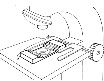
ObJervation cell for micro manipulation
A cell was needed in which specimens could be manipulated in water while
being viewed and filmed through a compound microscope. The cell had to be
shallow enough to fit between a 25X objective and a normal condenser, yet provide inside it ample lateral and vertical clearance for microtools. To allow tools to be inserted it had to be open at the sides yet the openings had to be small to keep
evaporation to a minimum. Fig. 1 shows a pattern satisfying these requirements.
The material used, "Darvic" rigid PVC 1), is easily worked with wood- or metal-
cutting tools. It is water-repellent, so that water in the cell is held between upper
and lower glass coverslips and does not readily run from the open sides. Steps of
different heights at the front and back of the cell provide alternative supporting
ledges for the upper coverslip, so that with a shorter slip the cell is shallower. Both upper and lower coverslips can be gummed in place with a tacky paraffin-vaseline mixture (Doncaster, 1962), but we prefer not to gum the upper coverslip, so that when it is touched inadvertently by the microtools, it moves and the tools are not broken. The upper slip can be made narrower to give more clearance for micro-
tools, or wider to limit evaporation. The cell is also suitable for hanging-drop
preparations. Our observation cell was milled from solid Darvic sheet 1 cm thick
and the central hole was bored and rebated on a lathe. Alternatively, shaped laminae of thinner Darvic can be bonded together with such adhesives as "Surasolve Solvent Cement" 2) or "I.S.-12 Cyanoacrylate Adhesive" 3).
1) I.C.I. Ltd., Marketed by G. H. Bloore Ltd., 480 Honeypot Lane, Stanmore, Middlesex, England. 2) Durapipe Fittings Ltd., Winnock Rd., West Drayton, Middx., England.
262
Fig. 1. Microscopical observation cell for micromanipulation. Suggested length, 75 mm; width, 30 mm; lower step height, 3 mm; upper step height, 2 mm.
Microcannulae
Two types of microcannula were used: one to insert in'-o nematodes through the cuticle or stoma in attempts to record internal hydrostatic pressure, the other to hold neamatodes securely by suction for observation and experimental procedures.
A piece of "Pyrex" tubing about 15 cm long, 3 mm o.d. and 2 mm bore was heated to dull red over a small gas burner and drawn out by hand to 0.5 mm o.d. The two resulting cannulae were broken apart and the tip of each gently drawn out with forceps while passing it steadily through the flame of a very small gas burner (e.g. a large-bore hypodermic needle clamped upright). The tip was broken off and flamed to produce a holding cannula with an aperture of 20-50 , (Fig. 2, A & B, right). A holding cannula filled with observation medium (ringer, tap or
distilled water) was mounted in a micromanipulator and connected by thin-walled
rubber tubing to a fixed, small-bore, smooth-acting hypodermic syringe. Under the microscope the tip of the microcannula was brought close to the subject and suction applied by the syringe, drawing the nematode into or onto the end of the cannula. The nibber tube flattened and exerted a slight, steady suction, which held
nematodes placed across the cannula tip in place for minutes or hours. When a
nematode had been drawn into a cannula, further suction was unnecessary and
263
cannula aperture could be enlarged by breaking off the tip and repolishing in the
flame; nematodes of different sizes could thus be accommodated. Fig. 2A shows
an arrangement for feeding a mononchid, with the mononch in a wide cannula and a tylenchid, the potential prey, held across a finer one.
Once drawn into a holding cannula, mononchs alternated locomotory lashing with quiescent periods during which they did not respond to touch stimuli. While
being held, one swallowed a small stylet-bearing nematode within two min; another
attempted to eat a second, larger, mononch.
To construct penetrant cannulae and holders much smaller than 20 , diameter, the preliminary pulls were made in the same way as for the larger holding cannulae.
A further controlled pull, with strong local heating, was then required. For this
operation some kind of microforge had to be used (e.g. the De Fonbrune 4) or
the homemade unit described by Thaysen & Morris (1947). After the first two
pulls, the drawn-out end was made into a hook for attaching a weight or spring.
A light pull (about 5 gm wt) was applied and the bright red heating filament was brought near the glass, which thinned and parted; the filament was then
rapidly withdrawn. This left a short, shouldered point tapering to less than 1 fL
diameter. The tip was stubbed under water beneath the microscope to enlarge it to a sharp-edged hole of the required diameter (usually 5-10 /j,). The abrupt taper minimised capillarity and viscosity effects and ensured maximum strength. Mounted
on a micromanipulator and connected by rigid polyethylene, nylon 5) or thick-
walled rubber tube to a micrometer syringe, short penetrant microcannulae could
easily be inserted through a nematode's body wall into the pseudocoelom, gut
or elsewhere. Microcannulae of this pattern but with heat-polished tips serve as
suction holders (Fig. 2A, left) and when placed in a nematode stoma (Fig. 2B) induce a swallowing reaction.
Gla.r.r microknive.r
To make microdissection knives a cleanly cut edge of plate glass was lightly
tapped or gripped with pliers and thin slivers broken off onto black paper. Under
a dissecting microscope, conchoidal or pointed sharp-edged fragments were
selected. A microcannula, as described above (unfinished or broken) served as a
handle, to which a sliver was attached in the desired orientation. Blades may be
fused to the handles with a microforge or stuck with epoxy resin glue (e.g. "Aral-
dite"). Two large examples are illustrated in Fig. 2, C & D.
ZUSAMMENFASSUNG
Methoden zur Handhabung kleiner Nematoden
Es werden Einzelheiten von Aufbau und Wirkungsweise einiger Apparate mitgeteilt, die für die Beobachtung und Handhabung kleiner Nematoden unter dem Mikroskop benutzt werden. Eine Beobachtungskammer ist einfach aus starrem PVC hergestellt. Ihre offenen Seiten erlauben das
4) Agents in Britain: Scientific Techniques Ltd., Reliant Works, Brockham, Betchworth, Surrey, England.
264
Anbringen von Mikrowerkzeugen, und ihre Tiefe kann stufenweise verändert werden. Mit Ansaug- kanülen aus Glas (20-50 µ Öffnungsweite), die mit Injektionsspritzen verbunden sind, lassen sich Nematoden sicher halten. Feinere Mikrokanülen mit Öffnungen von 5-10 µ können für Druck- messungen durch die Cuticula geführt werden. Mikropräpariermesser werden aus Glassplittern gemacht, die an Glasstäbe angeschmolzen oder angeklebt werden.
REFERENCES
DONCASTER, C. C. (1962). Sealing microscopical water mounts with soft wax. Nematologica 7, 258.
THAYSEN, A. C. & MORRIS, A. R. (1947). The preparation of microtools for the micromanipulator.