IRANIAN JOURNAL OF DIABETES AND OBESITY, VOLUME 11, NUMBER 1, SPRING 2019
28
Effect of Exercise Training on Tropomodulin-2 Gene Expression in
Cerebellum of Diabetic Rats
Seyed Jalal Taherabadi1, Masoud Rahmati1*, Rahim Mirnasuri1, Abdolreza Kazemi2
Introduction
iabetic peripheral neuropathy (DPN) is a serious complication associated with both type 1 and type 2 diabetes which is simply defined as “the presence of symptoms and/or signs of peripheral nerve dysfunction in people with diabetes after exclusion of other causes” (1). In addition to damaging the peripheral nervous system
(PNS), neurochemical, electrophysiological, structural and behavioral studies have provided evidences that DM has profound degenerative effects on central nervous system (CNS) such as impairments in motor control, learning and memory which referred to as diabetic encephalopathy (2). Unlike to the extensive research on DPN (3), little effort has
D
1. Department of Sport Sciences, Lorestan University, Khoram Abad, Iran.
2. Department of Sport Sciences, Vali-E-Asr University of Rafsanjan, Kerman, Iran.
*Correspondence:
Masoud Rahmati, PhD, Department of Sport Sciences, Lorestan University, Khoram Abad, Iran.
Tel:(98) 912 452 5538 Email:rahmati.mas@lu.ac.ir
Received: 17 February 2019
Accepted: 21 June 2019
Published in August 2019
Abstract
Objective: It is well documented that exercise training (ET) imposes beneficial effects on diabetes mellitus and its complication such as diabetic peripheral neuropathy (DPN). Regarding the importance of tropomodulin-2 (TMOD2) in nervous system plasticity, this protein may be recognized as a candidate mechanism for ET-induced neuroplasticity. The aim of this study was to investigate the effects of ET on cerebellar gene expression of TMOD2 in rats with DPN.
Materials and Methods: Animals were randomly divided into three groups: healthy control (C), diabetic control (DC) and diabetic trained (DT). Diabetes was induced by a single intraperitoneal injection of streptozotocin. Behavioral nociception assessment was carried out by Von Frey Filaments and tail-flick tests. TMOD2 gene expression was assessed by real time-PCR.
Results: The mRNA levels of TMOD2 increased to 0.50-fold (P-value: 0.005) in comparison of the sedentary controls after 6 weeks of DPN. Also, TMOD2 gene expression in DT group was decreased to -0.68-fold changes in comparison of the C group (P-value: 0.001). In addition, the TMOD2 gene expression in DT group was lower than the DC group (P-value: 0.0001).
Conclusion: The TMOD2 mRNA level in rat’s cerebellum was affected by ET and DPN, but its exact physiological roles were not clarified. Hence, identifying the importance of TMOD2 in DPN needs further research.
Keywords: Diabetic peripheral neuropathy, Exercise training, Tropomodulin 2, Plasticity
IRANIAN JOURNAL OF DIABETES AND OBESITY, VOLUME 11, NUMBER 1, SPRING 2019 29
been made to recognize the pathology of diabetic encephalopathy.
The actin cytoskeleton as the major components of the neural scaffold, is involved in many pivotal cellular processes in CNS such as cellular morphogenesis, motility, division and intracellular transport and comprise an important part of neuronal development (4) and neurogenesis (5). Polymerization and depolymerization of actin is a rate-limiting mechanical step of actin cytoskeleton dynamic which are controlled by numerous actin-regulatory proteins such as tropomodulins (TMODs) (6). It was demonstrated that the tropomodulins-2 (TMOD2), the neuronal-specific isoform of TMODs, has pivotal role in CNS and is related to various function such as the formation of new synaptic structures, so that TMOD2-lacking mice exhibited enhanced long-term potentiation (7). Increased neurite extension (8), reduced sensorimotor gating, and impaired learning and memory functions are other disorders arises from lack of TMOD2 expression (7). It has been demonstrated that, TMOD2 expression is altered in several diseases (9) such as fetal down syndrome (10) and down-regulated in hippocampal tissue of the patients with mesial temporal lobe epilepsy (11). However, on the basis of our knowledge, the expression of this protein in DPN state has not been investigated yet. It has been demonstrated that, actin dynamics and actin cytoskeleton contribute to neurodegenerative diseases (12,13). It is presumed that, because of degenerative effect of diabetes in brain (14), DPN as a symptom of neurodegeneration might be associated with impairment of actin dynamics and its regulatory proteins.
Increasing evidence has demonstrated that exercise training (ET) is associated with multiple health benefits in animal with type 1 (T1D) (1) and type 2 diabetes (T2D) so that increased physical activity profoundly decrease the risk of complications and improve glycemic control and blood lipid profiles (15). In addition, ET attenuates peripheral risk factors such as dyslipidemia,
hypertension and cardiovascular disease, which converge to cause neurodegeneration (16). ET also has considerable effects on structure and function of nervous system, defined as neuroplasticity (17). These beneficial effects of ET on CNS mediated by increased level of blood circulation, neurotrophic factors and the immune system function (18). Moreover, while immune system function is impaired in DPN (19), positive immunomodulatory effect of exercise were well demonstrated both in T1D and inflammation (20). Besides this, many mechanisms of the exercise benefits in DPN state have remained unclear. Due to the important role of TMOD2 in regulating actin dynamics, it seems that this protein plays an important role in nervous system plasticity response to various stimulants and conditions such as DPN. Considering profound effects of ET on neuroplasticity despite its unknown mechanisms, this protein may be a key factor in exercise-induced neuroplasticity in DPN. The aim of this study was to evaluate the effect of DPN and ET on the expression of TMOD2.
Materials and Methods
A total of 40 adult male wistar rats were supplied from Razi Institute (Karaj, Iran) and housed four-per-cage in animal lab under standard conditions (12- hour light/dark cycle in a room at temperature of 20- 25°C) with access to food and water. All institutional (registered as LUNS.REC.1395.170 ethical code at Lorestan University of Medical Sciences) and animal research health guidelines were followed. The animals were randomly divided into three groups: (1) healthy control (C, N= 6), 2) diabetic control (DC, N= 17) and (3) diabetic trained (DT, N= 17).
For acclimatization and reaching optimal weight (at least 300 g), all of the rats were kept in animal lab for two weeks before the experiments. Subsequently, after an overnight fasting, diabetes was induced through a single intraperitoneal injection of streptozotocin
30 IRANIAN JOURNAL OF DIABETES AND OBESITY, VOLUME 11, NUMBER 1, SPRING 2019
(STZ) (45 mg/kg; Sigma, St. Louis, MO) solution (dissolved in 0.5 mol/L citrate buffer at pH 4.0). Two days later, diabetes was confirmed by measuring tail vein blood glucose level (> 350 mg/ld.) The animals without any trace of hyperglycemic were excluded from the rest of the samples (Due to the lack of hyperglycemic sign and death, 7 and 8 rats were excluded from the DC and DT groups respectively). In addition, all along the study course, blood glucose levels were controlled once every two weeks.
Treadmill training protocol: The practiced treadmill training protocol was consisted of 6 weeks of moderate-intensity (50- 55% of maximal oxygen consumption) endurance aerobic training on a leveled motor-driven treadmill (Model T510E, Diagnostic and Research, Taoyuan, Taiwan). In the first week, the speed and duration of the treadmill running were 10 meters/minute (m/min) and 10 min per day, respectively. The figures were then increased gradually until the fifth week, ending up with training speed and duration of 18 m/min and 30 min per day, respectively, on the last training session. For stabilizing the obtained adaptations, training speed and duration were kept constant at the sixth week (21).
Behavioral nociceptive tests
For modeling the DPN in rats we used the procedures introduced by Calcutt (2004) which demonstrated that short-term diabetes induced by the STZ is best considered as models of DPN (22). In present study, for examination of DPN, two weeks after confirmation of diabetes, behavioral nociception tests were assessed using Von Frey Filaments and tail-flick tests and the diabetic animals demonstrating thermal hyperalgesia and mechanical allodynia were considered as diabetic neuropathic rats (23). To ensure perdurability of DPN, behavioral nociceptive tests were evaluated once every two weeks, and the animals without DPN symptoms were excluded (Due to the lack of thermal hyperalgesia and mechanical allodynia
modeling, 4 and 3 rats were excluded from the DC and DT groups respectively).
Von Frey Filaments test: In order to evaluate mechanical allodynia, rats were placed in a transparent plastic box with a metallic grid floor. After 30 minutes of acclimatization to the environment, Von Frey Filaments (Stoelting Co., Wood Dale, IL) of different stiffness values ranging from 2 grams to 60 grams in weight were applied to plantar surface of the hind paw. The minimum Von Frey filament stiffness at which the animal responded through jumping, vocalization, shaking or flicking its paw was recorded as paw withdrawal threshold (PWT).
Tail-flick test: One hour before testing, the rats were transferred to the behavioral test section and gently placed in plastic restrainer. Using a tail-flick instrument (LE-7406, Spain), radiant heat was focused on approximately 5 cm from the distal end of the rat’s tail, and the time at which the animal flicked his tail in response to the heat was recorded as latency response. A cutoff time of 20 seconds was used to prevent tissue damage. Each behavioral nociceptive test was performed three times with 5-minute intervals. Final latency response was obtained by averaging the three latency responses obtained, and designated as Tail Flick Latency (TFL).
Two days after the last exercise session in the 6th week of training, 3 rats for each group (Due to casualties in the end of study) were anesthetized by inhalation of 2% halothane in a mixture of 20% O2 and 80% CO2 (24) and
their cerebellar tissue was removed under sterile conditions and immediately perfused in 4% paraformaldehyde.
RNA extraction and cDNA synthesis
Cerebellum samples (N= 3 for each group) were directly homogenized in 1 mL of TRIzol (Invitrogen, Carlsbad, CA, USA) with a homogenizer added. Total RNA was isolated from the cerebellum using QlAzol® Lysis Reagent (Germany, Qiagen) and chloroform (Germany, Qiagen) according to the manufacturer instructions. Briefly, about 50IRANIAN JOURNAL OF DIABETES AND OBESITY, VOLUME 11, NUMBER 1, SPRING 2019 31
mg of the cerebellum was separately homogenized in QlAzol® Lysis Reagent (1:10) and then centrifuged at 12000 g for 10 minutes at 4°C. After that, the product was mixed with chloroform (ratio of 1:5) and centrifuged at 12000 g for 10 minutes at 4°C. Finally, after separating the associated water and mineral contents, RNA-containing part was mixed with isopropanol (1:5) and left at room temperature for 10 minutes. Then it was centrifuged at 12000 g for 10 minutes at 4°C. RNA-containing pellet was washed and diluted in RNase- free water. RNA concentration was quantitated by spectrophotometry (Eppendorf, Germany) and 260/280 nm ratios were 1.8 - 2 times the desired purity. cDNA synthesis was performed using Quanti Tect Reverse Transcription Kit (Qiagen, Germany) based on the manufacturer instructions.
Real-time PCR
Real-time PCR experiments were conducted on a Premix SYBR Green II (Qiagen, Germany). Reaction mixture was composed of 1 µL of cDNA, 1 µL of forward primer, 1 µL of reverse primer, 7 µL of DEPC water and 10 µL of Syber Green. All samples were measured in duplicate and primers were designed in accordance to TMOD2 and GAPDH genes in gene bank of NCBI and a German company, Qiagen. The real-time PCR was carried out with the following oligonucleotide primers: TMOD2 [5-CCTGTCTCCTTCAACTCTCTTC-3
(forward) and
5-CAAGATCCACAACCAGAGGC-3
(reverse)], and GAPDH
[5-AAGTTCAACGGCACAGTCAAGG-3
(forward) and
5-CATACTCAGCACCAGCATCACC-3
(reverse)]. Thermal treatment involved the following cycles: 95°C for 10 minutes, 95°C for 15 s, and 60°C for 1 min (40 repetitions). TMOD2 gene was normalized to GAPDH levels and calculated by 2 -∆∆CT.
Statistical analysis
Statistical analyses were conducted by SPSS software (version 19, SPSS Inc., Chicago, IL, USA). Normality of data distribution was assessed by Shapiro-Wilk test. Differences between different groups were analyzed based on ANOVA results followed by LSD post-hoc. Statistical significance level was set to P -value< 0.05. The data was reported as mean± standard error of mean (SEM) values.
Results
Table 1 showed the mean of blood glucose levels. The blood sugar levels of the animals in all groups were similar at the onset of the study, but the blood sugar levels were significantly higher in the animals in DC and DT groups rather than those in C group in the first (P-value: 0.001, 0.001), third (P-value: 0.001, 0.001) and sixth (P-value: 0.001, 0.001) weeks. The result demonstrated successful and profound induction of diabetes in animals by STZ, so that the hyperglycemia remained
Table 1. Description of blood glucose, paw withdrawal threshold and tail flick latency in control (C), diabetic control (DC) and diabetic trained (DT) groups
Groups Baseline 2th week 4th week 6th week
Blood glucose (mg/dl)
C 101.166± 0.600 100.666± 0.666 101.0± 0.516 101.333± 0.494
DC 100.0± 0.632 399.833± 3.487 412.666± 3.912 430.5.0± 2.50
DT 99.333± 1.282 378.666± 3.747 398.0± 4.487 419.0± 4.457
Paw withdrawal threshold (g)
C 54.283± 2.557 57.766±1.913 56.166±2.424 56.449±2.251
DC 57.766± 1.913 24.433±1.566 21.333±2.951 21.333±2.951
DT 58.116± 1.883 25.871±0.174 26.666±0.421 33.333±5.333
Tail flick latency (s)
C 7.114±0.334 7.0833±0.302 6.798±0.297 6.936±0.207
DC 6.965±0.360 4.716±0.208 4.133±0.380 3.816±0.241
DT 6.916±0.202 4.999±0.488 4.745±0.275 5.848±0.200
The data were reported as mean ± standard error of the mean (SEM).
32 IRANIAN JOURNAL OF DIABETES AND OBESITY, VOLUME 11, NUMBER 1, SPRING 2019
consistently throughout the course of study and ET was not capable of damping these abnormalities.
Nociceptive behaviors
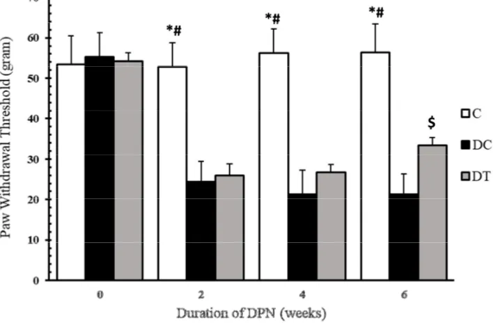
In the present paper, thermal hyperalgesia and mechanical allodynia were assessed by Von Frey filament and tail-flick tests to examine nociceptive behaviors of the diabetic animals. The diabetic animals with neuropathic pain were selected for DC and DT groups and the animals showing no nociceptive behaviors were excluded from these groups. Figures 1-2, and table 1 show PWT and TFL, respectively. As these figures suggest, mechanical allodynia and thermal hyperalgesia are evident in the animals in DC and DT groups, as compared to those in C group in the first (P-value: 0.001, 0.001), third (P-value: 0.001, 0.001) and sixth (P-value: 0.001, 0.001) weeks. Moreover, these nociceptive behaviors persisted throughout the experiments, except for the 6th week when ET managed to damp mechanical allodynia in diabetics rats, as compared to the
animals in DC group (P-value: 0.017). We insisted in using STZ-diabetic neuropathy on the basis of a previous report claiming that chronic pain shares global gene expression changes and some molecular pathobiology with neurodegeneration (25), and also it might be a functional examination of neural deterioration.
TMOD2 mRNA in cerebellum
mRNA levels of TMOD2 in cerebellum of diabetic rats were measured after exposure to 6 weeks of submaximal endurance training. In order to compare the levels of TMOD2 gene expression among C, DC and DT groups, one-way ANOVA was used. Results showed that, mRNA levels of TMOD2 increased to 0.50-fold changes (P-value: 0.005) of that of the sedentary controls after 6 weeks of DPN, indicating that DPN causes an increase in TMOD2 gene expression in cerebellum. Also, TMOD2 gene expression in DT group was decreased to -0.68-fold changes of that of the C group (P-value: 0.001), i.e. ET attenuated
Figure 1. Mechanical allodynia in C, DC and DT groups before STZ injection and after two, four and six weeks of diabetes induction
Compared to C, * and # marks represent significant differences from DC (P-value= 0.001) and DT (P-value= 0.001) groups, respectively. $ demonstrates that mechanical allodynia is damped in DT group, as compared to DC group (P-value= 0.017), but it is still significantly differentfrom that of C group (P-value= 0.001)
IRANIAN JOURNAL OF DIABETES AND OBESITY, VOLUME 11, NUMBER 1, SPRING 2019 33
the increased mRNA levels of TMOD2 in cerebellum of rats with DPN. In addition the TMOD2 gene expression in DT group was lower than the DC group (P-value: 0.0001). These results demonstrate that, the gene expression of TMOD2 in rat’s cerebellum was affected by ET and DPN (Figure 3).
Discussion
In this study we observed that DPN is associated with increased gene expression of TMOD2 in cerebellum. Although it has been demonstrated that TMOD2 plays important roles in neurodegeneration disease (26), but its changes in DPN which may impose prominent effect on pathology of neural degeneration, are not explored yet. The present research is the first to evaluate TMOD2 gene expression in cerebellum of rats with DPN. We supposed that this abnormal expression is a compensatory mechanism by CNS to strengthen neural network or increase its integrity respond to neurophysiological and
structural deterioration in DPN state. In confirm this claim evidence from animal research demonstrated that TMOD2 has dramatic effects on neuritogenesis, synaptic plasticity, neurite formation and neuronal development (27). For example, Sussman et al. (1994) reported elevated levels of gene and protein expression of TMOD2 in the dentate gyrus of hippocampus following prolonged seizure activity induced by kainic acid (28). They claimed that this elevation might be related to the formation of new synaptic structures and could be a mechanism for neuronal organization and plasticity. In addition, Gray et al. (2016) explored modulating effects of TMOD2 in the morphology of nervous system by using
L29E/L134D mutations of TMOD2
overexpression and they observed that, the TMOD2 overexpression increases average number of primary dendrites, number of mature dendritic spines, dendritic termini and total dendritic length, without changing average length of dendritic branches (9). They
Figure 2. Thermal hyperalgesia in C, DC and DT groups before STZ injection and after two, four and six weeks of diabetes induction
Compared to C, * and # marks represent significant differences from DC (P-value= 0.001) and DT (P-value= 0.001) groups, respectively. In addition, $ demonstrates that ET can slightly affect thermal hyperalgesia in diabetic rats, as compared to C group (P-value= 0.006).
34 IRANIAN JOURNAL OF DIABETES AND OBESITY, VOLUME 11, NUMBER 1, SPRING 2019
concluded that, up regulation of TMOD2 may be a means by which neurons increase the number and strength of dendritic spines, and TMOD2 might be a positive regulator for dendritic arbor complexity. These findings let to us to suppose that the TMOD2 up regulation might be an approach to confronting with diabetic encephalopathy. In line with our assumption, Kumar and colleagues (2011) investigated the mechanisms of cognitive deficits in diabetes mellitus (DM) and observed that DM up regulates muscarinic receptors expression, gene expression of GLUT3 and insulin receptor mRNA in brainstem. They concluded that these expressional changes might be characteristics of diabetic encephalopathy (29). Also in our previous study, we observed that in DPN state, the levels of KIF5B (a fast anterograde motor protein) mRNA were significantly increased in spinal cord and in sciatic nerve (30). In the other side decreased the level of TMOD2 expression is associated with neural diseases such as mesial temporal lobe epilepsy (11).
Another finding of the present study was the damping effect of treadmill ET on TMOD2 mRNA levels in cerebellum of DT group, as compared to those of DC group. Various forms of ET can have beneficial effects on structure and function of CNS in physiologic and pathologic conditions (31). Particularly in DPN state, ET could adapt CNS in such a way that, several functional, structural and behavioral impairments induced by hyperglycemia could be inhibited or adjusted once an ET program was implemented (32). Following the same line of reasoning, it was demonstrated that ET enhances cell formation and survival, long-term potentiation, spatial learning, synaptic strength, and memory functions (33-35). Our previous study showed that, ET was able to damp increased levels of KIF5B in spinal cord and sciatic nerve in DPN; accordingly, it is concluded that ET is an efficient tool to adjust the impairment of neural machinery system in DPN state (36). Although the exact mechanisms behind these beneficial effects of ET on DPN are not well understood, possible mechanisms include
Figure 3. TMOD2 mRNA levels in C, DC and DT groups
Compared to C, * mark shows that in DPN state the gene expression of TMOD2 in cerebellum has increased significantly (P-value= 0.005). In addition, # mark indicates that, 6 weeks of ET in DPN-engaged animals attenuated the increased gene expression compared to C group (P-value= 0.001). Also, significant difference is seen in TMOD2 mRNA between DT and DC groups ($) (P-value= 0.0001).
IRANIAN JOURNAL OF DIABETES AND OBESITY, VOLUME 11, NUMBER 1, SPRING 2019 35
insulin sensitivity and facilitation of muscle metabolism (37), enhancement of muscle and adipocyte insulin-stimulated glucose uptake (38), increased capillary supply and blood flow capacity to nervous system and skeletal muscle , and elevation of neurotrophic factors and insulin-like growth factor (IGF-1) production (39). Also, ET has been seen to be capable of decreasing chronic inflammation (40) and promoting microvascular function (41).
Further, along with the damping effect of ET on DPN-induced elevation of TMOD2 gene expression, the hyperglycemia did not change. It was accepted that, it is impossible for STZ diabetic rats to show normal glycemic in response to ET. It was also observed that mechanical allodynia and thermal hyperalgesia were not modified in response to ET, except for the 6th week of treadmill ET when nociception was attenuated but not eliminated in DT group. In our previous study, we observed that four weeks of treadmill running is not adequate for damping thermal hyperalgesia in tail-flick test (42), although several papers have reported that ET hinders the onset of diabetic pain (43) and reduces tactile hypersensitivity (44), mechanical allodynia (45) and hyperalgesia (46). These conflicts are likely due to different models, intensities and durations of exercise and/or pain stimuli (46). For example, acute swimming exercise decreases the nociception induced by thermal and chemical stimuli while chronic endurance exercise leads to significant improvement of mechanical allodynia and thermal hyperalgesia in diabetic rats (44). However, a great deal of discrepancy exists in the literature on effects of exercise on pain sensitivity, and the studies using electrical or pressure stimulation for nociception assay exhibit less discrepancy than those applying temperature tests (47).
It is well recognized that DM is associated with chronic cerebellum disorders which cause several abnormalities such as loss of pain,
impaired touch perception and decreased position sense (48), reduced locomotor activity (49), Purkinje cell degeneration and mitochondria deficiency (50). Similar to previous studies, in this paper it was observed that the cerebellum is affected by DM, in that its TMOD2 expression were up regulated, and ET can adjust these elevation. Although pathogenesis of diabetes-induced cerebellar degeneration is yet to be explored, mechanisms of DPN are thought to be involved in this process (50). Moreover, among a few studies addressing the effects of ET on the cerebellum functions in DM, Arantes et al. (2013) claimed that, swimming exercise may improve decreased balance performance of diabetic rats compared to the that of control group (49).
Conclusions
In the present study, it was observed that, cerebellar gene expression of TMOD2 was elevated in DPN state. As a non-pharmacologic therapeutic intervention, ET could modulate this elevation in diabetic rats. It seems that, TMOD2 plays important roles in CNS adaptation through actin dynamics, even though an exact physiologic role played by TMOD2 in diabetic encephalopathy is not clear. Hence, identifying the importance of TMOD2 in neurodegenerative diseases such as DPN needs further research.
Acknowledgments
We would like to thank Dr. Zohre Mazaheri for their assistance with the collection of my data.
Funding
This study was supported by Research Grant Lorestan University. This article is part of the exercise physiology PhD thesis.
Conflict of Interest
The Authors have no conflict of interest.
36 IRANIAN JOURNAL OF DIABETES AND OBESITY, VOLUME 11, NUMBER 1, SPRING 2019
References
1. Liu J, Feng L, Ma D, Zhang M, Gu J, Wang S, et al. Neuroprotective effect of paeonol on cognition deficits of diabetic encephalopathy in streptozotocin-induced diabetic rat. Neuroscience letters. 2013;549:63-8.
2. Mooradian AD. Diabetic complications of the central nervous system. Endocrine reviews. 1988;9(3):346-56.
3. Pop-Busui R, Boulton AJ, Feldman EL, Bril V, Freeman R, Malik RA, et al. Diabetic neuropathy: a position statement by the American Diabetes Association. Diabetes care. 2017;40(1):136-54. 4. Gomez TM, Letourneau PC. Actin dynamics in
growth cone motility and navigation. Journal of neurochemistry. 2014;129(2):221-34.
5. Kronenberg G, Gertz K, Baldinger T, Kirste I, Eckart S, Yildirim F, et al. Impact of actin filament stabilization on adult hippocampal and olfactory bulb neurogenesis. Journal of Neuroscience. 2010;30(9):3419-31.
6. Yamashiro S, Gokhin DS, Kimura S, Nowak RB, Fowler VM. Tropomodulins: pointed‐end capping proteins that regulate actin filament architecture in diverse cell types. Cytoskeleton. 2012;69(6):337-70.
7. Cox PR, Fowler V, Xu B, Sweatt JD, Paylor R, Zoghbi HY. Mice lacking Tropomodulin-2 show enhanced long-term potentiation, hyperactivity, and deficits in learning and memory. Molecular and Cellular Neuroscience. 2003;23(1):1-12.
8. Moroz N, Guillaud L, Desai B, Kostyukova AS. Mutations changing tropomodulin affinity for tropomyosin alter neurite formation and extension. PeerJ. 2013;1:e7.
9. Gray KT, Suchowerska AK, Bland T, Colpan M, Wayman G, Fath T, et al. Tropomodulin isoforms utilize specific binding functions to modulate
dendrite development. Cytoskeleton.
2016;73(6):316-28.
10. Sun Y, Dierssen M, Toran N, Pollak DD, Chen W-Q, Lubec G. A gel-based proteomic method reveals several protein pathway abnormalities in fetal Down syndrome brain. Journal of proteomics. 2011;74(4):547-57.
11. Yang J, Czech T, Felizardo M, Baumgartner C, Lubec G. Aberrant expression of cytoskeleton proteins in hippocampus from patients with mesial
temporal lobe epilepsy. Amino Acids.
2006;30(4):477-93.
12. Fulga TA, Elson-Schwab I, Khurana V, Steinhilb ML, Spires TL, Hyman BT, et al. Abnormal bundling and accumulation of F-actin mediates tau-induced neuronal degeneration in vivo. Nature cell biology. 2007;9(2):139-48.
13. Benitez-King G, Ramirez-Rodriguez G, Ortiz L, Meza I. The neuronal cytoskeleton as a potential therapeutical target in neurodegenerative diseases
and schizophrenia. Current drug targets CNS and neurological disorders. 2004;3(6):515-33.
14. Sowani A. Diabetes Mellitus and Brain. ECAB Reviews in Neurology 2013-E-Book. 2014:52. 15. Ji L, Zhang H, Potter GG, Zang Y-F, Steffens DC,
Guo H, et al. Multiple Neuroimaging Measures for Examining Exercise-induced Neuroplasticity in Older Adults: A Quasi-experimental Study. Frontiers in aging neuroscience. 2017;9.
16. Cotman CW, Berchtold NC, Christie L-A. Exercise builds brain health: key roles of growth factor
cascades and inflammation. Trends in
neurosciences. 2007;30(9):464-72.
17. Hötting K, Röder B. Beneficial effects of physical exercise on neuroplasticity and cognition. Neuroscience & Biobehavioral Reviews. 2013;37(9):2243-57.
18. Petzinger GM, Fisher BE, McEwen S, Beeler JA, Walsh JP, Jakowec MW. Exercise-enhanced neuroplasticity targeting motor and cognitive circuitry in Parkinson's disease. The Lancet Neurology. 2013;12(7):716-26.
19. Reddemma Sandireddy VGY, Areti A,
Komirishetty P, Kumar A. Neuroinflammation and Oxidative Stress in Diabetic Neuropathy: Futuristic Strategies Based on These Targets. International journal of endocrinology. 2014;2014.
20. Codella R, Lanzoni G, Zoso A, Caumo A, Montesano A, Terruzzi IM, et al. Moderate intensity training impact on the inflammatory status and glycemic profiles in NOD mice. Journal of diabetes research. 2015;2015.
21. Chae C-H, Kim H-T. Forced, moderate-intensity treadmill exercise suppresses apoptosis by increasing the level of NGF and stimulating phosphatidylinositol 3-kinase signaling in the
hippocampus of induced aging rats.
Neurochemistry international. 2009;55(4):208-13. 22. Calcutt NA. Modeling diabetic sensory neuropathy
in rats. Pain Research: Springer; 2004. p. 55-65. 23. Fox A, Eastwood C, Gentry C, Manning D, Urban
L. Critical evaluation of the streptozotocin model of painful diabetic neuropathy in the rat. Pain. 1999;81(3):307-16.
24. Kohler I, Meier R, Busato A, Neiger-Aeschbacher G, Schatzmann U. Is carbon dioxide (CO2) a useful short acting anaesthetic for small laboratory animals? Laboratory Animals. 1999;33(2):155-61. 25. Wang H, Sun H, Della Penna K, Benz R, Xu J,
Gerhold D, et al. Chronic neuropathic pain is accompanied by global changes in gene expression and shares pathobiology with neurodegenerative diseases. Neuroscience. 2002;114(3):529-46. 26. Jellinger KA. Recent advances in our understanding
of neurodegeneration. Journal of neural transmission. 2009;116(9):1111-62.
37
IRANIAN JOURNAL OF DIABETES AND OBESITY, VOLUME 11, NUMBER 1, SPRING 2019 27. Fath T, Fischer RS, Dehmelt L, Halpain S, Fowler
VM. Tropomodulins are negative regulators of neurite outgrowth. European journal of cell biology. 2011;90(4):291-300.
28. Represa A, Tremblay E, Ben-Ari Y. Sprouting of mossy fibers in the hippocampus of epileptic human and rat. Excitatory Amino Acids and Neuronal Plasticity: Springer; 1990. p. 419-24. 29. Kumar TP, Paul J, Antony S, Paulose C.
Expression of cholinergic, insulin, vitamin D receptors and GLUT 3 in the brainstem of streptozotocin induced diabetic rats: effect of treatment with vitamin D3. Neurochemical research. 2011;36(11):2116.
30. Rahmati M, Gharakhanlou R, Movahedin M, Mowla S, Khazani A, Fouladvand M, et al. Treadmill training modifies KIF5B motor protein in the STZ-induced diabetic rat spinal cord and sciatic
nerve. Archives of Iranian medicine.
2015;18(2):94.
31. Ströhle A, Schmidt DK, Schultz F, Fricke N, Staden T, Hellweg R, et al. Drug and exercise treatment of Alzheimer disease and mild cognitive impairment: a systematic review and meta-analysis of effects on cognition in randomized controlled trials. The American Journal of Geriatric Psychiatry. 2015;23(12):1234-49.
32. Singleton JR, Smith AG, Marcus RL. Exercise as Therapy for Diabetic and Prediabetic Neuropathy. Current diabetes reports. 2015;15(12):1-8.
33. Trejo JL, Carro E, Torres-Alemán I. Circulating insulin-like growth factor I mediates exercise-induced increases in the number of new neurons in the adult hippocampus. The Journal of Neuroscience. 2001;21(5):1628-34.
34. Van Praag H, Christie BR, Sejnowski TJ, Gage FH. Running enhances neurogenesis, learning, and long-term potentiation in mice. Proceedings of the
National Academy of Sciences.
1999;96(23):13427-31.
35. Kim H-B, Jang M-H, Shin M-C, Lim B-V, Kim Y-P, Kim K-J, et al. Treadmill exercise increases cell proliferation in dentate gyrus of rats with streptozotocin-induced diabetes. Journal of Diabetes and its Complications. 2003;17(1):29-33. 36. Rahmati M, Gharakhanlou R, Movahedin M,
Mowla SJ, Khazani A, Fouladvand M, et al. Treadmill Training Modifies KIF5B Motor Protein in the STZ-induced Diabetic Rat Spinal Cord and Sciatic Nerve. Archives of Iranian medicine. 2015;18(2):94-101.
37. Richter EA, Garetto LP, Goodman MN, Ruderman NB. Muscle glucose metabolism following exercise in the rat: increased sensitivity to insulin. Journal of Clinical Investigation. 1982;69(4):785.
38. Peres SB, de Moraes SMF, Costa CE, Brito LC, Takada J, Andreotti S, et al. Endurance exercise training increases insulin responsiveness in isolated
adipocytes through IRS/PI3-kinase/Akt pathway. Journal of Applied Physiology. 2005;98(3):1037-43.
39. Knaepen K, Goekint M, Heyman EM, Meeusen R. Neuroplasticity—exercise-induced response of peripheral brain-derived neurotrophic factor. Sports Medicine. 2010;40(9):765-801.
40. Ford ES. Does exercise reduce inflammation? Physical activity and C-reactive protein among US adults. Epidemiology. 2002;13(5):561-8.
41. Kluding PM, Pasnoor M, Singh R, Jernigan S, Farmer K, Rucker J, et al. The effect of exercise on neuropathic symptoms, nerve function, and cutaneous innervation in people with diabetic peripheral neuropathy. Journal of Diabetes and its Complications. 2012;26(5):424-9.
42. Taherabadi SJ, Heidarianpour A, Basereh M. Effects of Submaximal Endurance Training and Vitamin D3 Supplementation on Pain Threshold in Diabetic Rats. Zahedan Journal of Research in Medical Sciences. 2013;15(7):22-5.
43. Chen Y-W, Hsieh P-L, Chen Y-C, Hung C-H, Cheng J-T. Physical exercise induces excess hsp72 expression and delays the development of hyperalgesia and allodynia in painful diabetic neuropathy rats. Anesth Analg. 2013;116(2):482-90.
44. Shankarappa SA, Piedras‐Rentería ES, Stubbs EB. Forced‐exercise delays neuropathic pain in experimental diabetes: effects on voltage‐activated calcium channels. Journal of neurochemistry. 2011;118(2):224-36.
45. Cobianchi S, Casals-Diaz L, Jaramillo J, Navarro X. Differential effects of activity dependent treatments on axonal regeneration and neuropathic pain after peripheral nerve injury. Experimental neurology. 2013;240:157-67.
46. Rossi DM, Valenti VE, Navega MT. Exercise training attenuates acute hyperalgesia in streptozotocin-induced diabetic female rats. Clinics. 2011;66(9):1615-9.
47. Koltyn KF. Analgesia following exercise. Sports medicine. 2000;29(2):85-98.
48. Peeyush KT, Gireesh G, Jobin M, Paulose C. Neuroprotective role of curcumin in the cerebellum of streptozotocin-induced diabetic rats. Life sciences. 2009;85(19):704-10.
49. Arantes LM, Bertolini NO, de Moura RF, de Mello MAR, Luciano E. Insulin concentrations in cerebellum and body balance in diabetic male rats: Aerobic training effects. Physiology & behavior. 2013;118:58-62.
50. Hernández-Fonseca JP, Rincón J, Pedreañez A, Viera N, Arcaya JL, Carrizo E, et al. Structural and ultrastructural analysis of cerebral cortex, cerebellum, and hypothalamus from diabetic rats. Experimental Diabetes Research. 2009;2009.