R E S E A R C H
Open Access
Identification and characterization of a novel
gene,
c1orf109
, encoding a CK2 substrate that is
involved in cancer cell proliferation
Shan-shan Liu
1, Hong-xia Zheng
1, Hua-dong Jiang
1, Jie He
1, Yang Yu
2, You-peng Qu
3, Lei Yue
3, Yao Zhang
3and
Yu Li
1*Abstract
Background:In the present study we identified a novel gene,Homo Sapiens Chromosome 1 ORF109(c1orf109, GenBank ID: NM_017850.1), which encodes a substrate of CK2. We analyzed the regulation mode of the gene, the expression pattern and subcellular localization of the predicted protein in the cell, and its role involving in cell proliferation and cell cycle control.
Methods:Dual-luciferase reporter assay, chromatin immunoprecipitation and EMSA were used to analysis the basal transcriptional requirements of the predicted promoter regions. C1ORF109 expression was assessed by western blot analysis. The subcellular localization of C1ORF109 was detected by immunofluorescence and immune colloidal gold technique. Cell proliferation was evaluated using MTT assay and colony-forming assay.
Results:We found that twocis-acting elements within the crucial region of thec1orf109promoter, one TATA box and one CAAT box, are required for maximal transcription of thec1orf109gene. The 5′flanking region of the
c1orf109gene could bind specific transcription factors and Sp1 may be one of them. Employing western blot analysis, we detected upregulated expression ofc1orf109in multiple cancer cell lines. The protein C1ORF109 was mainly located in the nucleus and cytoplasm. Moreover, we also found that C1ORF109 was a phosphoprotein
in vivoand could be phosphorylated by the protein kinase CK2in vitro. Exogenous expression of C1ORF109 in breast cancer Hs578T cells induced an increase in colony number and cell proliferation. A concomitant rise in levels of PCNA (proliferating cell nuclear antigen) and cyclinD1 expression was observed. Meanwhile, knockdown of
c1orf109by siRNA in breast cancer MDA-MB-231 cells confirmed the role ofc1orf109in proliferation.
Conclusions:Taken together, our findings suggest that C1ORF109 may be the downstream target of protein kinase CK2 and involved in the regulation of cancer cell proliferation.
Keywords:Promoter, Transcription, CK2 kinase,c1orf109, Proliferation
Background
CK2 (formerly known as casein kinase II) is a ubiquitous, highly conserved and messenger-independent protein
serine/threonine kinase composed of two catalytic α
subunits (αα,αα′, orα′α′) and two regulatoryβsubunits in eukaryotic cells [1,2]. To date, more than 300 potential substrates located in various compartments of the cell have been identified [3]. A unique property of CK2 is that
it can use both ATP and GTP as the phosphate donor. CK2 plays a global role in cell cycle progression, cell growth and proliferation, cell survival and cell death [4-8]. Lack of any on/off regulatory mechanism, CK2 is constitu-tively active in cells. It was postulated that the intracellular dynamic shuttling of CK2 might represent a general mechanism of its regulation [9]. Emerging evidence shows that CK2 signaling is dysregulated in many human dis-eases, including cancer. CK2 is upregulated in all cancers that have been examined [10-12]. Although the kinase has been studied for over 50 years, its physiological role and
* Correspondence:[email protected] 1
Department of Life Science and Engineering, Harbin Institute of Technology (HIT), Harbin 150001, People’s Republic of China
Full list of author information is available at the end of the article
regulatory mechanism have not been thoroughly elucidated.
The identification of cancer associated molecular altera-tions has exploited many insights into the roles of onco-genes or tumor suppressor onco-genes in cancer progression. Previously, we obtained an unknown cDNA fragment
named OPB7-1, which had different expression levels in
two human lung cancer cell lines with different metastasis potentials [13]. Next, we mapped it to human chromosome 1p34 by radiation hybridization mapping [14]. Bioinfor-matic methods, RACE (rapid amplification of cDNA ends)
and sequencing were performed to obtain the 3′ and 5′
ends of the gene from normal human lung tissue. BLASTN results revealed that this cDNA sequence was homologous withHomo Sapiens Chromosome 1 ORF109(c1orf109, Gen-Bank ID: NM_017850.1). The mRNA sequences of c1orf109 are divided into five exons by four introns. The hypothetical protein C1ORF109 consists of 203 amino acids, and the predicted molecular weight and pI are 23.4kD and 5.47 respectively. However, no functional study onc1orf109has been reported.
In order to investigate the biological function ofc1orf109 in the cell, we analyzed the putative promoter and the biological features using bioinformatic tools. Meanwhile, we identified the existence and subcellular location of en-dogenous C1ORF109 protein. In addition, we also investi-gated the role of c1orf109 gene involving in cancer cell proliferation.
Methods
Cell lines and reagents
HEK293, HeLa, MDA-MB-231 and Hs 578 T cells were purchased from American Type Culture Collection (ATCC). All cells were cultured in accordance with the recommendations of ATCC. Oligonucleotides were synthe-sized by Invitrogen. Anti-Flag M5 and C1ORF109 anti-bodies were from Sigma-Aldrich. Anti-phosphoserine antibodies were from BD. Anti-PCNA and anti-cylcinD1 antibodies were from Abcam plc.
Generation ofc1orf109promoter-luciferase constructs
PCR amplification was performed with c1orf109-specific
primers to clone the putative c1orf109 5′ proximal
pro-moter. An approximately 1.8 kb fragment that contained the immediate 5′-flanking sequence of the putative c1orf109promoter (Genbank ID: AC104336) was amplified. This 1.8 kb fragment was subcloned into the pGL3-basic vector (Promega). The complete sequence was identified with sequencing by the 3130 Genetic Analyzer (Applied
Biosystems). Progressive 5′ deletions and site-directed
mutations of putative cis-elements were achieved by PCR
with the primers listed in Table 1.
Transient transfection and dual-luciferase assay
HEK293 cells were transiently transfected with various c1orf109 promoter-luciferase constructs by Lipofectamine 2000 Reagent (Invitrogen). About 2 × 10⁵HEK293 cells in each well of a 24-well plate were transfected with 1.0μg of
each pGL3-c1orf109promoter construct plus 50 ng of the
phRL-SV40 vector. The firefly luciferase activity was exam-ined 24 hr after transfection using the Dual-Luciferase Re-porter Assay System (Promega). Renilla luciferase activity was used as an internal control. Each experiment was repeated at least three times.
Electrophoretic mobility shift assay (EMSA)
EMSA was performed using Chemiluminescent EMSA
Kit (Beyotime). Briefly, 5 μg of nuclear extract was
incubated with 10 ng of each biotin-labeled probe in binding buffer for 30 min at room temperature. Meanwhile, reactions contained a 100-fold excess of the same unlabeled probe, and other unrelated probes were used to determine specific and nonspecific bind-ing. Furthermore, specific antibodies against Sp1 for supershift assay were performed in other reactions. Then the reaction mixtures were separated in a 4 % nondenaturing polyacrylamide gel in 0.5 × TBE at 60 V for 2 hours. Then the DNA/protein complex was transferred to nylon membrane, conjugated with Streptavidin-HRP, visualized with ECL, and detected by the Odyssey Fc Imaging System. The probes used for EMSA are listed in Table I.
Chromatin immunoprecipitation (ChIP) assay
ChIP assays were performed as described previously [15] with slight modification. About 1 × 10⁷ cells were fixed with 0.8 % formaldehyde for 10 min, lysed in 150μl Buffer A (10 mM Tris–HCl, pH 8.0, 10 mM NaCl, 0.2 % NP40) for 10 min on ice. Spin down the precipitation and resus-pend in 1 ml Buffer B (50 mM Tris–HCl, pH 8.0, 10 mM EDTA, 1 % SDS). The lysate was fragmented by sonication to yield fragments between 200 bp and 1000 bp, and then centrifuged at 13,000 g for 15 min at 4°C. The supernatant was whole cell extract (WCE). Threeμg of Sp1
anti-body was added into tubes containing 200 μl WCE plus
300 μl Buffer C (16.7 mM Tris–HCl, pH 8.0, 167 mM NaCl, 1.2 mM EDTA, 0.01 % SDS, 1.1 % Triton X-100). After incubation, the antibody complexes were collected with protein A agarose beads and subjected to serial washes. Cross-linked chromatin was reversed at 65°C in the presence of 200 mM NaCl for 5 hr. The DNA frag-ments were then purified using chloroform-isoamyl alco-hol. The PCR primers used to amplify the endogenous c1orf109 promoter were listed in Table 1. PCR products were then run on an agarose gel and photographed.
Meanwhile, DNA fragment extracted from 200 μl WCE
(ChIP InpF/R) that amplified DNA sequences from ~60 bp to ~900 bp downstream of the the transcriptional start site (TSS) was used as negative control.
Phosphorylation by CK2in vitro
The phosphorylation of the recombinant full-length C1ORF109 protein by CK2 in vitro was detected using the Casein Kinase 2 Assay Kit (Upstate). This assay is based on phosphorylation of a CK2 substrate using the transfer
of the γ-phosphate of [γ-32P]-ATP by CK2 kinase. The
phosphorylated substrate was separated from the residual [γ-32
P]-ATP using P81 phosphocellulose paper, and [32P] incorporation into the substrate was measured using a scintillation counter and expressed as the calculated pmol phosphate incorporated into CK2 substrate peptide/min/ ng of CK2.
To further verify C1ORF109 phosphorylation by CK2, about 0.1μg of recombinant full-length C1ORF109 protein was incubated with human CK2 (Upstate) in Hybrid Buffer
(25 mM Tris–HCl, pH 7.5, 25 mM NaCl, 5 mM MgCl2,
1 mM DTT) and 0.1 mM ATP plus 4 μCi [γ-32P]-ATP
(3000 Ci/mM) for 30 min at 25°C [16], and fractionated by SDS-PAGE. Dried Coomassie blue-stained gels were ana-lyzed by the Storage Phosphor System (Cyclone).
siRNA andc1orf109stably expressing cells
siRNA oligonucleotides were synthesized, and the
sequences of the siRNA for human c1orf109 was
5′-UGGAAUGGUUGCAGGAUAUTT3′. A non-targeting
siRNA, 5′-UUCUCCGAACGUGUCACGUTT-3′, was
used as a negative control. MDA-MB-231 cells were trans-fected with siRNA oligonucleotides using Lipofectamine 2000 Reagent (Invitrogen). Wild typec1orf109was cloned into the pcDNA3.1-Flag vector. Hs578T cells were stably
transfected with pcDNA3.1 or c1orf109 using
Lipofecta-mine 2000 Reagent followed by G418 (Merck) selection.
Cell proliferation assay
Proliferation was analyzed using MTT assay and colony-forming assay. In the MTT assay, 3 × 10³cells were plated in 100μl media per well in 96-well dishes, the medium was
removed and replaced with 100 μl fresh culture medium
containing 1.2 mM MTT at the indicated time points. The reaction was incubated at 37°C for 4 hr. Next, 100 μl of SDS-HCl solution (10 % SDS, 0.01 M HCl) was added to each well. After incubation at 37°C for 4 hr, each sample was mixed using a pipette, and absorbance was read at 570 nm. In the colony-forming assay, cells were plated in 6-well dishes at 500 cells/well. Every 4 days, the medium was replaced with fresh medium. When the colonies were clearly visible (after about two weeks), they were stained with crystal violet and counted.
Immunoblotting
Cells were lysed in RIPA lysis buffer (50 mM Tris–HCl, pH 7.4, 150 mM NaCl, 1 % NP-40, 0.1 % SDS, and 0.5 %
sodium deoxycholate) containing 10 μg/ml aprotinin,
10 μg/ml leupeptin, and 1 mM PMSF. Equal amounts of
Table 1 Sequences of oligonucleotides used in promoter cloning and site-directed mutagenesis
Oligonucleotide Sequence (5′to 3′) Purpose
PR CGAGATCTCGTGCCTGGCTACTGAGTCGC promoter cloning PF-1795 CGACGCGTCGAGTTGTGGTCCAGGCTTGTTTCCC promoter cloning PF-428 CGACGCGTCGTTCCAGCCTCTCGGTTTCAGGG promoter cloning PF-216 CGACGCGTCGCTAACAGGACATGCCACCAC promoter cloning PF-177 CGACGCGTCGCCGCAGGCTGACAAATGAGAAG promoter cloning PF-93 CGACGCGTCGCCACATGTTGGACTACAGTAC promoter cloning CAAT I mutR TGGGACTGGATGTTGGGACCG mutagenesis CAAT II mutR TTAAACTGGGTGGCGGTGGTG mutagenesis CAAT III mutR GACACTGTGTATCACAACCAACTGGC mutagenesis TATA mutR CGAGATCTCTGCCTGGCTACTGAGTCGCGAAAATCTCTCGTAGTG mutagenesis ChIP1F AGAGCGGCTCTACAGTCAAC ChIP ChIP1R TATTGCAGAGCCGCCACAAGGC ChIP ChIP2F GAATGATAGAGGAGCAGG ChIP ChIP2R ATCCTCAGGCACCCAGCAGAC ChIP ChIP3F TTCCAGCCTCTCGGTTTCAGGG ChIP ChIP3R CTGCCTGGCTACTGAGTCGCGA ChIP ChIP InpF GGGTTCTCACGCTTTGGCTGTC ChIP ChIP InpR CCGCTCTTTTAAATCTGGGA ChIP GC box ACCCGGCTCCGCCCTGGCCGGCT EMSA
cell lysates were electrophoresed in 12 % SDS-polyacryl-amide gels, and proteins were transferred to a nitrocellulose membrane. Membranes were blocked with 5 % defatted milk and probed with the indicated primary antibodies, and were incubated with secondary antibodies conjugated with horseradish peroxidase. The ECL western blotting analysis system was used to detect the substrates.
Flow cytometric analysis
Cells were harvested and fixed in 70 % ice-cold ethanol
for 10 minutes and incubated with RNase A (100μg/ml)
and propidium iodide (50 μg/ml) for 30 minutes, and
1 × 10⁴cells from each sample were subjected to fluores-cence-activated cell sorter scan (Becton Dickinson) analysis.
Statistical analysis
Statistical analysis was conducted using the two-tailed Student’s t test and one-way ANOVA where appropriate. The data were presented as means ± S.D. obtained from three independent experiments. Results were considered to be statistically significant atP<0.05.
Results and discussion
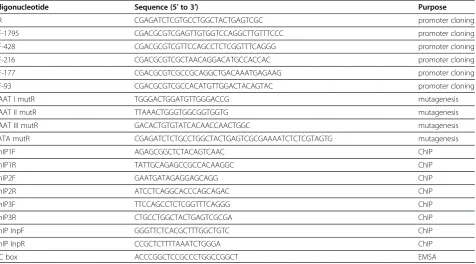
Identification ofcis-acting elements in thec1orf109 promoter region
Becausec1orf109is a novel gene, its mechanism of regu-lation is unclear. To analyze the putative promoter of
the humanc1orf109gene, an approximately 1.8 kb DNA
sequence located upstream of the TSS was cloned as described in the Methods and Materials section.
Nucleo-tide sequence analysis of the 5′ flanking region of the
c1orf109 gene using MatInspector online software
revealed the presence of one TATA box (at−48 bp) and
three CAAT boxes (at −135 bp, -200 bp, -293 bp
re-spectively), as shown in Figure 1A. Progressive 5′
dele-tions of the c1orf109 gene promoter constructs were
generated to identify transcriptional regulatory elements (Figure 1B). All truncated constructs were transiently transfected into HEK293 cells. Firefly luciferase activity was normalized by co-transfection with a Renilla lucifer-ase vector. Meanwhile, the promoter-less pGL3-basic vector was used as a negative control. Significant lucifer-ase activity was observed after transfection of the con-struct containing the proximal 93 bp region upstream of the TSS. Transfection of sequences further upstream, from−177 to−428 bp, resulted in a significant increase in promoter activity, whereas transfection of the prox-imal 41 bp did not generate luciferase activity. The
results indicate that the region from −41 to −177 bp
contains positive regulatory elements essential for achieving maximalc1orf109promoter activity.
To further identify the functional significance of the potential transcription factor binding sites within the
region of −41 to −177 bp, including the putative CAAT
boxes and TATA box, serial site-directed mutation con-structs were used to analyze their effects on luciferase ac-tivity in HEK293 cells. Disruption of the CAAT I and TATA box sites caused impaired promoter activity by ap-proximately 44 to 47 percent. In contrast, mutations of the CAAT box II or CAAT box III sites did not affect c1orf109promoter activity (Figure 2). Therefore, we con-clude that CAAT box I and TATA box act as important cis-acting elements within thec1orf109promoter.
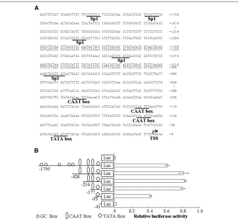
Participation of Sp1 inc1orf109transcription
Sp1 is a transcription factor that either enhances or represses the activity of promoters of genes involved in differentiation, cell cycle progression, and oncogenesis [17]. The presence of several potential GC boxes sug-gested that transcriptional factor Sp1 may be involved in the transcriptional regulation of c1orf109 gene. To con-firm whether Sp1 directly interact withc1orf109promoter, chromatin immunoprecipitation (ChIP) assay was per-formed. HeLa cells were fixed, lysed and fragmented as described in methods and materials. DNA was optimally sheared with a distribution of fragments from 200 to1000 bp, as shown in Figure 3A. Immunoprecipitation of DNA/ protein complexes using antibodies against Sp1 was followed by PCR amplification. As shown in Figure 3B, anti-Sp1 antibody was capable of immunoprecipitating the
c1orf109 promoter fragment containing the GC box 4
(Figure 3B, lane 9); however, primers ChIP InpF/R failed to produce a PCR product (Figure 3B, lanes 4, 8 and 12), indicating that Sp1 directly interacted with c1orf109 pro-moter region.
To detect whether Sp1 interacts directly with the po-tential GC boxes, an electrophoretic mobility shift assay (EMSA) was performed. Oligonucleotides corresponding
to the binding sites for Sp1 in the c1orf109 promoter
were designed (Table I). According to the results of EMSA (Figure 3C), the mobility of labeled probes corre-sponding to GC box 4 was shifted in the presence of nu-clear protein prepared from HeLa cells. The binding specificity of each probe was verified by supershift when we added anti-Sp1 anitibody or excessive unlabeled oligonucleotide competitors. These data suggest that the CAAT box and TATA box are required for achieving the basal transcription of thec1orf109gene. The 5′flanking
region of the c1orf109 gene could bind specific
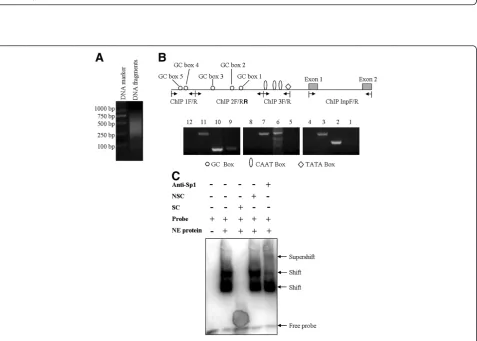
Upregulation of C1ORF109 in multiple cancer cell lines
Previously, we have reported that c1orf109 exhibited an
increased expression in lung cancer tissues compared to
paired adjacent non-tumor tissues using in situ
hybridization with specific RNA probes [14]. To further identify the existence and expression pattern of the puta-tive protein C1ORF109 in the cell, the expression of c1orf109 in 11 breast cancer cell lines and a melanoma
cell line were detected by immunoblotting. Meanwhile, a non-tumorigenic epithelial cell line (MCF10A) [18] and an immortalized human keratinocyte cell line (HaCaT) [19] were used as control. As shown in Figure 4A, C1ORF109 levels were upregulated in tumorigenic cell lines compared to the control, especially in the cell lines derived from metastatic sites, such as the cell lines derived from pleural effusion (MB-436,
Figure 2Effects of site-directed mutations on thec1orf109promoter activity.HEK293 cells were transiently transfected with the−428 bp region ofc1orf109promoter constructs with different mutations of CAAT or TATA box sites. The firefly luciferase activity was assayed 24 hr after transfection and normalized to Renilla luciferase activity. Values represent the means ± S.D. of three independent experiments. *P<0.05 versus the wild type control.
Figure 3Characterization of the transcription factors responsible for Sp1 binding sites in thec1orf109gene promoter. (A)Distribution of DNA fragments by sonication. DNA fragments were sheared with a distribution of fragments from 200 to1000 bp.(B)Chromatin
immunoprecipitation assay using HeLa cell lysates. Chromatin was immunoprecipitated using antibody against Sp1. Lanes 1, 5 and 9 were PCR products from immunoprecipitation using primers ChIP 1 F/R, ChIP 2 F/R and ChIP 3 F/R respectively. Lanes 2, 6 and 10 were PCR products from WCE using primers ChIP 1 F/R, ChIP 2 F/R and ChIP 3 F/R respectively. Lane 3, 7 and 11 were PCR products using ChIP InpF/R from WCE. Lanes 4, 8 and 12 were PCR products using ChIP InpF/R from immunoprecipitation. WCE, whole cell extract.(C)Biotin labeled oligonucleotides
MB-453, MDA-MB-231, MDA-MB-435 s, T-47D, and SK-BR-3), and ascites (ZR-75-30). Furthermore, we also found that C1ORF109 was overexpressed in hepatocellu-lar cancer tissues compared to paired adjacent non-tu-morous tissues by quantitative real-time PCR (qRT-PCR) (see Additional file 1). These findings indicate that
increased expression of C1ORF109 may be involved in cancer progression.
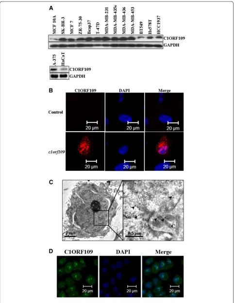
Subcellular localization of C1ORF109
The subcellular localization of the C1ORF109 protein was also examined using immunofluorescence. cDNA
(See figure on previous page.)
Figure 4C1ORF109 is upregulated in multiple cancer cells and located in both the nucleus and cytoplasm. (A)C1ORF109 expression in 11 human breast cancer cell lines and one melanoma cell lines were detected by western blot analysis. The non-tumorigenic epithelial cell line MCF 10A and an immortalized human keratinocyte cell line HaCaT were used as control.(B)Immunofluorescence analysis showed the subcellular localization of C1ORF109 using an anti-Flag tag antibody.(C)Immunoelectron microscope with colloidal gold showed the subcellular localization of C1ORF109 using an anti-V5 epitope antibody. Arrows indicate the specific binding of gold particles. Nu, nucleus. C, cytoplasm.(D)The subcellular localization of endogenous C1ORF109 was detected in HeLa cells using anti-C1ORF109 antibody.
was subcloned into a pCMV-Flag vector. Hs578T cells were cultured on a round coverslip and transiently transfected with pCMV-Flag-c1orf109. A mock vector was used as negative control. The cells were subse-quently fixed, incubated with TRITC-labeled antibodies, and analyzed by confocal microscopy. Positive signals were found mainly in the nucleus and cytoplasm. No signal was detected in the control cells (Figure 4B). To
confirm further the subcellular localization of
C1ORF109, an immune colloidal gold assay was per-formed in NIH3T3 cells that stably expressed V5 tagged C1ORF109. The samples were analyzed by transmission electron microscope, and the results showed that the
colloidal gold particles (diameter, ~15 nm) mainly loca-lized to the nucleus and cytoplasm (Figure 2C). In addition, the subcellular localization of endogenous C1ORF109 was detected in HeLa cells using anti-C1ORF109 antibodies (Figure 2D). These data indicate that C1ORF109 protein is mainly located in the nucleus and cytoplasm.
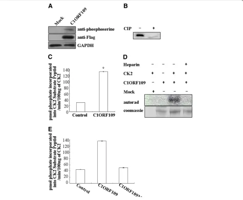
Phosphorylation of C1ORF109 by CK2in vitro
Since c1orf109 has been shown to be involved in cancer
progression, we focused on the biological functions of c1orf109in the cell. To determine the functional domain of C1ORF109 at the molecular level, the PROSITE method
was used to analyze the amino acid sequence, and three po-tential CK2 phosphorylation sites at serines 104, 134 and 182 were found. Therefore, C1ORF109 is predicted to be a phosphoprotein. The full-length C1ORF109 purified from HEK293 cells was recognized by phosphoserine anti-bodies (Figure 5A). Moreover, treatment of C1ORF109 immunoprecipitated from HEK293 cells with calf intestinal phosphatase (CIP) resulted in different electrophoretic mobility compared with untreated C1ORF109 (Figure 5B). Together, these data imply that C1ORF109 is a phospho-protein in eukaryotic cells.
To test whether C1ORF109 is a substrate of protein kinase CK2, the full-length C1ORF109 purified from
HEK293 cells was incubated with human CK2, [γ-32
P]-ATP in vitro, and the CPM was subsequently read in a scintillation counter. To exclude the influence of PKA, a PKA inhibitor cocktail was added to the reaction system. The results indicate that C1ORF109 is efficiently
phos-phorylated by CK2 in vitro (Figure 5C). Meanwhile,
C1ORF109 phosphorylation was abolished by heparin, which is a specific inhibitor of protein kinase CK2 (Fig-ure 5D). Next, serines 104, 134 and 182 were converted into nonphosphorylatable alanine residues yielding a mutant C1ORF109. C1ORF109Mut cannot be
phos-phorylated by CK2 in vitro (Figure 5E). These findings
suggest that C1ORF109 is specifically phosphorylated by CK2in vitro, and that it is a substrate of the protein kin-ase CK2.
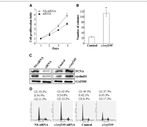
Involvement of C1ORF109 in cancer cell proliferation CK2, a ubiquitous protein serine/threonine kinase with hundreds of substrates, is essential for the modulation of cell growth and proliferation. Since C1ORF109 has been identified to be a substrate of CK2, it might play a role in the modulation of cell proliferation. To verify this postula-tion, MDA-MB-231 cells, which express endogenous C1ORF109 at high levels, were transiently transfected with c1orf109-siRNA to knock down endogenous C1ORF109
and showed a reduction in cell viability (P<0.05,
Figure 6A). However, Hs578T cells, which express low levels of endogenous C1ORF109, were stably transfected to overexpress exogenous C1ORF109 and showed a 5-fold increase in colony number in colony-forming assays (P<0.05, Figure 6B).
The D-type cyclins (Dl, D2 and D3) are key governors of the progression from G1 to S phase of the mammalian cell cycle. These three D-type cyclins are expressed in overlap-ping and apparently redundant fashion in proliferating tissues [20,21]. PCNA, a regulator of DNA replication and cell cycle control, is a well-defined cell proliferation par-ameter [22,23]. We tested the effect of C1ORF109 overex-pression or depletion on the exoverex-pression levels of PCNA and cyclinD1. Downregulation of PCNA and cyclinD1 were detected in C1ORF109-depleted cells. Meanwhile,
Hs578T cells stably expressing exogenous C1ORF109 showed increased expression of PCNA and cyclinD1 (Figure 6C). In addition, the effect of c1orf109 on cell cycle distribution was examined by flow cytometry. Cell cycle analysis of MDA-MB-231 cells transfected
with c1orf109-siRNA showed an increase in G1 phase
and a reduction in DNA synthetic activity (S phase), whereas stably expressing exogenous C1ORF109 in Hs578T cells resulted in fewer cells accumulating in G1 phase compared to the control (Figure 6D). These results indicate that upregulation of c1orf109 in breast cancer cells could promote cancer cell proliferation in vitro, which is mainly due to the acceleration of G1 to S phase transition.
Conclusions
In conclusion, our experiments show that the unknown genec1orf109encodes a CK2 substrate and is involved in the modulation of cell proliferation. More work will be required to identify the molecular mechanisms by which CK2 regulates the expression of C1ORF109 and then affects cell proliferation.
Additional file
Additional file 1:The expression ofc1orf109mRNA in hepatocellular carcinomas (HCCs) detected by quantitative real-time PCR.
Competing interests
The authors declare that they have no competing interest.
Acknowledgments
We thank Ji-lai Liu, Jie Su, and Zhu Wang for generatingc1orf109 promoter-luciferase constructs. This work was supported by the National Natural Science Foundation of China (No.30170516 and No.30871271).
Author details
1Department of Life Science and Engineering, Harbin Institute of Technology
(HIT), Harbin 150001, People’s Republic of China.2Laboratory of Medical
Genetics, Harbin Medical University, Harbin 150001, People’s Republic of China.3Bio-X Center, The Academy of Fundamental and Interdisciplinary
Science, Harbin Institute of Technology, Harbin 150080, People’s Republic of China.
Authors’contributions
SSL and YL designed the study and drafted the manuscript. SSL performed the transcriptional analyses, qRT-PCR, MTT assay and colony forming assay. HXZ, HDJ and JH carried out the immunofluorescence and immunoelectron microscopy with colloidal gold. YPQ, LY and YZ helped to collect the data. All authors read and approved the final manuscript.
Received: 14 November 2011 Accepted: 1 May 2012 Published: 1 May 2012
References
1. Litchfield DW:Protein kinase CK2: structure, regulation and role in cellular decisions of life and death.Biochem J2003,369:1–15. 2. Pinna LA:Protein kinase CK2: a challenge to canons.J Cell Sci2002,
115:3873–3878.
4. Lebrin F, Chambaz EM, Bianchini L:A role for protein kinase CK2 in cell proliferation: evidence using a kinase-inactive mutant of CK2 catalytic subunit alpha.Oncogene2001,20:2010–2022.
5. Ahmad KA, Wang G, Unger G, Slaton J, Ahmed K:Protein kinase CK2–a key suppressor of apoptosis.Adv Enzyme Regul2008,48:179–187.
6. Ahmed K, Gerber DA, Cochet C:Joining the cell survival squad: an emerging role for protein kinase CK2.Trends Cell Biol2002,12:226–230. 7. Wang G, Ahmad KA, Ahmed K:Impact of protein kinase CK2 on inhibitor
of apoptosis proteins (IAPs) in prostate cancer cells.Mol Cell Biochem 2008,316:91–97.
8. St-Denis NA, Litchfield DW:Protein kinase CK2 in health and disease: From birth to death: the role of protein kinase CK2 in the regulation of cell proliferation and survival.Cell Mol Life Sci2009,66:1817–1829. 9. Faust M, Montenarh M:Subcellular localization of protein kinase CK2–A
key to its function?Cell & Tissue Res2000,301:329–340.
10. Guerra B, Issinger OG:Protein kinase CK2 in human disease.Curr Medicinal Chem2008,15:1870–1886.
11. Tawfic S, Yu S, Wang H, Faust R, Davis A, Ahmed K:Protein kinase CK2 signal in neoplasia.Histol Histopathol2001,16:573–582.
12. Trembley JH, Wang G, Unger G, Slaton J, Ahmed K:CK2: A key player in cancer biology.Cell Mol Life Sci2009,66:1858–1867.
13. Meng XW, Li Y, Zhang GY, Wang RW, Li P:Molecular cloning of tumor metastasis related genes from human lung adenocarcinoma cells by mRNA differential display.Chinese J Med Genet1997,14:129–133. 14. Fan H, Li Y, Deng YQ, Chen YZ, Feng HC, Fu SB, Zhang GY, Li P:Cloning
and mapping analysis of cDNA fragmentOPB7-1gene in human lung adenocarcinoma.Chinese J Med Genet2003,20:156–159.
15. Fu XH, Liu DP, Xin L:A simple chromatin immunoprecipitation assay protocol.Prog Biochem Biophys2003,30(4):634–638.
16. Loizou JI, El-Khamisy SF, Zlatanou A, Moore DJ, Chan DW, Qin J, Sarno S, Meggio F, Pinna LA, Caldecott KW:The protein kinase CK2 facilitates repair of chromosomal DNA single-strand breaks.Cell2004,117:17–28. 17. Li L, Davie J:The role of Sp1 and Sp3 in normal and cancer cell biology.
Ann Anat2010,192:275–283.
18. Soule H, McGrath CM:Immortal human mammary epithelial cell lines. US Patent 5,026,637 dated Jun 25 1991.
19. Boukamp P, Petrussevska RT, Breitkreutz D, Hornung J, Markham A, Fusenig NE:Normal keratinization in a spontaneously immortalized aneuploid human keratinocyte cell line.J Cell Biol1988,106(3):761–771. 20. Sherr CJ:D-type cyclins.Trends Biochem Sci1995,20:187–190. 21. Sherr CJ:Cancer cell cycles.Science1996,274:1672–1677.
22. Maga G, Hubscher U:Proliferating cell nuclear antigen (PCNA): a dancer with many partners.J Cell Sci2003,116:3051–3060.
23. Strzalka W, Ziemienowicz A:Proliferating cell nuclear antigen (PCNA): a key factor in DNA replication and cell cycle regulation.Ann Bot2011,
107:1127–1140.
doi:10.1186/1423-0127-19-49
Cite this article as:Liuet al.:Identification and characterization of a
novel gene,c1orf109, encoding a CK2 substrate that is involved in cancer cell proliferation.Journal of Biomedical Science201219:49.
Submit your next manuscript to BioMed Central and take full advantage of:
• Convenient online submission
• Thorough peer review
• No space constraints or color figure charges
• Immediate publication on acceptance
• Inclusion in PubMed, CAS, Scopus and Google Scholar
• Research which is freely available for redistribution