Open Access
Research
Antitumor enhancement of celecoxib, a selective
Cyclooxygenase-2 inhibitor, in a Lewis lung carcinoma expressing
Cyclooxygenase-2
Won Park
1, Young Taek Oh*
2, Jae Ho Han
3and Hongryull Pyo
4Address: 1Department of Radiation Oncology, Samsung Medical Center, Sungkyunkwan University School of Medicine, Seoul, Korea,
2Department of Radiation Oncology, Ajou University Hospital, Suwon, Korea, 3Department of Pathology, Ajou University Hospital, Suwon, Korea
and 4Research Institute and Hospital, National Cancer Center, Goyang, Korea
Email: Won Park - wonp68@skku.edu; Young Taek Oh* - ohyoung@ajou.ac.kr; Jae Ho Han - hanpathol@yahoo.co.kr; Hongryull Pyo - quasar93@ncc.re.kr
* Corresponding author
Abstract
Background: The goal of this study was to determine the effects of a selective Cyclooxygenase (COX)-2 inhibitor on the inhibition of tumor growth and pulmonary metastasis in a Lewis Lung Carcinoma (LLC) animal model.
Methods: For immunoblot analysis of COX-2 and PGE2, cells were treated with irradiation in the presence or absence of celecoxib. The right thighs of male, 6-week old C57/BL mice were subcutaneously injected with 1 × 10⁶ LLC cells. The animals were randomized into one of six groups: (1) no treatment, (2) 25 mg/kg celecoxib daily, (3) 75 mg/kg celecoxib daily, (4) 10 Gy irradiation, (5) 10 Gy irradiation plus 25 mg/kg celecoxib daily, and (6) 10 Gy irradiation plus 75 mg/kg celecoxib daily. Mice were irradiated only once, and celecoxib was administered orally. Mice were irradiated with 4-MV photons once the tumor volume of the control group reached 500 mm3. All mice were sacrificed when the mean tumor volume of control animals grew to 4000 mm3. The left lobes of the lungs were extracted for the measurement of metastatic nodules.
Results: Irradiation resulted in a dose-dependent increase in PGE2 production. PGE2 synthesis decreased markedly after treatment with celecoxib alone or in combination with irradiation. Compared to mice treated with low dose celecoxib, mean tumor volume decreased significantly in mice treated with a high dose of celecoxib with or without irradiation. Mice treated with a high dose celecoxib alone, with irradiation alone, or with irradiation plus celecoxib had markedly fewer metastatic lung nodules than controls. The mean metastatic area was the smallest for mice treated with irradiation plus a high dose celecoxib.
Conclusion: Oral administration of high dose celecoxib significantly inhibited tumor growth, as compared to a low dose treatment. Radiotherapy in combination with high dose celecoxib delayed tumor growth and reduced the number of pulmonary metastases to a greater extent than celecoxib or radiotherapy alone.
Published: 11 November 2008
Journal of Experimental & Clinical Cancer Research 2008, 27:66 doi:10.1186/1756-9966-27-66
Received: 3 July 2008 Accepted: 11 November 2008
This article is available from: http://www.jeccr.com/content/27/1/66 © 2008 Park et al; licensee BioMed Central Ltd.
Background
Radiotherapy is a common treatment for localized can-cers. The radiation dose is important for tumor control. However, the therapeutic efficacy of radiotherapy is often limited by normal tissue damage within or nearby the field of radiation. In clinical practice, the radiation dose is optimized according to the probability of tumor control compared to the risks of complications due to the effects on normal tissue [1,2]. Combining chemotherapeutic agents concurrently with radiotherapy has improved tumor control and survival. However, this combined approach also increases systemic and local toxicities dur-ing radiotherapy. Because of the increased toxicity, the overall treatment duration of radiotherapy, in addition to chemotherapy, is usually prolonged when compared to the treatment time of radiotherapy alone [3,4]. This increased duration may decrease its efficacy for tumor control within the radiation field.
To further improve tumor response and reduce normal tissue toxicity from radiotherapy or chemoradiotherapy, many novel approaches have investigated several agents in preclinical and clinical settings. These approaches include those that selectively interfere with certain molec-ular processes and signaling pathways that regulate prolif-eration, survival, and function of normal cells. Because these agents are preferentially associated with specific sites of the cancer cells, their targeting is predicted to improve the tumor response to radiotherapy or chemoradiother-apy without additional toxicity to normal tissue. Among these agents, inhibition of cyclooxygenase (COX)-2 has been investigated as a potentially useful agent for the treatment of cancer.
COX-2 is normally present in cells and tissues of the brain and kidneys, but is induced in pathological states such as inflammation and tumors. COX-2 promotes carcinogene-sis, tumor proliferation, angiogenecarcinogene-sis, prevention of apoptosis, and immunosuppression [5]. COX-2 overex-pression has been associated with tumor behavior and prognosis in several cancers [6]. Selective inhibition of COX-2 activity in several animal models has been associ-ated with the decrease of new blood vessel production in tumors, a decrease in new vessel formation and an increase in tumor cell apoptosis. The selective inhibition of COX-2 activity has been associated with enhanced radi-ation sensitivity of tumors without enhancing the effects of radiation on normal tissue [7-9].
In this study, we evaluated the effect of a selective COX-2 inhibitor as a radiation sensitizer in order to inhibit tumor growth and pulmonary metastasis in a Lewis Lung Carci-noma (LLC) animal model.
Methods
Animals and Tumor Cells
Male, 6-week old C57/BL mice (Ajou animal laboratory, Suwon, Korea) were used for these experiments. The mice were acclimated for 1 week, and caged in groups of five or less in an air conditioned room. Mice were fed a diet of animal chow and water ad libitum. LLC cells were pur-chased from the American Type Tissue Collection. LCC cells were maintained in DMEM supplemented with 10% fetal bovine serum and penicillin-streptomycin. Cells were grown in monolayers in 100 mm dishes, and were maintained in a humidified 5% CO2 incubator at 37°C.
Celecoxib
Stock solutions of celecoxib were made by dissolving the compound in DMSO, then were stored at -20°C. Concen-trated drug stocks were diluted in DMEM before adminis-tration to cells or mice.
Immunoblot Analysis of COX-2
Cells were pretreated with 10 or 30 μM celecoxib for 1 h at 37°C. After treatment, the cells were irradiated at a dose of 5 Gy or 10 Gy. At 24 or 48 h post treatment, the cells were washed twice with PBS and lysed in buffer (Upstate). Supernatant protein concentrations were determined by Bradford assay using bovine serum albumin (BSA, Sigma Chemical Co.) as a standard. Aliquots of total protein (40
μg) was denatured and fractionated by SDS-polyacryla-mide gel electrophoresis (4–12% gels). The separated pro-teins were transferred to a 0.22 μm nitrocellulose membrane. The nonspecific binding sites were blocked for 1 h in 5% non-fat dry milk and in Tris-buffered saline (TBS). The membranes were incubated with monoclonal anti-COX-2 (610204, BD biosciences) and anti-α tubulin (Oncogene) for approximately 1 h at room temperature. The membranes were washed in buffer containing TBS plus 0.05% Tween-20 and incubated in the appropriate secondary antibody (P0447, Dakocytonation). Signals were detected using enhanced chemiluminescence (Pierce).
Determination of PGE2 Synthesis
1 × 10⁶ cells were either untreated, or treated with 30 μM celecoxib for 1 hr and then with 0, 5, or 10 Gy irradiation. After each treatment, supernatant PGE2 levels were assayed in triplicate. Determination of PGE2 levels by enzyme immunoassay was accomplished using a PGE2 monoclonal enzyme immunoassay kit (Cayman Chemi-cal). Quantification was performed according to the man-ufacturer's instructions.
In vivo Tumor Growth and Quantitation of Lung Metastases
right thighs of mice. The study groups (n = 12 per group) consisted of an untreated control (group 1), 25 mg/kg celecoxib daily (group 2), 75 mg/kg celecoxib daily (group 3), 10 Gy irradiation (group 3), 10 Gy irradiation plus 25 mg/kg celecoxib daily (group 5), and 75 mg/kg 10 Gy irradiation plus celecoxib daily (group 6).
Celecoxib was administered by lavage (0.1 mL) every afternoon from one day before the cell injection until the day of euthanasia or death. For tumor irradiation, mice were put under general anesthesia and restrained using adhesive tape and customized devices constructed from a 50 ml syringe. Once the tumors in the control group reached a mean volume of 500 mm3, the tumors in the right thighs were irradiated with 10 Gy using a 4 MV x-ray for one fraction. Following injection of the tumor cells, the primary tumors were measured three times a week at two perpendicular diameters using a Vernier caliper, and tumor volumes were evaluated based on the formula, vol-ume = 0.5 × a × b2 where a = length and b = width.
All mice were euthanized when the mean tumor volume in the control group reached 4000 mm3. The left lobes of the lungs were extracted, fixed in 10% formalin, and proc-essed for the quantitation of metastatic nodules. The number of metastatic nodules was measured in the maxi-mum sagittal plane from 5 μm paraffin-embedded lung tissue sections. The dimension of the outlined metastatic nodule was automatically calculated using commercial software (i solution DT, Seoul, Korea).
Statistical Analysis
The primary tumor volumes were expressed as the mean and standard deviation. Comparison of the area of the lung nodules and tumor volumes among the experimen-tal groups was determined by the Wilcoxon rank-sum test. The correlation between tumor volume and area of the metastatic lung nodules was evaluated by regression anal-ysis. P-values less than 0.05 were considered statistically significant. All statistical analyses were performed with the SAS® System (SAS 14.0, SAS Institute Inc., Cary, NC., USA).
Results
In vitro, Effect of the Selective COX-2 Inhibitor on LLC Cells
LLC cell COX-2 protein expression was confirmed by western blot analysis, which showed constitutive COX-2 expression (Figure 1). Differences in the amount of COX-2 protein expression were not observed after irradiation alone. However, COX-2 expression increased with celecoxib treatment alone and with irradiation plus celecoxib. Irradiation was associated with a dose-depend-ent increase in PGE2 production, as measured by enzyme immunoassay (Figure 2). PGE2 synthesis decreased
mark-edly after treatment with celecoxib alone or with celecoxib in combination with irradiation. Radiation treatment plus celecoxib did not increase PGE2 production when com-pared to celecoxib alone, regardless of the radiation dose.
In vivo Tumor Growth
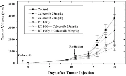
When the mean volume of the tumors in the control group reached 539.6 mm3 (487~589 mm3), 11 days after LLC cell injection, the animals in irradiation alone or celo-coxib plus irraditation were irradiated. At 20 days after injections, a protruding tumor and areas of denuded sur-face were found at the injection sites, especially in control mice. The mean tumor volume in the control group increased to 3802.2 mm3 (3466~4332 mm3), and all mice were euthanized as planned. At that time, the mean tumor volumes of groups 2, 3, 4, 5, and 6 were 3814.4 mm3 (2225~5421 mm3), 2757.6 mm3 (1995~3395 mm3), 1942.1 mm3 (1766~2275 mm3), 1874.3 mm3 (1341~2437 mm3), and 1319.3 mm3 (989~1815 mm3), respectively (Figure 3).
Compared to the control group, the mean tumor volume decreased significantly in all treatment groups, except for those treated with 25 mg/kg celecoxib daily (Table 1). The effect of celecoxib on the delay of tumor growth was dose-dependent. Compared to the mice treated daily with 25 mg/kg celecoxib, the mean tumor volumes decreased sig-nificantly in the mice treated daily with 75 mg/kg celecoxib, with or without irradiation (p = 0.0135, p = 0.0144). The delay in tumor growth was evident in the irradiated groups, regardless of the celecoxib dose.
In vivo Lung Metastasis
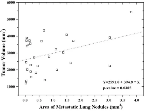
The prevalence of detected lung metastases from groups 1 to 6 were 100%, 100%, 75.0%, 50.0%, 87.5%, and 25%, respectively. The mean area of the metastatic lung nodules did not differ between mice treated with low dose celecoxib (25 mg/kg) only and the control group (Table 2). However, the area of metastatic lung nodules decreased significantly in mice treated with high dose celecoxib (75 mg/kg) alone or with irradiation with or without celecoxib. Among the mice treated with irradia-tion, the mean metastatic area was smaller than in the control group. The mean metastatic area was the smallest in mice treated with irradiation and 75 mg/kg celecoxib daily (group 6); however, the difference was only margin-ally significant (p = 0.0675). The area of metastatic lung nodules significantly correlated with tumor volume, regardless of treatment (Figure 4).
Discussion
has been associated with tumor differentiation, tumor size, stage, and metastasis. In several cancers, patients with COX-2 overexpression have a poor prognosis [10-14]. Denkert et al. immunohistochemically evaluated COX-2 expression from breast cancer specimens. They reported more lymph node metastasis, larger tumors, poor differ-entiation, increased vascular invasion, and negative estro-gen receptor status in patients with elevated COX-2 expression. COX-2 overexpression was reported to be of borderline significance for disease-free survival (relative risk 1.90). Sheehan et al. showed that high COX-2 expres-sion correlates with advanced stage disease and larger tumors in patients with colorectal cancer.
COX-2 associates with tumor growth, infiltration, and metastasis in preclinical experiments. Tsujii et al. studied the phenotypic and biochemical changes associated with COX-2 expression. Rat intestinal epithelial cells were infected with a COX-2 expression vector oriented in the sense (RIE-S) or anti-sense (RIE-AS) direction. RIE-S cells demonstrated increased adhesion to extracellular matrix proteins and inhibition of apoptosis, compared with RIE-AS cells. After human colon cancer cells (Caco-2) were transfected with a COX-2 expression vector, Caco-2 cells acquired increased invasiveness compared with parental
cells [15,16]. Liu et al. showed that transgenic mice over-expressing COX-2 in the mammary gland, expressed COX-2 during pregnancy and lactation. Multiparous transgenic mice, but not virgin mice, exhibited a high inci-dence of focal mammary gland hyperplasia, dysplasia, and transformation into metastatic tumors [17].
Nonsteroidal anti-inflammatory drugs (NSAID) are widely used for relief of inflammatory pain worldwide. Several population-based reports have demonstrated a 40% decrease in the death rate in persons with digestive tract cancers who regularly used aspirin, compared to those who did not. Clinical trials with NSAID in patients with Familial Adenomatous Polyposis clearly demon-strated that NSAID treatment shrunk pre-existing adeno-mas [18-20]. In addition, indomethacin offered palliative support to patients with advanced solid tumors, and pro-longed their mean survival compared to placebo-treated patients [21]. The mechanism of NSAID action was unknown until in 1971, when Vane proposed that NSAIDs primarily suppress inflammation by inhibiting COX and by limiting the production of PG [22]. NSAIDs non-selectively inhibit the activities of both COX-1 and COX-2. COX-1 inhibition causes the adverse effects of NSAIDs on the gastrointestinal tract [23,24]. In a
rand-Celecoxib effects on COX-2 protein expression
Figure 1
omized, placebo-controlled endoscopic evaluation, Sche-iman et al. reported that the short-term use of nonselective COX inhibitors associated with a signifi-cantly greater ulceration rate, compared with a placebo [25].
The development of selective COX-2 inhibitors, with decreased potential for gastrointestinal toxicity, has stim-ulated additional investigations. Numerous studies on the antineoplastic effects of selective COX-2 inhibitors have been performed. Williams et al. proposed that celecoxib reduced the viability of cell lines, including LLC cells, from the induction of apoptosis and the growth of tumors in vivo, and had no effect on apoptosis or the epithelium of the normal gut [26]. Leahy et al. reported that a reduc-tion in proliferareduc-tion and an increase in apoptosis were observed in colorectal tumor cells in response to celecoxib [27]. Connolly et al. proposed that in vitro COX inhibitors decreased vascular endothelial growth factor production and increased apoptosis of tumor cells, as well as a reduced primary tumor weight, the number of lung metastases, and microvessel density in primary tumors in mice [28]. Kobayashi et al. showed that selective COX-2 inhibition in colon cancer cell lines reduced the diameter of the tumor vessels as well as the number and size of the metastatic nodules in the lung. In addition,
dose-depend-ent selective COX-2 inhibition reduced the size of meta-static tumors [9].
In our study, we used western blot analysis to confirm that a COX-2 inhibition increased COX-2 expression. The effect of celecoxib on the delay of tumor growth was dose-dependent, regardless of irradiation. High dose celecoxib (75 mg/kg celecoxib daily) significantly reduced the tumor volume, compared to control and low dose celecoxib groups. The rate of lung metastasis was decreased by about 25% in the high dose celecoxib group, compared to the control group. In addition, the area of metastatic lung nodules decreased significantly in the high dose celecoxib group.
With irradiation, COX-2 expression increased in tumors and associated with increased PGE2 levels. Steinauer et al. observed a dose-dependent increase in COX-2, following irradiation. PGE2 levels in irradiated cells were higher than in controls, and decreased when combined with a COX-2 inhibitor [29]. The mechanisms underlying the radiation-enhancing effects of COX-2 inhibitors include (1) an accumulation of cells in the G2/M phases of the cell cycle which are considered to be sensitive to irradiation; (2) a reduction of PG-induced immunosuppressive activ-ity caused by antitumor immunologic responses capable of potentiating tumor responses to radiation; and (3) Celecoxib effects on PGE2 production
Figure 2
direct effects on tumor neovascularization [7,30,31]. Shin et al. reported that celecoxib's radiation-enhancing effect was observed in COX-2 expressing cells but was not observed in COX-2 non-expressing cells. The
radiation-enhancing effects disappeared in cells treated with COX-2-specific siRNA [32]. LLC cells express COX-2 [26]. In this study, we confirmed COX-2 expression by western blotting. Unlike Steinauer et al., irradiation, regardless of
Table 1: Wilcoxon rank-sum test for mean tumor volume at 20 days after tumor cell injection in control and treatment groups (p-value).
Groups Control Celecoxib 25 mg/kg Celecoxib 75 mg/kg RT RT + Celecoxib 25 mg/ kg
RT + Celecoxib 75 mg/ kg
Control - 0.9761 0.0004 < 0.0001 < 0.0001 < 0.0001
Celecoxib 25 mg/kg 0.9761 - 0.0135 < 0.0001 0.0002 < 0.0001
Celecoxib 75 mg/kg 0.0004 0.0135 - 0.0004 0.0023 < 0.0001
RT < 0.0001 < 0.0001 0.0004 - 0.7238 0.0001
RT + Celecoxib 25 mg/ kg
< 0.0001 0.0002 0.0023 0.7238 - 0.0144
RT + Celecoxib 75 mg/ kg
< 0.0001 < 0.0001 < 0.0001 0.0001 0.0144
-RT: radiation
Celecoxib effects the in vivo tumor growth of Lewis lung carcinoma cells.
Figure 3
the dosage, did not increase COX-2 expression. However, PGE2 levels increased with irradiation in a dose-depend-ent manner. Tumor growth was delayed as a result of irra-diation, especially in mice treated by irradiation and high dose celecoxib, where tumor growth was markedly delayed. The area of metastatic lung nodules was signifi-cantly smaller in mice treated by irradiation, regardless of celecoxib dose, than in the control group. Lung metastases were detected in only 25% of the mice treated by irradia-tion plus high dose celecoxib, as compared to 100% of the control mice.
The surgical removal of a primary tumor or radiation in order to eradicate a primary tumor can result in rapid growth and metastasis [33,34]. Camphausen et al. reported that a single or hypofractionated irradiation pro-tocol for the eradication of primary LLC cells increased the
number of surface lung metastases in irradiated animals, as compared to controls. However, in our study, the number of lung metastases decreased from 100% in con-trol mice to 50% in mice treated with irradiation alone. The differences between these two studies may be due to the doses used, i.e., whether it was a curative dose or not. We did not irradiate the primary tumor for eradication because we were studying the effects of a selective COX-2 inhibitor as a radiation sensitizer for the inhibition of tumor growth and pulmonary metastasis.
In conclusion, a high dose oral treatment of celecoxib sig-nificantly inhibited tumor growth, compared to a low dose treatment. In mice treated with radiotherapy and a high dose celecoxib, delay of tumor growth and reduction of pulmonary metastases were more prominent than in the mice treated with celecoxib or radiotherapy alone.
Regression analysis showed a significant correlation between the area of metastatic lung nodules and tumor volume regardless of treatment.
Figure 4
However, further studies are needed to evaluate the effect of selective COX-2 inhibitors combined with conven-tional fractionation or hypofractionation radiotherapy on various cancer cell lines with regard to the delay of tumor growth and inhibition of metastasis.
Competing interests
The authors declare that they have no competing interests.
Authors' contributions
H.P. participated in the design of the study. J.H.H. carried out pathological analysis in the animal experiment. Y.T.O. participated in its design and coordination and helped to draft the manuscript. W.P. designed the study, performed the experiments and wrote the manuscript.
Acknowledgements
This work was supported by a grant from "Department of medical sciences, the graduate school, Ajou University" in 2003.
References
1. Emami B, Lyman J, Brown A, Coia L, Goitein M, Munzenrider JE, Shank B, Solin LJ, Wesson M: Tolerance of normal tissue to therapeu-tic irradiation. Int J Radiat Oncol Biol Phys 1991, 21:109-122. 2. Marks LB: The impact of organ structure on radiation
response. Int J Radiat Oncol Biol Phys 1996, 34:1165-1171. 3. Al-Sarraf M, LeBlanc M, Giri PG, Fu KK, Cooper J, Vuong T,
Forast-iere AA, Adams G, Sakr WA, Schuller DE, Ensley JF: Chemoradio-therapy versus radioChemoradio-therapy in patients with advanced nasopharyngeal cancer: phase III randomized Intergroup study 0099. J Clin Oncol 1998, 16:1310-1317.
4. Girinsky T, Rey A, Roche B, Haie C, Gerbaulet A, Randrianarivello H, Chassagne D: Overall treatment time in advanced cervical carcinomas: a critical parameter in treatment outcome. Int J Radiat Oncol Biol Phys 1993, 27:1051-1056.
5. Koki AT, Masferrer JL: Celecoxib: a specific COX-2 inhibitor with anticancer properties. Cancer Control 2002, 9:28-35. 6. Choy H, Milas L: Enhancing radiotherapy with
cyclooxygenase-2 enzyme inhibitors: a rational advance? J Natl Cancer Inst 2003,
95:1440-1452.
7. Milas L, Furuta Y, Hunter N, Nishiguchi I, Runkel S: Dependence of indomethacin-induced potentiation of murine tumor radi-oresponse on tumor host immunocompetence. Cancer Res
1990, 50:4473-4477.
8. Williams CS, Mann M, DuBois RN: The role of cyclooxygenases in inflammation, cancer, and development. Oncogene 1999,
18:7908-7916.
9. Kobayashi H, Gonda T, Uetake H, Higuchi T, Enomoto M, Sugihara K:
JTE-522, a selective COX-2 inhibitor, interferes with the growth of lung metastases from colorectal cancer in rats due to inhibition of neovascularization: a vascular cast model study. Int J Cancer 2004, 112:920-926.
10. Sheehan KM, Sheahan K, O'Donoghue DP, MacSweeney F, Conroy RM, Fitzgerald DJ, Murray FE: The relationship between cycloox-ygenase-2 expression and colorectal cancer. JAMA 1999,
282:1254-1257.
11. Graffney DK, Holden J, Davis M, Zempolich K, Murphy KJ, Dodson M: Elevated cyclooxygenase-2 expression correlates with diminished survival in carcinoma of the cervix treated with radiotherapy. Int J Radiat Oncol Biol Phys 2001, 49:1213-1217. 12. Buskens CJ, van Rees BP, Sivula A, Reitsma JB, Haglund C, Bosma PJ,
Offerhaus GJ, van Lanschot JJ, Ristimaki A: Prognostic significance of elevated cyclooxygenase 2 expression in patients with adenocarcinoma of the esophagus. Gastroenterology 2002,
122:1800-1807.
13. Kim YB, Kim GE, Cho NH, Pyo HR, Shim SJ, Chang SK, Park HC, Suh CO, Park TK, Kim BS: Overexpression of cyclooxygenase-2 is associated with a poor prognosis in patients with squamous cell carcinoma of the uterine cervix treated with radiation and concurrent chemotherapy. Cancer 2002, 95:531-539. 14. Denkert C, Winzer KJ, Muller BM, Weichert W, Pest S, Kobel M,
Kristiansen G, Reles A, Siegert A, Guski H, Hauptmann S: Elevated expression of cyclooxygenase-2 is a negative prognostic fac-tor for disease free survival and overall survival in patients with breast carcinoma. Cancer 2003, 97:2978-2987.
15. Tsujii M, DuBois RN: Alterations in cellular adhesion and apop-tosis in epithelial cells overexpressing prostaglandin endoperoxide synthase 2. Cell 1995, 83:493-501.
16. Tsujii M, Kawano S, DuBois RN: Cyclooxygenase-2 expression in human colon cancer cells increases metastatic potential.
Proc Natl Acad Sci USA 1997, 94:3336-3340.
17. Liu CH, Chang SH, Narko K, Trifan OC, Wu MT, Smith E, Hauden-schild C, Lane TF, Hla T: Overexpression of cyclooxygenase-2 is sufficient to induce tumorigenesis in transgenic mice. J Biol Chem 2001, 276:18563-18569.
18. Waddell WR, Loughry RW: Sulindac for polyposis of the colon.
J Surg Oncol 1983, 24:83-87.
19. Thun MJ, Namboodiri MM, Calle EE, Flanders WD, Heath CW Jr:
Aspirin use and risk of fatal cancer. Cancer Res 1993,
53:1322-1327.
20. Fossilien E: Molecular pathology of cyclooxygenase-2 in neo-plasia. Ann Clin Lab Sci 2000, 30:3-21.
21. Lundholm K, Gelin J, Hyltander A, Lonnroth C, Sandstrom R, Svanin-ger G, Korner U, Gulich M, Karrefors I, Norli B: Anti-inflamma-tory treatment may prolong survival in undernourished patients with metastatic solid tumors. Cancer Res 1994,
54:5602-5606.
22. Vane JR: Inhibition of prostaglandin synthesis as a mechanism of action for aspirin-like drugs. Nat New Biol 1971, 231:232-235.
Table 2: Analysis of metastatic lung nodules according to control and treatment groups.
Groups Areas of metastatic nodule (mean, mm2) p-value
Control 0.61–4.04 (1.09)
-Celecoxib 25 mg/kg 0.05–3.81 (1.48) 0.3607
Celecoxib 75 mg/kg 0.07–1.37 (0.81) 0.0295
RT 0.20–0.45 (0.31) 0.0181
RT + Celecoxib 25 mg/kg 0.04–1.64 (0.50) 0.0238
RT + Celecoxib 75 mg/kg 0.03–0.22 (0.13) 0.0675
Publish with BioMed Central and every scientist can read your work free of charge "BioMed Central will be the most significant development for disseminating the results of biomedical researc h in our lifetime."
Sir Paul Nurse, Cancer Research UK
Your research papers will be:
available free of charge to the entire biomedical community
peer reviewed and published immediately upon acceptance
cited in PubMed and archived on PubMed Central
yours — you keep the copyright
Submit your manuscript here:
http://www.biomedcentral.com/info/publishing_adv.asp
BioMedcentral
23. Fosslien E: Adverse effects of nonsteroidal anti-inflammatory drugs on the gastrointestinal system. Ann Clin Lab Sci 1998,
28:67-81.
24. Warner TD, Giuliano F, Vojnovic I, Bukasa A, Mitchell JA, Vane JR:
Nonsteroid drug selectivities for cyclo-oxygenase-1 rather than cyclo-cxygenase-2 are associated with human gastroin-testinal toxicity: a full in vitro analysis. Proc Natl Acad Sci USA
1999, 96:7563-7568.
25. Scheiman JM, Cryer B, Kimmey MB, Rothstein RI, Riff DS, Wolfe MM:
A randomized, controlled comparison of ibuprofen at the maximal over-the-counter dose compared with prescrip-tion-dose celecoxib on upper gastrointestinal mucosal injury. Clin Gastroenterol Hepatol 2004, 2:290-295.
26. Williams CS, Watson AJ, Sheng H, Helou R, Shao J, DuBois RN:
Celecoxib prevents tumor growth in vivo without toxicity to normal gut: lack of correlation between in vitro and in vivo models. Cancer Res 2000, 60:6045-6051.
27. Leahy KM, Ornberg RL, Wang Y, Zweifel BS, Koki AT, Masferrer JL:
Cyclooxygenase-2 inhibition by celecoxib reduces prolifera-tion and induces apoptosis in angiogenic endothelial cells in vivo. Cancer Res 2002, 62:625-631.
28. Connolly EM, Harmey JH, O'Grady T, Foley D, Roche-Nagle G, Kay E, Bouchier-Hayes DJ: Cyclo-oxygenase inhibition reduces tumour growth and metastasis in an orthotopic model of breast cancer. Br J Cancer 2002, 87:231-237.
29. Steinauer KK, Gibbs I, Ning S, French JN, Armstrong J, Knox SJ: Radi-ation induces upregulRadi-ation of cyclooxygenase-2 (COX-2) protein in PC-3 cells. Int J Radiat Oncol Biol Phys 2000, 48:325-328. 30. Furuta Y, Hunter N, Barkley T Jr, Hall E, Milas L: Increase in radi-oresponse of murine tumors by treatment with indometh-acin. Cancer Res 1988, 48:3008-3013.
31. Milas L, Kishi K, Hunter N, Mason K, Masferrer JL, Tofilon PJ:
Enhancement of tumor response to gamma-radiation by an inhibitor of cyclooxygenase-2 enzyme. J Natl Cancer Inst 1999,
91:1501-1504.
32. Shin YK, Park JS, Kim HS, Jun HJ, Kim GE, Suh CO, Yun YS, Pyo H:
Radiosensitivity enhancement by celecoxib, a cyclooxygen-ase (COX)-2 selective inhibitor, via COX-2-dependent cell cycle regulation on human cancer cells expressing differen-tial COX-2 levels. Cancer Res 2005, 65:9501-9509.
33. O'Relly MS, Holmgren L, Shing Y, Chen C, Rosenthal RA, Moses M, Lane WS, Cao Y, Sage EH, Folkman J: Angiostatin: a novel angio-genesis inhibitor that mediated the suppression of metas-tases by Lewis lung carcinoma. Cell 1994, 79:315-328. 34. Camphausen K, Moses MA, Beecken WD, Khan MK, Folkman J,