R E S E A R C H A R T I C L E
Open Access
Development and evaluation of adsorption
sheet (HD safe sheet-U) using active carbon
for the purpose of the preventing the
contamination diffusion of urinary excreted
anticancer drug
Junya Sato
1,2,3*, Haruka Ohkubo
1, Yuki Sasaki
1, Makoto Yokoi
4, Yasunori Hotta
4and Kenzo Kudo
1,2Abstract
Background:Certain amount of anticancer drugs is excreted in the urine of patients receiving anticancer
drugs, and urinary scattering including anticancer drugs at excretion has become a route of anticancer drug contamination. Therefore, we developed an active carbon sheet (HD safe sheet-U) that prevented diffusion by adsorbing anticancer drugs including that excreted in urine. The present study conducted a performance evaluation of this sheet.
Methods: The adsorption performance of active carbon to anticancer drug in the urine was evaluated by
determining concentration changes in the active carbon suspension (5 mg/mL) of 14 kinds of anticancer drugs (cyclophosphamide, ifosfamide, carboplatin, cisplatin, methotrexate, 5-fluorouracil, cytarabine, gemcitabine, doxorubicin, epirubicin, paclitaxel, docetaxel, etoposide, and irinotecan) diluted with artificial urine. Adhesion of the anticancer drug dropping on the sheet to a slipper sole was evaluated because urine including anticancer drugs is scattered on the floor, which can spread by adhering to shoe soles of patients and healthcare workers. The performance of the active carbon sheet was compared with two other types of medical adsorption sheets use d as control sheets. Anticancer drugs diluted with artificial urine (1 mL) were dropped on the active carbon sheet and the two control sheets. The sheets were trod with slippers made by polyvinyl chloride. The adhered anticancer drug was wiped off and its quantity was determined.
Results:A remarkable decrease in anticancer drug concentrations, except for cisplatin, was detected by mixture of active carbon in the artificial urine (0–79.6%). The quantity of anticancer drug adhesion to slipper soles from the active carbon sheet was significantly lower compared with that observed for the two control sheets for eight kinds of anticancer drugs (cyclophosphamide, ifosfamide, carboplatin, methotrexate, cytarabine, gemcitabine, doxorubicin, and docetaxel). There was no adhesion in cyclophosphamide and docetaxel. Furthermore, the quantities of adhesion in cytarabine, gemcitabine, doxorubicin, paclitaxel, and irinotecan were lower than determination limit.
Conclusion:Active carbon might be effective in adsorbing urinary anticancer drugs. The active carbon sheet adsorbed urinary excreted anticancer drugs, and use of such sheets might prevent diffusion of contamination due to urinary excreted anticancer drugs.
Keywords:Active carbon, Anticancer drug contamination, Adsorption sheet, HD safe sheet-U
* Correspondence:[email protected]
1
Department of pharmacy, Iwate Medical University Hospital, 19-1 Uchimaru, Morioka, Iwate 020-8505, Japan
2Department of Clinical Pharmaceutics, School of Pharmacy, Iwate Medical University, 2-1-1 Nishitokuta, Yahaba, Iwate 028-3694, Japan
Full list of author information is available at the end of the article
Background
Anticancer drug exposure of healthcare workers and patients’ family members becomes health hazard risk. Although anticancer drug may be exposed in extremely small amounts compared with the amount administered to patients, studies have reported increased risk of acute toxicity, including diarrhea, cough, hair loss, exanthem [1–3], miscarriage as a reproductive toxicity [4], and car-cinogenicity [5, 6]. Anticancer drugs are scattered during the processes of preparation, administration, disposal of administration equipment at the pharmacy and at patients’ bedside. These scattered anticancer drugs can be inhaled, come in contact with the skin, or are acci-dentally ingested by healthcare workers or patients’ fam-ily members (https://www.cdc.gov/niosh/docs/2004-165/ pdfs/2004-165.pdf ). The use of tools such as biological safety cabinet (BSC), closed system, and personal protec-tion equipment (PPE) such as masks, gloves, and gowns are effective in protecting against exposure during the preparation and administration. Another contamination process is scattering of anticancer drugs through pa-tients’ excrement. All body fluids such as urine, stool, vomit, saliva, sweat, and blood are considered patient excrement. In particular, urine is the most concerned ex-crement for diffusion because urine has a large volume and can easily scatter depending on the excretion pos-ture, patient gender, and inappropriate handling during urine collection. Although urinary excretion of antican-cer drugs may considerably vary by individual, most anticancer drugs are excreted in the unchanged form in the urine. The International Society of Oncology Phar-macy Practitioners guidelines recommend that PPE should be used during handling of excrement, including urine, within <48 h of administration (http://www.oncos ystems.com.tr/dosyalar/ISOPP_Standards_of_ Practice-Safe Handling of Cy totoxics.pdf ). In addition, for pre-vention of urinary scattering, the guidelines recommend the use of Western style toilet stool for urination for both men and women, sitting down during urination for men, and flushing after closing the cover of the toilet for patients receiving chemotherapy [7]. However, antican-cer drug contamination of patients’ home and hospital lavatories has been reported. Yuki et al. surveyed anti-cancer drug contamination in home lavatories of pa-tients with breast cancer [8]. Cyclophosphamide (CPA) at 0.04–8.35 ng/cm2
and 0.08–1.53 ng/cm2 were de-tected on the toilet seat and floor, respectively. These contamination levels are similar to those reported in various other contamination surveys conducted in hos-pital pharmacies [9, 10]. Moreover, Yuki et al. surveyed human exposure of family members who lived with patients receiving CPA administration [11]. CPA was detected at 17–252 ng per member in the urine in five members from 10 patient families. These findings
suggested that healthcare workers and family members were more likely to be exposed to anticancer drugs owing to contact with patients’ body fluids including urine. Therefore, PPE should be used when healthcare workers and family members come in close contact with patients receiving chemotherapy. Morimoto et al. reported platinum contamination in a lavatory near an outpatient chemotherapy unit of a hospital [12]; plat-inum contamination of the floor of the toilet stool for women was <15–360 ng whereas that of the floor of the toilet stool for men was remarkably high (990–3000 ng). Urine may directly scatter on urination or scattered at a flash bulb. Moreover, anticancer drugs scattered on the lavatory floors adhered to slipper soles and seemed to spread outside the lavatory as well. Thus, when antican-cer drug solution to be administered is scattered, use of a spill kit including PPE and inactivating agent is recom-mended to prevent contamination and diffusion while handling [13]. A wipe cleaning method using sodium hy-droxide, sodium hypochlorite, and ozone waters is used for the preparation or administration environment, such as inside BSCs or hospital rooms where anticancer agent contamination occurs routinely [14–16]. However, this may lead to other problem such as damage to tiles, toilet stool, or other metal equipment, need for neutralization, and other uncertain degradation effects associated with the use of these inactivating agents in the lavatory. Such antiexposure methods for patients’urine do not seem to be adequately performed in hospital and home settings. Therefore, a new method to prevent scattering of urin-ary anticancer drugs is warranted.
We developed active carbon sheets that adsorb the scattering splash during anticancer drug preparation (product name; HD safe sheet) [17]. Seven kinds of anticancer drugs diluted in a preparation concentra-tion were dropped on an active carbon sheet; none of the drugs exhibited any adhesion to the infusion sur-face. In addition, the concentrations of these antican-cer drugs were remarkably decreased in the active carbon suspension used for the active carbon sheet. We concluded that the active carbon sheet contrib-uted to diffusive prevention of contamination by an active carbon adsorbing the anticancer drug scattered during preparation.
Methods
Anticancer drug and artificial urine
The following anticancer drugs (pharmaceutical prod-ucts) were used: cyclophosphamide hydrate (Endoxan® 500 mg, Shionogi & Co., Ltd. Osaka, Japan; CPA), ifosfa-mide (Ifoifosfa-mide® 1 g, Shionogi & Co., Ltd. Osaka, Japan; IFM), Carboplatin (Carboplatin 150 mg/15 mL, Pfizer Inc., Tokyo, Japan; CBDCA), cisplatin (Randa Inj 10 mg/20 mL, Nippon Kayaku Co., Ltd. Tokyo, Japan; CDDP), Methotrexate (Methotrexate® Injection 200 mg, Pfizer Inc., Tokyo, Japan; MTX), 5-fluorouracil (5-FU Injection 1000 mg, Kyowa Hakko Kirin Co. Ltd., Tokyo, Japan; 5-FU), cytarabine (Cylocide® N Injection 1 g/ 50 mL, Nippon Shinyaku Co. Ltd., Kyoto, Japan; Ara-C), gemcitabine hydrochloride (Gemcitabine I.V. infusion 200 mg [Yakult], Yakult Honsha Co. Ltd., Tokyo, Japan; GEM), doxorubicin hydrochloride (Doxorubicin Hydro-chloride Injection 50 mg [NK], Nippon Kayaku Co. Ltd., Tokyo, Japan; ADR), epirubicin hydrochloride (Farmoru-bicin® RTU Inj 10 mg, Pfizer Inc., Tokyo, Japan; Epi-ADR), paclitaxel (Taxol Injection 30 mg, Bristol-Myers Squibb Company, Tokyo, Japan; PTX), docetaxel (Doce-taxel Injection 20 mg/1 mL, Nippon Chemiphar Co. Ltd., Tokyo, Japan; DTX), etoposide (Etoposide Intraven-ous Infusion 100 mg [SANDOZ], Sandoz. Co. Ltd., Tokyo, Japan; VP-16), and irinotecan hydrochloride (Topotecin Intravenous Drip Infusion 100 mg, Daiichi
Sankyo Co. Ltd., Tokyo, Japan; CPT-11). These
anticancer drugs were diluted with artificial urine or injectable distilled water.
The artificial urine was prepared according to Japanese Industrial Standard T3214 [18] by using the following composition and preparation method: urea 25.0 g, sodium chloride 9.0 g, anhydrous sodium dihydrogen phosphate 2.5 g, ammonium chloride 3.0 g, potassium dihydrogenphosphate (anhydride) 2.5 g, creatinine 2.0 g, and sodium sulfite (hydrate) 3.0 g were dissolved in 1.0 L of distilled water and regulated with phosphoric acid to achieve a pH of 6.6.
Active carbon and HD safe sheet-U
Active carbon used in this study was prepared by Futamura Chemistry Co. Ltd. (Nagoya, Japan).
Materials for the active carbon were derived from a palm husk (average capillary diameter: 1.98 nm), with the following performance: specific surface area:
1,684 m2/g and iodine adsorption performance:
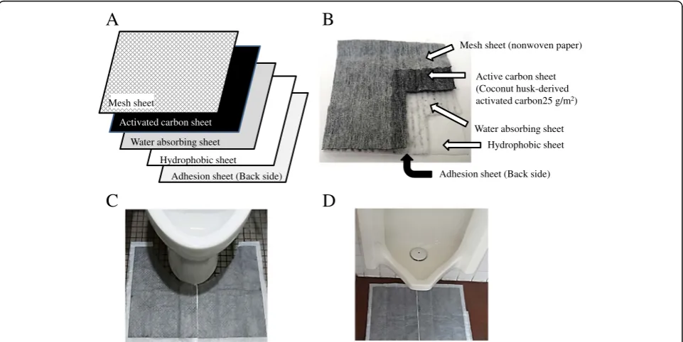
1,580 mg/g. The HD safe sheet-U including this active carbon was also prepared by Futamura Chemistry Co. Ltd. A photograph, structure, and directions for use of HD safe sheet-U were indicated in Fig. 1. Structur-ally, HD safe sheet-U consisted of a nonwoven, ad-sorption layer that included active carbon by a density of 25 g/m2, a water (urine) absorption layer, an opacity film, and a back-side adhesive tape from the top.
Mesh sheet (nonwoven paper)
Active carbon sheet (Coconut husk-derived activated carbon25 g/m2)
Hydrophobic sheet Water absorbing sheet
A
Activated carbon sheet
Hydrophobic sheet Water absorbing sheet
Adhesion sheet (Back side) Adhesion sheet (Back side)
C
B
D
Mesh sheet
Adsorptive evaluation of active carbon for urinary anticancer drug
Various anticancer drugs were diluted with artificial urine or distilled water following four series of diluted concentrations (CPA: 100–2,000 μg/mL, IFM: 20– 1,000 μg/mL, CBDCA: 100–5,000 μg/mL, CDDP: 1– 100μg/mL, MTX: 50–6,000μg/mL, 5-FU: 10–1,000μg/ mL, Ara-C: 50–1,000 μg/mL, GEM: 10–300 μg/mL, ADR: 50–1,000μg/mL, Epi-ADR: 50–1,000 μg/mL, VP-16: 10–500 μg/mL, and CPT-11: 50–1,500 μg/mL). Active carbon was added to the anticancer drug solution at a concentration of 5 mg/mL. The mixture was shaken by a warm bath maintained at 25 °C for 1 h and centri-fuged (20,800g for 5 min). Anticancer drug concentra-tions were determined in the supernatant. For control, a mixture that did not include active carbon was treated under the same conditions and anticancer drug concen-trations were determined in the supernatant.
Evaluation of anticancer drug diffusive prevention performance
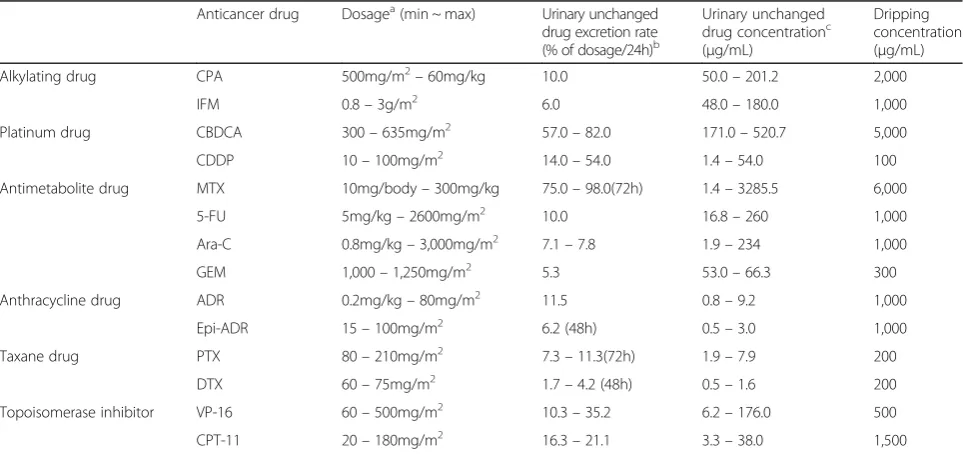
The diffusive prevention performance of urinary anti-cancer drug scattered on the sheet was evaluated based on adhesion of anticancer drugs to slippers soles from the sheet with dropped anticancer drugs. The dropping concentration of the anticancer drug set higher than urinary unchanged drug concentra-tion. Unchanged anticancer drug concentrations in the urine were calculated as dose × urinary unchanged drug excretion rate ÷ urine volume. The dose was calculated with coverage dose by insurance in the
Japanese normal body weight or surface area (58 kg or 1.73 m2) [19, 20]. The urinary unchanged drug excretion rate was referred from a value described in the package insert of each pharmaceutical product. The daily mean urine volume was assumed as 1,730 mL/1.73 m2 [21]. Table 1 summarizes the esti-mated urinary unchanged drug excretion concentra-tions and the dropping concentration per sheet of each anticancer drug. A comparison with HD safe sheet-U was performed with two medical absorbing sheets (control sheet 1: Pitapa sheet , Vilene Create Co. Ltd., Tokyo, Japan; control sheet 2: Absocare sheet, Lily Co. Ltd., Niigata, Japan). Three types of sheets were cut for 10 cm in every direction, and 1 mL of anticancer drug diluted with artificial urine was dropped on the sheets. Subsequently, an experi-menter (body weight: 50 kg) stepped on the sheet in slippers made from vinyl chloride for 10 s. The anti-cancer drug that adhered to the slipper soles was wiped off using 5 × 5 cm of cotton and 5 mL of dis-tilled water, and the wiped off recovery solution was centrifuged (20,800g, 5 min). An anticancer drug con-centration of the supernatant was determined. An anticancer drug adhesion to the slipper sole without using any sheet was determined as follows. An anti-cancer drug was dropped on a stainless steel bat dir-ectly. The stainless steel bat was stepped on with slippers. The drug that adhered to the slipper sole was wiped off and its concentration was determined. The recovery rate of this wipe off method was assessed by dropping an anticancer drug directly on
Table 1Urinary excretion concentration of the anticancer drug and dripping concentration
Anticancer drug Dosagea(min ~ max) Urinary unchanged drug excretion rate (% of dosage/24h)b
Urinary unchanged drug concentrationc (μg/mL)
Dripping concentration (μg/mL)
Alkylating drug CPA 500mg/m2–60mg/kg 10.0 50.0–201.2 2,000
IFM 0.8–3g/m2 6.0 48.0–180.0 1,000
Platinum drug CBDCA 300–635mg/m2 57.0–82.0 171.0–520.7 5,000
CDDP 10–100mg/m2 14.0–54.0 1.4–54.0 100
Antimetabolite drug MTX 10mg/body–300mg/kg 75.0–98.0(72h) 1.4–3285.5 6,000
5-FU 5mg/kg–2600mg/m2 10.0 16.8–260 1,000
Ara-C 0.8mg/kg–3,000mg/m2 7.1–7.8 1.9–234 1,000
GEM 1,000–1,250mg/m2 5.3 53.0–66.3 300
Anthracycline drug ADR 0.2mg/kg–80mg/m2 11.5 0.8–9.2 1,000
Epi-ADR 15–100mg/m2 6.2 (48h) 0.5–3.0 1,000
Taxane drug PTX 80–210mg/m2 7.3–11.3(72h) 1.9–7.9 200
DTX 60–75mg/m2 1.7–4.2 (48h) 0.5–1.6 200
Topoisomerase inhibitor VP-16 60–500mg/m2 10.3–35.2 6.2–176.0 500
CPT-11 20–180mg/m2 16.3–21.1 3.3–38.0 1,500
a
; Insurance application dose in Japan,b
; Report value in each pharmaceutical products IF,c
the slipper sole. The anticancer drug on the slipper
sole was wiped off and its concentration was
determined.
Anticancer drug determination
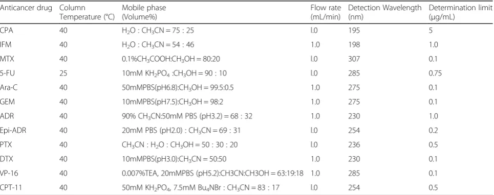
CBDCA and CDDP concentrations were determined using an atomic absorption photometer (AA-7000, Shimadzu Co. Ltd., Kyoto, Japan). As determination con-dition, an absorptivity of a wavelength of 265.9 nm due to a platinum atom ionized by the furnace method was detected. A standard curve was formed using a platinum standard solution (Kanto Chemical Co. Inc., Tokyo, Japan), and the determination limit was 0.05 μg/mL. Anticancer drugs except CBDCA and CDDP were deter-mined using high-performance liquid chromatography with ultraviolet absorptiometry (D-2000 Elite System, Hitachi High-Technologies Co. Ltd., Tokyo, Japan). An octadecyl silicagel column (YMC-Pack ODS-A, 5 μm, 6 × 150 nm, YMC Co. Ltd., Kyoto, Japan) was used for determination. The measure method of these anticancer drugs used the method of mention for Japanese Pharmacopoeia or product interview form, previous art-icle. The details of determination condition were shown in Table 2. The standard curve in each measurement set 6–8 concentrations. We used standard curve with straightness coefficient of correlation (r2) = 0.99 or more. The reproducibility of the measurement including within-day and day-to-day was less than ±5% of setting concentrations.
Statistics
Four sample preparations were conducted for both ex-periments. Each value was provided as mean ± standard deviation. For adsorption performance to the active car-bon of urinary anticancer drug, the anticancer drug
residual ratio for setting concentrations was com-pared. For comparing diffusive prevention perform-ance of urinary anticperform-ancer drug scattered on the sheet, one-way analyses of variance (ANOVA) was performed; when this was significant, multiple com-parisons were performed between the three sheets (Fisher’s least significant difference test). A hazard ratio of <5% represented a statistically significant dif-ference. Statistical analysis was performed using Excel statistics 2012 (Social Survey Research Information, Co. Ltd., Tokyo, Japan).
Results
Adsorptive evaluation of active carbon to urinary anticancer drug
The adsorption performance of active carbon to urinary anticancer drug was indicated as a residual ratio after mixture with active carbon, as indicated in Figs. 2 and 3. All anticancer drugs except CDDP showed a tendency toward decrease of the residual rate as the concentra-tions lowered (0–79.6%). The changes in anticancer drug concentrations by mixture of artificial urine or water alone were not as remarkable when compared with mix-ture of active carbon (Additional file 1: Appendix data 1 and 2). The change in residual rates of most drug con-centrations were less than ±25%, and the anticancer drug that showed the most exceptional decrease in this rate was 100μg/mL of CPA diluted with urine (−36.1%). Moreover, the influence of artificial urine on adsorption performance of active carbon was not remarkable. The decrease of adsorptive performance by urine in most drug concentrations was <20%, and the difference of
−31.7% in the residual rate was maximum exceptionally in 100μg/mL of DTX.
Table 2HPLC-UV analysis condition
Anticancer drug Column Temperature (°C)
Mobile phase (Volume%)
Flow rate (mL/min)
Detection Wavelength (nm)
Determination limit (μg/mL)
CPA 40 H2O : CH3CN = 75 : 25 l.0 195 5
IFM 40 H2O : CH3CN = 54 : 46 1.0 198 1.0
MTX 40 0.1%CH3COOH:CH3OH = 80:20 l.0 307 0.1
5-FU 25 10mM KH2PO4:CH3OH = 90 : 10 l.0 285 0.75
Ara-C 40 50mMPBS(pH6.8):CH3OH = 99.5:0.5 1.0 275 0.1
GEM 40 10mMPBS(pH7.5):CH3OH = 98:2 1.0 275 0.1
ADR 40 90% CH3CN:50mM PBS (pH3.2) = 68 : 32 1.0 230 1.0
Epi-ADR 40 20mM PBS (pH2.0) : CH3CN = 69 : 31 l.0 254 0.2
PTX 40 CH3CN : H2O : CH3OH = 50 : 30 : 20 l.0 236 0.5
DTX 40 10mMPBS(pH3.0):CH3CN = 50:50 1.0 230 0.1
VP-16 40 0.007%TEA, 20mMPBS (pH5.2):CH3CN:CH3OH = 63:19:18 1.0 285 0.1
CPT-11 40 50mM KH2PO4, 7.5mM Bu4NBr : CH3CN = 83 : 17 l.0 254 0.5
Evaluation of anticancer drug diffusive prevention performance
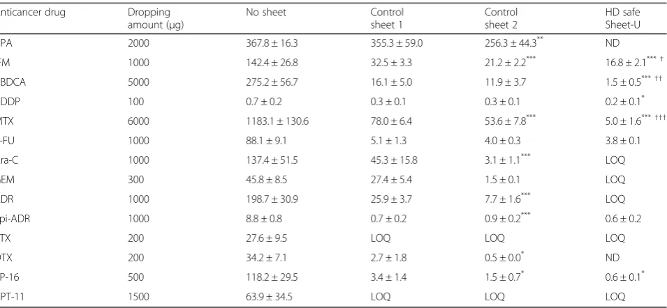
The wipe-off recovery rates of the anticancer drugs that adhered to slipper soles were summarized in Table 3. Wipe-off recovery rates of all anticancer drugs except CDDP, Ara-C, Epi-ADR, and VP-16 were >80%. The coefficient of variation of the wipe-off recovery rate was <20% in each anticancer drug. Table 4 summarizes the quantity of anticancer drug adhering to slipper soles from each sheet of the anticancer drug dropped. The an-ticancer drug adhesion quantity of HD safe sheet-U was significantly lower than that of control sheet 1 for CPA, IFM, CBDCA, CDDP, MTX, Ara-C, GEM, ADR, DTX, and VP-16. Moreover, the anticancer drug adhesion quantity of HD safe sheet-U was significantly lower than that of control sheet 2 for CPA, IFM, CBDCA, MTX, Ara-C, GEM, ADR, and DTX. However, the anticancer drug adhesion quantity of HD safe sheet-U was not sig-nificantly lower than those of the two control sheets for 5-FU and Epi-ADR. Furthermore, the anticancer drug adhesion quantity of HD safe sheet-U and both control sheets was less than the determination limit (<2.5 μg) for PTX and CPT-11.
Discussion
As for the condition not to lay all sheets, all anticancer drugs attached to the slippers sole with remarkable quantity. Therefore, it seemed to be important for pre-venting diffusion of the contamination by laying an absorbent sheet around the toilet stool. HD safe sheet-U, developed in this study, immediately absorbs scattered urine, and active carbon adsorbs the urinary excreted anticancer drug. Moreover, adhesion quantity of an anti-cancer drug from slipper soles trod on the HD safe sheet-U was lower than that of both or one of the exist-ing sheet for 10 of the 14 anticancer drugs used in this study. From these results, the adhesion prevention per-formance to the slippers sole of the scattered anticancer drug of the HD safe sheet-U seemed to be superior to an existing control sheets. A decrease in anticancer drug concentrations for most anticancer drugs except CDDP was found by mixing the drug with active carbon in arti-ficial urine (−20.4 – −99.7%) even at comparatively higher concentrations of the anticancer drug. Based on these results, adsorption of active carbon seemed suc-cessful in inducing a decrease in anticancer drug Fig. 2Residual ratio of anticancer drugs (CPA, IFM, CBDCA, CDDP,
MTX, 5-FU, and Ara-C) in active carbon suspension. Bar indicates residual ratio for setting concentrations as mean ± standard deviation (%) (n= 4)
adhesion from anticancer drugs dropped on the sheet. In urine, the solute including urea and creatinine etc is included for approximately 5%. However, adsorption was noted even when an anticancer drug was diluted with artificial urine. The urinary influence on anticancer drug adsorption performance of active carbon did not seem to have a considerable problem.
In this study, there was a difference in adsorption abil-ity to HD safe sheet-U depending on the kind of the an-ticancer drug. As this reason, the adsorption mechanism of the active carbon, molecular weight and the polarity of the anticancer drug were thought to affect it. Gener-ally, the ability for adsorption of the active carbon was
caused by van der waals power on hydrophobic surface of the active carbon, and target molecules was attracted and adsorbed physically. The active carbon has a large number of the capillary called mesoporous and micro-porous, which make large contact area to target mole-cules. It is also important for adsorption by the active carbon that target particle diameter is smaller than these capillary because the target particle must get into the capillary porous. A material with nonpolar, and insolu-bility in water is easy to adsorb comparatively in
aque-ous solutions. Therefore, it was thought about
adsorptive of ionized CDDP in distilled water is poor while being a small molecule.
Capillary action hold hold
Mesh sheet (nonwoven paper) Active carbon sheet
Water absorbing sheet
Hydrophobic sheet
Anticancer drug
Urine
Active carbon
absorption
Adhesion sheet (Back side)
Lavatory floor
Fig. 4Mechanism by which HD safe seat-U adsorbs urine and urinary anticancer drugs. Indicates the assumed mechanism of action of HD safe sheet-U. The scattered urine passes through a mesh sheet and is aspirated to an active carbon bed, absorbing a water layer by the capillary phenomenon. The urinary anticancer drug molecules coming in contact with active carbon during the aspiration process will be adsorbed by active carbon. The anticancer drug once adsorbed by active carbon is isolated from slipper soles by using a mesh sheet
Table 3Wipe off recovery of dropped anticancer drug from the slipper sole
Anticancer drug Dropped amount (μg) Recovery (μg) Recovery (%) CV (%)
CPA 2000 1947.2 ± 261.5 97.4 ± 13.1 13.4
IFM 1000 1094.6 ± 87.8 109.5 ± 8.8 8.0
CBDCA 5000 4240.5 ± 618.2 84.8 ± 12.4 14.6
CDDP 100 68.2 ± 11.5 68.2 ± 11.5 16.9
MTX 6000 4890.6 ± 413.3 81.5 ± 6.9 8.5
5-FU 1000 873.2 ± 33.4 87.3 ± 3.3 3.8
Ara-C 1000 726.3 ± 117.3 72.6 ± 11.7 16.1
GEM 300 300.4 ± 27.3 100.1 ± 9.1 9.1
ADR 1000 899.4 ± 121.1 89.9 ± 12.1 13.5
Epi-ADR 1000 475.5 ± 40.8 47.6 ± 4.1 8.6
PTX 200 200.0 ± 18.9 100.0 ± 9.5 9.5
DTX 200 171.1 ± 14.9 85.5 ± 7.4 8.7
VP-16 500 308.7 ± 46.8 61.7 ± 9.4 15.2
CPT-11 1500 1213.2 ± 159.5 80.9 ± 10.6 13.1
The difference in anticancer drug adhesion quantity between the sheets was thought to be caused by the fol-lowing material and structural differences in the three types of sheets other than the adsorption ability with ac-tive carbon. Control sheet 1 was a sheet for the purpose of preventing floor contamination and fall by liquids such as blood, body fluids, and contaminated water scattered in a hospital environment. Control sheet 1 constituted a laminating waterproofing resin and a water-absorptive, nonwoven, and adhesive back side. It was the thinnest with favorable ease of conveni-ence. However, it was not able to often absorb 1 mL of artificial urine in the present study conditions. The prevention performance of anticancer drug diffusion from scattered urine in control sheet 1 did not seem adequate. Control sheet 2 was a medical sheet for the purpose of preventing body fluid diffusion during sur-gery and wound care. Control sheet 2 had three lam-inar structures consisting of nonwoven, absorbing pulp, and an impermeability film. Control sheet 2 rapidly absorbed 1 mL of artificial urine; however, ad-hesion of anticancer drugs to slipper soles was ob-served for all anticancer drugs. The structure of HD safe sheet-U comprised an active carbon bed and su-perabsorbent polymer for the purpose of adsorption of urinary anticancer drugs and a large volume of urine. In preliminary examination, the maximum water absorbing quantity of control sheet 1, 2 and HD safe sheet-U were 0.021, 0.078, and 1.059 g/cm2
, respectively. Among the three sheets, HD safe sheet-U seemed to most rapidly absorb 1 mL of dropped artificial urine.
There was lower quantity of adhesion of most antican-cer drugs from HD safe sheet-U to slipper soles com-pared with the two control sheets. For anticancer drug adhesion prevention performance of HD safe sheet-U, following three processes seemed to function: The scat-tered urine passes through a mesh sheet and is aspirated to an active carbon bed, followed by absorption of water layer through the capillary phenomenon. The urinary anticancer drug molecules coming in contact with active carbon during the aspiration process are adsorbed by active carbon. The anticancer drug once adsorbed by active carbon is isolated from slipper soles by using a mesh sheet. This mechanism of action of HD safe sheet-U was indicated in Fig. 4.
Active carbon is widely used for environmental clarifi-cation such as deodorization, waste water or gas dis-posal, and decoloration refinement of food. Moreover, active carbon has medical applications such as drug ad-sorption from gastrointestinal tract in cases of poisoning and for adsorption of uremic toxins in chronic renal fail-ure [22, 23]. To our knowledge, apart from our previous report, no other study has utilized active carbon to ad-sorb an anticancer drug and to prevent diffusion. These results indicate that active carbon is effective in prevent-ing diffusion of anticancer drug contamination through urine for the first time. For the clinical application of the HD safe sheet-U, we thought that a price should not dis-turb the popularity. An active carbon is relatively low-cost material, and the low-cost as HD safe sheet-U will be several hundred yen per 1 sheet (60 × 90 cm).
The present study had certain limitations. Difference in the adhesion to the slippers sole and adsorption by Table 4Adhesion to the slipper sole of urinary anticancer drug dropped on the sheet
Anticancer drug Dropping
amount (μg)
No sheet Control
sheet 1
Control sheet 2
HD safe Sheet-U
CPA 2000 367.8 ± 16.3 355.3 ± 59.0 256.3 ± 44.3** ND
IFM 1000 142.4 ± 26.8 32.5 ± 3.3 21.2 ± 2.2*** 16.8 ± 2.1***†
CBDCA 5000 275.2 ± 56.7 16.1 ± 5.0 11.9 ± 3.7 1.5 ± 0.5***††
CDDP 100 0.7 ± 0.2 0.3 ± 0.1 0.3 ± 0.1 0.2 ± 0.1*
MTX 6000 1183.1 ± 130.6 78.0 ± 6.4 53.6 ± 7.8*** 5.0 ± 1.6***†††
5-FU 1000 88.1 ± 9.1 5.1 ± 1.3 4.0 ± 0.3 3.8 ± 0.1
Ara-C 1000 137.4 ± 51.5 45.3 ± 15.8 3.1 ± 1.1*** LOQ
GEM 300 45.8 ± 8.5 27.4 ± 5.4 1.5 ± 0.1 LOQ
ADR 1000 198.7 ± 30.9 25.9 ± 3.7 7.7 ± 1.6*** LOQ
Epi-ADR 1000 8.8 ± 0.8 0.7 ± 0.2 0.9 ± 0.2*** 0.6 ± 0.2
PTX 200 27.6 ± 9.5 LOQ LOQ LOQ
DTX 200 34.2 ± 7.1 2.7 ± 1.8 0.5 ± 0.0* ND
VP-16 500 118.2 ± 29.5 3.4 ± 1.4 1.5 ± 0.7* 0.6 ± 0.1*
CPT-11 1500 63.9 ± 34.5 LOQ LOQ LOQ
active carbon depending on the anticancer drug might be affect by the difference of wiped off recovery ratio and the determination limit. In Epi-ADR and VP-16, the lower recovery rate by the adsorption to cotton might lead the adhesion to the slippers bottom underestimated. Also, in CPA, IFM, and ADR, the determination limits were comparatively higher than others. This seemed to affect the interpretation of results. Tashiro et al investi-gated a cut-off of the environmental contamination of CPA, IFM and the GEM as 10 ng/mL [24]. Analysis with the determination limit of the ng/mL order using mass spectrograph is necessary for the future clinical evalua-tions. The validity of concentration and volume of the anticancer drug dropped on the sheets might need to be reconsidered in future studies. Urinary anticancer drug concentrations were calculated using dose, urinary ex-cretion rate, and standard urine volume. Anticancer drug concentrations dropped on the sheet were higher than urinary concentrations considering the change in excretion rates and urine volumes. However, urinary ex-cretion concentrations of anticancer drugs seem highly individual and are affected by dose, route of administra-tion, and hepatic and renal functions of patients. More-over, the urine concentrations at the time of maximum concentration (Tmax) may be higher than the mean concentrations calculated from an excretion rate for 24 h. Furthermore, depending on the use, situation, and lavatory shape, the urine volume to be scattered may be greater than the volume experimentally (1 mL) dropped on the sheet. Active metabolites may be included in the urinary excretion of an anticancer drug; for instance, active metabolites of CPA include 4-hydroxy cyclophos-phamide and aldophoscyclophos-phamide, whereas those of IFM, MTX, CPT-11, and Epi-ADR include 4-hydroxy ifosfa-mide and aldoiphosphaifosfa-mide, 7-OH- methotrexate, SN-38, and epirubicinol, respectively. Active metabolites often have lower urinary concentrations compared with the unchanged drug, but these may have adverse effects on human exposure. We did not consider the adhesive-ness of these urinary active metabolites in this study. This study was performed under experimentally con-trolled conditions. It was shown that HD safe sheet-U was superior in urinary anticancer drug adsorption char-acteristics as compared with the existing sheet in this condition. However, the final endpoint about the per-formance of the HD safe sheet-U seemed to be nonpro-liferation performance of environmental anticancer drug contamination and human exposure. Therefore, add-itional studies are warranted in the future to evaluate ef-fects on environmental contamination and human exposure of anticancer drugs by using HD safe sheet-U in routine clinical practice. We had not yet determined at the exchange frequency of the HD safe sheet-U. Prob-ably everyday exchange might be an ideal from hygienic
perspective. However, it should be determined for the exchange cycle in consideration of maintenance of the adsorption performance by clinical evaluations.
Conclusions
Active carbon might be able to adsorb the various anti-cancer drugs excreted in urine. Use of active carbon seems the unprecedented exposure-lowering method. HD safe sheet-U including active carbon might adsorb anticancer drugs from the scattered urine of cancer patients receiving chemotherapy, thus encloses contam-ination. Use of HD safe sheet-U might be effective in lowering anticancer drug exposure of healthcare workers and patients’ family members through urine including excreted anticancer drugs.
Additional file
Additional file 1:Appendix 1 and 2. Adsorption properties of the activated carbon to the urinary anticancer drug. (ZIP 103 kb)
Abbreviations
5-FU:5-Fluorouracil; ADR: Doxorubicin; ANOVA: Analysis of variance; Ara-C: Cytarabine; BSC: Biological safety cabinet; CBDCA: Carboplatin; CDDP: Cisplatin; CPA: Cyclophosphamide; CPT-11: Irinotecan;
DTX: Docetaxel; Epi-ADR: Epirubicin; GEM: Gemcitabine; IFM: Ifosfamide; MTX: Methotrexate; PPE: Personal protection equipment; PTX: Paclitaxel; VP-16: Etoposide
Acknowledgment Not applicable
Funding
This study was performed by Iwate Medical University and the Futamura chemistry Co. Ltd. The HD safe sheet-U was made by a fund of the Futamura chemistry Co., Ltd. The experiment was performed by a fund of Iwate Medical University.
Availability of data and materials
The dataset supporting the conclusions of this article is included within the article.
Authors’contributions
JS designed this concept originally and performed analytical experiment, statistical analyses, writing the manuscript. HO and YS helped for sample making and assay of anticancer drug. MY and YH developed HD safe seat U experimentally. KK provided interpretation and discussion of the data. All authors have read and approved the final manuscript.
Authors’information
JS Ph.D.; Master of Hospital Pharmacy, Lecture of School of Pharmacy, Iwate Medical University, JSPHCS-Certified Oncology Pharmacist (JOP), JSPHCS-Certified Senior Oncology Pharmacist (JSOP), and JPPS-Certified Pharmacist in Palliative Pharmacy (BCPPP). HO and YS; Student of School of Pharmacy, Iwate Medical University. MY and YH is research fellow of Futamura chemistry Co., Ltd. KK. Ph.D.; Proressor, School of Pharmacy, Iwate Medical University.
Competing interests
The authors declare that they have no competing interests.
Ethics approval and consent to participate Not applicable.
Publisher’s Note
Springer Nature remains neutral with regard to jurisdictional claims in published maps and institutional affiliations.
Author details
1Department of pharmacy, Iwate Medical University Hospital, 19-1 Uchimaru, Morioka, Iwate 020-8505, Japan.2Department of Clinical Pharmaceutics, School of Pharmacy, Iwate Medical University, 2-1-1 Nishitokuta, Yahaba, Iwate 028-3694, Japan.3Department of pharmacy, Shizuoka Cancer Center, 1007 Shimonagakubo, Nagaizumi-cho, Sunto-gun, Shizuoka 411-8777, Japan. 4Department of function group, Technology team, Futamura chemistry Co., Ltd., 2-29-16 Meieki, Nakamuraku, Nagoya, Aichi 450-0002, Japan.
Received: 1 March 2017 Accepted: 26 May 2017
References
1. Valanis BG, Vollmer WM, Labuhn KT, Glass AG. Association of antineoplastic drug handling with acute adverse effects in pharmacy personnel. Am J Hosp Pharm. 1993;50:455–62.
2. Valanis BG, Vollmer WM, Labuhn KT, Glass AG. Acute symptoms associated with antineoplastic drug handling among nurses. Cancer Nurs. 1993;16:288–95.
3. Krstev S, PerunicićB, VidakovićA. Work practice and some adverse health effects in nurses handling antineoplastic drugs. Med Lav. 2003;94:432–9. 4. Lawson C, Rocheleau M, Whelan A, Lividoti Hibert N, Grajewski B,
Spiegelman D, et al. Occupational exposures among nurses and risk of spontaneous abortion. Am J Obstet Gynecol. 2012;206:327.e1–8. 5. Skov T, Maarup B, Olsen J, Rørth M, Winthereik H, Lynge E. Leukaemia and
reproductive outcome among nurses handling antineoplastic drugs. Br J Ind Med. 1992;49:855–61.
6. Hansen J, Olsen JH. Cancer morbidity among Danish female pharmacy technicians. Scand J Work Environ Health. 1994;20:22–6.
7. Safe Handling of Hazardous Drugs, 2nd edition. Martha Polovich, ed. Oncology Nursing Society: Pittsburgh; 2011. p.47–50. https://www.ons.org/ store/books/safe-handling-hazardous-drugs-second-edition.
8. Yuki M, Takase K, Sekine S, Ishida T. Evaluation of surface contamination with cyclophosphamide in the home setting of outpatients on cancer chemotherapy. J Nurs Educ Pract. 2014;4:16–23.
9. Sato J, Mori M, Sasaki T, Nihei S, Kumagai M, Nakayama S, et al. Field survey of the anticancer drug contamination in the preparation environment. Usefulness of the 5-FU monitoring by the coupon method. Yakugaku Zasshi. 2014;134:751–6.
10. Nakano H, Yoshida J, Komori K, Mochizuki C, Nakata Y, Tei G, et al. Evaluation of the safety precaution to occupational exposure to antineoplastic drugs in the department of pharmacy. Jpn J Pharm Health Care Sci. 2013;39:1–9.
11. Yuki M, Ishida T, Sekine S. Secondary exposure of family members to cyclophosphamide after chemotherapy of outpatients with cancer: a pilot study. Oncol Nurs Forum. 2015;42:665–71.
12. Morimoto S, Fujii C, Yoshida J, Hata H, Terui K, Anami S, et al. Evaluation of contamination level of antineoplastic drugs due to excrement in hospital. J Jpn Soc Hosp Pharm. 2012;48:1339–43.
13. American Society of Health System Pharmacists. Guidelines on handling hazardous drugs. Am J Health Syst Pharm. 2006;63:1172–93.
14. Gonzalez R, Massoomi F. Manufacturers’recommendations for handling spilled hazardous drugs. Am J Health Syst Pharm. 2010;67:1985–6. 15. Hama K, Tanaka S, Hashida T. Evaluation of decontamination methods of
antineoplastic drugs using ozone water and sodium hypochlorite. Jpn J Pharm Health Care Sci. 2015;41:740–9.
16. Mochizuki C, Fujikawa I, Teii G, Yoshi J. A comparison of cleaning solutions in the biological safety cabinet for preparation of anti-cancer agents. J Jpn Soc Hosp Pharm. 2008;44:601–4.
17. Sato J, Yoshida S, Furukawa M, Kudo K. Development and evaluation of the preparation work sheet with anticancer drug adsorption characteristics. Jpn J Pharm Health Care Sci. 2016;42:317–27.
18. Japanease industrial standards committee. Urethral catheters (JIST3214). 2011. p. 6.
19. The national nutrition survey in Japan. Ministry of health and welfare, Japan. Tokyo: Dai-Ichi Shuppan; 2008.
20. Du Bois D, Du Bois F. A formula to estimate the approximate surface area if height and weight be known. Nutrition. 1916;5:303–13.
21. Uemura O. Comments to clarify the concept of glomerular filtration rate. Jpn J Pediatr Nephrol. 2014;27:76–80.
22. Toyoguchi T, Ebihara M, Ojima F, Hosoya J, Nakagawa Y. In vitro study of the adsorption characteristics of drugs. Biol Pharm Bull. 2005;28:841–4. 23. Schulman G, Berl T, Beck GJ, Remuzzi G, Ritz E, Arita K, et al. Randomized
placebo-controlled EPPIC trials of AST-120 in CKD. J Am Soc Nephrol. 2015; 26:1732–46.
24. Tashiro Y, Kudoda J, Yamamoto S, Izuta M, Takemoto M, Kito N, et al. Antineoplastic drug preparation using compounding aseptic contaminent isolators and robotic system. Jpn J Pharm Health Care Sci. 2016;43:209–14.
• We accept pre-submission inquiries
• Our selector tool helps you to find the most relevant journal
• We provide round the clock customer support
• Convenient online submission
• Thorough peer review
• Inclusion in PubMed and all major indexing services
• Maximum visibility for your research
Submit your manuscript at www.biomedcentral.com/submit