R E S E A R C H
Open Access
Restriction enzyme digestion of host DNA
enhances universal detection of parasitic
pathogens in blood via targeted amplicon
deep sequencing
Briana R. Flaherty
1,2†, Eldin Talundzic
3†, Joel Barratt
1,2, Kristine J. Kines
1, Christian Olsen
4, Meredith Lane
1,5,
Mili Sheth
6and Richard S. Bradbury
1*Abstract
Background:Targeted amplicon deep sequencing (TADS) of the 16S rRNA gene is commonly used to explore and
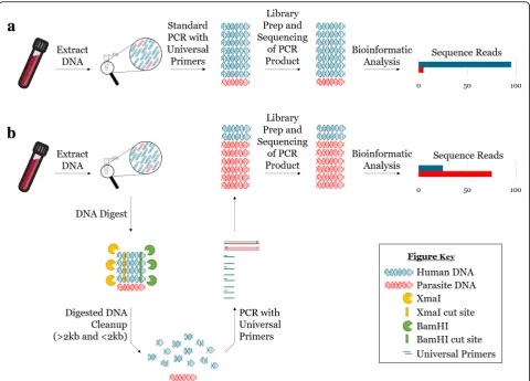
characterize bacterial microbiomes. Meanwhile, attempts to apply TADS to the detection and characterization of entire parasitic communities have been hampered since conserved regions of many conserved parasite genes, such as the 18S rRNA gene, are also conserved in their eukaryotic hosts. As a result, targeted amplification of18S rRNAfrom clinical samples using universal primers frequently results in competitive priming and preferential amplification of host DNA. Here, we describe a novel method that employs a single pair of universal primers to capture all blood-borne parasites while reducing host18S rRNAtemplate and enhancing the amplification of parasite18S rRNAfor TADS. This was achieved using restriction enzymes to digest the 18S rRNA gene at cut sites present only in the host sequence prior to PCR amplification.
Results:This method was validated against 16 species of blood-borne helminths and protozoa. Enzyme digestion prior
to PCR enrichment and Illumina amplicon deep sequencing led to a substantial reduction in human reads and a corresponding 5- to 10-fold increase in parasite reads relative to undigested samples. This method allowed for discrimination of all common parasitic agents found in human blood, even in cases of multi-parasite infection, and markedly reduced the limit of detection in digested versus undigested samples.
Conclusions:The results herein provide a novel methodology for the reduction of host DNA prior to TADS and establish
the validity of a next-generation sequencing-based platform for universal parasite detection.
Keywords:Molecular parasitology, Amplicon sequencing, Blood microbiota, Parasite biodiversity
Background
Several studies have applied next-generation sequencing (NGS) technologies to the investigation of parasite diversity and ecology using various methods to identify all parasites present in a given host [1–5]. Much of this work has depended on metagenomic and metatranscriptomic approaches, including whole genome shotgun sequen-cing of entire microbial communities [1–3]. Although such approaches are frequently applied to viral and
bacterial communities [6–9], direct sequencing of parasite DNA from clinical samples poses challenges with regard to sensitivity and specificity since the concentration of parasite DNA present is often markedly lower in pro-portion to host DNA. Removal of host DNA via prefer-ential cutting of methylated host sequences using modification-dependent restriction endonucleases has previously been used to increase the recovery of Plas-modium falciparumDNA during whole genome sequen-cing of human blood samples [10]. Unfortunately, this method is only applicable in organisms that do not undergo DNA cytosine methylation, such as apicom-plexan parasites [11, 12], and would be ineffective for * Correspondence:isl5@cdc.gov
†Briana R. Flaherty and Eldin Talundzic contributed equally to this work. 1Parasitic Diseases Branch, Division of Parasitic Diseases and Malaria, Centers for
Disease Control and Prevention, 1600 Clifton Road, Atlanta, GA 30329, USA Full list of author information is available at the end of the article
© The Author(s). 2018Open AccessThis article is distributed under the terms of the Creative Commons Attribution 4.0
International License (http://creativecommons.org/licenses/by/4.0/), which permits unrestricted use, distribution, and
reproduction in any medium, provided you give appropriate credit to the original author(s) and the source, provide a link to the Creative Commons license, and indicate if changes were made. The Creative Commons Public Domain Dedication waiver (http://creativecommons.org/publicdomain/zero/1.0/) applies to the data made available in this article, unless otherwise stated. Flahertyet al. Microbiome (2018) 6:164
detecting C5-methylating eukaryotic pathogens [13–15]. Another common approach to increase the capture of parasite DNA relies on using a set of pathogen-specific primers in conjunction with a strand-displacing DNA polymerase to achieve selective whole genome amplifica-tion [16, 17]. However, species-specific methods such as this are difficult to adapt to broader analyses of whole parasite communities.
Over the past decade, targeted amplicon deep sequen-cing (TADS) of the 16S rRNA gene has frequently been used to study and characterize bacterial microbiomes [18–22]. A similar approach, using universal PCR primers to target a conserved parasite gene for TADS, would be amenable to studies of parasite communities. Unfortunately, such an approach is usually compromised by the overabundance of host DNA, as common primer targets are conserved across higher order eukaryotic spe-cies, including metazoan parasites. A recent study sought to overcome this challenge by utilizing host DNA blocking primers in the assessment of parasite biodiversity in the feces of wild rats [5]. However, this method was rarely able to achieve species-level identification, and application of the method to assess helminth biodiversity ultimately re-quired worm isolation from fecal samples and amplifica-tion with class-specific primers [4].
To overcome the challenge of host DNA interference, we designed a TADS method that utilizes restriction en-zymes to reduce amplification of host DNA template prior to PCR and NGS. Using universal primers to target the 18S rRNA gene in a region containing restriction enzyme cut sites only present in the host sequence, host18S rRNA template was digested, and PCR enrichment of the host sequence was reduced to allow enhanced detection of parasite18S rRNA. This method achieved a substantial re-duction in reads belonging to the host and a 5- to 10-fold increase in parasite reads following NGS. The method was validated using 16 species of human blood-borne parasites and was effective in detecting both single and mixed para-site infections. Limit of detection analyses showed consist-ent reduction in LOD for digested versus undigested samples, where positive results were achieved for speci-mens with parasitemias as low as ~ 7 parasites per micro-liter for digested samples versus a low of ~ 40 parasites per microliter for undigested samples. This method pro-vides a single assay for detection of all major blood-borne parasites found in humans and represents a promising new tool for the study of parasite communities.
Results Assay design
Primers were designed to amplify a region of the 18S rRNA gene approximately 200 base pairs in length that is highly conserved across eukaryotic organisms yet contains sufficient diversity at the nucleotide level to allow accurate
species identification and differentiation. The selected amplification region possesses BamHI and XmaI restric-tion enzyme cut sites only in the human host sequence (Additional file 1: Figure S1), which allows cleavage of host template and reduced amplification of host DNA during PCR amplicon enrichment prior to Illumina ampli-con deep sequencing (Fig. 1). Following sequencing, paired and trimmed reads were mapped to a database of human and parasite18S rRNAsequences, and the number of mapped reads per parasite species was counted. Given the sensitivity of Illumina sequencing, the occurrence of Illumina index cross-talk, and because some DNA cross-contamination can be expected in samples that are extracted and processed together [23–25], we established a conservative, dual-criterion system to differentiate “noise”from a true positive sequencing result. This system utilizes a minimum cutoff for positivity based on the aver-age proportion of contaminating reads obtained per nega-tive control specimen over multiple replicate analyses (assuming 60–80 samples are multiplexed in a single li-brary, see the “Methods” section for further details). In addition, a species-specific shifting maximum cutoff value was established to be applied on a per specimen basis and to account for minor changes in the degree of index cross-talk and variations in the number of reads generated per specimen/experiment. Using this dual-criterion sys-tem, specimens were considered positive only if more than 20 reads mapped to the respective parasite reference se-quence and if the number of parasite derived reads mapped to that reference sequence also exceeded the shifting maximum cutoff value.
Since total sequencing reads can vary from run to run, results were normalized to allow comparisons between experiments. Reads were normalized according to the total number of paired reads per sample (after trimming) and reported as reads per thousand. To assess the im-pact of restriction enzyme reduction of competitive host template DNA and the capacity to detect a variety of blood parasites, this technique was applied to clinical blood specimens with and without prior restriction endonuclease treatment. For these experiments, paired DNA specimens (i.e., restriction digested specimens and their respective undigested partner) were sequenced in the same Illumina library so that direct comparisons could be made between the two conditions.
Assay validation
were analyzed in an identical fashion to human clinical samples. Most blood-borne parasites known to infect humans were included in our analysis; however, we were unable to obtain clinical samples/isolates forTrypanosoma brucei subsp. gambiense and several rare human filarial blood parasites.Bioinformatic analysis of the complete18S rRNA sequence from several blood parasites identified BamHI and/or XmaI cut sites outside the region of amplifi-cation, varying slightly in their location and frequency be-tween parasite taxa. As such, the resulting restriction fragments would vary in size for different blood parasites. Consequently, post-digestion DNA for all validations was divided into two equal parts and cleaned using a Monarch PCR & DNA Cleanup Kit selecting for both > 2 kb and < 2 kb DNA products, respectively. Prior to DNA extraction and restriction digestion, all samples were spiked with cat blood containing 3.4 × 10⁶Cytauxzoon felisparasites as an extraction, amplification, and sequencing internal control.
Following TADS, a substantial reduction in reads mapping to the human host reference was observed in the digested samples compared to undigested samples (Fig.2a). Furthermore, the digested samples showed a 5- to 10- fold increase in the number of parasite-spe-cific reads (Fig. 2a and Additional file 2: Figure S2). All samples assayed passed our set criteria for positiv-ity except for twoWuchereria bancroftiblood samples. For these samples, the failure to detect W. bancrofti was attributed to the formation of a large, non-uni-form clot that made DNA extraction problematic (Additional file 3: Figure S3a-c). Nevertheless, these data suggest that reduction of host DNA background via restriction enzyme digestion improves detectability of parasite DNA for the universal detection of para-sites in blood. Analysis of post-digestion size selection found no statistical difference between > 2 kb and < 2 kb cleanup conditions (p= 0.0631).
Fig. 1Reduction of host DNA by restriction enzyme digestion enhances PCR amplification of parasite DNA. DNA extraction from parasite-infected whole blood yields a DNA sample containing high amounts of host DNA (blue) and low amounts of parasite DNA (bright red).aPerforming conventional PCR on this sample, using universal primers, amplifies primarily host DNA (blue), and yields sequencing reads almost entirely belonging to the host.bIn contrast, restriction enzyme digestion of host DNA prior to PCR alters the ratio of host to parasite DNA in the initial sample, allowing for selective amplification of parasite DNA (bright red) and resulting in an increase in the relative number of parasite amplicons post-PCR and an increase in the sensitivity of parasite detection via NGS
Digestion reduces host reads by 50% or more
To determine the extent to which enzyme digestion re-duces human host background, a series of dilutions was prepared using human DNA donated by healthy volun-teers and parasite DNA obtained from a 3D7 P. falcip-arum culture. Samples contained 0.2 ng/μL P. falciparum DNA, or approximately 8600 parasites per microliter, as well as human DNA diluted to a
concentration of 3, 2.5, 2, 1.5, 1, 0.5, or 0 ng/μL. Bio-informatic analysis indicated no BamHI/XmaI cut sites within 2 kbp of the PCR amplification region for P. fal-ciparum3D7, so all post-digestion samples were cleaned according to the > 2 kb size selection protocol. In un-digested samples, 80–100% of sequencing reads mapped to the human host reference sequence with only 0–20% of reads mapping toP. falciparum(Fig. 2b). Meanwhile, Fig. 2Digestion of host DNA increases the sensitivity of parasite detection in parasite-positive human blood samples. (a) Restriction enzyme digestion yields a marked reduction in human18S rRNAreads per thousand (left panel, greyscale diamonds) and a 5- to 10-fold increase in parasite reads per thousand (right panel, colored circles) in digested relative to undigested samples (n= 3 biological replicates, mean ± SD, samples were normalized according to the reads per thousand for reads derived from human host and parasite separately, with the central dotted line reflective of a zero fold change, which marks the undigested samples before treatment with restriction enzymes). No statistical difference was found for size selection (i.e., > 2 kb vs. < 2 kb) (two-way ANOVA,p= 0.0631). (b) Proportional composition of human DNA dilutions in undigested (ud) and digested (d) samples demonstrates an average 2-fold reduction in human DNA and a 5-fold increase in parasite reads post-digestion (black bars =C. felis, dark grey bars = H. sapiens, light grey bars =P. falciparum, concentration of 3D7 DNA includesP. falciparumandH. sapiensDNA from 3D7 cultures which contain human blood products, two-way ANOVA with Sidak’s multiple comparisons posttest,p< 0.0001,n= 3, mean ± SD)
the composition of digested sample reads was only 40– 50% human and 50–60% P. falciparum, reflecting a greater than or equal to 2-fold decrease in human reads and an average 5-fold increase in parasite reads post-digestion (Fig.2b).
As an additional control, a mock restriction digestion was performed in triplicate wherein DNA extracted from human blood spiked with cultured 3D7 parasites was in-cubated for the same period and at the same temperature as identical specimens subjected to a true restriction en-zyme digestion. These samples were subsequently purified using a Monarch Cleanup Kit (> 2 kb) and PCR amplified according to the protocol described. Post-sequencing ana-lysis found no statistical difference in the number of 3D7-derived sequencing reads obtained between mock digested DNA specimens and their paired undigested DNA samples, confirming that the increase in parasite reads in the restriction-digested samples is directly related to the action of the restriction enzymes on host DNA (Additional file4: Figure S4).
Detection of mixed parasite infections
To explore the effectiveness of this method for de-tecting mixed infections, a variety of mixed parasite blood samples were artificially produced by combin-ing previously diagnosed parasite-infected blood samples and subjecting them to analysis via the de-scribed method. The samples simulated all varieties of mixed malaria infections, including all the major
Plasmodium species that infect humans, together and in pairs, as well as other geographically pos-sible mixed infections. As before, we saw a dramatic decrease in human reads in digested relative to undigested samples and, in this case, a 2- to 15-fold increase in para-site reads post-digestion (Fig.3, left panel). These data es-tablish that this method can reliably detect parasites in mixed infections but suggest that competitive amplifica-tion occurs between the different 18S rRNAtypes, which may affect the sensitivity of detection for parasite species occurring at relatively low numbers in mixed parasite communities.
To further assess assay effectiveness in detecting mixed infections, a non-artificial (natural) mixed malaria infec-tion that had been previously diagnosed by the CDC Parasite Reference Diagnostic Laboratory was tested. Interestingly, in this case, enzyme digestion proved to be the deciding factor between an accurate and inaccurate assessment of the sample by NGS. While universal PCR and NGS of the undigested sample showed positive results for only P. falciparum, sample digestion led to a greater than 10-fold increase in aligned reads for Plasmodium malariae, a more accurate evaluation of this mixed para-site community (Fig.3, right panel).
Digestion improves the limit of detection of parasites in blood
Plasmodium knowlesi-infected rhesus macaque blood with 3.3% parasitemia (approximately 144,000 parasites per microliter) was utilized. Three aliquots of the sample were serially diluted in parasite-negative whole human blood to a parasitemia of 0.072 parasites per microliter and analyzed in biological triplicate. For the region
amplified, the rhesus macaque and human 18S rRNA target sequences are identical, and thus, sample diges-tion will be equivalent despite the host species being dif-ferent. Post-digestion samples were, again, cleaned only according to the > 2 kb size selection protocol. As ex-pected, the LOD for samples that had not undergone en-zyme digestion before PCR and sequencing was high, such that reads began to fall below baseline at parasite-mias between 720 and 72 parasites per microliter; mean-while, prior enzyme digestion caused samples to fall below baseline between 72 and 7.2 parasites per micro-liter (Fig. 4a). A trend line was fitted to the log-transformed data to determine a more precise LOD before and after enzyme digestion (Fig. 4b). Using this trend line, the LOD for undigested samples was extrapo-lated to 163 parasites per microliter while that of the digested samples was estimated at 15 parasites per microliter. As this estimate was extrapolated from a trend line rather than empirical data, a series of finer di-lutions (between 61.2 and 0.72 parasites per microliter) was performed to establish a more precise LOD (Fig.4c). With restriction enzyme digestion, it was confirmed that the assay can detect as few as 7.2 P. knowlesi parasites Fig. 4Enzyme digestion markedly lowers assay limit of detection. (a) Reads per thousand for undigested (gray) and digested (black) 10-fold serial dilutions ofP. knowlesiin whole human blood (n= 4, mean ± SD). (b) Log-transformation of reads per thousand from serially diluted samples suggests a limit of detection of 163 parasites per microliter for undigested samples (gray, r2= 0.9852) and 15 parasites per microliter for digested samples (black,r2= 0.9533) (n= 4 biological replicates, mean ± SD). (c) After deeper analysis, reads per thousand for undigested (gray) and digested (black) serial dilutions between 61 parasites per microliter and 0.72 parasites per microliter demonstrate a limit of detection of 40 to 60 parasites per microliter for undigested samples and 7 to 29 parasites per microliter for digested samples (two-way ANOVA with Sidak’s multiple comparisons posttest, ****p< 0.0001, ***p< 0.001, **p< 0.005
n= 3, mean ± SD)
Table 2Detection ofP. knowlesiin blood at different
concentrations following restriction enzyme treatment of DNA extracts
Pkreads/total number of reads Result
Pkparasites/μL R1 R2 R3 R1 R2 R3
61.2 54
10994 45566383 30568208 + + +
50.4 44
6160 31360180
305
28068 + + +
39.6 31
7562
231 47096
44
28514 + + +
28.8 26
9030
84 21680
95
23942 + + +
18 18
12074
24 15722
62
19654 – + +
7.2 12
20290
22 19068
43
63164 – + +
0.72 2
36190
2 17220
6
57076 – – –
Pk Plasmodium knowlesi,Rreplicate,+positive,−negative Note: The read counts listed here represent the values obtained after trimming and filtering
per microliter of blood, albeit inconsistently, as this LOD was achieved for only two out of three triplicate samples with the third replicate detecting only 28.8 para-sites per microliter (Table2). For the identical set of un-digested specimens, a consistent positive result was obtained only at the highest parasite concentration of 61.2 parasites per microliter, with one replicate detecting down to 39.6 parasites per microliter and a second de-tecting only 50.4 parasites per microliter. Based on this data, there is a 1.4- to 8.5-fold improvement in LOD fol-lowing restriction enzyme digestion.
Discussion
Potential applications of this method extend beyond detec-tion of parasitic pathogens in human blood. This method may allow for exploration of mammalian blood parasites for ecological, wildlife disease, zoonotic disease, and patho-gen discovery studies. To explore these potential applica-tions, the 18 s rRNA sequences of various classes of vertebrates relative to the human target DNA sequence were analyzed in silico. Both the BamHI and XmaI restric-tion enzyme cut sites, the PCR primer binding sites, and in some cases, the entire amplification region were conserved in mammals and birds (Additional file 5: Figure S5 and Additional file6: Table S1). Among the vertebrates analyzed were many common livestock and companion animals. Within the region of amplification, these animals shared greater than 98% identity with the human sequence, sug-gesting this methodology may be applied in animal hosts for agricultural and veterinary purposes.
It is also pertinent to consider the effectiveness of this method in exploring the mycobiome. A preliminary bio-informatic analysis of DNA sequences from several com-mon fungal organisms was conducted and found that neither the BamHI nor the XmaI cut sites were present in any fungi tested while primer binding sites were universally conserved (Additional file7: Figure S6 and Additional file8: Table S2). It may be inferred from these data that this plat-form will provide a tool for the detection and identification of fungal infections in eukaryotic hosts, as well. Further in-vestigation and validation of these additional applications is underway.
A recent study evaluating the LOD for several published Plasmodium species’ real time PCR (qPCR) assays re-ported LODs in the range of 0.3 to 2.5 parasites per microliter [26]. Conventional PCR and loop-mediated iso-thermal amplification (LAMP) assays are generally less sensitive than qPCR, with LODs usually falling between 1 and 20 parasites per microliter of blood [27, 28]. The LOD of the assay described herein falls between 7 and 29 parasites per microliter (Table2) and is therefore similar to those reported for conventional PCR assays. Rapid diagnostic tests for detection of malaria antigens are typic-ally less sensitive and are reportedly most suitable for
detecting parasitemias above 200 parasites per microliter [29]. Meanwhile, for Plasmodium species, it is estimated that a highly competent microscopist can detect approxi-mately 50 parasites per microliter of blood [30] while a typical microscopist using the WHO standardized method detects an average of 88 parasite per microliter [31]. Thus, the method described herein possesses an LOD for Plas-modium species similar to published conventional PCR assays, but boasts the added advantage of being able to de-tect and identify all human blood-borne parasitic patho-gens in a sample using a single test.
An important consideration regarding the sensitivity of this assay is the high amount of variation in 18S rRNAcopy number between different species of blood parasites. For example, the rDNA copy number of P. falciparum ranges from five to eight copies per hap-loid genome [32]. Other apicomplexan blood parasites possess a similarly low copy number, such as Plasmo-dium vivax which possesses four to eight copies [33] and Babesia microti with only two copies of rDNA [34]. It is reasonable to assume that this method will have increased sensitivity for parasite species with higher rDNA copy numbers, such asT. brucei(56 cop-ies), Trypanosoma cruzi (110 copies) and Leishmania donovani (166 copies) [33]. Further investigation will be required to establish limits of detection for other parasite species.
paired reads following NGS, it was not possible to con-firm the identity of this species as no reference M. per-stans 18S rRNA sequence for the region amplified is currently available in any genome database. However, a large number of unused paired reads (10,000–15,000) from the raw sequences following reference alignment to the human 18S rRNA reference suggested that a large amount of non-host eukaryotic DNA had been amplified in this sample. Further investigation revealed that 10,298 of these reads aligned with 100% identity to a sequence in GenBank designated as an18S rRNAsequence from a Filarioidea sp. (accession: KT907503.1). It is, therefore, proposed that these reads are likely derived fromM. per-stansDNA amplified from within that sample.
A limitation of this method is that it does not amplify regions with sufficient sequence variation to differentiate W. bancrofti from L. loa or to discriminate between some parasite subspecies, such asT. bruceisubsp. gam-biense and T. brucei subsp. rhodesiense or L. donovani subsp. donovani and L. donovani subsp. infantum. The demands of identifying a single region flanked by univer-sally conserved primer-binding sites and possessing re-striction enzyme recognition sites that cut only the host gene, was unfortunately restricting. However, infections with these agents are rare in most parts of the world and differentiation when such cases are detected may be undertaken by a further, species-specific PCR.
Conclusions
This universal detection platform offers a versatile and broad-spectrum method for the study of parasitic com-munities in human hosts. Improved sensitivity was ob-tained by employing restriction enzymes targeting host-specific cut sites to selectively limit the amplifica-tion of unwanted host DNA sequences and reduce com-petitive PCR amplification of host 18S rRNA. This method has been validated in biological triplicate for 16 human blood-borne parasites. Future exploration and optimization of this method for the study of parasite di-versity and the detection of parasitic disease in other eukaryotic hosts, as well as in other sample matrices such as tissue and feces, is warranted.
Methods Samples
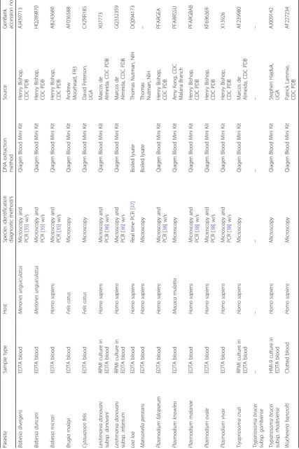
Human clinical blood samples used in this study were ori-ginally submitted to the CDC Parasitic Diseases Branch for confirmatory diagnosis of parasitic infections. Follow-ing diagnosis, samples were de-identified and frozen in 200 μL aliquots at −80 °C for use in assay development and validation. Samples containingP. falciparum,P. vivax, P. malariae, Plasmodium ovale, B. microti, and Babesia divergens were acquired in this way. For some rare blood-borne parasites, either animal blood or human
blood samples collected during previous research studies and stored at−80 °C were used. Bioinformatic analysis in-dicated that the restriction enzyme cut sites and the re-gion of amplification were identical to human for all relevant animal samples. Some rare blood parasites that could not be acquired as true clinical samples were recre-ated by spiking uninfected human blood with cultured parasites—L. donovani subspecies infantum, L. donovani subspeciesdonovani,T. cruzi, andT. bruceicultures were added to whole human blood at a ratio of 1:10. All blood samples were collected into EDTA anticoagulant except for the clotted bloodW. bancroftisample. The full details of source, matrix, parasite identification, and DNA extrac-tion methods are provided in Table1.
Assay design
In an effort to design universal parasite primers, Geneious bioinformatics software (Biomatters Inc., Newark, NJ, USA) was used to create an alignment of the 18S ribosomal RNA genes from the publicly available sequences of 24 protozoa, 17 helminths, and Homo sapiens. No18S rRNA sequences forM. perstanswere available for inclusion in the alignment. Restriction enzyme cut sites were analyzed bioinformatically, and 18 primers were designed and tested in 14 primer com-binations with 6 candidate restriction enzymes against a panel of four protozoan and one nematode, one trematode and two cestode helminth parasites (P. falciparum, Toxo-plasma gondii,T. brucei, C. felis, Brugia pahangi, Schisto-soma mansoni, Dipylidium caninum, and Taenia sp., relatively). From these analyses, one primer set was selected that amplifies an approximately 200 bp region of the 18S rRNA gene in all parasites tested, yielding a clearly visible band on a 1.5% agarose gel. Of the six restriction enzymes tested, BamHI-HF and XmaI (NEB, Ipswich, MA, USA) con-sistently yielded the highest degree of host DNA removal when paired with the selected primers.
Universal parasite detection assay
Aliquots of parasite-positive human blood samples at a volume of 200 μL were spiked 1:100 with C. felis -in-fected cat blood at a concentration of 1.7 million para-sites per microliter. DNA was extracted using the QIAamp DNA Blood Mini Kit (Qiagen, Redwood City, CA, USA) and eluted into 50 μL of PCR-grade water. Following extraction, DNA concentrations were deter-mined using a Qubit 2.0 Fluorometer with the Qubit dsDNA High Sensitivity Assay Kit (Life Technologies, Grand Island, NY, USA). One hundred fifty nanograms of DNA were subsequently digested via incubation with 20 units of BamHI-HF and 20 units of XmaI in 1X CutSmart Buffer (NEB) in a final volume of 50 μL for 2 h in a 37 °C water bath. Digested samples were divided into two equal volumes, and enzymes and buffers were
subsequently removed using the Monarch PCR & DNA Cleanup Kit (NEB). The Monarch kit enables template size selection (protocol dependent), and since there is uncertainty as to what the template size would be post-digestion, in most cases one sample aliquot was cleaned as a > 2 kb sample, and the second was cleaned as a < 2 kb sample according to the manufacturer’s in-structions. Both samples were then eluted in 10μL elu-tion buffer (NEB).
PCR was performed with the cleaned and digested samples in a reaction volume of 20 μL using Q5 High-Fidelity DNA Polymerase (NEB) according to the manufacturer’s instructions and supplementing with Q5 High GC Enhancer. The primer sequences are as fol-lows: CCGGAGAGGGAGCCTGAGA (forward) and GA GCTGGAATTACCGCGG (reverse). Samples were de-natured at 98.0 °C for 2 min followed by 30 cycles of 98.0 °C for 10 s, primer annealing at 67.0 °C for 30 s and extension at 72.0 °C for 45 s, and a final extension at 72.0 °C for 5 min. Following PCR, samples were ana-lyzed on a 1.5% agarose gel, cleaned with the Monarch PCR & DNA Cleanup Kit (NEB, < 2 kb), diluted 1:5 in elution buffer, and transferred to a 96-well plate for li-brary preparation and sequencing.
Library preparation and sequencing were performed by the CDC Biotechnology Core Facility’s Genome Sequen-cing Lab using the NEBNext Ultra DNA Library Prep Kit for Illumina (NEB), and multiplexing was performed using the NEBnext Multiplex Oligos for Illumina Index kit (NEB) or using TruSeq HT Adapter sets (Illumina). No more than 80 samples were multiplexed on a single MiSeq run to ensure sufficient and consistent sequencing depth for each sample. Runs were prepared using a MiSeq Re-agent Nano Kit v2 (PE250bp) (Illumina), and sequencing was performed on the Illumina MiSeq platform (Illumina). For all experiments, the paired digested and undigested (otherwise identical) samples were multiplexed on the same sequencing run to ensure they could be compared directly.
Bioinformatic analysis
Geneious bioinformatics software (www.geneious.com) was used to analyze the raw fastq files generated by the MiSeq sequencing runs. Reads were paired and subse-quently trimmed using the BBDuk plugin with a mini-mum quality of 35 and a minimini-mum read length of 150 base pairs. Paired and trimmed reads were then mapped to the reference alignment described below using a mini-mum mapping quality of 35, a minimini-mum overlap of 150, an allowance of zero mismatches per read, and a mini-mum overlap identity of 99%. To allow comparisons to be made between experiments and to assess the impact on restriction enzyme treatment for host template reduction, samples were normalized by dividing the species-specific
mapped reads by the total paired reads in the sample, multiplying that value by 1000, and reporting the final value as reads per thousand. To account for index cross-talk between DNA samples multiplexed on the same Illumina run, a series of careful analyses were conducted to establish both a minimum cutoff to be applied uni-formly across all samples as well as a shifting maximum cutoff to be applied on a sample-by-sample basis. Satisfac-tion of both criteria, as described below, would eliminate the occurrence of false-positive results.
Reference alignment
Geneious bioinformatics software was used to establish a set of blood parasite reference sequences from among the 18S rRNA gene sequences found on GenBank. This reference database was used for assay development and validation and utilized the parasite GenBank accession numbers listed in Table 1 and the Homo sapiens se-quence available under GenBank accession number HUMRGE. Since the region of amplification shares 100% sequence identity for L. d. donovani/L. d. infan-tum, T. b. rhodesiense/T. b. gambiense, and W. ban-crofti/L. loa, only one representative sequence was included for each of these pairs in the final reference alignment cohort.
Establishment of a minimum and maximum coverage cutoff value for positivity
A minimum coverage cutoff value of 20 reads was estab-lished for the specific protocol described in this study (i.e., using the Illumina MiSeq platform and a 500 cycle Nano Kit, and multiplexing 60 to 80 samples per se-quencing run). The following formula was used to deter-mine the minimum coverage cutoff:
μcontam all
ð Þ þ4ðS:D:Þ
½ μsample reads¼CUTOFFmin
where μcontam_all= the mean proportion of contaminat-ing reads (i.e., the mean proportion of reads from all blood negative samples, n= 18, from this study that mapped to a parasite reference sequence), S.D. = stand-ard deviation for μcontam(four standard deviations above
the mean was selected as the number of reads obtained for all samples represents a normal distribution), and
μsample_reads= the mean number of reads obtained for
each sample from 425 sequenced samples.
For this study, the minimum cutoff was calculated empir-ically and as follows (using all blood negatives in a sample):
0:0001
ð Þ þ4 0ð :00026Þ
½ 17;553¼19:855 reads
number of contaminating reads you might expect for any given specimen regardless of the parasite species under investigation, using this specific protocol.
Similarly, the maximum sliding cutoff was calculated for each individual sample using the following formula:
μrun contam
ð Þ þ4ðS:D:Þ
½ Sreads¼CUTOFFmax
where μrun_contam= the mean proportion of contaminating reads within the negative control samples included in this specific sequencing run (at least four negatives were included in each run), S.D. = standard deviation for μrun_contam, and
Sreads= the number of reads sequenced for the sample.
These coverage cutoffs take into account the index cross-talk (i.e., sample bleeding) for samples containing parasite reads that were multiplexed on the same se-quencing run [25]. Two examples are provided below for calculating CUTOFFmax:
(a). 0:0002
ð Þ þ4 0ð :00033Þ
½ 10;410¼15:823reads
(b). 0:0001
ð Þ þ4 0ð :00023Þ
½ 24;204¼24:688reads
In example A, the sliding maximum rule would sug-gest a cutoff of 16 reads. However, at this cutoff we can-not be confident that this is can-not due to index cross-talk [25]. Consequently, we use the value of CUTOFFmin(20 reads) to exclude false positive results. In example B, CUTOFFmax is used (25 reads) to account for the fact that as the number of reads increases (i.e., depth) the proportion of contaminating sequences will also in-crease. Cutoff values are always rounded up to the near-est whole number.
This dual criterion system was developed to account for certain variables that may affect the sequencing output. As discussed above, at greater sequencing depth, it is more likely that contaminating reads will be detected; the sliding CUTOFFmax accounts for this. Additionally, the composition of samples sequenced in a given run will have an impact on the number and composition of contaminat-ing reads present in negative control specimens as a result of index cross-talk. For example, if a run contains a large number of samples that are positive forP. falciparum, yet a small number of Leishmania positive samples, one would expect to see a larger proportion ofP. falciparum reads in the negative control specimens compared to Leishmaniareads. This is also the reason why the sliding CUTOFFmaxis calculated for each run and for each para-site species individually—it compensates for the diversity of specimens that may be included within and between
runs. Furthermore, if a single specimen out of 80 included on a single MiSeq run containsP. knowlesiDNA while all other samples are negative, it is possible that noP. know-lesireads will be detected in the negative samples. In this case, the sliding CUTOFFmax cannot be used so CUTOFFminis implemented.
Multiple sequence alignments
All multiple sequence alignments were performed using the MUltiple Sequence Comparison by Log-Expectation (MUSCLE) algorithm. GenBank accession numbers for human parasites can be found in Table 1, those for ver-tebrates in Additional file 6: Table S1 and those for fun-gal organisms in Additional file8: Table S2.
Assessment of host DNA removal by restriction enzyme treatment
A dilution series was prepared from DNA extracted from the buffy coat layer of whole blood provided by healthy human volunteers and parasite DNA from 3D7P. falcip-arum cultures. Samples were spiked with cat blood in-fected withC. felis prior to DNA extraction. Dilutions of human DNA were prepared at 3, 2.5, 2, 1.5, 1, 0.5, and 0 ng/μL and supplemented with 0.2 ng/μL DNA from a 3D7 P. falciparum culture (DNA equivalent of ~ 8600 parasite per microliter). Samples were then processed as described above by restriction digestion, PCR enrichment, and deep sequencing. Each digested sample was paired with an identical sample that was not restriction digested. The resulting sequencing reads were mapped against the reference database for quantification of parasite-derived reads. Note that for every experiment, an unquantified proportion of human reads was contributed by human products in theP. falciparum3D7 culture.
Limit of detection
For analysis of assay limit of detection, frozen samples of P. knowlesi from a non-human primate infection for which parasitemia had previously been determined by mi-croscopy at the CDC (~ 144,000 parasites per microliter) were serially diluted in parasite-free whole blood. Samples were processed and sequenced as described above, in trip-licate, with restriction digested samples paired with an identical undigested sample sequenced on the same run.
Sample acquisitions
P. knowlesi in rhesus macaque blood was generously provided by Amy Kong (CDC, Malaria Branch, Atlanta, GA, USA);Babesia duncaniin gerbil blood andB. diver-gens stabilate in human blood were kindly provided by Henry Bishop (CDC, Parasitic Diseases Branch, GA, USA); B. malayi microfilariae in feline blood were pro-vided by Andy Moorhead (Filariasis Resource Reagent Resource Center (FR3), Athens, GA, USA);W. bancrofti
microfilariae in human blood was provided by Patrick Lammie (CDC, Parasitic Diseases Branch, Atlanta, GA, USA); andC. felisin feline blood was provided by David Peterson (University of Georgia, Athens, GA, USA).L. d. infantum, L. d. donovani, and T. cruzi in culture were generously provided by Marcos deAlmeida (CDC, Para-sitic Diseases Branch, Atlanta, GA, USA), T. b. rhode-siense in culture was provided by Stephen Hajduk (University of Georgia, Athens, GA, USA). Finally,L. loa and M. perstans purified DNA was generously provided by Thomas Nutman (National Institutes of Health, Be-thesda, MD, USA). Funding for this work was provided by the CDC Advanced Molecular Detection initiative.
Additional files
Additional file 1:Figure S1.18S rRNANucleotide alignment showing primers designed to detect a region of the gene wherein XmaI and BamHI restriction enzyme cut sites are present only in in the human host sequence and not in any parasite sequences. (TIF 13161 kb)
Additional file 2:Figure S2.Scatterplots demonstrating human reads per thousand (x-axis) vs parasite reads per thousand (y-axis) for undigested samples (black), digested samples cleaned using the > 2 kb DNA cleanup protocol (red), and digested samples cleaned using the < 2 kb DNA cleanup protocol (blue). Plots demonstrate a shift in reads for all parasite species tested: (a)P. falciparum, (b)P. vivax, (c)P. ovale, (d)P. malariae, (e)P. knowlesi, (f)B. microti, (g)B. divergens, (h)B. duncani, (i)L. infantumsubspecies
infantum, (j)L. infantumsubspeciesdonovani, (k)T. cruzi, (l)T. brucei
subspeciesrhodesiense, (m)B. malayi, (n)W. bancrofti, (o)L. loa, and (p)C. felis. (TIF 5182 kb)
Additional file 3:Figure S3.Skewed results forW. bancroftiandL. loa
due to sample composition.W. bancroftisamples had been collected into vials lacking anticoagulant. Uneven distribution of microfilariae in the clotted samples led to variations in parasite DNA concentrations in each aliquot and inconsistent resultant reads (a). Nevertheless, reductions in human reads per thousand were consistent with other analyses at 1.5- to 2.5-fold (b) despite wide variations in parasite relative reads per thousand (c). Meanwhile,L. loasamples were provided as worm DNA. Because of the lack of human DNA background,L. loareads per thousand were consistently high (d), and relative reads indicated no fold-change between undigested and digested samples (e). Data shown represents results for 3 biological replicate runs. (TIF 6651 kb)
Additional file 4:Figure S4.Mock digestion of human blood spiked with cultured 3D7P. falciparum-parasites confirmed that there was no difference between the number of parasite reads detected between mock digested and undigested samples (shown here in units of parasite reads per thousand). Furthermore, for matched samples subjected to a true restriction digest, the number of parasite reads detected was significantly larger compared to the undigested and mock digested samples (1way ANOVA with Dunnett’s multiple comparisons test,p< 0.005,n= 3, mean ± SD). (TIF 1302 kb)
Additional file 5:Figure S5.18SrRNA Nucleotide alignment showing primer binding sites and both the XmaI and BamHI restriction enzyme cut sites are conserved in assessed vertebrates, including many livestock, companion animals, rodents and birds. Differences in sequence are shown in color. (TIF 15201 kb)
Additional file 6:Table S1.Common name, scientific name and Genbank accession number of vertebrates tested in silico and found to be suitable candidates for host reduction by this universal blood parasite detection method. (DOCX 15 kb)
Additional file 7:Figure S6.18S rRNANucleotide alignment showing conservation of primer binding sites but not restriction enzyme cut sites in assorted clinically relevant fungi. Although primer binding sites are
conserved in all fungal DNA sequences and the XmaI and BamHI restriction enzyme cut sites are present in the human sequence, neither cut site is found in any fungal organism tested, indicating this method may also have increased sensitivity for detecting fungi in eukaryotic hosts. (TIF 18499 kb)
Additional file 8:Table S2.Genbank accession numbers of fungi tested in silico and found to be suitable candidates for detection and identification by this universal blood parasite detection method. (DOCX 14 kb)
Abbreviations
NGS:Next-generation sequencing; TADS: Targeted amplicon deep sequencing
Acknowledgements
We thank Amy Kong, Henry Bishop, Andy Moorhead, Patrick Lammie, David Peterson, Marcos de Almeida, Stephen Hajduk, and Thomas Nutman for supplying clinical and cultured samples. We also gratefully acknowledge Samuel Thaseal for discussions and helpful suggestions.
Funding
This research was supported by a grant from the Centers for Disease Control and Prevention Office of Advanced Molecular Detection.
Availability of data and materials
The materials analyzed during the current study will be made available from the corresponding author on reasonable request. All raw reads have been made publicly available by submission to the NCBI Sequence Read Archive (SRA) and can be accessed under BioProject accession numbers
PRJNA437674 and PRJNA476473.
Authors’contributions
BRF optimized the experimental design, performed the majority of experiments, analyzed the data, and generated the figures. ET conceived the method, developed the experimental design, and assisted with data analysis. JB designed the dual-criterion test for positivity and assisted with data analysis and editing of the manuscript. KJK performed human DNA dilution experiments. CO assisted with conception of the method and data analysis. MS performed all DNA library preparations and Illumina sequencing experiments. ML assisted with LOD and mock digestion experiments. RSB conceived the project, ob-tained funding, supervised the study, and assisted with conception of the method and experimental design. The manuscript was written by BRF, ET, and RSB. All authors read and approved the final manuscript.
Ethics approval and consent to participate
Ethics approval for the use of anonymized, de-identified, non-reidentifiable blood samples as non-engaged research was granted by Centers for Disease Control and Prevention Division of Parasitic Diseases and Malaria Human Subjects Review, approval number 2016-314. The findings and conclusions in this report are those of the authors and do not necessarily represent the official position of the Centers for Disease Control and Prevention/the Agency for Toxic Substances and Disease Registry.
Competing interests
E.T., R.S.B., C.O., and B.R.F. have submitted a patent (E-113-2017/0; I-024-16) for the use of restriction enzymes to reduce host DNA in TADS analyses. The authors declare that they have no competing interests.
Publisher’s Note
Springer Nature remains neutral with regard to jurisdictional claims in published maps and institutional affiliations.
Author details
1Parasitic Diseases Branch, Division of Parasitic Diseases and Malaria, Centers for
Disease Control and Prevention, 1600 Clifton Road, Atlanta, GA 30329, USA. 2
Ravinia Drive, Atlanta, GA 30346, USA.6Biotechnology Core Facility, Centers for Disease Control and Prevention, 1600 Clifton Road, Atlanta, GA 30329, USA.
Received: 9 February 2018 Accepted: 29 August 2018
References
1. Bonnet S, Michelet L, Moutailler S, Cheval J, He C, Eloit M. Identification of parasitic communities within European ticks using next-generation sequencing. PLoS Negl Trop Dis. 2014;8:1–6.
2. Larsen PA, Hayes CE, Williams CV, Junge RE, Razafindramanana J, Mass V, et al. Blood transcriptomes reveal novel parasitic zoonoses circulating in Madagascar’s lemurs. Biol Lett. 2016;12:20150829.
3. Srivathsan A, Ang A, Vogler AP, Meier R. Fecal metagenomics for the simultaneous assessment of diet, parasites, and population genetics of an understudied primate. Front Zool Frontiers in Zoology. 2016;13:17. 4. Tanaka R, Hino A, Tsai IJ, Palomares-rius JE, Yoshida A, Ogura Y, et al.
Assessment of Helminth biodiversity in wild rats using 18S rDNA based metagenomics. PLoS One. 2014;9:1–11.
5. Hino A, Maruyama H, Kikuchi T. A novel method to assess the biodiversity of parasites using 18S rDNA Illumina sequencing; parasitome analysis method. Parasitol Int Elsevier BV. 2016;65:572–5.
6. Cleary B, Brito IL, Huang K, Gevers D, Shea T, Young S, et al. Detection of low-abundance bacterial strains in metagenomic datasets by eigengenome partitioning. Nat Biotechnol. Nature Publishing Group. 2015;33:1053–60. 7. Didelot X, Walker AS, Peto TE, Crook DW, Wilson DJ. Within-host evolution
of bacterial pathogens. Nat Rev Microbiol. 2016;14:150–62. 8. Delwart EL. Viral metagenomics. Rev Med Virol. 2007;17:115–31. 9. Li Y, Wang H, Nie K, Zhang C, Zhang Y, Wang J, et al. VIP: an integrated
pipeline for metagenomics of virus identification and discovery. Sci Rep. Nature Publishing Group. 2016;6:1–10.
10. Oyola SO, Gu Y, Manske M, Otto TD, Alcock D, Macinnis B, et al. Efficient depletion of host DNA contamination in malaria clinical sequencing. J Clin Microbiol. 2013;51:745–51.
11. Gissot M, Choi SW, Thompson RF, Greally JM, Kim K.Toxoplasma gondiiand
Cryptosporidium parvumlack detectable DNA cytosine methylation. Eukaryot Cell. 2008;7:537–40.
12. Choi SW, Keyes MK, Horrocks P. LC/ESI-MS demonstrates the absence of 5-methyl-2′-deoxycytosine inPlasmodium falciparumgenomic DNA. Mol Biochem Parasitol. 2006;150:350–2.
13. Rojas MV, Galanti N. DNA methylation in Trypanosoma cruzi. FEBS Lett. 1990;263:113–6.
14. Figueiredo LM, Cross GAM, Janzen CJ. Epigenetic regulation in African trypanosomes: a new kid on the block. Nat Rev Microbiol. 2009;7:504–13. 15. Gao F, Liu X, Wu X-P, Wang X-L, Gong D, Lu H, et al. Differential DNA
methylation in discrete developmental stages of the parasitic nematode Trichinella spiralis. Genome Biol. BioMed Central Ltd. 2012;13:R100. 16. Oyola SO, Ariani CV, Hamilton W, Kekre M, Amenga-Etego L, Ghansah A, et
al. Whole genome sequencing of Plasmodium falciparum from dried blood spots using selective whole genome amplification. Malar J. BioMed Central. 2016;15:597.
17. Fisch K, Lescano AG, Baldeviano GC, Durand S, Gerbasi V, Sutherland CJ, et al. Selective whole-genome amplification is a robust method that enables scalable whole-genome sequencing ofPlasmodium vivaxfrom unprocessed clinical samples. Am Soc Microbiol. 2017;8:e02257–16.
18. Yatsunenko T, Rey FE, Manary MJ, Trehan I, Dominguez-Bello MG, Contreras M, et al. Human gut microbiome viewed across age and geography. Nature. 2012;486:222–7.
19. Jovel J, Patterson J, Wang W, Hotte N, O’Keefe S, Mitchel T, et al. Characterization of the gut microbiome using 16S or shotgun metagenomics. Front Microbiol. 2016;7:1–17.
20. Jiang B, Liang X, Chen Y, Ma T, Liu L, Li J, et al. Integrating next-generation sequencing and traditional tongue diagnosis to determine tongue coating microbiome. Sci Rep. 2012;2:936.
21. Shin J, Lee S, Go MJ, Lee SY, Kim SC, Lee CH, et al. Analysis of the mouse gut microbiome using full-length 16S rRNA amplicon sequencing. Sci Rep. Nature Publishing Group. 2016;6:1–10.
22. Al-Shehri SS, Sweeney EL, Cowley DM, Liley HG, Ranasinghe PD, Charles BG, et al. Deep sequencing of the 16S ribosomal RNA of the neonatal oral microbiome: a comparison of breast-fed and formula-fed infants. Sci Rep. 2016;6:1–12.
23. Schirmer M, Amore RD, Ijaz UZ, Hall N, Quince C. Illumina error profiles: resolving fine-scale variation in metagenomic sequencing data. BMC Bioinformatics. 2016;17:125.
24. Schirmer M, Ijaz UZ, Amore RD, Hall N, Sloan WT, Quince C. Insight into biases and sequencing errors for amplicon sequencing with the Illumina MiSeq platform. Nucleic Acids Res. 2015;43:e37.
25. MacConaill LE, Burns RT, Nag A, Coleman HA, Slevin MK, Giorda K, et al. Unique, dual-indexed sequencing adapters with UMIs effectively eliminate index cross-talk and significantly improve sensitivity of massively parallel sequencing. BMC Genomics. 2018;19:1–10.
26. Alemayehu S, Feghali KC, Cowden J, Komisar J, Ockenhouse CF, Kamau E. Comparative evaluation of published real-time PCR assays for the detection of malaria following MIQE guidelines. Malar J. 2013;12:1–8.
27. Piera KA, Aziz A, William T, Bell D, González IJ, Barber BE, et al. Detection of Plasmodium knowlesi, Plasmodium falciparum and Plasmodium vivax using loop-mediated isothermal amplification (LAMP) in a co-endemic area in Malaysia. Malar J. 2017;16:29.
28. Echeverry DF, Deason NA, Davidson J, Makuru V, Xiao H, Niedbalski J, et al. Human malaria diagnosis using a single-step direct-PCR based on the Plasmodium cytochrome oxidase III gene. Malar J. 2016;15:128. 29. Ponce C, Kaczorowski F, Perpoint T, Miailhes P, Sigal A, Javouhey E, et al.
Diagnostic accuracy of loop-mediated isothermal amplification (LAMP) for screening patients with imported malaria in a non-endemic setting. Parasite. 2017;24:1–10.
30. Chilton D, Malik ANJ, Armstrong M, Kettelhut M, Parker-Williams J, Chiodini PL. Use of rapid diagnostic tests for diagnosis of malaria in the UK. J Clin Pathol. 2006;59:862–6.
31. Joanny F, Löhr SJ, Engleitner T, Lell B, Mordmüller B. Limit of blank and limit of detection of Plasmodium falciparum thick blood smear microscopy in a routine setting in Central Africa. Malar J. 2014;13:234.
32. Mercereau-puijalon O, Barale J, Bischoff E. Three multigene families in Plasmodium parasites: facts and questions. Int J Parasitol. 2002;32:1323–44. 33. Torres-machorro AL, Hern R, Mar A, Imelda L. Ribosomal RNA genes in
eukaryotic microorganisms: witnesses of phylogeny? FEMS Microbiol Rev. 2010;34:59–86.
34. Cornillot E, Hadj-kaddour K, Dassouli A, Noel B, Duclos A, Augagneur Y, et al. Sequencing of the smallest Apicomplexan genome from the human pathogenBabesia microti. Nucleic Acids Res. 2012;40:9102–14. 35. Bonnet SI, Jouglin M, Vétérinaire ÉN. Transstadial and transovarial
persistence ofBabesia divergensDNA inIxodes ricinusticks fed on infected blood in a new skin-feeding technique. Parasitology. 2007;134:197–207. 36. de Almeida ME, Steurer FJ, Koru O, Herwaldt BL, Pieniazek NJ, Silva AJ.
Identification ofLeishmaniaspp. by molecular amplification and DNA sequencing analysis of a fragment of rRNA internal transcribed spacer 2. J Clin Microbiol. 2011;49:3143–9.
37. Fink DL, Fahle GA, Fischer S, Fedorko DF, Nutman TB. Toward molecular Parasitologic diagnosis: enhanced diagnostic sensitivity for filarial infections in mobile populations. J Clin Microbiol. 2011;49:42–7.
38. Rougemont M, Van SM, Sahli R, Hinrikson HP, Bille J, Jaton K. Detection of fourPlasmodiumSpecies in blood from humans by 18S rRNA gene subunit-based and species-specific real-time PCR assays. J Clin Microbiol. 2004;42: 5636–43.