R E S E A R C H A R T I C L E
Open Access
Targeted high throughput sequencing in clinical
cancer Settings: formaldehyde fixed-paraffin
embedded (FFPE) tumor tissues, input amount
and tumor heterogeneity
Martin Kerick
1†, Melanie Isau
1,2†, Bernd Timmermann
1, Holger Sültmann
3, Ralf Herwig
1, Sylvia Krobitsch
1,
Georg Schaefer
4,5, Irmgard Verdorfer
5,6, Georg Bartsch
4, Helmut Klocker
4, Hans Lehrach
1and Michal R Schweiger
1*Abstract
Background:Massively parallel sequencing technologies have brought an enormous increase in sequencing throughput. However, these technologies need to be further improved with regard to reproducibility and applicability to clinical samples and settings.
Methods:Using identification of genetic variations in prostate cancer as an example we address three crucial challenges in the field of targeted re-sequencing: Small nucleotide variation (SNV) detection in samples of formalin-fixed paraffin embedded (FFPE) tissue material, minimal amount of input sample and sampling in view of tissue heterogeneity.
Results:We show that FFPE tissue material can supplement for fresh frozen tissues for the detection of SNVs and that solution-based enrichment experiments can be accomplished with small amounts of DNA with only minimal effects on enrichment uniformity and data variance.
Finally, we address the question whether the heterogeneity of a tumor is reflected by different genetic alterations, e.g. different foci of a tumor display different genomic patterns. We show that the tumor heterogeneity plays an important role for the detection of copy number variations.
Conclusions:The application of high throughput sequencing technologies in cancer genomics opens up a new dimension for the identification of disease mechanisms. In particular the ability to use small amounts of FFPE samples available from surgical tumor resections and histopathological examinations facilitates the collection of precious tissue materials. However, care needs to be taken in regard to the locations of the biopsies, which can have an influence on the prediction of copy number variations. Bearing these technological challenges in mind will significantly improve many large-scale sequencing studies and will - in the long term - result in a more reliable prediction of individual cancer therapies.
Background
According to the world health organization (WHO) malignant neoplasms are the most common cause of death worldwide in 2010 [1]. We now know that human solid tumors, which account for the majority of all human cancers, result from the accumulation of
numerous genetic and epigenetic alterations that finally lead to the deregulation of protein-encoding genes [2-10].
Previous efforts to identify protein-encoding cancer genes were limited by insufficient technologies to detect genomic alterations on a global scale. Over the last years more advanced technologies such as next genera-tion sequencing (NGS) technologies have been devel-oped to detect the various patterns of mutations and rearrangements in individual cancer genomes revealing * Correspondence: mschweig@molgen.mpg.de
†Contributed equally
1
Max Planck Institute for Molecular Genetics, Ihnestr. 63-73, 14195 Berlin, Germany
Full list of author information is available at the end of the article
the complexity of tumor genetics [11]. These NGS tech-nologies promise to bring about a revolution in cancer genomics such that it becomes feasible to describe the complex genetic networks underlying tumors and thus to identify pathomechanisms of tumor progression and therapy resistance [12-16].
In this regard first whole genome sequences have been published. For example, sequencing of a cytogenetically normal acute myeloid leukemia genome has revealed eight somatic mutations [14]. Within a similar range is the profile of a sequenced breast tumor with 32 non-synonymous somatic mutations [15]. Recently the com-plete genomes of lung cancer and melanoma cell lines have been analyzed and indicate correlations between DNA repair mechanisms and mutational spectra [17,18]. However, even though the power of next generation sequencing (NGS) technologies is enormous, remarkably few studies on cancer genomes have been published so far. This is mainly due to the fact that NGS is still rela-tively cost - and time - intensive and that bioinformatics analyses of tumor tissues are not only challenging, but also need a lot of time - this is likely to be the major bottleneck in the future. One solution to these draw-backs is to increase the sequencing output by focusing on coding DNA regions [11,19,20]. Several targeted DNA enrichment technologies to reduce sequence com-plexity are available [21-27]. These technologies have been mainly developed using large amounts of input DNA generated from blood samples. To identify somatic mutations in solid tumors, DNA has to be extracted from tissues; with often limited access and amounts of extracted DNA. Formalin fixed and paraffin embedded (FFPE) tissue samples, which are archived on a routine basis in pathology departments, could render more and rare conditions accessible. Although FFPE tissue was successfully used for low-coverage whole genome sequencing and copy number detection it is not known if it can be taken for SNV and InDel detection after tar-geted enrichment strategies [28].
Here, we have specifically addressed cancer-relevant technical questions for targeted sequencing in cancer genomics. We investigated whether FFPE tissue mate-rial can be used for targeted re-sequencing applica-tions. We further evaluated the reproducibility and uniformity of the experiments and the effect of modifi-cations such as DNA input amounts. Finally we addressed the question whether the heterogeneity of the tumor as seen by a pathologist is reflected by dif-ferent mutation patterns or copy number alterations, e. g. if the localization of the biopsy matters. For this, we established quality standards for targeted re-sequencing experiments which can be also used for round-robin tests in clinical diagnostics.
Methods
Prostate tissue collection and preparation
Frozen and paraffin-embedded prostate tissue samples were obtained from five patients who had undergone radical prostatectomy at the Department of Urology, Innsbruck Medical University [29,30]. Immediately after surgery, the prostate specimens were cooled and sent to the pathologist, who performed a rapid section and iso-lated a prostate slice that was embedded in Tissue-Tek OCT Compound (Sakura Finetek, Staufen, Germany), snap-frozen in liquid nitrogen, and stored at -80°C until use. Pathological and clinical data were retrieved from the clinical databases and patients health records. The study was approved by the ethics committee at the Innsbruck Medical University and is in compliance with the Helsinki Declaration (UN3174 and AM3174).
Isolation of DNA samples from prostate tissues
with repeatedly mixing during the first hours of incuba-tion. After sample isolation the DNA amount was deter-mined by UV spectroscopy using a Nanodrop instrument (PEQLAB Biotechnology, Erlangen Ger-many) and the quality was assessed by calculating the A260/280 ratio, which had to be≥1.8.
For isolation of DNA from paraffin-embedded tissue specimens the EZ1 DNA tissue kit (Qiagen) procedure was slightly modified. Combined sections of each sample were suspended in 200μl of sample extraction buffer G2. Samples were incubated for 5 min at 75°C with vig-orous mixing (1400 rpm) on a thermomixer (Eppen-dorf). Thereafter the incubation temperature was lowered to 56°C and 10 μl of protease K solution (Roche) were added. Incubation at 56°C with continuous shaking was continued for an hour. During that hour samples were suspended 2-3 times by pipetting up and down several times to facilitate dissolution of the tissue samples. Afterwards additional 40μl of protease K solu-tion were added and the incubasolu-tion at 56°C was contin-ued over night. On the next morning additional 20μl of protease solution was added and the incubation with shaking continued for 1 hour. Then the samples were centrifuged in a table centrifuge (Eppendorf) at 10000 g for 1 min to pellet all insoluble material and the super-natant was transferred to a fresh 2 ml sample tube.
DNA was isolated with an EZ1 BioRobot (Qiagen) equipped with an EZ1 DNA Paraffin Section Card using the EZ1 DNA Tissue Kit according to the instructions for this instrument. At the end of the purification proce-dure the DNA was eluted in 50 μl of RNAse/DNAse free water and the DNA concentration was measured using a nanodrop photometer (Peqlab, Erlangen Germany).
DNA capturing of selected regions (3.9 Mb and 52 Mb)
The library preparation was performed according to Agilent’s SureSelect protocol for Illumina single end sequencing with slight modifications. In brief, 0.5-3.0μg of genomic DNA was sheared for 90 sec on a Covaris™ instrument set (duty cycle 20%, intensity 5 and 200 cycles per burst). The fragmented DNA (200-300 bp) was re-quantified with the Agilent 2100 Bioanalyzer 7500 chip. The following end repair reaction was per-formed to generate blunt-end fragments with 5’ -phos-phorylated ends. For the adapter ligation the “A” bases were added to the 3’-end of the DNA fragment. The
adapters (5’GATCGGAAGAGCTCGTATGCCGTCTT
CTGCTTG3’) and (5’
ACACTCTTTCCCTA-CAC-GACGCTCTTCCGATCT3’) were used in a 10:1 molar ratio to raw genomic DNA.
The ligation products were purified and size selected with a range of 200-350 bp by agarose gel electrophor-esis at 120 V for 1 h. The amplification of the library
was performed with the Phusion High-Fidelity PCR master mix with HF buffer (Finnzymes) using Illumina PCR primers 1.1
(5’AATGATACGGCGACCACCGAGATCTACACT
CTTTCCCTACACGACGCTTCCGATCT3’) and 2.1
(5’CAA-GCAGAAGACGGCATACGAGCTCTTCCG
ATCT3’) for 14 cycles.
For the hybrid selection the libraries were adjusted to 500 ng in 3.4 μl H2O and added to the SureSelect Block solutions. This mixture was heated at 95°C for 5 min and held for 5 min at 65°C. The library was then mixed with the prewarmed hybridization buffer (5 min at 65°C) and SureSelect oligo capture library mix (2 min at 65°C). After 24 h incubation at 65°C, the hybridization mix was added to 500 ng (50 μl) of M-280 streptavidin Dynabeads (Invitrogen), and the incubation was continued for 30 min at room tem-perature (RT). The beads were pulled down and washed once at RT for 15 min with 500μl of SureSe-lect wash buffer 1, followed by three 10 min washes at 65°C with 500μl of prewarmed SureSelect wash buffer 2. Hybrid-selected DNA was eluted with 50 μl of Elu-tion buffer and incubated for 10 min at RT. After the pull down of the beads, the supernatant was trans-ferred to a tube containing 50 μl of Neutralization buffer and the samples were desalted and concen-trated on a QIAquick MinElute column and subse-quently eluted in 30 μl Elution buffer. The post amplification step was performed with the Herculase polymerase and the SureSelect GA PCR-Primer-mix for 14 cycles.
Quality control and NGS Sequencing
Quantification of the SureSelect captured library: Before sequencing, the samples were re-quantified with two methods. First, the size and concentration was checked on the Agilent 2100 Bioanalyzer and in a second step the enrichment efficiency was estimated by qPCR (Applied Biosystems) using a primerset for an enriched exon (fw: ATCCCGGTTGTTCTTCTGTG and rv: TTCTGGCTCTGCTGTAGGAAG) and a primerset in an intron region as a negative control (fw: AGGTTT GCTGAGGAACCTTGA and rv: ACCGAAACATCCT GGCTACAG). In general the CT-values of target and control fragments differed by 6 to 10, thus confirming a very good enrichment of our target regions.
Affymetrix SNParray
Cryo-embedded tissue material was genotyped on the Affymetrix 6.0 array, according to the manufacturer’s protocol. Array positions with a quality score (p-value) < 0.01 were used as a‘gold standard’for the comparison with the sequencing data. Sequencing data positions within the enriched regions were used if their coverage exceeded 3-fold. This generated 6, 127 and 6, 122 posi-tions for cryo and FFPE tissue, respectively, that were eligible for comparison. To determine false positive and false negative rates, we set the array data as standard and distinguished between reference call and SNP call depending on the array data.
Bioinformatics analyses
Alignment: Raw reads were mapped to the golden path version hg19 using the bwa 0.5.8 alignment tool with default parameters. Sequences were deposited at the
European Genome-phenome Archive [EGA:
EGAS00001000136]. Enrichment statistics were calcu-lated for target regions extended by 100 bp on either side. A read had to have at least one base within the tar-get region to be evaluated“on target”.
Coverage uniformity: The coefficient of variation was calculated for normalized mean coverages per exon. Normalization was done by a fixed factor per tissue sample to adjust the median coverage over all exons to the same level across all samples. For each two way comparison per exon we plotted the mean coverage of the exon with lower coverage on the x-axis. To examine the GC content dependent coverage for FFPE prepara-tions for all exons the GC content was counted and exons were combined according to their GC content in step sizes of 0.1%. The basewise average exon coverage was averaged within each bin.
Normalized coverage-distribution plots were calcu-lated as follows: The mean coverage per exon was divided by the overall mean coverage of all exons as normalized coverage (x-axis). The fraction of bait-cov-ered exons in the genome achieving coverages equal or lower than the overall mean coverage is indicated on the y-axis.
Sorted coverage plots: Exons were sorted by their mean coverage and plotted along the x-axis. Coverage was plotted on the y-axis using a log10 scale.
Variant detection and comparison: Initial SNV and InDel detection was done using samtools 0.1.8 for each sample separately. Detected SNVs were required to have a Phred-scaled SNV probability greater or equal 20 and the SNV had to be present in at least 15% of all reads at a given position. A two step procedure was then applied to call the SNVs for comparison. SNVs detected by our criteria in one preparation were then examined in the second preparation to see if the SNV was found in at
least one read. Discordant positions were determined by complimentary comparisons: SNVs called in preparation A had not to be found in preparation B or vice versa. Divergent positions for the snap frozen versus FFPE comparison could be stratified into false positive and false negative, assuming the snap frozen preparation as reference. For somatic SNV detection from two biopsies of the same prostate cancer tumor the Phred-scale cut-off was required to be greater or equal to 20 and the SNV was required to be found in both tumor foci in at least 4 reads but not in the corresponding benign tissue with a minimal coverage of 10 fold.
Determination of copy number variations
After the DNA fragments were mapped aligned DNA read frequencies were determined for chromosomal intervals (bins) of 55-190 Kb. Interval sizes were chosen individually for each chromosome so that a minimal count of 600 reads per bin was achieved to ensure even data variance across the genome. The log2 ratio of tumor versus benign counts per bin was calculated and normalized by setting the genome wide median of the ratios to zero. To visualize copy number changes we cal-culated a running median of 20 bins using the lowess function in R. Differences in copy number between the two foci of one tumor were visualized by calculating the difference of the two running median vectors. Differ-ences greater or equal 0.2 were highlighted.
Results
FFPE tissue can be used for targeted DNA capturing experiments and SNV detection
Thousands of patient samples are stored in pathology departments as formalin fixed and paraffin embedded (FFPE) tissues and provide an excellent source for mole-cular genetic studies. Previously we have shown that whole genome sequencing can be performed with this material [28,31].
Since many large-scale sequencing projects are now directed towards exome sequencing strategies, the ques-tion remains whether targeted re-sequencing on FFPE tissue might be possible. We therefore divided prostate tissue samples of radical prostatectomy specimens and stored one part as snap frozen tissue blocks, the other as FFPE material (Figure 1, Additional file 1, Methods). We used material from both preservation technologies for DNA extraction and subsequent hybridization for DNA capturing followed by Illumina sequencing of the
DNA. However, for the subsequent library preparation step double stranded DNA is required. We found that an initial heating step to 75°C for 5 min is sufficient to melt the paraffin and preserve dsDNA. (Detailed proto-cols for the preparation of DNA, libraries and the cap-turing are provided in the Material and Method section.) For both DNA materials - snap frozen as well as FFPE stored - we identified an average of 75% of the sequencing reads located within the whole exome target region and more than 99% of the regions were captured by at least one read (Table 1, Additional file 2, Table S3). Both preservation technologies have similar cover-age profiles and we found a high degree of correlation of enrichment per exon between experiments, which is depicted as coefficient of variation (Figure 1A, Addi-tional file 1 Figure S1A, B). Even though the coefficient of variation is low within the same preparation technol-ogy, e.g. snap frozen versus snap frozen and FFPE versus
FFPE, the variation is higher between snap frozen and FFPE. Thus, one should preferably remain within the same tissue preparation technology for one set of experiments (Additional file 1, Figure S1C). It is known that FFPE tissues are prone to spontaneous deamination of guanine and cytosine during the tissue preservation process and/or during storage and that the Illumina sequencing technology is biased for underrepresentation and reduced quality at loci with extreme base composi-tions [32]. In this regard, we found a slight, but not sig-nificant, shift in the GC-dependent coverage profile between the snap-frozen and FFPE tissue (Figure 1B, Additional file 1, Figure S1D).
achieved very similar accuracies above 98%, even at cov-erages down to 10× (Figure 1D).
Next we investigated the reproducibility of single nucleotide variation (SNV) detection in snap frozen ver-sus FFPE tissues. We found 179 (1.2%) discordant loci investigating positions with at least 20-fold coverage. The potential artifacts can be grouped into false posi-tives, e.g. a SNV is found in FFPE tissue without evi-dence in snap frozen tissue and false negative SNVs, where a SNV is found in snap frozen but not in FFPE material. Of the discordant loci we found 149 (0.99%) potential false positives with all but four that can be explained by processes likely to occur during formalin fixation, as e.g. deamination (C > T, A > G, 76 Loci, 53%). As false negative SNVs, namely SNVs found in snap frozen preparations but not FFPE preparations, we found 30 loci (0.2%) at a coverage level of greater than 20×. We next addressed the question if the differences detected can be overcome using more stringent coverage cutoffs (Figure 1C). While at 40× coverage 12 (0.19%) discordant loci were found, no discordance is left at 80× coverage. This also holds true for the custom designed sequencing of a 3.9 Mb region in tumor tissues (Addi-tional file 1, Figure S1E).
In addition to SNVs we also detected insertions and deletions (InDels) and compared InDels detected in snap frozen versus FFPE tissues at a coverage cutoff of 20×. Discordant positions were found more frequent for InDels as opposed to SNVs with 8 (1.17%) loci as false
positive and 4 (0.58%) loci as false negative positions. Again, higher coverage levels led to a lower percentage of discordant InDels, with no differences found at a cover-age level of 40× (Figure 1C, Additional file 1, Figure 1F).
Targeted sequence enrichment for small amounts of input DNA
An important objective of technology development is to lower the amount of input DNA required. To this end, we used the targeted enrichment of 3.9 Mb distributed over 12, 366 independent regions and performed enrich-ment experienrich-ments with three different amounts of DNA (500 ng, 1500 ng, 3000 ng) obtained from frozen prostate cancer tissues. We found the enrichment efficiency to be similar for all three DNA amounts: Approximately 80% of sequencing reads mapped to the target regions and more than 98% of targets were hit at least once (Table 1, Additional file 2, Table S3). All three preparations had very similar global coverage profiles. In addition we found a high degree of correlation of enrichment per exon between experiments with a coefficient of variation lower than 0.2 at 20× coverage for all comparisons made (Figure 2A, Additional file 1, Figure S2). The enrichment is highly uniform and reproducible across several experi-ments (Additional file 1, Figure S2, S3).
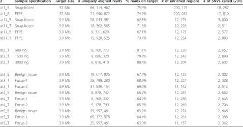
Since smaller amounts of DNA might lead to a decreased sample complexity, and thereby to increased data variance, we calculated and visualized the variant/ reference ratio distributions for different DNA Table 1 Sequencing statistics
ID sample specification Target size # uniquely aligned reads % reads on target # of enriched regions # of SNVs called (20×)
Pat1_B Snap-frozen 52 Mb 66, 114, 467 75.4% 200, 175 18, 287
Pat1_B FFPE 52 Mb 71, 590, 872 74.7% 200, 032 17, 810
Pat11_B Snap-frozen 3.9 Mb 28, 043, 981 62.8% 12, 274 3, 000
Pat11_T Snap-frozen 3.9 Mb 18, 302, 565 71.3% 12, 226 2, 511
Pat11_B FFPE 3.9 Mb 9, 311, 629 67.1% 12, 175 2, 577
Pat11_T FFPE 3.9 Mb 15, 928, 525 73.7% 12, 254 2, 883
Pat2_T 500 ng 3.9 Mb 8, 760, 773 81.1% 12, 220 2, 652
Pat2_T 1500 ng 3.9 Mb 9, 686, 320 79.9% 12, 243 2, 848
Pat2_T 3000 ng 3.9 Mb 6, 810, 410 80.4% 12, 204 2, 602
Pat3_B Benign tissue 3.9 Mb 19, 617, 926 67.7% 12, 122 2, 002
Pat3_T Focus-1 3.9 Mb 28, 798, 280 68.9% 12, 227 2, 328
Pat3_T Focus-2 3.9 Mb 31, 939, 154 69.6% 11, 142 2, 513
Pat4_B Benign tissue 3.9 Mb 8, 878, 742 66.2% 12, 281 2, 662
Pat4_T Focus-1 3.9 Mb 8, 768, 332 64.2% 12, 288 2, 645
Pat4_T Focus-2 3.9 Mb 9, 178, 790 65.3% 12, 269 2, 706
Pat5_B Benign tissue 3.9 Mb 25, 957, 461 63.2% 12, 274 2, 646
Pat5_T Focus-1 3.9 Mb 65, 372, 578 64.4% 12, 261 2, 388
Pat5_T Focus-2 3.9 Mb 25, 957, 461 63.0% 11, 157 2, 342
amounts at a coverage level of 50× for SNVs and InDels (Figure 2B). In an ideal situation a heterozygous position would have 50% reads showing the variant - a variant/reference ratio of 0.5. Based on the ratios we find a slightly broader distribution for small amounts of input DNA which is also shifted towards higher ratios. The slightly lowered complexity of the samples with decreased DNA input amounts is also reflected in the number of unique start sites: For 500 ng input material we received 40% of the expected unique start sites (calculated in relation to the 3.9 Mb target region), for 1500 ng 54% and for 3000 ng 62%. This needs to be considered when the input amounts are reduced and when homozygous versus heterozygous gene loci are compared. In comparison to SNV call-ings, InDels do not follow the expected bimodal distri-bution for variant/reference ratios but resemble rather a Bernoulli distribution (Figure 2B). Based on these findings, we chose to discard InDels with variant/refer-ence ratios lower than 15% from further analysis.
loci alike. For InDels we found concordance rates of 98% at above 20× coverage (Figure 2D), and we observed much smaller differences between known and unknown positions (Additional file 1, Figure S5).
Distinct biopsies from a single tumor have identical somatic SNV profiles in selected prostate cancer
candidate genes, but differ in their copy number patterns
A long-standing question of cancer research is whether biopsies are true representatives for the tissue of origin. This is of particular interest since many solid tumors grow as distinct tumor foci. We therefore addressed the questions whether biopsies from prostate tumors are uniform or if they are associated with different muta-tional patterns or different copy number variations. Prostate cancer is a prototype tumor to address this problem. The majority of these tumors are multifocal and in many cases two or more distinct, locally sepa-rated tumor foci can be identified [30,33].
We designed a target gene set of 1121 genes carefully selected by association to prostate cancer, cancer in gen-eral and signal transduction pathways. DNA was isolated from two different loci for each tumor in addition to matched benign tissue from frozen radical prostatect-omy specimens of three prostate cancer patients. Selec-tion of different foci was based on differences of histological and morphological phenotypes and was per-formed and controlled on the basis of HE stainings and P63/AMACR double immunostainings. The basal cell marker P63 decorates only benign glands whereas AMACR is a marker for tumor cells. In each case the two tumor foci analyzed displayed different histopatho-logical gradings, in two cases Gleason patterns 3+4 in the low grade focus and 4+5 in the high grade focus, the third case displayed an additional tertiary pattern 5 in the high grade focus (See also Table 2 and Additional file 1, Methods).
Targeted enrichment with subsequent sequencing was performed with these nine tissue samples. We found the enrichment efficiency to be very similar for all samples: Approximately 69% of sequencing reads mapped to the target region and about 99% of targets were hit at least once (Table 1 and Additional file 2, Table S3). In addi-tion, the coverage profiles were very similar for all patients as demonstrated by the cumulative normalized coverage plot and the coefficients of variation (Figure 3A, B).
For a comparison of the SNV profiles we used a two step procedure for loci covered in both preparations at a minimal coverage level of 20×. First, called SNVs for focus A were required to have at least 15% of reads con-taining the SNV. In the second step focus B was then analyzed and a SNV was considered concordant if the SNV was found in at least one read of focus B.
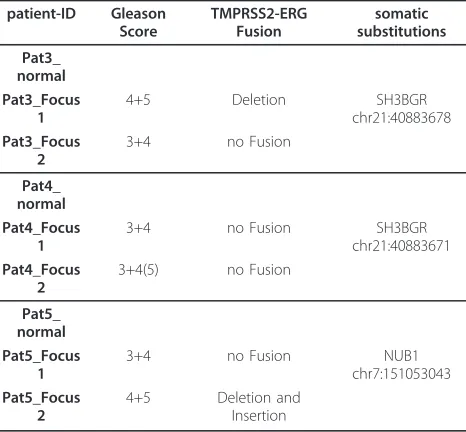
Although the SNVs differed substantially between patients, we found no discordant position in any two foci of the same tumor in the three patients at this level of stringency. We also determined the concordance of SNV profiles at smaller coverage levels (Figure 3C). At a minimal coverage of 5× we observed 0.4% discordant loci at maximum but this difference is most likely caused by an amplification bias rather than by real dif-ferences, since the number of discordant foci quickly diminishes with rising coverage demands. We analyzed small InDels in a similar way and found again higher rates of discordance as compared to SNVs (Figure 3D). Except for one discordant locus found in Patient 5, no discordances were found when higher coverage cutoffs were used. We also investigated potential somatic SNVs by comparing each individual focus with its matched benign tissue. We found one somatic SNV for each of the three patients. This mutation was identified in both tumor foci but not in the benign tissue (Table 2).
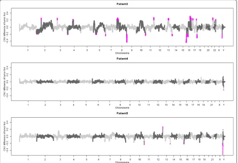
In addition to the SNV profiles for the different tumor foci we also investigated the copy number variations within each focus. For this, we generated low-coverage whole genome sequencing profiles for the two tumor foci and the corresponding benign tissue from each patient. We determined potential somatic copy number variations by comparing each tumor focus with the matched benign tissue (Additional file 1, Figure S6). Received copy number variations were then compared between the two foci and the difference was plotted genome-wide (Figure 4). For patient 3 we found clear differences on chromosome 4, 10 and 13 between both foci with regard to CNVs (Additional file 1, Figure S6 Table 2 TMPRSS-ERG Fusion status and somatic
mutations of the different foci analysed
patient-ID Gleason Score
TMPRSS2-ERG Fusion
somatic substitutions
Pat3_ normal Pat3_Focus
1
4+5 Deletion SH3BGR
chr21:40883678 Pat3_Focus
2
3+4 no Fusion
Pat4_ normal Pat4_Focus
1
3+4 no Fusion SH3BGR
chr21:40883671 Pat4_Focus
2
3+4(5) no Fusion
Pat5_ normal Pat5_Focus
1
3+4 no Fusion NUB1
chr7:151053043 Pat5_Focus
2
4+5 Deletion and
A-C). In comparison, the biopsies taken from patient 4 and 5 seemed more homogeneous as no differences of the CNV profiles were apparent between the two tumor foci. Marked differences for patient 5 are located towards chromosome ends and visual inspection pro-posed the individual CNVs to be artefacts (Additional file 1, Figure S6 D).
Discussion
Next-generation technologies such as targeted re-sequencing platforms are powerful tools for identifying genetic variations in cancer samples. Using prostate can-cer as an example, we have assessed the use of different kinds and amounts of tissue samples for identifying genetic variations. In particular, we have investigated three aspects which are frequently addressed from oncologists and pathologists:
The first is whether or not it is possible to use FFPE material in addition to snap frozen material. The use of FFPE material would open up a large collection of
damage done by the FFPE preparation has a random distribution across all DNA fragments and can be cor-rected by sequencing depth. Since coverage levels of 80× and higher can easily be reached by targeted re-sequencing approaches, we recommend to use such high coverages when analyzing FFPE material. The same holds true for false negative SNVs. Keeping in mind that SNV detection is the main focus of DNA sequence analysis in cancer, the detection of small insertions and deletions becomes increasingly impor-tant. We therefore investigated if preparation of DNA from FFPE tissue may have an adverse effect on InDel detection. While the relative amount of discordant InDel positions is about 7 times higher than the amount of discordant SNV positions, we observed the same low discrepancy rates at higher coverage levels. Again, no discordance was found at a coverage level of 80×. Taken together, snap frozen tissues remain the preferred source of DNA, but FFPE tissue can be used for SNV and InDel detection instead if the coverage is
increased. Furthermore, for certain clinically relevant questions, like for the detection of germline variants, e.g. when for a snap frozen tumor tissue no adequate matching benign tissue material is available, FFPE tis-sues can be used. In this case, the positive error rate obtained with FFPE material plays an inferior role.
The second methodological issue relates to the amount of material required. Decreasing the input amount of DNA to 500 ng still yielded good enrichment results, an even coverage and a highly reproducible call-ing of known genetic variants. However, we find increased redundant reads (reads with identical first positions) and a slightly higher variance of variant/refer-ence ratios with decreased amounts of starting material. This suggests that - with these enrichment technologies - the minimal amount of input DNA cannot easily be reduced beyond 500 ng. Notably, the comparison among average and high amounts of DNA (1.5μg vs 3μg) per-formed better than a comparison including the lowest amount of DNA (500 ng).
While InDels detected show a variant/reference ratio distribution clearly deviating from the expected bimodal distribution and visible differences for the three DNA amounts, InDels are still highly reproducible above a coverage level of 45× for all amounts of DNA. We con-clude that a decrease to 500 ng of input DNA is possi-ble, but the benefit has to be weighed against the high coverage demands and potential challenges to SNV and InDel categorization.
The third challenge presented in our study consists of the heterogeneity of tumor tissue. In order to obtain results representative for the whole tumor, the amount and location of biopsies necessary is unknown. So far, it is not decided whether primary prostate cancers have a multifocal origin and thus are composed of multiple genetically distinct cancer cell clones or not. Currently, an independent clonal nature of multiple foci is consid-ered since healthy men below 40 years frequently show presence of focal histological aberrations [34-36] many of which give rise only to latent prostate cancer, while clonal evolution of a few foci paves the way to clinically detectable disease [33,37-40]. On the other hand, pros-tate cancer metastases from different locations but from the same patient show a surprisingly similar pattern with regard to copy number alterations [41-43]. Experi-ments available to address this question include the determination of the DNA ploidy, micro-satellite analy-sis, c-myc amplifications with FISH, DNA methylation or the TMPRSS2-ERG fusion status on separate tumors within the same prostate [44]. In our hands, using sam-ples derived from different foci within one prostate tumor and performing DNA re-sequencings of prostate cancer relevant genes, we found almost identical distri-butions of mutations within different foci of the same patient. Notably, SNV profile concordance was 100% for all three patients at coverage levels above 20×. Even tumor parts with different TMPRSS2-ERG gene fusion status are remarkably identical with regard to small nuclear variations. In addition, focusing on somatic mutations, we find no differences between different tumor foci. However, although we focused on prostate cancer candidate genes, the low number of somatic mutations in prostate cancer and the fact that we only analyzed ~10% of the exome prohibit a generalized con-clusion. Recent studies, such as Taylor et al with 0.31, Kan et al with 0.33, and Berger et al with 0.9 non-synonymous mutations per Mb, suggest low somatic mutation rates per Mb for prostate cancer [8,9,45]. In line with this somatic mutation frequency we found only one somatic mutation for each of the three patients. The sensitivity of current re-sequencing approaches might further explain the missing focal diversity. Irrespective of the low frequency of somatic mutations we detected in the tumor samples we found
large aberrations in copy number. We have used a whole genome re-sequencing approach to detect somatic copy number variations for each focus and compared the two foci from the same tumor. Interestingly, for one patient with clear differences in the TMPRSS2-ERG fusion pattern, we also find significant differences between the two foci, whereas for two other patients no significant CNVs can be detected. Along this line Navin et al. used a modified comparative genomic hybridiza-tion (CGH) technology to study the clonal composihybridiza-tion of breast tumors and found a large proportion of mono-genomic tumors and only a small fraction of tumors with a heterogenomic foci structure [46]. Our results would implicate that the location of biopsies taken within tumors is of minor relevance for the detection of mutations, but plays a major role for the detection of copy number variations. Within this direction, recent publications also suggest that genomic rearrangements are a major genetic factor underlying prostate cancer [47]. Since we did not perform 3D reconstructions of the whole tumors our approach cannot be used to answer the question of multifocal origin of heteroge-neous prostate tumors. Even for the estimation of tumor heterogeneity our studies are most likely an underesti-mation, because we are investigating tissue samples with a complex composition of single cells. Thus, the genetic profiles are the sums over all cells contained within the section and might mask the true tumor heterogeneity. At the moment we are extending our analysis onto a single cell level to further gain insight into the evolu-tionary architecture of prostate tumors. With this we might be able to pin down the true tumor composition and we might even identify tumor stem cells on a genetic level. However, since we find differences between different biopsies from the same tumor on a copy number level, we can conclude that several biop-sies need to be investigated to gain insight into the genomic context of prostate cancers based the overall tumor heterogeneity.
Furthermore, with the technologies described we are now in the progress to extend our analyses to large sample cohorts from pathology departments where we can select tissue specimens from specific clinical studies. This enables us to address clinical relevant questions such as progression and therapy resistance of tumors which is an important step towards the application of targeted re-sequencing approaches as routine diagnostic tools in oncology.
Conclusion
and reproducibility of the data in regard to tumor het-erogeneity. Our optimized protocols guide through each of these issues and provide data for an optimal strategy for the usage in clinical settings. We show that FFPE material can be used with higher coverages as substitu-tion of cryo-frozen tissue and that it is in particular use-ful for the determination of germline variations when tumor tissues have already been sequenced. Lowering the amount of input material results in an increase of redundant reads and a slightly higher variance of var-iant/reference ratios, but can be overcome to a certain degree with adequate analysis tools. Finally, the tumor heterogeneity plays an important role for the detection of copy number variations, but is of minor importance for the detection of somatic variations. This implies that the sampling of tumor tissues is of major importance and needs to be taken into consideration for clinical diagnostic purposes.
Additional material
Additional file 1: Supplementary Methods, Figure Legends S1-S6, Table Legends S1-S3. Figures S1-S6
Additional file 2: Tables S1-S3.
Acknowledgements and Funding
We would like to thank Nada Kumer, Anna Kosiura, Sonia Paturej, Isabelle Kuehndahl, Ilona Hauenschild, Irma Sottsas and Christof Seifarth for excellent technical assistance.
This work was supported by a grant of the Austrian Nationalstiftung and the Austria Wirtschaftsservice GmbH in the framework of the IMGuS research program (Institute for Medical Genome Research and Systems Biology, Vienna); and the German Federal Ministry of Education and Research (01GS08105, 01GS08111 and Predict)
Author details
1Max Planck Institute for Molecular Genetics, Ihnestr. 63-73, 14195 Berlin,
Germany.2Free University Berlin, Department of Biology, Chemistry and
Pharmacy, Takustrasse 3, 14195 Berlin, Germany.3Unit Cancer Genome Research, DKFZ German Cancer Research Center and National Center for Tumor Diseases, Im Neuenheimer Feld 460, 69120 Heidelberg, Germany.
4Innsbruck Medical University, Department of Urology, Anichstr. 35, A 6020
Innsbruck, Austria.5Innsbruck Medical University, Department of Pathology, Muellerstr. 40, A-6020 Innsbruck, Austria.6Innsbruck Medical University,
Department for Medical Genetics, Molecular and Clinical Pharmacology, Division of Human Genetics, Schöpfstraße 4, 6020 Innsbruck, Austria.
Authors’contributions
Clinical samples and discussions were provided by GS, IV, GB and HK. MK, MI, BT and MRS carried out the experiments, data analyses were done by MK, MI, HK and MRS; HS and MRS conceived and coordinated the project; MK, MI, HS, RH, SK, HK, HL and MRS wrote the paper. All authors read and approved the final version.
Competing interests
The authors declare that they have no competing interests.
Received: 20 March 2011 Accepted: 29 September 2011 Published: 29 September 2011
References
1. Jemal A, Bray F, Center MM, Ferlay J, Ward E, Forman D:Global cancer statistics.CA Cancer J Clin61:69-90.
2. Bell DW:Our changing view of the genomic landscape of cancer.J Pathol2010,220:231-243.
3. Greenman C, Stephens P, Smith R, Dalgliesh GL, Hunter C, Bignell G, Davies H, Teague J, Butler A, Stevens C,et al:Patterns of somatic mutation in human cancer genomes.Nature2007,446:153-158.
4. Jones S, Zhang X, Parsons DW, Lin JC, Leary RJ, Angenendt P, Mankoo P, Carter H, Kamiyama H, Jimeno A,et al:Core signaling pathways in human pancreatic cancers revealed by global genomic analyses.Science2008,
321:1801-1806.
5. Parsons DW, Jones S, Zhang X, Lin JC, Leary RJ, Angenendt P, Mankoo P, Carter H, Siu IM, Gallia GL,et al:An integrated genomic analysis of human glioblastoma multiforme.Science2008,321:1807-1812.
6. Sjoblom T, Jones S, Wood LD, Parsons DW, Lin J, Barber TD, Mandelker D, Leary RJ, Ptak J, Silliman N,et al:The consensus coding sequences of human breast and colorectal cancers.Science2006,314:268-274. 7. Wood LD, Parsons DW, Jones S, Lin J, Sjoblom T, Leary RJ, Shen D,
Boca SM, Barber T, Ptak J,et al:The genomic landscapes of human breast and colorectal cancers.Science2007,318:1108-1113.
8. Taylor BS, Schultz N, Hieronymus H, Gopalan A, Xiao Y, Carver BS, Arora VK, Kaushik P, Cerami E, Reva B,et al:Integrative genomic profiling of human prostate cancer.Cancer Cell2010,18:11-22.
9. Kan Z, Jaiswal BS, Stinson J, Janakiraman V, Bhatt D, Stern HM, Yue P, Haverty PM, Bourgon R, Zheng J,et al:Diverse somatic mutation patterns and pathway alterations in human cancers.Nature2010,466:869-873. 10. Yu J, Mani RS, Cao Q, Brenner CJ, Cao X, Wang X, Wu L, Li J, Hu M, Gong Y,
et al:An integrated network of androgen receptor, polycomb, and TMPRSS2-ERG gene fusions in prostate cancer progression.Cancer Cell 2010,17:443-454.
11. Timmermann B, Kerick M, Roehr C, Fischer A, Isau M, Boerno ST, Wunderlich A, Barmeyer C, Seemann P, Koenig J,et al:Somatic mutation profiles of MSI and MSS colorectal cancer identified by whole exome next generation sequencing and bioinformatics analysis.PLoS One2010,
5:e15661.
12. Ding L, Ellis MJ, Li S, Larson DE, Chen K, Wallis JW, Harris CC, McLellan MD, Fulton RS, Fulton LL,et al:Genome remodelling in a basal-like breast cancer metastasis and xenograft.Nature2010,464:999-1005. 13. Lee W, Jiang Z, Liu J, Haverty PM, Guan Y, Stinson J, Yue P, Zhang Y,
Pant KP, Bhatt D,et al:The mutation spectrum revealed by paired genome sequences from a lung cancer patient.Nature2010,465:473-477. 14. Ley TJ, Mardis ER, Ding L, Fulton B, McLellan MD, Chen K, Dooling D,
Dunford-Shore BH, McGrath S, Hickenbotham M,et al:DNA sequencing of a cytogenetically normal acute myeloid leukaemia genome.Nature2008,
456:66-72.
15. Shah SP, Morin RD, Khattra J, Prentice L, Pugh T, Burleigh A, Delaney A, Gelmon K, Guliany R, Senz J,et al:Mutational evolution in a lobular breast tumour profiled at single nucleotide resolution.Nature2009,461:809-813. 16. Schweiger MR, Kerick M, Timmermann B, Isau M:The power of NGS
technologies to delineate the genome organization in cancer: from mutations to structural variations and epigenetic alterations.Cancer Metastasis Rev2011.
17. Pleasance ED, Cheetham RK, Stephens PJ, McBride DJ, Humphray SJ, Greenman CD, Varela I, Lin ML, Ordonez GR, Bignell GR,et al:A comprehensive catalogue of somatic mutations from a human cancer genome.Nature2010,463:191-196.
18. Pleasance ED, Stephens PJ, O’Meara S, McBride DJ, Meynert A, Jones D, Lin ML, Beare D, Lau KW, Greenman C,et al:A small-cell lung cancer genome with complex signatures of tobacco exposure.Nature2010,
463:184-190.
19. Krawitz PM, Schweiger MR, Rodelsperger C, Marcelis C, Kolsch U, Meisel C, Stephani F, Kinoshita T, Murakami Y, Bauer S,et al:Identity-by-descent filtering of exome sequence data identifies PIGV mutations in hyperphosphatasia mental retardation syndrome.Nat Genet2010,
42:827-829.
20. Ng SB, Turner EH, Robertson PD, Flygare SD, Bigham AW, Lee C, Shaffer T, Wong M, Bhattacharjee A, Eichler EE,et al:Targeted capture and massively parallel sequencing of 12 human exomes.Nature2009,
21. Okou DT, Steinberg KM, Middle C, Cutler DJ, Albert TJ, Zwick ME:
Microarray-based genomic selection for high-throughput resequencing.
Nat Methods2007,4:907-909.
22. Albert TJ, Molla MN, Muzny DM, Nazareth L, Wheeler D, Song X, Richmond TA, Middle CM, Rodesch MJ, Packard CJ,et al:Direct selection of human genomic loci by microarray hybridization.Nat Methods2007,
4:903-905.
23. Hodges E, Xuan Z, Balija V, Kramer M, Molla MN, Smith SW, Middle CM, Rodesch MJ, Albert TJ, Hannon GJ, McCombie WR:Genome-wide in situ exon capture for selective resequencing.Nat Genet2007,39:1522-1527. 24. Porreca GJ, Zhang K, Li JB, Xie B, Austin D, Vassallo SL, LeProust EM,
Peck BJ, Emig CJ, Dahl F,et al:Multiplex amplification of large sets of human exons.Nat Methods2007,4:931-936.
25. Gnirke A, Melnikov A, Maguire J, Rogov P, LeProust EM, Brockman W, Fennell T, Giannoukos G, Fisher S, Russ C,et al:Solution hybrid selection with ultra-long oligonucleotides for massively parallel targeted sequencing.Nat Biotechnol2009,27:182-189.
26. Weise A, Timmermann B, Grabherr M, Werber M, Heyn P, Kosyakova N, Liehr T, Neitzel H, Konrat K, Bommer C,et al:High-throughput sequencing of microdissected chromosomal regions.Eur J Hum Genet2010,
18:457-462.
27. Briggs AW, Good JM, Green RE, Krause J, Maricic T, Stenzel U, Lalueza-Fox C, Rudan P, Brajkovic D, Kucan Z,et al:Targeted retrieval and analysis of five Neandertal mtDNA genomes.Science2009,325:318-321.
28. Schweiger MR, Kerick M, Timmermann B, Albrecht MW, Borodina T, Parkhomchuk D, Zatloukal K, Lehrach H:Genome-wide massively parallel sequencing of formaldehyde fixed-paraffin embedded (FFPE) tumor tissues for copy-number- and mutation-analysis.PLoS One2009,4:e5548. 29. Bartsch G, Horninger W, Klocker H, Pelzer A, Bektic J, Oberaigner W,
Schennach H, Schafer G, Frauscher F, Boniol M,et al:Tyrol Prostate Cancer Demonstration Project: early detection, treatment, outcome, incidence and mortality.BJU Int2008,101:809-816.
30. Horninger W, Berger AP, Rogatsch H, Gschwendtner A, Steiner H, Niescher M, Klocker H, Bartsch G:Characteristics of prostate cancers detected at low PSA levels.Prostate2004,58:232-237.
31. Wood HM, Belvedere O, Conway C, Daly C, Chalkley R, Bickerdike M, McKinley C, Egan P, Ross L, Hayward B,et al:Using next-generation sequencing for high resolution multiplex analysis of copy number variation from nanogram quantities of DNA from formalin-fixed paraffin-embedded specimens.Nucleic Acids Res2010,38:e151.
32. Aird D, Ross MG, Chen WS, Danielsson M, Fennell T, Russ C, Jaffe DB, Nusbaum C, Gnirke A:Analyzing and minimizing PCR amplification bias in Illumina sequencing libraries.Genome Biol2011,12:R18.
33. Aihara M, Wheeler TM, Ohori M, Scardino PT:Heterogeneity of prostate cancer in radical prostatectomy specimens.Urology1994,43:60-66, discussion 66-67.
34. Yatani R, Kusano I, Shiraishi T, Hayashi T, Stemmermann GN:Latent prostatic carcinoma: pathological and epidemiological aspects.Jpn J Clin Oncol1989,19:319-326.
35. Sakr WA, Grignon DJ, Crissman JD, Heilbrun LK, Cassin BJ, Pontes JJ, Haas GP:High grade prostatic intraepithelial neoplasia (HGPIN) and prostatic adenocarcinoma between the ages of 20-69: an autopsy study of 249 cases.In Vivo1994,8:439-443.
36. Shiraishi T, Watanabe M, Matsuura H, Kusano I, Yatani R, Stemmermann GN:
The frequency of latent prostatic carcinoma in young males: the Japanese experience.In Vivo1994,8:445-447.
37. Bostwick DG, Shan A, Qian J, Darson M, Maihle NJ, Jenkins RB, Cheng L:
Independent origin of multiple foci of prostatic intraepithelial neoplasia: comparison with matched foci of prostate carcinoma.Cancer1998,
83:1995-2002.
38. Macintosh CA, Stower M, Reid N, Maitland NJ:Precise microdissection of human prostate cancers reveals genotypic heterogeneity.Cancer Res 1998,58:23-28.
39. Mehra R, Han B, Tomlins SA, Wang L, Menon A, Wasco MJ, Shen R, Montie JE, Chinnaiyan AM, Shah RB:Heterogeneity of TMPRSS2 gene rearrangements in multifocal prostate adenocarcinoma: molecular evidence for an independent group of diseases.Cancer Res2007,
67:7991-7995.
40. Clark J, Attard G, Jhavar S, Flohr P, Reid A, De-Bono J, Eeles R, Scardino P, Cuzick J, Fisher G,et al:Complex patterns of ETS gene alteration arise
during cancer development in the human prostate.Oncogene2008,
27:1993-2003.
41. Shah RB, Mehra R, Chinnaiyan AM, Shen R, Ghosh D, Zhou M, Macvicar GR, Varambally S, Harwood J, Bismar TA,et al:Androgen-independent prostate cancer is a heterogeneous group of diseases: lessons from a rapid autopsy program.Cancer Res2004,64:9209-9216.
42. Mehra R, Tomlins SA, Yu J, Cao X, Wang L, Menon A, Rubin MA, Pienta KJ, Shah RB, Chinnaiyan AM:Characterization of TMPRSS2-ETS gene aberrations in androgen-independent metastatic prostate cancer.Cancer Res2008,68:3584-3590.
43. Liu W, Laitinen S, Khan S, Vihinen M, Kowalski J, Yu G, Chen L, Ewing CM, Eisenberger MA, Carducci MA,et al:Copy number analysis indicates monoclonal origin of lethal metastatic prostate cancer.Nat Med2009,
15:559-565.
44. Barry M, Perner S, Demichelis F, Rubin MA:TMPRSS2-ERG fusion heterogeneity in multifocal prostate cancer: clinical and biologic implications.Urology2007,70:630-633.
45. Berger MF, Lawrence MS, Demichelis F, Drier Y, Cibulskis K, Sivachenko AY, Sboner A, Esgueva R, Pflueger D, Sougnez C,et al:The genomic complexity of primary human prostate cancer.Nature470:214-220. 46. Navin N, Krasnitz A, Rodgers L, Cook K, Meth J, Kendall J, Riggs M,
Eberling Y, Troge J, Grubor V,et al:Inferring tumor progression from genomic heterogeneity.Genome Res2010,20:68-80.
47. Michael FBerger MSL, Francesca Demichelis, Drier KC, Andrey YSivachenko, Andrea Sboner, Esgueva DP, Carrie Sougnez, Robert Onofrio, Scott LCarter, Park LH, Lauren Ambrogio, Timothy Fennell, Melissa Parkin, Saksena DV, Alex HRamos, Trevor JPugh, Jane Wilkinson, Fisher WW, Scott Mahan, Kristin Ardlie, Jennifer Baldwin, W. Simons NK, Theresa YMacDonald, Philip WKantoff, Chin SBG, Mark BGerstein, Todd RGolub, Meyerson AT, Eric SLander, Gad Getz, Mark A, Rubin LAG:The genomic complexity of primary human prostate cancer.Nature2011.
Pre-publication history
The pre-publication history for this paper can be accessed here: http://www.biomedcentral.com/1755-8794/4/68/prepub
doi:10.1186/1755-8794-4-68
Cite this article as:Kericket al.:Targeted high throughput sequencing in clinical cancer Settings: formaldehyde fixed-paraffin embedded (FFPE) tumor tissues, input amount and tumor heterogeneity.BMC Medical Genomics20114:68.
Submit your next manuscript to BioMed Central and take full advantage of:
• Convenient online submission
• Thorough peer review
• No space constraints or color figure charges
• Immediate publication on acceptance
• Inclusion in PubMed, CAS, Scopus and Google Scholar
• Research which is freely available for redistribution