R E S E A R C H
Open Access
Discovery and replication of a peripheral
tissue DNA methylation biosignature to
augment a suicide prediction model
Makena L. Clive
1, Marco P. Boks
2, Christiaan H. Vinkers
2, Lauren M. Osborne
1, Jennifer L. Payne
1, Kerry J. Ressler
3,4,5,
Alicia K. Smith
4, Holly C. Wilcox
1,6and Zachary Kaminsky
1,6,7*Abstract
Background:Suicide is the second leading cause of death among adolescents in the USA, and rates are rising. Methods to identify individuals at risk are essential for implementing prevention strategies, and the development of a biomarker can potentially improve prediction of suicidal behaviors. Prediction of our previously reportedSKA2 biomarker for suicide and PTSD is substantially improved by questionnaires assessing perceived stress or anxiety and is therefore reliant on psychological assessment. However, such stress-related states may also leave a biosignature that could equally improve suicide prediction. In genome-wide DNA methylation data, we observed significant overlap between waking cortisol-associated and suicide-associated DNA methylation in blood and the brain, respectively.
Results:Using a custom bioinformatic brain to blood discovery algorithm, we derived a DNA methylation biosignature that interacts withSKA2methylation to improve the prediction of suicidal ideation in our existing suicide prediction model across both blood and saliva data sets. This biosignature was independently validated in the Grady Trauma Project cohort and interacted with HPA axis metrics in the same cohort. The biosignature showed a relationship with immune status by its correlation with myeloid-derived cell proportions in all data sets and with IL-6 measures in a prospective postpartum depression cohort. Three probes showed significant correlations with the biosignature: cg08469255 (DDR1), cg22029879 (ARHGEF10), and cg24437859 (SHP1), of whichSHP1methylation correlated with immune measures.
Conclusions:We conclude that this biosignature interacts withSKA2methylation to improve suicide prediction and may represent a biological state of immune and HPA axis modulation that mediates suicidal behavior.
Keywords: Suicide, Biomarker, Epigenetics, DNA methylation, SKA2, HPA axis, Illumina HM450 microarray, Childhood trauma
Background
Suicide accounts for 1.4 % of worldwide deaths annually, posing a serious public health issue [1]. Based on 2014 data, it is the second leading cause of death among ado-lescents, and the tenth leading cause of death for all ages in the USA [2]. Given the rising rates of suicide in the USA, methods to identify individuals at risk for imple-menting prevention strategies are urgently needed [3].
Recently, our laboratory identified a DNA methylation mark that is associated with suicide in a postmortem brain tissue cohort at a CpG (cg13989295) located within a single nucleotide polymorphism (SNP), rs7208505, in the spindle- and kinetochore-associated protein 2 (SKA2) where the reference allele of rs7208505 eliminates the CpG. The observed epigenetic association with suicide was replicated in two additional brain tissue cohorts and with suicidal behaviors including suicidal ideation (SI) and attempt (SA) in peripheral blood in three living cohorts [4]. In our original work, gene expression of SKA2 was correlated with DNA methylation at this position and was significantly decreased in suicide decedents. Several recent * Correspondence:[email protected]
1
Department of Psychiatry and Behavioral Sciences, Johns Hopkins University School of Medicine, Baltimore, MD 21205, USA
6Department of Mental Health, Johns Hopkins Bloomberg School of Public
Health, Baltimore, MD 21218, USA
Full list of author information is available at the end of the article
independent studies have observed decreased expression of SKA2in both the blood of violent suicide completers [5] and in the prefrontal cortex of suicide victims [4, 6], the latter of which was also associated with decreased protein levels.
The SKA2 protein is thought to interact with the hypothalamic-pituitary-adrenal (HPA) axis by chaperon-ing the glucocorticoid receptor (GR) from the cytoplasm to the nucleus upon cortisol binding [7]. Once in the nucleus, the GR can interact with genomic DNA and influence gene expression involved in negative feedback regulation of the HPA axis response. In two independent cohorts with high levels of childhood trauma, elevated
SKA2 DNA methylation in peripheral blood before administration of the TRIER social stress test was signifi-cantly associated with a blunted post-test cortisol level, while SKA2 DNA methylation before the dexametha-sone suppression test (DST) was significantly associated with elevated post-test cortisol levels [8, 9]. These data support the interpretation that SKA2 DNA methylation state may be an important contributor to individual stress response.
In an attempt to identify at-risk individuals, we previ-ously generated a suicide prediction model, which de-scribes suicidal behavior as a function of both genotype and methylation at the single nucleotide polymorphism (SNP) rs7208505 in SKA2 which interacts with a state level metric of stress or trait level metric of anxiety to confer risk [4]. Notably, some studies demonstrate that state level stress can be influenced by trait level anxiety [10]. Model predictive accuracies vary between ~70 and 85 % in various cohorts and are consistent with SKA2 gene expression-based prediction accuracies reported by other groups [8, 11]. The statistical interaction with stress is likely related to the physiological role SKA2 plays in mediating HPA axis activity. In this context, it is hypothesized that epigenetic variation ofSKA2may rep-resent an underlying trait vulnerability of the HPA axis that must interact with a state of stress to elicit risk. In our previous work, we have identified significant interac-tions of SKA2 with various self-reported psychological scales to influence suicide risk. The scales vary by study cohort and include the Child Trauma Questionnaire (CTQ), the Perceived Stress Scale, waking salivary cor-tisol levels, and various metrics of anxiety including self-reported binary estimates and those quantified by the Self-Report for Childhood Anxiety Related Disor-ders (SCARED) [4, 8, 9]. Furthermore, our work and others have noted an increased model efficacy in sub-groups of individuals having experienced childhood trauma [8, 9, 12, 13]. It is possible that high values in the stress metrics represent a biological state that may be related to HPA axis function. Despite the possibility to assess these states using questionnaires, the use of
self-reported scales has many drawbacks including a lack of standardization across studies, variability in psycho-metric properties, and variability in the subjective rating of stress levels. In the clinical context, the administration of questionnaires requires time and patient compliance.
Recent attempts have been made to circumvent the use of psychological assessments and develop biomarker proxies [11, 14]. A challenge for the identification of peripheral tissue-based epigenetic biomarkers in the context of psychiatry is the generalizability of the identi-fied peripheral epigenetic variation in the brain. We have hypothesized previously that circulating steroid hor-mones such as cortisol may mark peripheral tissue DNA on the epigenetic level while affecting behavior through central nervous system (CNS) specific actions [15–17]. In support of this hypothesis, the initial objective of this study was to evaluate if cortisol-associated DNA methy-lation levels in peripheral tissues (blood and saliva) are enriched among suicide-associated DNA methylation levels in the brain.
While systemic factors like cortisol may influence epi-genetic patterns across-tissues and may represent rele-vant biomarkers interacting with SKA2, we did not wish to limit our analysis to cortisol-associated probes alone. Thus, the second major objective of this study was to generate an epigenetic biosignature of SKA2-interacting state markers in a bioinformatically driven and unbiased manner and to understand the underlying biological context driving any identified biosignatures.
Results
Overrepresentation of peripheral cortisol-associated loci among brain-associated suicide genes
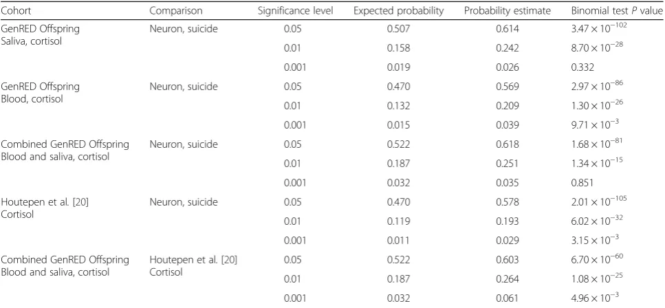
levels of significance (Table 1) [18, 19]. Given the import-ance of dysregulated cortisol biology to suicidal behaviors, cortisol-associated methylation probes in peripheral blood (N= 18) and saliva (N= 20) from the GenRED Offspring cohorts were assessed for an overrepresentation with those probes significantly associated with completed suicide separately in postmortem prefrontal cortical neurons and non-neurons (N= 45). Cortisol-associated probes within genes or gene regulatory sites were significantly overrepresented among prefrontal neuron suicide-associated genes and genes previously identified as associated with cortisol stress reactivity (Table 2) [20]. These findings indicate that there may be common pathways between cortisol biology and suicidal behavior and that the epigenetic marks of suicide-associated hor-monal changes may be detectable in peripheral tissues.
Algorithmic identification of SKA2-interacting biosignature for DNA methylation-based suicidal behavior prediction In light of the above findings, our strategy to approach our second objective of generating a biosignature of
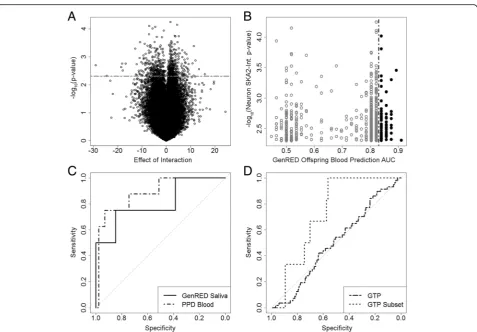
SKA2-interacting state markers was to identify epigen-etic variation interacting with SKA2 that was consistent across brain and peripheral tissues. The full algorithm is explained in Additional file 4: Figure S1. Briefly, DNA methylation in prefrontal cortex neurons at each probe
was assessed for statistical interaction with rs7208505 CpG methylation (chr17:59110368, hg38) in a linear model controlling for age, sex, and rs7208505 genotype and identified 669 probes below aPvalue cutoff of 0.005 (Fig. 1a, Additional file 4: Result S1). Of these 669 probes, 72 exhibited an AUC prediction for SA in the top 25th percentile (AUC > 0.825) in the GenRED Off-spring blood cohort (Fig. 1b; Table 3). The methylation at these 72 probes was used to train a principal compo-nent analysis (PCA) model on the GenRED Offspring blood data, which was then used to predict PCA models in the other data sets. The first eigenvector was used to assess suicidal behavior prediction in the original predic-tion model, replacing the stress measure with the PCA first eigenvector. All steps were evaluated by the Monte Carlo method and found to be statistically significant (P< 0.001, Additional file 4: Result S1). This approach predicted SI in PPD cohort blood with an AUC of 0.88 (95 % CI 0.75–1; P= 0.041) and in GenRED Off-spring saliva with an AUC of 0.81 (95 % CI 0.59–1;
P= 0.011) (Fig. 1c; Table 4). These high-prediction AUCs provide evidence that the PCA first eigenvector may represent a methylation SKA2 “interaction bio-signature” that is predictive of suicidal behavior in the existing suicide prediction model and replaces the need for a stress questionnaire.
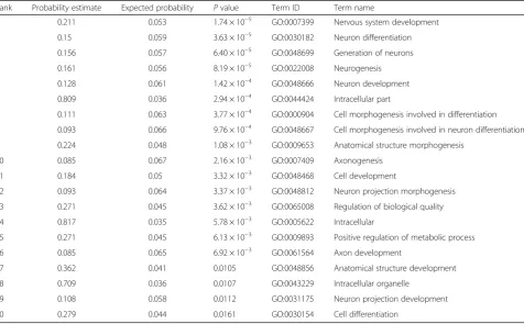
Table 1Gene ontology results
Rank Probability estimate Expected probability Pvalue Term ID Term name
1 0.211 0.053 1.74 × 10⁻⁵ GO:0007399 Nervous system development
2 0.15 0.059 3.63 × 10⁻⁵ GO:0030182 Neuron differentiation
3 0.156 0.057 6.40 × 10⁻⁵ GO:0048699 Generation of neurons
4 0.161 0.056 8.19 × 10⁻⁵ GO:0022008 Neurogenesis
5 0.128 0.061 1.42 × 10⁻⁴ GO:0048666 Neuron development
6 0.809 0.036 2.94 × 10⁻⁴ GO:0044424 Intracellular part
7 0.111 0.063 3.77 × 10⁻⁴ GO:0000904 Cell morphogenesis involved in differentiation
8 0.093 0.066 9.76 × 10⁻⁴ GO:0048667 Cell morphogenesis involved in neuron differentiation
9 0.224 0.048 1.08 × 10⁻³ GO:0009653 Anatomical structure morphogenesis
10 0.085 0.067 2.16 × 10⁻³ GO:0007409 Axonogenesis
11 0.184 0.05 3.32 × 10⁻³ GO:0048468 Cell development
12 0.093 0.064 3.37 × 10⁻³ GO:0048812 Neuron projection morphogenesis
13 0.271 0.045 3.62 × 10⁻³ GO:0065008 Regulation of biological quality
14 0.817 0.035 5.78 × 10⁻³ GO:0005622 Intracellular
15 0.271 0.045 6.13 × 10⁻³ GO:0009893 Positive regulation of metabolic process
16 0.085 0.065 6.92 × 10⁻³ GO:0061564 Axon development
17 0.362 0.041 0.0105 GO:0048856 Anatomical structure development
18 0.709 0.036 0.0107 GO:0043229 Intracellular organelle
19 0.108 0.058 0.0112 GO:0031175 Neuron projection development
20 0.279 0.044 0.0161 GO:0030154 Cell differentiation
Independent validation of SKA2 model interaction biosignature performance
The interaction biosignature model was validated using methylation array data from the Grady Trauma Project (GTP), a sample of urban minorities with low socioeco-nomic status and high rates of traumatic experience and PTSD. On the entire sample, the prediction model generated an AUC of 0.50 (95 % CI 0.42–0.58;P= 0.724) for prediction of SI in all 376 individuals. Based on recent publications that have provided evidence that both PTSD and substance abuse may confound SKA2 methylation [9, 21], we selected a subset of the GTP sample with no history of PTSD or drug use (N= 115; 6 cases, 109 controls), where a combination of SKA2 and the interaction biosignature predicted SI with an AUC of 0.73 (95 % CI 0.59–0.87; P= 0.050) (Fig. 1d; Table 4) [12]. Although our interaction biosignature model was unsuccessful in suicidal behavior prediction across the entire GTP cohort, prediction was successful in a subset without PTSD. This altered suicidal behavior prediction with PTSD is consistent with previously published findings [9].
Association of interaction biosignature metrics with HPA axis function
To improve our understanding of the biological rele-vance of the interaction biosignature, we assessed bio-signature loci for a relationship with various metrics of HPA axis function in two cohorts with high levels of childhood trauma as assessed by the CTQ. The inter-action biosignature eigenvector interacted with CTQ
scores to associate with post-test AUC cortisol levels following the administration of the TRIER social stress test (biosignature β= 3446.9 ± 1631.2, P= 0.038; CTQ β=−40.6 ± 12.9, P= 0.002; interaction β=−92.8 ± 45.0,
P= 0.043, F = 4.5, df = 4/81, model P= 0.038) (Additional file 4: Figure S2A). In the GTP sample subset, the interaction biosignature eigenvector interacted with CTQ scores to associate with the natural log of the day 2 cortisol following administration of the DST (biosignature β=−6.4 ± 2.8,P= 0.027; CTQβ= 0.096 ± 0.037,P= 0.012; interaction β= 0.22 ± 0.095, P= 0.027, F = 2.4, df = 4/49, modelP= 0.027) (Additional file 4: Figure S2B). Taken to-gether, the data suggest thatSKA2interaction biosignature values associate with early life trauma status to influence HPA axis sensitivity.
Assessment of biological relevance of SKA2 model interaction biosignature
We reasoned that the biological underpinnings of our
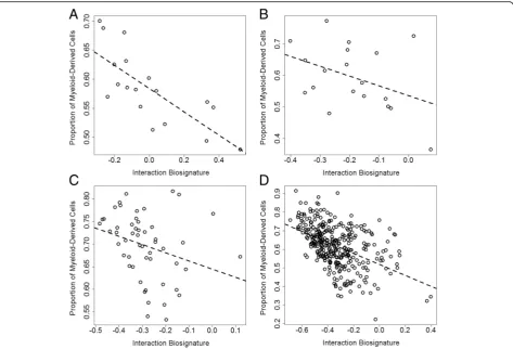
SKA2interaction biosignature may be related to variation in peripheral immune cells, as inflammation may be influ-enced by state factors like stress. The predicted proportion of granulocyte and monocyte content (myeloid-derived cells) showed a negative association with the interaction biosignature across all data sets (Fig. 2a–d), with significant correlations in GenRED Offspring blood (rho =−0.76,
P= 2.7 × 10⁻⁴), PPD cohort blood (rho =−0.29,P= 0.034), and GTP blood (rho =−0.57, P= 2.4 × 10⁻⁷) and a non-significant association in GenRED Offspring sal-iva (rho =−0.39, P= 0.092). Substituting the propor-tion of myeloid-derived cells for the interacpropor-tion
Table 2Overrepresentation analysis
Cohort Comparison Significance level Expected probability Probability estimate Binomial testPvalue
GenRED Offspring Saliva, cortisol
Neuron, suicide 0.05 0.507 0.614 3.47 × 10−102
0.01 0.158 0.242 8.70 × 10⁻²⁸
0.001 0.019 0.026 0.332
GenRED Offspring Blood, cortisol
Neuron, suicide 0.05 0.470 0.569 2.97 × 10−86
0.01 0.132 0.209 1.30 × 10⁻²⁶
0.001 0.015 0.039 9.71 × 10⁻³
Combined GenRED Offspring Blood and saliva, cortisol
Neuron, suicide 0.05 0.522 0.618 1.68 × 10−81
0.01 0.187 0.251 1.34 × 10⁻¹⁵
0.001 0.032 0.035 0.851
Houtepen et al.[20] Cortisol
Neuron, suicide 0.05 0.470 0.578 2.01 × 10−105
0.01 0.119 0.193 6.02 × 10⁻³²
0.001 0.011 0.029 3.15 × 10⁻³
Combined GenRED Offspring Blood and saliva, cortisol
Houtepen et al.[20] Cortisol
0.05 0.522 0.603 6.70 × 10−60
0.01 0.187 0.264 1.08 × 10⁻²⁵
0.001 0.032 0.061 4.96 × 10⁻³
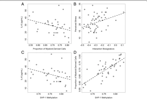
biosignature in the prediction model yielded compar-able predictions of SI (Additional file 4: Figure S3; Table 4) in GenRED Offspring saliva (AUC = 0.79; 95 % CI 0.56–1; P= 0.28), PPD cohort blood, (AUC = 0.83; 95 % CI 0.61–1; P= 0.003), and GTP subset (AUC = 0.73; 95 % CI 0.55–0.9; P= 0.99). The PPD cohort (trimester >1) showed a non-significant correl-ation between peripheral blood interleukin-6 (IL-6) levels and the predicted myeloid-derived cell proportion (Fig. 3a; rho =−0.32, P= 0.054); however, there was no correlation between IL-6 and the interaction biosignature (rho = 0.05,P= 0.80). Increased granulocyte and monocyte counts along with altered IL-6 levels may indicate increased inflammation and imply that our interaction biosignature could be an indicator of an immune state involved in sui-cide etiology [22]. The PPD cohort also showed a signifi-cant correlation between perceived stress and the interaction biosignature (Fig. 3b; rho = 0.33, P= 0.019), suggesting that the interaction biosignature may represent a biological state of both stress and inflammation.
Identification of probes driving the SKA2 model interaction biosignature
Each of the 72 probes comprising the interaction bio-signature was tested for correlation to the first eigen-vector of the PCA model across each data set to identify subsets of probes that may be driving a majority of the variation. Three probes exhibited sig-nificant correlations (P< 0.05) consistent across all co-horts (Additional file 4: Table S3): cg08469255 (DDR1), cg22029879 (ARHGEF10), and cg24437859 (SHP1). Microarray-derived DNA methylation values were vali-dated by pyrosequencing in select loci (Additional file 4: Figure S4). Methylation at cg24437859 used in place of the interaction biosignature predicted SI in GenRED Offspring saliva with an AUC of 0.77 (95 % CI 0.54–1;
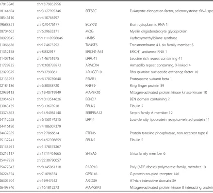
Table 3Probes from interaction biosignature contributing to the PCA model
Probe ID Location (hg19) Gene symbol Gene name
cg01060471 chr10:103911733 NOLC1 Nucleolar and coiled body phosphoprotein 1
cg01252219 chr12:110302105 GLTP Glycolipid transfer protein
cg02068690 chr2:25600451 DTNB Dystrobrevin, beta
cg02097235 chr16:1116799 SSTR5-AS1 SSTR5 antisense RNA 1
cg02246725 chr11:2014127 HOTS H19 opposite tumor suppressor
cg02340818 chr8:145808436 ARHGAP39 Rho GTPase activating protein 39
cg02464608 chr3:122631723 SEMA5B Semaphorin 5B
cg02516957 chr8:128011063
cg02953125 chr2:1079100 SNTG2 Syntrophin, gamma 2
cg03198117 chrX:152939967 PNCK Pregnancy up-regulated nonubiquitous CaM kinase
cg03351894 chrX:48686200 ERAS ES cell expressed Ras
cg03782771 chr4:152801238
cg03887528 chr2:231090531 SP140 SP140 nuclear body protein
cg06363485 chr6:41207376 TREML4 Triggering receptor expressed on myeloid cells like 4
cg06774087 chr5:79647614 CRSP8P Mediator complex subunit 27 pseudogene
cg07483709 chr10:29439573
cg07589300 chr4:1404128
cg07787977 chr1:962651 AGRN Agrin
cg08119631 chrX:118822815 SEPT6 Septin 6
cg08469255 chr6:30851069 DDR1 Discoidin domain receptor tyrosine kinase 1
cg08674415 chr16:34430905
cg08720806 chr11:125142671 PKNOX2 PBX/knotted 1 homeobox 2
cg08795752 chr9:136293271 ADAMTS13 ADAM metallopeptidase with thrombospondin type 1 motif, 13
cg09105334 chr17:15683060
cg11450546 chr19:43965012 LYPD3 LY6/PLAUR domain containing 3
cg11496113 chr5:34627766
cg11780096 chr2:178976254 RBM45 RNA binding motif protein 45
cg11880367 chr10:80141181 LINC00856
cg12378817 chr10:133961235 JAKMIP3 Janus kinase and microtubule interacting protein 3
cg12622680 chr7:158819620 LINC00689
cg12833118 chr4:2572546
cg12967319 chr14:101291997 MEG3 Maternally expressed 3 (non-protein coding)
cg13466253 chr20:3731334 HSPA12B Heat shock 70kD protein 12B
cg13798679 chr1:36617570 TRAPPC3 Trafficking protein particle complex 3
cg14439102 chr9:80360719 GNAQ Guanine nucleotide binding protein (G protein), q polypeptide
cg14509196 chr15:25494565 SNORD115-44 Small nucleolar RNA, C/D box 115-44
cg15071166 chr17:3771325 CAMKK1 Calcium/calmodulin-dependent protein kinase kinase 1, alpha
cg15302379 chr10:102821848 KAZALD1 Kazal-type serine peptidase inhibitor domain 1
cg15508401 chr2:239997380 HDAC4 Histone deacetylase 4
cg15677087 chr20:61584850 SLC17A9 Solute carrier family 17, member 9
cg15838142 chr17:77184010 RBFOX3 RNA binding protein, fox-1 homolog 3
cg16900102 chr7:98283412
cg16943635 chr11:62067691 SCGB1D4 Secretoglobin, family 1D, member 4
Table 3Probes from interaction biosignature contributing to the PCA model(Continued)
cg17813840 chr15:79852956
cg18144654 chr3:127995346 EEFSEC Eukaryotic elongation factor, selenocysteine-tRNA-specific
cg18546110 chr4:10763497
cg19688321 chrX:70476177 BCYRN1 Brain cytoplasmic RNA 1
cg20704602 chr6:29635371 MOG Myelin oligodendrocyte glycoprotein
cg20929545 chr11:118958046 HMBS Hydroxymethylbilane synthase
cg21066636 chr17:4675292 TM4SF5 Transmembrane 4 L six family member 5
cg21352158 chr8:832917 ERICH1-AS1 ERICH1 antisense RNA 1
cg21407196 chr1:46751975 LRRC41 Leucine rich repeat containing 41
cg21729235 chrX:100739272 ARMCX4 Armadillo repeat containing, X-linked 4
cg22029879 chr8:1790861 ARHGEF10 Rho guanine nucleotide exchange factor 10
cg22133973 chr6:170789640 PSMB1 Proteasome subunit beta 1
cg22184136 chr6:30038720 RNF39 Ring finger protein 39
cg22939113 chr19:40719949 MAP3K10 Mitogen-activated protein kinase kinase kinase 10
cg22954621 chr10:13514626 BEND7 BEN domain containing 7
cg23043139 chr3:13678918 FBLN2 Fibulin 2
cg23374863 chr14:94984140 SERPINA12 Serpin family A member 12
cg24112628 chr6:150174215 LRP11 Low-density lipoprotein receptor-related protein 11
cg24416190 chr4:186007379
cg24437859 chr12:7066614 PTPN6 Protein tyrosine phosphatase, non-receptor type 6
cg25132241 chr14:92396859 FBLN5 Fibulin 5
cg25133951 chr1:178575267
cg25215117 chr17:11461665 SHISA6 Shisa family member 6
cg25447359 chr22:30790057
cg25477843 chr8:145061318 PARP10 Poly (ADP-ribose) polymerase family, member 10
cg26224354 chr7:1096374 GPR146 G protein-coupled receptor 146
cg26305504 chr19:947612 ARID3A AT-rich interactive domain 3A
cg26493346 chr16:1812273 MAPK8IP3 Mitogen-activated protein kinase 8 interacting protein 3
Table 4Prediction model results
Cohort N Outcome Cases Controls Interaction AUC 95 % CI Pvalue
GenRED Offspring blood 18 Attempt 4 14 Biosignature 0.768 0.47–1 0.052
GenRED Offspring saliva 20 Ideation 5 15 Biosignature 0.807 0.59–1 0.011
PPD cohort blood 51 Ideation 8 43 Biosignature 0.884 0.75–1 0.041
GTP blood 376 Ideation 63 313 Biosignature 0.500 0.42–0.58 0.724
GTP subset blood 115 Ideation 6 109 Biosignature 0.727 0.59–0.87 0.050
GenRED Offspring blood 18 Attempt 4 14 % myeloid 0.732 0.40–1 0.28
GenRED Offspring saliva 20 Ideation 5 15 % myeloid 0.788 0.56–1 0.28
PPD cohort blood 51 Ideation 8 43 % myeloid 0.834 0.61–1 0.003
GTP subset blood 115 Ideation 6 109 % myeloid 0.728 0.55–0.9 0.99
SKA2identified in our data. This relationship was investi-gated further in the PPD cohort, where plasma cytokine levels were available. CpG methylation at cg24437859 cor-related with IL-6 levels (rho =−0.37, P= 0.035; Fig. 3c) and the predicted myeloid-derived cell proportions (rho = 0.74,P= 1.4 × 10⁻⁸; Fig. 3d) in PPD cohort blood collected during the second or third trimester.
Discussion
In this study, we used brain, saliva, and whole-blood DNA methylation data of several cohorts to derive a bio-signature of a stress state that may aid in the prediction of suicide. Using a statistically oriented approach that analyzed cross-tissue epigenetic reprogramming by cor-tisol and interaction with the previous reported SKA2 suicide biomarker, we generated an epigenetic biosigna-ture. To assess the effects of cortisol on DNA methyla-tion patterns, we performed a pathway enrichment analysis of genes with methylation significantly associ-ated with AUC weekday cortisol in the GenRED Off-spring samples, which revealed an enrichment of neural developmental pathways. This data is consistent with the
of DNA methylation in the brain, while leaving measur-able marks in the periphery that enmeasur-able the biomarker-based prediction of suicidal ideation and behaviors.
We present a biosignature that is representative of probes with a significant interaction with both SKA2 genotype and methylation in prefrontal neurons and is predictive of suicidal ideation in three cohorts. Using this biosignature in interaction with SKA2 can replace the stress questionnaire metrics used as interactive co-variates in our original suicide prediction model. We used Monte Carlo-based testing for significance by gen-erating a similar PCA eigenvector from randomly se-lected sets of 72 probes. Randomly sese-lected probes yielded predictions inferior to that of the biosignature in almost all bootstraps, suggesting that improved model prediction accuracy is not due to the underlying data structure. The biosignature performance did not reach significance by this method in saliva, possibly due to the confounding influence of buccal-derived cell types influ-encing the variation generated at biosignature loci. This interaction biosignature showed correlation with percent granulocyte and monocyte (both myeloid lineage-derived
Substituting the percentage of myeloid-derived cells for the interaction biosignature in the prediction model was successful in predicting suicidal ideation in all of the co-horts, suggesting that this interaction biosignature may be indicative of a biological state that interacts with the trait ofSKA2genotype and methylation to influence behavior.
In further efforts to reduce these 72 probes to a smaller number that would help us better understand the biology and facilitate practical implementation, we assessed the genes displaying epigenetic variation most closely mimick-ing the PCA first eigenvector. We discovered that there were three probes within the 72-probe interaction bio-signature with significant correlations to the interaction biosignature in all cohorts: cg22029879, cg08469255, and cg24437859. ARHGEF10 (rho guanine nucleotide ex-change factor 10; cg22029879) was identified as one of the 21 genes located on chromosome 8p, a region that is thought to contribute to neuropsychiatric disorders, in-cluding depression [38]. Although little evidence exists tying ARHGEF10 to suicide etiology, this locus may be worthy of further investigation due to its consistent asso-ciation with the interaction biosignature across all data sets.DDR1(discoidin domain receptor tyrosine kinase 1; cg08469255) is primarily involved in cell adhesion and extracellular matrix remodeling but also has known roles in immune and inflammatory pathways.DDR1was shown in a cell culture model to induce the expression of cyclo-oxygenase (COX2), which is involved in the synthesis of prostaglandins and has a known role in inflammation [39].
COX2also activates the NF-κB pathway, which is involved in inflammatory pathways and cytokine production [39] and has been shown as a downstream target ofDDR1 to cause infiltrating macrophages to produce chemokines. Additionally,DDR1was also shown in a mouse model of kidney obstruction to mediate the development of inflam-mation and fibrosis following kidney injury [40]. Given this evidence, DDR1 methylation could account for the correlation of myeloid-derived cell content with the inter-action biosignature and could also represent a target for further investigation.
SHP1 (protein tyrosine phosphatase, non-receptor type 6; cg24437859) has been implicated in modulating neutro-phil recruitment to inflamed tissues through modulation of the phosphoinositol pathway [41, 42] and has been shown to play an inhibitory role in cytokine-induced acti-vation of the HPA axis through the JAK-STAT pathway [43]. Furthermore,SHP1methylation correlated with IL-6 in the PPD cohort as well as the myeloid-derived cell pro-portion in all cohorts, altogether demonstrating biological evidence for the statistical interaction with HPA axis rele-vantSKA2 identified in our data. Critically, IL-6 contrib-utes to hematopoietic stem cell fate decisions and helps to differentiate myeloid from non-myeloid cells [42, 44]. As such, the possibility remains that epigenetic variation in
genes likeSHP1may be important not only for differenti-ation of hematopoietic stem cells into myeloid cells but for regulation of pro-inflammatory cytokines and may moder-ate the influence of pro-inflammatory cytokines on HPA axis activity. This supposition is supported by data demon-strating thatSKA2interaction biosignature data interacted with CTQ scores to predict HPA axis responsivity in two stress challenges from multiple data sets. The relationship between the interaction biosignature signals and HPA axis sensitivity is very similar to previously reported findings related to the influence of SKA2 DNA methylation on HPA axis activity from the same cohorts [8, 9].
observed in many studies showing increased levels of C-reactive protein and IL-6 in the blood of both mili-tary and non-milimili-tary cohorts [30–35]. The relationship betweenSKA2 methylation and PTSD should be studied further to better understand the impact on suicidal behaviors.
This study has many limitations. Sodium bisulfite modification cannot distinguish between 5-methyl cyto-sine (5-MC) and 5-hydroxy methyl cytocyto-sine (5-HMC) levels. Like 5-MC, 5-HMC can vary in the brain in re-sponse to stress [47, 48] and has been identified in vari-ous psychiatric disorders [49, 50]. Brain tissue analyses have the potential to be confounded by postmortem factors such as the method and timing of tissue preser-vation. Psychiatric diseases can often be co-morbid with other illnesses such as cancer and heart disease, among others [51, 52]. Despite the implication that inflamma-tory factors may be interacting with SKA2, we did not assess for the health status of the study subjects and any potential impact this might have on our results. This study is limited by using small samples that are not representative of the general population and are biased towards controls due to a low ratio of cases to controls and only validated findings in a single independent sam-ple in which suicidal behavior is only predicted in small subsets. Ideally, these findings would be further validated in a large sample that is more representative of the gen-eral population to prove its usefulness in prediction of suicidal behavior.
Conclusions
We present a biosignature that predicts suicide consist-ently across multiple, highly variable data sets, specific-ally youth at high risk for depression, pregnant women at high risk for PPD, and middle-aged urban population with high incidence of trauma and PTSD. This biosigna-ture is cross-tissue in that it predicts suicidal behavior in both blood and saliva samples and is based on probes that are associated with suicide in prefrontal neurons. To our knowledge, this is the first prediction model to date that works in both blood and saliva and the first suicide prediction model to use only DNA methylation to predict suicidal behavior. Additionally, correlations of the interaction biosignature with myeloid proportion and stress metrics may indicate a fuller integration of suicide etiology into the existingSKA2suicide prediction model. Finally, this biosignature allows us to predict suicidal behavior without using a stress questionnaire or assessment. Although the biosignature produces lower prediction AUCs than the stress questionnaires, it repre-sents a single measure that allows us to predict suicidal behavior across all data sets, providing consistency and better allowing for comparison across populations. Ultimately, this work will add to the development of
early diagnostics tests that may aid in the early identifi-cation and prevention of suicide.
Methods Human samples
Peripheral blood and saliva samples were obtained from separate individuals in the GenRED Offspring cohort from Johns Hopkins [4, 53–56]. Postmortem prefrontal cortex neurons (cases,N= 22; controls,N= 23) were ob-tained as previously described [4], data available at NCBI Gene Expression Omnibus (GEO) accession GSE41826. Peripheral blood samples (cases,N= 8; controls,N= 43) were obtained from consenting individuals in a Johns Hopkins prospective cohort of pregnant women (PPD cohort), as previously described [57], data available from GEO accession GSE44132. Data from individuals in the Grady Trauma Project (cases, N= 63; controls,
N= 313) were downloaded from the NCBI GEO ac-cession GSE72680 [53–56]. Data on TRIER social stress test cohort (N= 85) was downloaded from GEO accession GSE77445 [20]. All cohorts used in model generation and validation are described in detail in Additional file 4: Table S1.
DNA methylation analysis
Study data was derived from genome-wide DNA methy-lation data using the Infinium HumanMethymethy-lation450 BeadChip Array (Illumina Inc., San Diego, CA). DNA methylation profiles for GenRED Offspring cohort blood (cases, N= 4; controls, N= 14) and saliva (cases,
N= 5; controls, N= 15), respectively, were generated as described below.
Infinium chip assay
Data acquisition
Data were extracted using Methylation Module of Geno-meStudio v1.0 Software and processed using the “minfi” and “wateRmelon”packages in R [58, 59]. Raw data was trimmed of probes failing quality assessment, followed by scale-based data correction for Illumina type I relative to type II probes. Methylated and un-methylated inten-sity values were then quantile normalized separately prior to the calculation of the β (beta) value based on the following definition:
βvalue = (signal intensity of methylation-detection probe)/(signal intensity of methylation−detection probe + signal intensity of non-methylation-detection probe + 100).
Sodium bisulfite pyrosequencing
Microarray data was validated at select probes in the GenRED Offspring saliva cohort to corroborate array data (Additional file 4: Figure S4). Bisulfite conversion was carried out using EZ DNA Methylation-Gold Kit (Zymo Research, Irvine, CA) according to the manufac-turer’s instructions on N= 51 subjects from the Johns Hopkins Prospective cohort. Nested PCR amplifications were performed with a standard PCR protocol in 25 μl volume reactions containing 3–4 μl of sodium bisulfite-treated DNA, 0.2 μM primers, and master mix contain-ing Taq DNA polymerase (Sigma-Aldrich, St. Louis, MO). Primer sequences can be found in Additional file 4: Table S2. PCR amplicons were processed for pyrose-quencing analysis according to the manufacturer’s stand-ard protocol using a PyroMark Q96 MD system (QIAGEN, Germantown, MD) with Pyro Q-CpG 1.0.9 software (QIAGEN) for CpG methylation quantification. Only data passing internal quality checks for sodium bisulfite conversion efficiency, signal-to-noise ratio, and the observed versus expected match of the predicted pyrogram peak profile using reference peaks were incor-porated in subsequent analyses. Data generated derive from one technical replicate.
Blood analysis in PPD cohort
Participant blood was collected at each visit in four 10-ml EDTA tubes. Blood samples were non-fasting, and collection times were arranged at the convenience of the participant. All occurred during the working day (9:00 am to 5:00 pm). Cytokine levels have a recognized circa-dian rhythm but are lowest during the daytime; we were unable to control further for time of day in our analyses [60]. Samples were immediately centrifuged at 4 °C for 30 min. The plasma was then aliquoted in 2-mL micro-centrifuge tubes, snap frozen on dry ice, and immedi-ately stored in a−80 °C freezer. Cytokines were analyzed using BD Cytometric Bead Array. Plasma samples from
patients were diluted 1:10 and incubated with capture beads coated with antibodies specific for IL-6. The beads were then incubated with a phycoerythrin-conjugated detection reagent containing antibodies specific to each capture bead. The capture bead + analyte + detection re-agent complexes produced an individual fluorescent signal for each cytokine and were acquired on a FACSCalibur instrument. The data were analyzed using FCAP Array software. The limit of detection for IL-6 was 1.6 pg/mL. Proportions of our samples that fell below the limit of detection were as follows: IL-6, 18.9 %. Samples that fell below the limit of detection were coded“0.” All samples were run in duplicate, and the coefficient of variation between samples was <10 %. Analyses were repeated using the lowest detectable dose for those below the limit of detection, and results did not change.
Statistical analysis
All statistical tests were performed in R (https://www.r-project.org/). Cross-reactive Illumina probes were re-moved from data prior to further analysis [61]. Using an Anderson-Darling test from the “nortest” package [62], all distributions of data that rejected the null hypothesis of normality were subsequently evaluated with non-parametric tests. Variance between case and control groups in each sample was assumed to be equal. All stat-istical tests performed were two tailed, and a P< 0.05 was considered significant. All statistics presented are a result of either a linear regression model or a Monte Carlo method permutation test (1000 permutations). Unless otherwise specified, ± denotes the standard de-viation of the mean. Cell sub-fraction percentages were quantified for CD8 T cells, CD4 T cells, B cells, NK cells, monocytes, and granulocytes using an algorithm designed by Houseman et al. for the quantification of the cell types using DNA methylation proxies [63]. A buccal cell epigenetic profile was derived by taking the mean of N= 109 buccal-derived HM450 microarray profiles from a data set in GEO accession GSE25892. Incorporation of the buccal-derived profile at N= 500 HM450 loci into the Houseman algorithm generated training set, and incorporation of a buccal covariate was used to retrain the Houseman algorithm to quan-tify buccal profiles.
Additional files
Additional file 1:Title: GenRED Offspring Blood Cortisol Associated Probes. Description: Table containing all probes nominally associated with AUC weekday cortisol in the GenRED Offspring blood cohort (N = 18) with the corresponding regression coefficient (β) andp-value. (XLSX 860 kb)
Additional file 3:Title: Combined GenRED Offspring Blood & Saliva Cortisol Associated Probes. Description: Table containing all probes nominally associated with AUC weekday cortisol in the combined GenRED Offspring blood and saliva cohorts (N= 38) with the corresponding regression coefficient (β) andp-value. (XLSX 1086 kb)
Additional file 4:Supplementary Data. Description: Contains supplementary results, figures, and tables. (DOCX 2399 kb)
Abbreviations
ARHGEF10:Rho guanine nucleotide exchange factor 10; AUC: Area under the curve; CNS: Central nervous system; CpG: Cytosine-guanine dinucleotide; CTQ: Childhood Trauma Questionnaire; DDR1: Discoidin domain receptor tyrosine kinase 1; DST: Dexamethasone suppression test; GenRED: Genetics of Recurrent Early-Onset Depression; GEO: NCBI Gene Expression Omnibus; GR: Glucocorticoid receptor; GTP: Grady Trauma Project; HM450: Infinium HumanMethylation450 BeadChip array; HPA axis: Hypothalamic-pituitary-adrenal axis; IL-6: Interleukin 6; PCA: Principal component analysis; PTSD: Post-traumatic stress disorder; SA: Suicidal attempt; SCARED: Self-Report for Childhood Anxiety Related Disorders; SHP1: Protein tyrosine phosphatase, non-receptor type 6; SI: Suicidal ideation; SKA2: Spindle- and kinetochore-associated protein 2; SNP: Single nucleotide polymorphism
Acknowledgements
We would like to thank the Solomon R. and Rebecca D. Baker Foundation and the James Wah Award for Mood Disorders for their generous support of this research.
Funding
This work was funded in part by NIMH 1R21MH094771-01 and a Russell Military Scholars Award to ZAK from the Johns Hopkins Military and Veterans Health Institute and an NIEHS Training Grant ES07141 to MLC.
Availability of data and materials
Numerous data sets supporting the findings in this study are available at the Gene Expression Omnibus under accession numbers GSE41826, GSE44132, GSE72680, and GSE77445. Other supporting data are available from the authors upon reasonable request.
Authors’contributions
MLC and ZAK generated, analyzed, and interpreted the data and prepared the manuscript. MPB, CHV, LMO, JLP, KJR, AKS, and HCW provided the data and assisted in data analysis and manuscript preparation. All authors read and approved the final manuscript.
Competing interests
Dr. Kaminsky is listed on patents for use of epigenetic information at the SKA2locus to predict suicidal behavior and PTSD and received consultant fees from Janssen Research and Development, LLC. Dr. Kaminsky is a co-inventor listed on a patent for DNA methylation at biomarker loci related to PPD. Under a former option agreement between Physician’s Choice Laboratory Services and the Johns Hopkins University, the University was entitled to fees associated with a PPD biomarker invention mentioned in this article. Dr. Kaminsky was also an unpaid speaker to Physician’s Choice Laboratory Services. This arrangement has been reviewed and approved by the Johns Hopkins University in accordance with its conflict of interest policies. All other authors declare no biomedical financial interests or potential conflicts of interest.
Consent for publication Not applicable.
Ethics approval and consent to participate
All research was performed in accordance with the 1975 Declaration of Helsinki. Human subject research for the GenRED Offspring cohort, prospective cohort, and PRC cohort was conducted under IRB protocol # 00015387, # 00008149, and # 000000354, and subjects were collected with funding from the American Foundation for Suicide Prevention to H.C.W., NIDA R01DA09897 to W.W.E., and NIMH K23 MH074799-01A2 to J.P. Human tissue was obtained from the NICHD Brain and Tissue Bank for Developmental Disorders and the University of Maryland, Baltimore, MD.
Author details
1Department of Psychiatry and Behavioral Sciences, Johns Hopkins University
School of Medicine, Baltimore, MD 21205, USA.2Department of Psychiatry,
Brain Center Rudolf Magnus, University Medical Center Utrecht (UMCU), Utrecht, The Netherlands.3Department of Psychiatry, McLean Hospital,
Harvard Medical School, Boston, MA 02478, USA.4Department of Psychiatry
and Behavioral Sciences, Emory University School of Medicine, Atlanta, GA 30322, USA.5Howard Hughes Medical Institute, Chevy Chase, MD, USA.
6Department of Mental Health, Johns Hopkins Bloomberg School of Public
Health, Baltimore, MD 21218, USA.7The Mood Disorder Center, Johns
Hopkins University, 720 Rutland Avenue, Ross Research Building 1070, Baltimore, MD 21205, USA.
Received: 24 August 2016 Accepted: 20 October 2016
References
1. Suicide Data [online]. World Health Organization. 2016. Available from URL: http://www.who.int/mental_health/prevention/suicide/suicideprevent/en/. Accessed 30 June 2016.
2. Web-based Injury Statistics Query and Reporting System (WISQARS) [online]. Centers for Disease Control and Prevention: National Center for Injury Prevention and Control. 2016. Available from URL: http://www.cdc.gov/ injury/wisqars. Accessed 30 June 2016.
3. Pringle B, Colpe LJ, Heinssen RK, Schoenbaum M, Sherrill JT, Claassen CA, et al. A strategic approach for prioritizing research and action to prevent suicide. Psychiatr Serv. 2013;64(1):71–5.
4. Guintivano J, Brown T, Newcomer A, Jones M, Cox O, Maher BS, et al. Identification and replication of a combined epigenetic and genetic biomarker predicting suicide and suicidal behaviors. Am J Psychiatry. 2014;171(12):1287–96.
5. Niculescu AB, Levey D, Le-Niculescu H, Niculescu E, Kurian SM, Salomon D. Psychiatric blood biomarkers: avoiding jumping to premature negative or positive conclusions. Mol Psychiatry. 2015;20:286–8.
6. Pandey GN, Rizavi HS, Zhang H, Bhaumik R, Ren X. The expression of the suicide-associated gene SKA2 is decreased in the prefrontal cortex of suicide victims but not of nonsuicidal patients. Int J
Neuropsychopharmacology. 2016;19(8):1–10.
7. Rice L, Charlotte E, Eccles J, Garside H, Sommer P, Kay P, Blackhall FH, et al. Identification and functional analysis of SKA2 interaction with the glucocorticoid receptor. J Endocrinol. 2008;198:499–509.
8. Boks MP, Rutten BP, Geuze E, Houtepen LC, Vermetten E, Kaminsky Z, et al. SKA2 methylation is involved in cortisol stress reactivity and predicts the development of posttraumatic stress disorder (PTSD) after military deployment. Neuropsychopharmacology. 2015;41(5):1350–6.
9. Kaminsky Z, Wilcox HC, Eaton WW, Van Eck K, Kilaru V, Jovanovic T, et al. Epigenetic and genetic variation at SKA2 predict suicidal behavior and post-traumatic stress disorder. Transl Psychiatry. 2015;5:e627.
10. Chung KC, Springer I, Kogler L, Turetsky B, Freiherr J, Derntl B. The influence of androstadienone during psychosocial stress is modulated by gender, trait anxiety and subjective stress: an fMRI study. Psychoneuroendocrinology. 2016;68:126–39.
11. Niculescu AB, Levey DF, Phalen PL, Le-Niculescu H, Dainton HD, Jain N, et al. Understanding and predicting suicidality using a combined genomic and clinical risk assessment approach. Mol Psychiatry. 2015;20(11):1266–85. 12. Sadeh N, Spielberg JM, Logue MW, Wolf EJ, Smith AK, Lusk J, et al. SKA2
methylation is associated with decreased prefrontal cortical thickness and greater PTSD severity among trauma-exposed veterans. Mol Psychiatry. 2016;21(3):357–63.
13. Sadeh N, Wolf EJ, Logue MW, Hayes JP, Stone A, Griffin LM, et al. Epigenetic variation at Ska2 predicts suicide phenotypes and internalizing psychopathology. Depress Anxiety. 2016;33(4):308–15.
14. Levey DF, Niculescu EM, Le-Niculescu H, Dainton HL, Phalen PL, Ladd TB, et al. Towards understanding and predicting suicidality in women: biomarkers and clinical risk assessment. Mol Psychiatry. 2016;21(6):768–85.
15. Guintivano J, Kaminsky ZA. Role of epigenetic factors in the development of mental illness throughout life. Neurosci Res. 2016;102:56–66.
17. Osborne L, Clive M, Kimmel M, Gispen F, Guintivano J, Brown T, et al. Replication of epigenetic postpartum depression biomarkers and variation with hormone levels. Neuropsychopharmacology. 2016;41(6):1648–58. 18. Reimand J, Kull M, Peterson H, Hansen J, Vilo J. g:Profiler—a web-based
toolset for functional profiling of gene lists from large-scale experiments. Nucleic Acids Res. 2007;35(Web Server issue):W193–200.
19. Reimand J, Arak T, Vilo J. g:Profiler—a web server for functional interpretation of gene lists (2011 update). Nucleic Acids Res. 2011;39(suppl 2):W307–15.
20. Houtepen LC, Vinkers CH, Carrillo-Roa T, Hiemstra M, van Lier PA, Meeus W, et al. Genome-wide DNA methylation levels and altered cortisol stress reactivity following childhood trauma in humans. Nat Commun. 2016;7:10967. 21. Boks MP, Rutten BP, Geuze E, Houtepen LC, Vermetten E, Kaminsky Z, et al. SKA2 methylation is Involved in cortisol stress reactivity and predicts the development of posttraumatic stress disorder (PTSD) After Military Deployment. Neuropsychopharmacology. 2015;41(5):1350–6.
22. Rosenblat JD, Cha DS, Mansur RB, McIntyre RS. Inflamed moods: a review of the interactions between inflammation and mood disorders. Prog Neuro-Psychopharmacol Biol Psychiatry. 2014;53:23–34.
23. Naninck EFG, Hoeijmakers L, Kakava-Georgiadou N, Meesters A, Lazic SE, Lucassen PJ, et al. Chronic early life stress alters developmental and adult neurogenesis and impairs cognitive function in mice. Hippocampus. 2015;25(3):309–28.
24. Bockmühl Y, Patchev AV, Madejska A, Hoffmann A, Sousa JC, Sousa N, et al. Methylation at the CpG island shore region upregulates Nr3c1 promoter activity after early-life stress. Epigenetics. 2015;10(3):247–57.
25. Hsiao Y-M, Tsai T-C, Lin Y-T, Chen C-C, Huang C-C, Hsu K-S. Early life stress dampens stress responsiveness in adolescence: evaluation of neuroendocrine reactivity and coping behavior. Psychoneuroendocrinology. 2016;67:86–99. 26. Baes CW, Martins CMS, Tofoli SMC, Juruena MF. Early life stress in depressive
patients: HPA axis response to GR and MR agonist. Front Psychiatry. 2014;5:2. 27. Lindqvist D, Janelidze S, Hagell P, Erhardt S, Samuelsson M, Lennart M, et al.
Interleukin-6 is elevated in the cerebrospinal fluid of suicide attempters and related to symptom severity. Biol Psychiatry. 2009;66(3):287–92.
28. Tonelli LH, Stiller J, Rujescu D, Giegling I, Schneider B, Maurer K, et al. Elevated cytokine expression in the orbitoprefrontal cortex of victims of suicide. Acta Psychiatr Scand. 2008;117(3):198–206.
29. Pandey GN, Rizavi HS, Ren X, Fareen J, Hoppensteadt DA, Roberts RC, et al. Proinflammatory cytokines in the prefrontal cortex of teenage suicide victims. J Psychiatr Res. 2012;46(1):57–63.
30. Gill J, Lee H, Barr T, Baxter T, Heinzelmann M, Rak H, et al. Lower health related quality of life in U.S. military personnel is associated with service-related disorders and inflammation. Psychiatry Res. 2014;216(1):116–22. 31. O’Donovan A, Chao LL, Paulson J, Samuelson KW, Shigenaga JK, Grunfeld C,
et al. Altered inflammatory activity associated with reduced hippocampal volume and more severe posttraumatic stress symptoms in Gulf War veterans. Psychoneuroendocrinology. 2015;51:557–66.
32. Heath NM, Chesney SA, Gerhart JI, Goldsmith RE, Luborsky JL, Stevens NR, et al. Interpersonal violence, PTSD, and inflammation: potential psychogenic pathways to higher C-reactive protein levels. Cytokine. 2013;63(2):172–8.
33. Michopoulos V, Rothbaum AO, Jovanovic T, Almli LM, Bradley B, Rothbaum BO, et al. Association of CRP genetic variation and CRP level with elevated PTSD symptoms and physiological responses in a civilian population with high levels of trauma. Am J Psychiatry. 2014;172(4):353–62.
34. Eraly SA, Nievergelt CM, Maihofer AX, et al. Assessment of plasma c-reactive protein as a biomarker of posttraumatic stress disorder risk. JAMA Psychiatry. 2014;71(4):423–31.
35. Plantinga L, Bremner JD, Miller AH, Jones DP, Veledar E, Goldberg J, et al. Association between posttraumatic stress disorder and inflammation: a twin study. Brain Behav Immun. 2013;30:125–32.
36. Gladkevich A, Kauffman HF, Korf J. Lymphocytes as a neural probe: potential for studying psychiatric disorders. Prog Neuropsychopharmacol Biol Psychiatry. 2004;28:559–76.
37. Pandey GN. Biological basis of suicide and suicidal behavior. Bipolar Disord. 2013;15:524–41.
38. Tabares-Seisdedos R, Rubenstein J. Chromosome 8p as a potential hub for developmental neuropsychiatric disorders: implications for schizophrenia, autism and cancer. Mol Psychiatry. 2009;14:563–89.
39. Das S, Ongusaha PP, Yang YS, Park J-M, Aaronson SA, Lee SW. Discoidin domain receptor 1 receptor tyrosine kinase induces cyclooxygenase-2 and
promotes chemoresistance through nuclear factor-κB pathway activation. Cancer Res. 2006;66(16):8123–30.
40. Guerrot D, Kerroch M, Placier S, Vandermeersch S, Trivin C, Mael-Ainin M, et al. Discoidin domain receptor 1 is a major mediator of inflammation and fibrosis in obstructive nephropathy. Am J Pathol. 2011;179(1):83–91. 41. Stadtmann A, Block H, Volmering S, Abram C, Sohlbach C, Boras M, et al.
Cross-talk between Shp1 and PIPKIgamma controls leukocyte recruitment. J Immunol. 2015;195(3):1152–61.
42. Watson NB, Schneider KM, Massa PT. SHP-1-dependent macrophage differentiation exacerbates virus-induced myositis. J Immunol. 2015;194(6):2796–809.
43. Bousquet C, Susini C, Melmed S. Inhibitory roles for SHP-1 and SOCS-3 following pituitary proopiomelanocortin induction by leukemia inhibitory factor. J Clin Invest. 1999;104(9):1277–85.
44. Lodish H. Molecular cell biology. 5th ed. New York: W. H. Freeman and Co; 2003. 45. de Kloet CS, Vermetten E, Geuze E, Kavelaars A, Heijnen CJ, Westenberg
HGM. Assessment of HPA-axis function in posttraumatic stress disorder: pharmacological and non-pharmacological challenge tests, a review. J Psychiatr Res. 2006;40(6):550–67.
46. van Zuiden M, Kavelaars A, Vermetten E, Olff M, Geuze E, Heijnen C. Pre-deployment differences in glucocorticoid sensitivity of leukocytes in soldiers developing symptoms of PTSD, depression or fatigue persist after return from military deployment. Psychoneuroendocrinology. 2015;51:513–24. 47. Papale LA, Li S, Madrid A, Zhang Q, Chen L, Chopra P, et al. Sex-specific
hippocampal 5-hydroxymethylcytosine is disrupted in response to acute stress. Neurobiol Dis. 2016;96:54–66.
48. Li S, Papale LA, Zhang Q, Madrid A, Chen L, Chopra P, et al. Genome-wide alterations in hippocampal 5-hydroxymethylcytosine links plasticity genes to acute stress. Neurobiol Dis. 2016;86:99–108.
49. Dong E, Gavin DP, Chen Y, Davis J. Upregulation of TET1 and downregulation of APOBEC3A and APOBEC3C in the parietal cortex of psychotic patients. Transl Psychiatry. 2012;2:e159.
50. Tseng PT, Lin PY, Lee Y, Hung CF, Lung FW, Chen CS, et al. Age-associated decrease in global DNA methylation in patients with major depression. Neuropsychiatr Dis Treat. 2014;10:2105–14.
51. Kang HJ, Kim SY, Bae KY, Kim SW, Shin IS, Yoon JS, et al. Comorbidity of depression with physical disorders: research and clinical implications. Chonnam Med J. 2015;51(1):8–18.
52. Kapfhammer HP. Comorbid depressive and anxiety disorders in patients with cancer. Nervenarzt. 2015;86(3):291–292, 294–298, 300–291. 53. Edgar R, Domrachev M, Lash AE. Gene Expression Omnibus: NCBI gene
expression and hybridization array data repository. Nucleic Acids Res. 2002;30(1):207–10.
54. Gillespie CF, Bradley B, Mercer K, Smith AK, Conneely K, Gapen M, et al. Trauma exposure and stress-related disorders in inner city primary care patients. Gen Hosp Psychiatry. 2009;31(6):505–14.
55. Ressler KJ, Mercer KB, Bradley B, Jovanovic T, Mahan A, Kerley K, et al. Post-traumatic stress disorder is associated with PACAP and the PAC1 receptor. Nature. 2011;470(7335):492–7.
56. Binder EB, Bradley RG, Liu W, Epstein MP, Deveau TC, Mercer KB, et al. Association of FKBP5 polymorphisms and childhood abuse with risk of posttraumatic stress disorder symptoms in adults. JAMA. 2008;299(11):1291–305.
57. Guintivano J, Arad M, Gould TD, Payne JL, Kaminsky ZA. Antenatal prediction of postpartum depression with blood DNA methylation biomarkers. Mol Psychiatry. 2014;19:560–7.
58. Aryee MJ, Jaffe AE, Corrada-Bravo H, Ladd-Acosta C, Feinberg AP, Hansen KD, et al. Minfi: a flexible and comprehensive Bioconductor package for the analysis of Infinium DNA methylation microarrays. Bioinformatics. 2014;30(10):1363–9.
59. Pidsley R, Wong CCY, Volta M, Lunnon K, Mill J, Schalkwyk LC. A data-driven approach to preprocessing Illumina 450K methylation array data. BMC Genomics. 2013;14:293.
60. Dinarello CA. Proinflammatory cytokines. Chest. 2000;118(2):503–8. 61. Y-a C, Lemire M, Choufani S, Butcher DT, Grafodatskaya D, Zanke BW, et al.
Discovery of cross-reactive probes and polymorphic CpGs in the Illumina Infinium HumanMethylation450 microarray. Epigenetics. 2013;8(2):203–9. 62. Gross J, Ligges U. nortest: tests for normality. R package version 1.0-4 edn. 2015. 63. Houseman EA, Accomando WP, Koestler DC, Christensen BC, Marsit CJ,