R E S E A R C H
Open Access
Evaluation of intraarterial and intravenous
cisplatin chemotherapy in the treatment of
metastatic osteosarcoma using an
orthotopic xenograft mouse model
Bernhard Robl
1, Sander Martijn Botter
1, Giovanni Pellegrini
2, Olga Neklyudova
1and Bruno Fuchs
1*Abstract
Background:Osteosarcoma is the most common primary malignancy of bone. Its treatment relies on the administration of neoadjuvant and adjuvant chemotherapy combined with surgery. Alternative to common intravenous (i.v.) administration of chemotherapeutic drugs, clinical studies also evaluated the benefit of intraarterial (i.a.) administrations. However, conflicting results were obtained when both routes of administration of cisplatin (CDDP), a gold standard drug in osteosarcoma treatment, were compared. In order to overcome clinical confounding factors, we evaluated both routes of drug administration in a mouse model of experimental osteosarcoma.
Methods:We directly compared i.v. versus i.a. drug infusions of cisplatin (CDDP), in an orthotopic xenograft mouse model of metastatic osteosarcoma. We performed tumor monitoring using caliper and micro computed
tomography and measured tumor perfusion using laser speckle contrast imaging. Histopathological changes were evaluated using hematoxylin and eosin staining as well as immunohistochemistry (cleaved PARP-1, CD31, HIF-1α). Results:First, an effective concentration of 4 mg/kg i.a. CDDP was determined that significantly reduced primary tumor volume. We used this concentration of i.a. CDDP and compared it to infusions of i.v. CDDP. Systemic (i.v.) CDDP only showed minor suppression of tumor growth whereas local (i.a.) CDDP strongly inhibited tumor growth and destruction of cortical bone in the tumor-bearing hind limb. Inhibition of tumor growth was linked to a reduced blood perfusion and resulted in increased amounts of tumor necrosis after i.a. CDDP. After treatment with i.a. CDDP, remaining viable tumor tissue responded by increasing expression of HIF-1α. Side effects due to
administration of CDDP were minor, showing no differences in kidney damage between i.v. and i.a. CDDP. However, increased epidermal apoptosis in the foot was an indirect marker for locally increased concentrations of CDDP. Conclusions:Our findings demonstrate the great potential of local administration of cytotoxic chemotherapeutics, such as CDDP. Consequently, we provide a preclinical basis for a renewed interest in the clinical use of i.a.
chemotherapy in osteosarcoma therapy.
Keywords:Intraarterial, Cisplatin, Osteosarcoma, Intravenous, Metastasis
* Correspondence:research@balgrist.ch
1Laboratory for Orthopedic Research, Department of Orthopedics, Balgrist
University Hospital, Forchstrasse 340, Zurich 8008, Switzerland Full list of author information is available at the end of the article
Background
Bone cancers are among the deadliest cancers in adolescents, with osteosarcoma as its most common representative [1, 2]. Subsequent to the introduction of chemotherapy in the early 1970s, 5-year survival rates of osteosarcoma patients with localized disease increased from 20 % to above 60 % [1, 3]. Standard of care for osteosarcoma patients currently includes systemic, intravenous (i.v.) administration of neoad-juvant chemotherapy, combined with surgical resec-tion of the primary tumor, followed by adjuvant chemotherapy. However, 5-year survival rates plat-eaued at 60 % and survival rates of patients with metastatic disease remained unchanged at a low 20–30 % until today [3].
In osteosarcoma treatment, the use of neoadjuvant chemotherapy is considered valuable, yet without dir-ectly improving event-free survival compared to immedi-ate surgery [4]. Especially pathologic analysis of the tumor response has important prognostic value. Good responders (i.e. 90 % or greater tumor necrosis) achieve up to two times better survival rates compared to pa-tients with poor histologic responses [5–7]. In addition, neoadjuvant chemotherapy reduces tumor volumes prior to surgical resection, facilitating limb sparing procedures [8]. Therefore, neoadjuvant chemotherapy with highly cytotoxic drugs in osteosarcoma is commonly accepted as standard of care. One of the chemotherapeutics always included in today’s osteosarcoma treatment regimens is cisplatin (Cis-diamminedichloroplatinum, CDDP). However, its use is limited by systemic toxic-ities such as ototoxicity and nephrotoxicity [9, 10].
Therefore, local, yet controlled application of CDDP may be advantageous. One way to achieve this goal is by local drug administration via the tumor feeding artery. Such intraarterial (i.a.) drug administrations were already successfully performed in the 1980s. These studies con-firmed a higher bioavailability of the drug after i.a. infu-sion of CDDP [11–13], explaining superior histological response rates in osteosarcoma [14–16]. For instance, Wilkins et al. achieved a good response in 87 % of the patients if patients with localized osteosarcomas were treated with i.a. CDDP and i.v. doxorubicin [14], com-pared to only 41 % in similar patient cohorts where i.v. CDDP and i.v. doxorubicin were used [17], and 71 % in case of a three/four-drug regimen comprising metho-trexate [6, 17–21]. In addition to better response rates, studies from the St. Luke’s Medical Center achieved 10-year survival rates of between 82 and 93 % using i.a. CDDP [14, 22]. These survival rates compare favorably to other studies with maximum 10-year survival rates of, at best, 64 % with an i.v. two-drug regimen [17, 23] or up to 70 % with an i.v. three/four-drug regimen [3, 24]. Similarly, canine osteosarcoma patients showed superior
responses when CDDP was infused via the tumor feed-ing artery compared to i.v. infusions [25].
Although these results demonstrate a clear added value of i.a. CDDP in osteosarcoma treatment, a clinical trial comparing both routes of CDDP administration was unable to show a benefit of i.a. chemotherapy [10]. This discrepancy might be explained by the design of the study, dose adaptations, administration of multiple drugs (standard of care) or its multi-institutional ap-proach. In another study comparing i.a. versus i.v. CDDP, superior tumor responses with i.a. CDDP were only seen in the context of a three-drug regimen, and not as part of a four-drug regimen [20]. However, tumor response rates with the three-drug regimen comprising i.a. (77 %) were similar to the rates found with a four-drug regimen (81 %).
In summary, these studies demonstrate the difficulty of evaluating the“true”efficacy of i.a. CDDP due to con-founding factors such as administration of different combinations of chemotherapeutics and the large differ-ence in reported survival rates (between 50 % and 71% already for i.v. CDDP) per treatment center [6, 26]. In addition, tumor heterogeneity and side effect manage-ment make it difficult to reliably interpret the results of trials comparing both methods in a clinical setting. In this study, encouraged by the initial promising clinical benefits of i.a. chemotherapy, we investigated, under ex-perimentally controlled conditions, the effects of local (i.a.) versus systemic (i.v.) CDDP in a preclinical mouse model of osteosarcoma.
Methods
Cell culture and transduction
Human OS 143B cells (CRL-8303) were obtained from American Type Culture Collection (ATCC, USA) and cultured in DMEM (4.5 g/L glucose)/HamF12 (1:1) medium (Invitrogen, USA) supplemented with 10 % heat-inactivated fetal calf serum at 37 °C in a humidified atmosphere containing 5 % CO2. Previously, 143B cells
were transduced with the LacZ gene [27]. In this study, 143B/LacZ cells were additionally infected with retro-viral particles containing the mCherry sequence integrated into a pQCIXH backbone, similar as de-scribed elsewhere [28]. The original mCherry-containing pcDNA3.1 plasmid was a kind gift from Prof. M. Rudin (Institute of Biomedical Engineering, University and ETH Zurich). After transduction, 143B cells were se-lected in tissue culture medium with 1200 μg/ml of G418 (Merck, Germany) and 400 μg/ml of hygromycin (Merck, Germany) to stably express LacZ and mCherry.
Animal care
Germany) were maintained in enriched individually venti-lated cages with light/dark-cycles of 12 h/12 h. After deliv-ery, animals were kept for at least a week without any interventions. Food and water was provided to the mice ad libitum. Animal care and experimental procedures were in accordance with the institutional guidelines and approved by the Ethics Committee of the Veterinary Department, Canton of Zurich, Switzerland (License Number 64/2013).
Orthotopic tumor induction in mice
143B cells were grown to subconfluence, detached with Trypsin/PBS/0.05 % EDTA, resuspended in PBS/0.05 % EDTA and kept on ice until injected. Before tumor cell injections (TCIs), mice were anesthetized using injection anesthesia. TCIs into left hind limbs were performed similar to as described elsewhere [29]. Briefly, holes were pre-drilled into the medullar cavity of left tibias using sterile needles, before 105 143B cells were injected. After TCIs, mice were monitored weekly for development of primary tumors (see below). Once mice started limping due to the tumor burden, 0.1 mg/kg of intraperitoneal (i.p.) Buprenor-phine (Temgesic; Reckitt Benckiser, UK) was given twice daily. At the end of the study, mice were sacrificed and lung metastases were counted as described [28].
Tumor monitoring Primary tumor monitoring
After TCIs, mice were monitored weekly using caliper and fluorescence measurements, similar as described [28]. Once human 143B osteosarcoma cells established measureable primary tumors (unambigous mCherry sig-nal and a volume greater than 25 mm3), drug treatment was started. mCherry tumor fluorescence was measured using an IVIS Lumina XR imaging system (Caliper Life Sciences, Inc., USA) and quantified with Living Image v3.1 software (Xenogen Corporation, USA).
Micro computed tomography
Micro computed tomography (microCT) using a Sky-Scan1176 microCT system (SkyScan/Bruker, Billerica, USA) equipped with a 0.5 mm aluminum filter was con-ducted to yield high-resolution tomographs of mouse hind limbs. Scans were obtained from each animal at the end of the study at a working source voltage of 50 kV and a source current of 500 μA yielding a final image pixel size of 17.7 μm. Frame averaging of three and ex-posure times of 210 ms per projection were set. Each shot required a source rotation step of 0.7° yielding scan times of approximately 8 min per mouse. Post-acquisition three-dimensional image reconstitution was done in NRecon software v1.6.9.18 (Skyscan/Bruker, USA). Reconstituted images were segmented and bone volumes were calculated using CTAn v1.13.11.0 (Sky-scan/Bruker, USA). For calculation of bone and tumor
volumes, the region between the distal end of the patella (“start of selection”) and the bifurcation of tibia and fib-ula (“end of selection”) was used. Bone volumes were calculated using the following formula: Δcortical bone volume = bone volumetumor-limb- bone volumehealthy-limb.
Three-dimensional images of the mouse tibias were made in Ctvox v.2.7.0 (Skyscan/Bruker, USA).
Drug infusions
After induction anesthesia with 5 % isoflurane (Forane; AbbVie, Inc., USA), anesthesia was maintained with 2 % isoflurane during drug infusions. Mice were kept warm on a heating mat throughout the procedure. Intravenous infusions were performed via the tail vein using a 30G needle attached to a polyethylene catheter (Portex; Smiths Medical, Inc., USA) under control of a syringe pump (Legato; WPI, Inc., USA). Intraarterial infusions were performed similarly as described [30]. Briefly, after revealing the femoral artery proximal to the intratibial tumor, the femoral nerve and the femoral vein were pro-tected by inserting a nitrile strip. Subsequently, the fem-oral artery was cut and in-house-made, polyethylene catheters were inserted and manually held in place. Drug (2 or 4 mg/kg CDDP; Sandoz, Austria, in 0.9 % NaCl; B. Braun Medical, Inc., Germany, containing 0.8 % patent blue V; Guerbet, France) or vehicle (0.9 % NaCl contain-ing 0.8 % patent blue V) alone were infused in a total volume of 350μl within 2 min under control of a syringe pump (Legato; WPI, Inc., USA) for three times (every 72 h). All manipulations were performed under a stereo microscope (SZX 10; Olympus, Inc., Japan) placed in a sterile working environment. Success of the infusion was controlled through observing the distribution of the blue dye across the hind limb. After removal of the catheter, slight pressure was applied to the injection site in order to prevent bleeding and the site of surgery was flushed with 0.9 % NaCl. The wound was closed with non-degradable silk sutures (7–0 silk; B. Braun Medical, Inc.) in an intermittent pattern. Surgical procedures for an individual i.a. drug infusion took on average 52 min.
weight loss. One mouse of the i.a. vehicle group died dur-ing the third surgery for unknown reasons. Only one mouse from the i.a. CDDP group dropped out of the study, after being found dead in the cage for unknown reasons. Drop outs were excluded from the analysis.
Hind limb blood perfusion measurements
Laser speckle contrast imaging of the hind limbs of mice was conducted using a moorFLPI Full-Field Perfusion Imager (Moor instruments Ltd., UK) while mice were fixed in supine position. Imaging was done under low-light conditions on a heating pad set to 37 °C. Analysis of perfusion was done using the recorded flux (arbitrary units) images and the moorFLPI Review software v3.0 (Moor instruments Ltd.) by placing regions of interest (ROIs) where the primary tumor developed as well as in the corresponding region of the contralateral limb. Flux-ratios were calculated using the following formula: flux-ratio = fluxtumor/fluxcontralateralx 100 %.
Histological and immunohistochemical analysis
Shortly after euthanasia, primary tumors were cut in equal parts, one snap frozen and the other part decalci-fied, paraffin-embedded and stained using routine methods. All slides were scanned using a digital slide scanner (NAnoZoomer-XR C12000, Hamamatsu Pho-tonics K.K., Japan) and images were obtained using the corresponding NDP.view2 software. Quantitation of tumor necrosis was conducted using frozen and paraffin-embedded, hematoxylin and eosin (H&E)-stained sections of the tumors, assessing manually the proportion of necrotic tissues versus the total amount of tumor tissue available in the sections. Immunohisto-chemistry (IHC) was applied on frozen tumor sections to detect apoptotic cells (anti-cleaved PARP1 rabbit monoclonal antibody, #5625S, Cell Signaling Technology, Inc., USA; 1:50), HIF-1α (anti-HIF-1α rabbit polyclonal antibody, NB100479, Novus Biologicals, LLC; USA; 1:500), CD31 (anti-PECAM-1 rabbit polyclonal antibody, sc-1506-R, Santa Cruz Biotechnology, Inc., USA; 1:1000) and Von Willebrand Factor (factor VIII-related anti-gen (FVIII-Rag) rabbit polyclonal antibody, A0082, Dako-Agilent Technologies, Denmark; 1:100). All immunohisto-chemical stains were performed using a Dako Autostainer (Dako-Agilent Technologies). A minimum of five high power fields (10X magnification in NDP.view2) or if less, the maximum available tissue area were used for analysis using ImageJ v1.47 (U. S. National Institutes of Health).
In the H&E-stained kidney sections, at least 300 prox-imal tubules from four randomly selected cortical regions were analyzed by a veterinary pathologist (GP) and a re-searcher (BR) in a blinded fashion. Tubules exhibiting de-generative changes of the lining epithelial cells such as pyknosis, fragmentation and absence of the nucleus and
cytoplasmic hypereosinophilia were counted and normal-ized to the total number of healthy tubules using ImageJ v1.47 (U. S. National Institutes of Health, USA).
Apoptotic cells in the epidermis of tumor-bearing and tumor-free hind limbs were counted by a pathologist (GP) on the digital scans of the H&E-stained sections and expressed as average number of apoptotic cells per cm of skin (2 cm of epidermis evaluated in each limb). Apoptotic keratinocytes (AKs) exhibited a small, strongly basophilic, often fragmented nucleus and a round-up in-tensely eosinophilic cytoplasm. Apoptosis was confirmed using IHC for cleaved caspase-3 on paraffin-embedded sections (cleaved caspase 3 rabbit monoclonal anti-body, #9664, Cell Signaling Technology, Inc; 1:50).
Statistical analysis
The results were given as mean ± standard error of the mean (SEM) unless otherwise stated. If Gaussian distri-butions were assumed, population means were com-pared with one-way ANOVA (for analysis of metastases, bone volume, necrosis, IHC stains) or repeated measures two-way ANOVA (for analysis of body weights, tumor volumes, blood perfusion) using Prism 5 v5.01 software (GraphPad Software, Inc., USA) followed by Bonferroni posttests. Using Prism 5, Pearson correlation calculations (HIF-1α versus CD31) as well as the Kruskal-Wallis test (tubular degeneration) and the Wilcoxon matched pairs test (number of AKs) were performed. Fisher’s exact test was calculated using SPSS Statistics v22 (IBM, USA). All statistical tests were 2-sided andp< 0.05 was regarded as statistically significant.
Results
Establishment of an i.a. drug injection model for treating osteosarcoma
quality controls indicated homogeneous dye distribution after three infusions of 4 mg/kg i.a. CDDP, whereas vehicle and 2 mg/kg i.a. CDDP yielded inhomogeneous dye distri-butions within the region of tumor growth.
Osteosarcoma development dependent on the route of CDDP administration
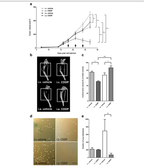
Next, a comparison between i.v. and i.a. CDDP infusions was conducted. Only i.a. CDDP (88 ± 31 mm3) inhibited tumor growth and caused regression of primary tumors, while tumors continued to grow in all other treatment groups (tumor volumes at 27 days post tumor cell injection: i.v. CDDP: 307 ± 25 mm3; i.a. vehicle: 375 ± 73 mm3; i.v. ve-hicle: 491 ± 44 mm3; two-way ANOVA:p< 0.0001; Fig. 2a). One day prior to sacrifice, mice were subjected to microCT scans. Tumor volumes measured within the resulting tomographs confirmed caliper measurements and yielded significantly smaller final tumor volumes in the group re-ceiving i.a. CDDP (54 ± 35 mm3) compared to volumes measured in other treatment groups (i.v. CDDP: 297 ± 29 mm3; i.a. vehicle: 286 ± 58 mm3; i.v. vehicle 479 ± 34 mm3; ANOVA:p< 0.0001). Osteosarcoma is known to be associated with pathological bone remodelling and
increased fracture risk, and thus, the structural integrity of the bone influences the quality of life of osteosarcoma pa-tients. 143B cell-derived osteosarcomas were shown to be-have mostly osteolytic in vivo and loss of cortical bone correlates with increasing tumor volume. Accordingly, ad-ministration of i.a. CDDP (87 ± 5 % of initial bone volume (before treatment)) led to the smallest loss of cortical bone compared to i.v. vehicle (75 ± 3 %), i.a. vehicle (68 ± 6 %) and i.v. CDDP (51 ± 2 %; ANOVA:p< 0.0001; Fig. 2b, c).
Finally, X-gal staining of lacZ tagged cells on the sur-face of lungs collected during necropsy (Fig. 2d) showed that systemic i.v. CDDP had no significant effect on metastatic spread towards the lung compared to i.v. ve-hicle control (i.v. CDDP: 202 ± 15; i.v. veve-hicle: 218 ± 41). In contrast, i.a. CDDP significantly reduced the number of lung metastases (i.a CDDP: 82 ± 42; i.a vehicle: 695 ± 300; ANOVA: p< 0.001; Fig. 2e). Of note, a nonsignifi-cant, but on average higher amount of metastases was found in i.a. vehicle versus i.v. vehicle group.
Effect of CDDP treatment on tumor blood perfusion
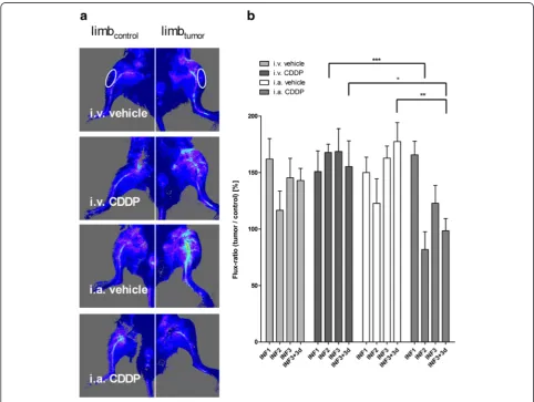
Tumor-associated vasculature was assessed in vivo via blood perfusion measurements. Primary tumor growth
induced an increase in perfusion of the tumor-bearing limbs compared to the contralateral control limbs (Fig. 3a). Following i.a. CDDP, a significant decrease in perfusion compared to i.v. CDDP or i.a. vehicle was ob-served (two-way ANOVA: p< 0.05; Fig. 3b). Interest-ingly, the largest reduction in perfusion were detected after i.a. CDDP infusions. At the end of the study, perfu-sion of the i.a. CDDP-treated limbs was close to physio-logical values, similar to the contralateral knee region. However, areas formerly infiltrated by osteosarcomas appeared poorly perfused, indicating the occurrence of ischemic tumor necrosis (e.g. Fig. 3a: i.a. CDDP).
Histologic response to CDDP chemotherapy
Assessment of tumor necrosis after neoadjuvant chemo-therapy is an established endpoint to evaluate the re-sponse to treatment in osteosarcoma patients. Figure 4a illustrates representative examples from each treatment
group which were used for the analysis of tumor necrosis. In case of two animals treated with i.a. CDDP, no tumor tissue could be found on cross sections of the tumor-bearing limb, indicating a strong anti-tumor effect (100 % of tumor necrosis was assumed). The largest mean tumor necrosis was detected after i.a. CDDP (68 ± 12 %) com-pared with i.a. vehicle (32 ± 8 %), i.v. CDDP (17 ± 2 %) or i.v. vehicle (21 ± 3 %, ANOVA:p< 0.01; Fig. 4b). Accord-ing to Salzer-Kuntschik, a good responder is defined by more than 90 % tumor necrosis [31]. With i.a. CDDP, a total of five (45 %) good responses was achieved, whereas no good responses were detected with i.a. vehicle, i.v. ve-hicle or i.v. CDDP (Fisher’s exact test:p< 0.01; Additional file 1). Tumor cell death in the H&E-stained sections con-sisted of multifocal to coalescing, variably sized areas of necrosis: these areas which are, to varying extents, inher-ent to any rapidly growing tumor (i.e. after i.v. vehicle) are likely indicative of ischemic cell death.
Influence of different routes of CDDP administration on remaining viable tumor tissue
Due to the anti-tumor efficacy of i.a. CDDP, smaller areas of viable tumor were evaluated after treatment: i.a. CDDP (median 0.6 mm2; interquartile range 0.1–4.0 mm2) compared with i.a. vehicle (7.1 mm2; 3.9–11.3 mm2), i.v. CDDP (10.0 mm2; 7.2.–12.7 mm2) and i.v. vehicle (11.9 mm2; 11.0–14.8 mm2). Furthermore, two animals from the i.a. CDDP group were excluded from all immuno-histochemical analyses involving viable primary tumor be-cause of a total absence of tumor tissue. Within regions of viable tumor, scattered neoplastic cells exhibited mor-phological features of apoptosis, such as cell shrink-age, nuclear pyknosis and fragmentation, as indicated by immunohistochemical stains for cleaved PARP-1, a marker for chemotherapy-induced apoptosis [32]. However, no significant differences in the number of cleaved PARP-1+ cells within areas of remaining viable tumor were detected between corresponding vehicle and treatment groups (Fig. 5a).
In order to see if the reduced limb perfusion also re-sulted in increased hypoxia, expression of HIF-1α, a pro-tein expressed under sub-physiological levels of oxygen, was studied. IHC of HIF-1α demonstrated intense stain-ing of remainstain-ing viable tumor tissue after i.a. CDDP (Fig. 5b). Furthermore, quantitation of HIF1-1α expres-sion demonstrated a significant increase in HIF-1αlevels in tumors after administration of i.a. CDDP (2.8 ± 0.7 % of remaining viable tumor tissue) compared with tumors exposed to i.a. vehicle (0.6 ± 0.6 %), i.v. CDDP (0.4 ± 0.2 %) or i.v. vehicle (0.3 ± 0.1 %; ANOVA: p< 0.001; Fig. 5c). High levels of HIF-1α indicate low oxygen
levels, subsequently triggering neovascularization. To this end, IHC for CD31 and factor VIII-related antigen (FVIII-RAg) was performed to characterize and quantify newly formed blood vessels within the neoplasms [33]. Examples of CD31 IHC are shown in Fig. 5d. In all groups, areas of viable tumor contained negligible num-bers of FVIII-Rag+ blood vessels, while the endothelial cells lining large vessels in the skeletal muscle and con-nective tissue adjacent to the osteosarcomas expressed FVIII-RAg (data not shown). Quantitation of the CD31+ areas within viable tumor tissue indicated a trend towards increased neovascularization after i.a. CDDP (4.1 ± 1.5 % of remaining viable tumor tissue) compared with the lower levels observed after i.a. vehicle (1.8 ± 0.7 %), i.v. CDDP (1.6 ± 0.2 %) or i.v. vehicle treatment (1.3 ± 0.2 %; Fig. 5e). Furthermore, CD31 IHC significantly correlated with HIF-1αIHC (Pearson’s r:p< 0.01; r = 0.62; Fig. 5f).
Side effects associated with different routes of CDDP administration
Similar to the “dose establishment study”, body weights were measured at regular intervals throughout the“ com-parison study” (Fig 6a). When comparing CDDP admin-istrations only (i.a. CDDP: 88 ± 2 % of body weight normalized to the weight at day of tumor cell injection versus i.v. CDDP: 85 ± 1 %), no significant difference in body weight development between i.a. or i.v. CDDP ad-ministration was demonstrated. In contrast, i.v. CDDP caused a significant drop in body weight compared with i.v. vehicle (97 ± 2 %), whereas no difference was found between i.a. CDDP and i.a. vehicle (90 ± 2 %; two-way ANOVA:p< 0.0001; Fig. 6a).
In general, application of CDDP is limited by a high incidence of severe nephrotoxicity characterized by de-generation or death of the proximal tubule epithelial cells [34, 35]. In this study, however, no histological ab-normality was recognized in the kidneys, except in three mice after i.a. CDDP, which exhibited mild acute tubular degeneration/necrosis, affecting only a small proportion of the renal tubules (Fig. 6b).
To assess whether i.a. CDDP resulted in higher apop-totic rates of non-malignant cells, suggestive of higher local concentrations of CDDP in the operated limb, the number of apoptotic keratinocytes within the skin of treated limbs and contralateral limbs were quantified. Results demonstrated lower numbers of apoptotic
keratinocytes (AK) after i.v. vehicle (0–0.05 AK/cm of skin; minimum - maximum of tumor bearing limb), i.v. CDDP (0–0.1 AK/cm) or i.a. vehicle treatment (0–0.05 AK/cm), in tumor-bearing limbs as well as in the corre-sponding contralateral limb (Fig. 6c). In contrast, signifi-cantly higher numbers of AKs were found after the administration of i.a. CDDP (0.35–7.6 AK/cm), yet in the tumor-bearing limb only (Wilcoxon matched pair test:p< 0.01; Fig. 6c).
Discussion
In this study, we present the successful establishment of i.a. drug administrations in a mouse model of experi-mental orthotopic osteosarcoma. Using CDDP as a gold
standard drug for osteosarcoma treatment, we showed that our setup of i.a. drug infusions is feasible and that primary and systemic disease could be inhibited in a concentration-dependent manner. Moreover, we demon-strated that i.a. CDDP is more effective in inhibiting osteosarcoma progression than equivalent concentra-tions of i.v. CDDP, as indicated by smaller primary tumor volumes, decreased destruction of cortical bone as well as decreased numbers of lung metastases.
Increased anti-tumor efficacy of i.a. CDDP infusions was also confirmed by histological analyses, where we dem-onstrated increased levels of tumor necrosis. Decreased tumor blood perfusion and increased hypoxia of the neoplasms after i.a. CDDP administration was demon-strated and explains, at least partially, the superior effi-cacy of localized CDDP delivery. Finally, we showed that i.a. CDDP causes increased levels of apoptotic keratino-cytes in the epidermis of tumor-bearing limbs, while
other systemic side effects were similar compared with i.v. CDDP.
Despite the use of larger animal model systems such as dogs [25] or sheep [36], the most commonly used model systems in osteosarcoma research are rodents [37]. To our knowledge, this is the first report of an i.a. infusion model in experimental, orthotopic osteosarcoma in mice, where we could demonstrate superior tumor con-trol as compared to routine i.v. infusions. Hence, our i.a. model can be used as a platform for the investigation of other small molecules whose systemic application is lim-ited by side effects. Especially for osteosarcomas of the limb, easy access to the tumor feeding artery offers a valu-able alternative to systemic CDDP infusions. However, i.a. infusions are not limited to tumors of the limb. Another recent study using a mouse model of metastatic brain tumors demonstrated advantages in tumor control with i.a. chemotherapy (internal carotid artery) compared with i.v. chemotherapy (tail vein), similar to our study [38]. This demonstrates that even difficult-to-access tumor en-tities can be treated with i.a. drug infusions.
Local application of small molecules such as CDDP of-fers several advantages compared with systemic applica-tion. First, higher tumor drug loads can be achieved after infusion of equivalent drug concentrations via the tumor-feeding artery [11], causing greater tumor necro-sis [13]. In line with this assumption, we demonstrated greater tumor necrosis after i.a. CDDP compared with i.v. CDDP administration. In contrast to Winkler et al., we were able to compare i.a. versus i.v. routes of admin-istration in the absence of clinical confounding factors such as changes in drug infusion times or stratification of patients (e.g. high-risk only) [10]. In our study, i.a. CDDP not only caused a reduction of the primary tumor volume but also minimized the loss of mineralized bone-an indicator for experimental osteosarcoma progression [39]. In contrast, loss of cortical bone was increased after i.v. CDDP treatment compared with i.v. vehicle. Like-wise, when sites of bone turnover in dogs were studied, bone remodeling was significantly influenced by the sys-temic administration of CDDP [40].
Systemic side effects, especially nephrotoxicity, are equal or reduced after local CDDP infusion compared to systemic application without a simultaneous reduction of the systemic potency of the drug [9, 20]. In our study, most animals showed no signs of nephrotoxicity. How-ever, we found evidence of mild nephrotoxicity, repre-sented by scattered tubular degeneration/necrosis in three animals treated with i.a. CDDP. Renal injury was minor and unlikely to have an impact on kidney func-tion. It is possible that hypovolemia, resulting from blood loss and/or insufficient rehydration after the re-peated surgical interventions, exacerbated the observed nephrotoxicity in these animals.
Skin necrosis is another side effect observed in human patients after i.a. administration of CDDP [10, 20]. How-ever, this usually does not lead to complications during the treatment and regenerates well. Likewise, our results indicated that i.a. CDDP led to increased numbers of apoptotic keratinocytes in the tumor-bearing limbs, but not in the healthy contralateral limbs. Elevated numbers of AK may indicate a higher local CDDP concentration in the proximity of the primary tumor and thus, help to explain the superior response after administration of i.a. CDDP compared with i.v. CDDP. Interestingly, some mild chemotherapy-induced toxicities were shown to be associated with improved osteosarcoma patient survival [23].
Consistent with the observations reported by Wilkins et al., where a reduction of the spongy tumor vascula-ture after i.a. CDDP was detected [14, 22], we also observed a general decrease in perfusion of the tumor region shortly after i.a. CDDP administration. This re-duction in tumor perfusion in addition to the higher local CDDP concentration may have further contributed to the regression of the experimental tumors and re-sulted in a good histological response (as defined by hav-ing at least 90 % tumor necrosis) in at least 45 % of the mice. Thus, destruction of the tumor vasculature seems to be a necessity for i.a. CDDP to successfully induce tumor necrosis potentially resulting in a high percentage of good histologic responses [14].
The reduction in tumor perfusion after i.a. CDDP might have caused the remaining viable tumor tissue to react by expressing increased levels of HIF-1α. It is known that constitutively active HIF-1αinduces neovas-cularization and increased expression of CD31 or VEGF [41–44]. Increased expression of HIF-1α in osteosar-comas after i.a. CDDP was paralleled by increased microvascular density assessed using IHC for CD31, a marker of immature endothelium. In addition to CD31, consecutive tissue sections were stained for FVIII-RAg, normally found in large, mature vessels [33]. The few scattered FVIII-Rag+ vascular structures found within the tumors were likely pre-existing and no difference was found among the different groups. In summary, our results indicate a response of the neoplasms towards is-chemic damage after i.a. CDDP by increasing HIF-1α -levels and potentially initiating neovascularization.
CDDP also successfully controlled the number of spon-taneous lung metastases. This is especially relevant in osteosarcoma, where controlling pulmonary metastases strongly influences patient survival [47–49].
One limitation of our study is the inherent variability due to any surgical procedure. Although the same sur-geon performed i.a. drug infusions, the parameters of i.a. infusions varied (e.g. duration of surgery or degree of blood loss). Especially the placement of the catheter is critical for a homogeneous drug distribution in subse-quent arterial branches [50] and thus, impacts success of therapy. In general, our study suggests a superior outcome in the chemotherapeutic response after i.a. delivery of CDDP, however, the individual outcomes must be interpreted alongside corresponding toxicoki-netic information.
Conclusions
Taken together, our study demonstrates the potential of i.a. CDDP in a clinically relevant osteosarcoma model. The superior primary tumor control of i.a. CDDP in our study demonstrates the potential of i.a. drug administra-tions as currently used in some clinics. Despite the greater technical requirements for i.a. drug infusions, we suggest that the potential of i.a. infusions in osteosar-coma treatment should be considered when evaluating (novel) compounds and combinations thereof. Especially for a rare disease such as osteosarcoma, we believe that our intraarterial therapy model can aid in the preclinical assessment of drug efficacy and thus, improve osteosar-coma patient treatment.
Additional file
Additional file 1:Histological response according to treatment. Groups the histological tumor response according to current clinical criteria for evaluation of tumor necrosis. (DOCX 14 kb)
Abbreviations
AK, apoptotic keratinocyte; CD31, cluster of differentiation 31; CDDP, cisplatin; FVIII-RAg, factor VIII related antigen; H&E, hematoxylin and eosin; HIF-1α, hypoxia inducible factor-1α; i.a, intraarterial; i.p, intraperitoneal; i.v, intravenous; IHC, immunohistochemistry; microCT, micro computed tom-ography; PARP-1, poly ADP ribose polymerase-1; SCID, severe combined immunodeficiency; TCIs: tumor cell injections
Acknowledgments
We would like to thank Prof. Anja Kipar for fruitful discussions regarding the histological analysis of the tumor material.
Funding
Our work is supported by the University of Zurich, the Schweizerischer Verein Balgrist (Zurich, Switzerland), the Walter L. & Johanna Wolf Foundation (Zurich, Switzerland), the Highly Specialized Medicine for Musculoskeletal Oncology program of the Canton of Zurich, the Zurcher Krebsliga (Zurich, Switzerland), the“Kind und Krebs”fund (Zollikerberg, Switzerland), and the Swiss National Science Foundation SNF Nr.310030_149649.
Availability of data and materials
The datasets supporting the conclusions of this article are included within the article and its additional file.
Authors’contributions
Conception and design: BR, SMB, BF. Development of methodology: BR, ON, BF. Acquisition of data: BR, GP, SMB, ON. Analysis and interpretation of data: BR, GP. Writing, review, and/or revision of the manuscript: BR, SMB, BF. Administrative, technical, or material support (i.e., reporting or organizing data, constructing databases): BR, GP, BF. Study supervision: BF. All authors read and approved the final manuscript.
Competing interests
The authors declare that they have no competing interests.
Consent for publication Not applicable.
Ethics approval and consent to participate
Animal care and experimental procedures were in accordance with the institutional guidelines and approved by the Ethics Committee of the Veterinary Department, Canton of Zurich, Switzerland (License Number 64/ 2013).
Author details 1
Laboratory for Orthopedic Research, Department of Orthopedics, Balgrist University Hospital, Forchstrasse 340, Zurich 8008, Switzerland.2Laboratory for Animal Model Pathology, Veterinary Pathology, Vetsuisse Faculty, Zurich, Switzerland.
Received: 12 May 2016 Accepted: 8 July 2016
References
1. Mirabello L, Troisi RJ, Savage SA. Osteosarcoma incidence and survival rates from 1973 to 2004: data from the Surveillance, Epidemiology, and End Results Program. Cancer. 2009;115(7):1531–43.
2. Howlader N, Noone AM, Krapcho M, Garshell J, Neyman N, Altekruse SF, Kosary CL, Yu M, Ruhl J, Tatalovich Z, Cho H, Mariotto A, Lewis DR, Chen HS, Feuer EJ, Cronin KA (eds). SEER Cancer Statistics Review, 1975-2010, Bethesda: National Cancer Institute. http://seer.cancer.gov/csr/1975_2010/, based on November 2012 SEER data submission, posted to the SEER web site, April 2013.
3. Allison DC, Carney SC, Ahlmann ER, Hendifar A, Chawla S, Fedenko A, Angeles C, Menendez LR. A meta-analysis of osteosarcoma outcomes in the modern medical era. Sarcoma. 2012;2012:704872.
4. Goorin AM, Schwartzentruber DJ, Devidas M, Gebhardt MC, Ayala AG, Harris MB, Helman LJ, Grier HE, Link MP. Presurgical chemotherapy compared with immediate surgery and adjuvant chemotherapy for nonmetastatic osteosarcoma: Pediatric Oncology Group Study POG-8651. J Clin Oncol Off J Am Soc Clin Oncol. 2003;21(8):1574–80.
5. Kager L, Zoubek A, Potschger U, Kastner U, Flege S, Kempf-Bielack B, Branscheid D, Kotz R, Salzer-Kuntschik M, Winkelmann W, et al. Primary metastatic osteosarcoma: presentation and outcome of patients treated on neoadjuvant Cooperative Osteosarcoma Study Group protocols. J Clin Oncol. 2003;21(10):2011–8.
6. Bacci G, Mercuri M, Longhi A, Ferrari S, Bertoni F, Versari M, Picci P. Grade of chemotherapy-induced necrosis as a predictor of local and systemic control in 881 patients with non-metastatic osteosarcoma of the extremities treated with neoadjuvant chemotherapy in a single institution. Eur J Cancer. 2005; 41(14):2079–85.
7. Hauben EI, Weeden S, Pringle J, Van Marck EA, Hogendoorn PC. Does the histological subtype of high-grade central osteosarcoma influence the response to treatment with chemotherapy and does it affect overall survival? A study on 570 patients of two consecutive trials of the European Osteosarcoma Intergroup. Eur J Cancer. 2002;38(9):1218–25.
8. Grimer RJ. Surgical options for children with osteosarcoma. Lancet Oncol. 2005;6(2):85–92.
intravenous chemoradiation for advanced head and neck cancer: Results of a randomized phase 3 trial. Cancer. 2010;116(9):2159–65.
10. Winkler K, Bielack S, Delling G, Salzer-Kuntschik M, Kotz R, Greenshaw C, Jurgens H, Ritter J, Kusnierz-Glaz C, Erttmann R, et al. Effect of intraarterial versus intravenous cisplatin in addition to systemic doxorubicin, high-dose methotrexate, and ifosfamide on histologic tumor response in
osteosarcoma (study COSS-86). Cancer. 1990;66(8):1703–10.
11. Stewart DJ, Benjamin RS, Zimmerman S, Caprioli RM, Wallace S, Chuang V, Calvo 3rd D, Samuels M, Bonura J, Loo TL. Clinical pharmacology of intraarterial cis-diamminedichloroplatinum(II). Cancer Res. 1983;43(2):917–20. 12. Jaffe N, Knapp J, Chuang VP, Wallace S, Ayala A, Murray J, Cangir A, Wang A,
Benjamin RS. Osteosarcoma: intra-arterial treatment of the primary tumor with cis-diammine-dichloroplatinum II (CDP). Angiographic, pathologic, and pharmacologic studies. Cancer. 1983;51(3):402–7.
13. Jaffe N, Raymond AK, Ayala A, Carrasco CH, Wallace S, Robertson R, Griffiths M, Wang YM. Effect of cumulative courses of intraarterial cis-diamminedichloroplatin-II on the primary tumor in osteosarcoma. Cancer. 1989;63(1):63–7.
14. Wilkins RM, Cullen JW, Camozzi AB, Jamroz BA, Odom L. Improved survival in primary nonmetastatic pediatric osteosarcoma of the extremity. Clin Orthop Relat Res. 2005;438:128–36.
15. Hong S, Shin SJ, Jung M, Jeong J, Lee YJ, Shin KH, Roh JK, Rha SY. Comparison of long-term outcome between doublet and triplet neoadjuvant chemotherapy in non-metastatic osteosarcoma of the extremity. Oncology. 2011;80(1–2):107–17.
16. Zhou Y, Huang Z, Wu S, Zang X, Liu M, Shi J. miR-33a is up-regulated in chemoresistant osteosarcoma and promotes osteosarcoma cell resistance to cisplatin by down-regulating TWIST. J Exp Clin Cancer Res. 2014;33:12. 17. Bramwell VH, Burgers M, Sneath R, Souhami R, van Oosterom AT, Voute PA,
Rouesse J, Spooner D, Craft AW, Somers R, et al. A comparison of two short intensive adjuvant chemotherapy regimens in operable osteosarcoma of limbs in children and young adults: the first study of the European Osteosarcoma Intergroup. J Clin Oncol Off J Am Soc Clin Oncol. 1992;10(10):1579–91. 18. Bielack SS, Smeland S, Whelan JS, Marina N, Jovic G, Hook JM, Krailo MD,
Gebhardt M, Papai Z, Meyer J, et al. Methotrexate, Doxorubicin, and Cisplatin (MAP) Plus Maintenance Pegylated Interferon Alfa-2b Versus MAP Alone in Patients With Resectable High-Grade Osteosarcoma and Good Histologic Response to Preoperative MAP: First Results of the EURAMOS-1 Good Response Randomized Controlled Trial. J Clin Oncol Off J Am Soc Clin Oncol. 2015;33(20):2279–87.
19. Ferrari S, Smeland S, Mercuri M, Bertoni F, Longhi A, Ruggieri P, Alvegard TA, Picci P, Capanna R, Bernini G, et al. Neoadjuvant chemotherapy with high-dose Ifosfamide, high-dose methotrexate, cisplatin, and doxorubicin for patients with localized osteosarcoma of the extremity: a joint study by the Italian and Scandinavian Sarcoma Groups. J Clin Oncol Off J Am Soc Clin Oncol. 2005;23(34):8845–52.
20. Bacci G, Ferrari S, Tienghi A, Bertoni F, Mercuri M, Longhi A, Fiorentini G, Forni C, Bacchini P, Rimondini S, et al. A comparison of methods of loco-regional chemotherapy combined with systemic chemotherapy as neo-adjuvant treatment of osteosarcoma of the extremity. Eur J Surg Oncol. 2001;27(1):98–104.
21. Bielack S, Kempf-Bielack B, Schwenzer D, Birkfellner T, Delling G, Ewerbeck V, Exner GU, Fuchs N, Gobel U, Graf N, et al. Neoadjuvant therapy for localized osteosarcoma of extremities. Results from the Cooperative osteosarcoma study group COSS of 925 patients. Klin Padiatr. 1999;211(4):260–70. 22. Hugate RR, Wilkins RM, Kelly CM, Madsen W, Hinshaw I, Camozzi AB.
Intraarterial chemotherapy for extremity osteosarcoma and MFH in adults. Clin Orthop Relat Res. 2008;466(6):1292–301.
23. McTiernan A, Jinks RC, Sydes MR, Uscinska B, Hook JM, van Glabbeke M, Bramwell V, Lewis IJ, Taminiau AH, Nooij MA, et al. Presence of
chemotherapy-induced toxicity predicts improved survival in patients with localised extremity osteosarcoma treated with doxorubicin and cisplatin: a report from the European Osteosarcoma Intergroup. Eur J Cancer. 2012; 48(5):703–12.
24. Tunn PU, Schmidt-Peter P, Pomraenke D, Hohenberger P. Osteosarcoma in children: long-term functional analysis. Clin Orthop Relat Res. 2004;421:212–7. 25. Powers BE, Withrow SJ, Thrall DE, Straw RC, LaRue SM, Page RL, Gillette EL.
Percent tumor necrosis as a predictor of treatment response in canine osteosarcoma. Cancer. 1991;67(1):126–34.
26. Petrilli AS, de Camargo B, Filho VO, Bruniera P, Brunetto AL, Jesus-Garcia R, Camargo OP, Pena W, Pericles P, Davi A, et al. Results of the Brazilian
Osteosarcoma Treatment Group Studies III and IV: prognostic factors and impact on survival. J Clin Oncol off J Am Soc Clin Oncol. 2006;24(7):1161–8. 27. Gvozdenovic A, Arlt MJ, Campanile C, Brennecke P, Husmann K, Born W,
Muff R, Fuchs B. Silencing of CD44 Gene Expression in Human 143-B Osteosarcoma Cells Promotes Metastasis of Intratibial Tumors in SCID Mice. PLoS One. 2013;8(4):e60329.
28. Arlt MJ, Banke IJ, Walters DK, Puskas GJ, Steinmann P, Muff R, Born W, Fuchs B. LacZ transgene expression in the subcutaneous Dunn/LM8 osteosarcoma mouse model allows for the identification of micrometastasis. J Orthop Res. 2011;29(6):938–46.
29. Husmann K, Arlt MJ, Jirkof P, Arras M, Born W, Fuchs B. Primary tumour growth in an orthotopic osteosarcoma mouse model is not influenced by analgesic treatment with buprenorphine and meloxicam. Lab Anim. 2015; 49(4):284–93.
30. Berndt K, Vogel J, Buehler C, Vogt P, Born W, Fuchs B. A new method for repeated drug infusion into the femoral artery of mice. J Am Assoc Lab Anim Sci. 2012;51(6):825–31.
31. Salzer-Kuntschik M, Brand G, Delling G. Determination of the degree of morphological regression following chemotherapy in malignant bone tumors. Pathologe. 1983;4(3):135–41.
32. Kaufmann SH, Desnoyers S, Ottaviano Y, Davidson NE, Poirier GG. Specific proteolytic cleavage of poly(ADP-ribose) polymerase: an early marker of chemotherapy-induced apoptosis. Cancer Res. 1993;53(17):3976–85. 33. Wang D, Stockard CR, Harkins L, Lott P, Salih C, Yuan K, Buchsbaum D,
Hashim A, Zayzafoon M, Hardy RW, et al. Immunohistochemistry in the evaluation of neovascularization in tumor xenografts. Biotech Histochem. 2008;83(3–4):179–89.
34. Jones TW, Chopra S, Kaufman JS, Flamenbaum W, Trump BF. Cis-diamminedichloroplatinum (II)-induced acute renal failure in the rat. Correlation of structural and functional alterations. Lab Invest. 1985; 52(4):363–74.
35. Kuhlmann MK, Burkhardt G, Kohler H. Insights into potential cellular mechanisms of cisplatin nephrotoxicity and their clinical application. Nephrol Dial Transplant. 1997;12(12):2478–80.
36. Harker G, Stephens F. A report on the comparative response of sheep epidermal squamous-cell carcinoma to intraarterial versus intravenous Cisplatin infusion. Int J Oncol. 1995;7(2):365–70.
37. Guijarro MV, Ghivizzani SC, Gibbs CP. Animal models in osteosarcoma. Front Oncol. 2014;4:189.
38. Kim B, Kim K, Im KH, Kim JH, Lee JH, Jeon P, Byun H. Multiparametric MR imaging of tumor response to intraarterial chemotherapy in orthotopic xenograft models of human metastatic brain tumor. J Neurooncol. 2016; 127(2):243–51.
39. Ohba T, Cole HA, Cates JM, Slosky DA, Haro H, Ando T, Schwartz HS, Schoenecker JG. Bisphosphonates inhibit osteosarcoma-mediated osteolysis via attenuation of tumor expression of MCP-1 and RANKL. J Bone Mineral Res Off J Am Soc Bone Mineral Res. 2014;29(6):1431–45.
40. Ehrhart N, Eurell JA, Tommasini M, Constable PD, Johnson AL, Feretti A. Effect of cisplatin on bone transport osteogenesis in dogs. Am J Vet Res. 2002;63(5):703–11.
41. Elson DA, Thurston G, Huang LE, Ginzinger DG, McDonald DM, Johnson RS, Arbeit JM. Induction of hypervascularity without leakage or inflammation in transgenic mice overexpressing hypoxia-inducible factor-1alpha. Genes Dev. 2001;15(19):2520–32.
42. Mouriaux F, Sanschagrin F, Diorio C, Landreville S, Comoz F, Petit E, Bernaudin M, Rousseau AP, Bergeron D, Morcos M. Increased HIF-1alpha expression correlates with cell proliferation and vascular markers CD31 and VEGF-A in uveal melanoma. Invest Ophthalmol Vis Sci. 2014;55(3):1277–83. 43. Musumeci G, Castorina A, Magro G, Cardile V, Castorina S, Ribatti D.
Enhanced expression of CD31/platelet endothelial cell adhesion molecule 1 (PECAM1) correlates with hypoxia inducible factor-1 alpha (HIF-1alpha) in human glioblastoma multiforme. Exp Cell Res. 2015;339(2):407–16. 44. Chiche J, Pommier S, Beneteau M, Mondragon L, Meynet O, Zunino B,
Mouchotte A, Verhoeyen E, Guyot M, Pages G, et al. GAPDH enhances the aggressiveness and the vascularization of non-Hodgkin’s B lymphomas via NF-kappaB-dependent induction of HIF-1alpha. Leukemia. 2015;29(5):1163–76. 45. Liotta LA, Kleinerman J, Saidel GM. Quantitative relationships of intravascular
tumor cells, tumor vessels, and pulmonary metastases following tumor implantation. Cancer Res. 1974;34(5):997–1004.
47. Nataraj V, Rastogi S, Khan SA, Sharma MC, Agarwala S, Vishnubhatla S, Bakhshi S. Prognosticating metastatic osteosarcoma treated with uniform chemotherapy protocol without high dose methotrexate and delayed metastasectomy: a single center experience of 102 patients. Clin Transl Oncol. 2016.
48. Rasalkar DD, Chu WC, Lee V, Paunipagar BK, Cheng FW, Li CK. Pulmonary metastases in children with osteosarcoma: characteristics and impact on patient survival. Pediatr Radiol. 2011;41(2):227–36.
49. Mizuno T, Taniguchi T, Ishikawa Y, Kawaguchi K, Fukui T, Ishiguro F, Nakamura S, Yokoi K. Pulmonary metastasectomy for osteogenic and soft tissue sarcoma: who really benefits from surgical treatment? Eur J Cardiothorac Surg. 2013;43(4):795–9.
50. van den Hoven AF, Lam MG, Jernigan S, van den Bosch MA, Buckner GD. Innovation in catheter design for intra-arterial liver cancer treatments results in favorable particle-fluid dynamics. J Exp Clin Cancer Res. 2015;34:74.
• We accept pre-submission inquiries
• Our selector tool helps you to find the most relevant journal
• We provide round the clock customer support
• Convenient online submission
• Thorough peer review
• Inclusion in PubMed and all major indexing services
• Maximum visibility for your research
Submit your manuscript at www.biomedcentral.com/submit