R E S E A R C H
Open Access
The leukemogenic fusion gene
MLL-AF9
alters microRNA expression pattern and
inhibits monoblastic differentiation via
miR-511 repression
Katrin K. Fleischmann
1*, Philipp Pagel
2, Julia von Frowein
1, Thomas Magg
1, Adelbert A. Roscher
3and Irene Schmid
1Abstract
Background:In this study we explored the role of microRNAs (miRNAs) as mediators of leukemogenic effects of the fusion geneMLL-AF9, which results from a frequent chromosomal translocation in infant and monoblastic acute myeloid leukemia (AML).
Methods:We performed a specific and efficient knockdown of endogenousMLL-AF9in the human monoblastic AML cell line THP1.
Results:The knockdown associated miRNA expression profile revealed 21MLL-AF9dependently expressed miRNAs. Gene ontology analyses of target genes suggested an impact of these miRNAs on downstream gene regulation via targeting of transcriptional modulators as well as involvement in many functions important for leukemia maintenance as e.g. myeloid differentiation, cell cycle and stem cell maintenance. Furthermore, we identified one of the most intensely repressed miRNAs, miR-511, to raiseCCL2expression (a chemokine ligand important for immunosurveillance), directly target cyclin D1, inhibit cell cycle progression, increase cellular migration and promote monoblastic differentiation. With these effects, miR-511 may have a therapeutic potential as a pro-differentiation agent as well as in leukemia vaccination approaches.
Conclusions:Our study provides new insights into the understanding of miRNAs as functional mediators of
the leukemogenic fusion gene MLL-AF9 and opens new opportunities to further investigate specific
therapeutic options for AML via the miRNA level.
Keywords: Leukemia, AML, MLL, MLL-AF9, microRNA, miR-511, cyclin D1, CCND1, Differentiation, Monocytic differentiation
Background
MLL-AF9 (alias KMT2A-MLLT3) is a fusion gene result-ing from the chromosomal translocation t(9;11)(p22;q23). It is sufficient to initiate acute leukemia in murine models [1, 2] and is the most frequent fusion gene in infant acute myeloid leukemia (AML) and especially associated with monoblastic AML (FAB-classification M5) [3, 4]. For this
type of leukemia with a persisting poor prognosis, new targeted therapies are needed.
MLL and AF9 wildtype proteins lead to transcrip-tional initiation (MLL) and elongation (AF9) of target genes [5, 6], while the fusion protein MLL-AF9 is believed to combine these properties, leading to increased activa-tion of target genes [7]. Novel therapeutic strategies which aim to directly intervene with MLL-AF9 functions are under investigation but struggle with the complexity to target the involved proteprotein and proteDNA in-teractions and with the obstacle to avoid targeting of the important wildtype protein functions [5, 7].
* Correspondence:katrin.fleischmann@med.uni-muenchen.de
1Division of Pediatric Hematology and Oncology, Children’s Research Center,
Dr. von Hauner Children’s Hospital, Ludwig-Maximilians-Universität München, Lindwurmstrasse 2a, 80337 Munich, Germany
Full list of author information is available at the end of the article
MicroRNAs (miRNAs) are important cellular posttran-scriptional gene regulators and are involved in presum-ably all physiological and pathophysiological cellular processes [8], including oncological state, progression and clinical outcome [9–11].
Although technical challenges – predominantly the specific, efficient and safe delivery of miRNA modulators to the tumor site – remain to be overcome, miRNA therapeutics are believed to hold huge promise for can-cer treatment [12]. In this context, a comprehensive un-derstanding of the role of miRNAs within the complex regulatory networks in malignant cells is a prerequisite for the development of combinatorial therapeutic strat-egies employing miRNA modulators [12].
As fusion gene breakpoints vary between individual patients, siRNA-targeting of oncogenic fusion genes themselves would have to be tested and optimized in each case and thus does not represent an opportune therapeutic option. Finding common, relevant down-stream miRNA targets of MLL-AF9 instead might yield new alternative therapeutic routes. This approach has the benefit that oncogenic transcription factors or other protein classes which are currently non-druggable by e.g. small molecule drugs might be targeted via miRNA modulators. Another advantage of miRNA therapeutics is the fact that not only single gene transcripts can be targeted (e.g. via target site blockage) but also whole ar-rays of mRNAs, which may cooperate to create an onco-genic phenotype (e.g. via miRNA mimics or inhibitors).
In order to identify new alternative drug targets for MLL-AF9 positive AML, we thus aimed to explore the
role and function of miRNAs as mediators of
leukemogenic effects of the fusion gene MLL-AF9. To this end, a differential miRNA expression profile was generated after knockdown of endogenous MLL-AF9 in the monoblastic AML cell line THP1 and revealed 21 miRNAs predicted to be involved in leukemogenic func-tions. Additionally, we provide evidence for a relevant role of miR-511 in the context of monoblastic differenti-ation. To our knowledge, this is the first experimental study that comprehensively analyzes the effects of an MLLfusion gene on miRNA expression.
Methods
Cells, cell cultivation, small interfering RNA and transfection
Cultivation and siRNA treatment of THP1 cells (DSMZ GmbH, Braunschweig, Germany) was performed as pre-viously reported [7]. Briefly, the cell line was
authenti-cated via MLL-AF9 breakpoint sequencing, was
routinely checked for mycoplasma contamination and transfected with 50 nM Silencer Select siRNAs (Life Technologies, Carlsbad, CA, USA) and Dreamfect (OZ Biosciences, Marseille, France) leading to transfection
efficiencies and survival rates of 93 % each. Experimental incubations lasted eight days with repeated transfections on day 0, 3, and 6. Prior to each transfection event, cell densities were determined and cells were reseeded at 5 × 10⁴cells per ml [7].
THP1 miRNA mimic transfections were performed as described for siRNA transfections but with 30 nM Ambion Pre-miR miRNA Precursors (hsa-miR-511-5p AM10237, neg Control #1 AM17110, neg Control #2 AM17111) and experimental incubations up to day nine again with repeated transfections on day 0, 3, and 6.
Human CD14+ monocytes (C-12909, PromoCell GmbH, Heidelberg, Germany) were cultivated for 48 h in mono-nuclear cell medium (PromoCell) prior to immunostaining and RNA isolation. Human CD34+ progenitor cells (C-12921, PromoCell) were directly taken to RNA isolation.
RNA isolation and reverse transcription quantitative PCR assays and arrays
Total RNA was extracted with miRNeasy Mini Kit (Qia-gen, Hilden, Germany) according to the supplier’s proto-col. For miRNA profiling, RNA of five independent experiments was pooled for each of the four distinct siRNA treatments to reduce inter-experimental variation.
previously described [17]. Of the 664 distinct miRNAs detectable by this LDA, 72 are measured in duplicate. MiRNAs were removed from the candidate list if their replicate did not show concordant regulation. MiRNA profiling data have been deposited in and approved by NCBI’s Gene Expression Omnibus (GEO) [18] and are available under the accession number GSE70525.
For LDA validation, pooled RNA samples from two additional, independent replicate experiments was sub-jected to quantitative real time PCR via single TaqMan® miRNA assays after megaplex reverse transcription with TaqMan MicroRNA Reverse Transcription Kit (Life Technologies) and preamplification according to the manufacturers’instructions. For this purpose, the follow-ing miRNA assays were used (Life technologies, assay IDs given in parentheses): hsa-miR-214 (002306), − 219-5p (000522), −369-3p (000557),−432 (001026), −511-5p (001111), −539 (001286), −576-5p (002350), −582-3p (002399), −589 (002409), −758 (001990) and −760 (002328). RNU6B (001093), RNU44 (001094) and RNU48 (001006) assays were tested as internal references. RNU48 showed the least intra- and inter-experimental variance and thus was used as internal reference. Spear-man’s rank correlation coefficient including p-value was calculated with the statistical language R version 2.11.1 [13]. MiR-511 quantification in further experiments was performed via TaqMan miRNA assays after concurrent (2-plex) reverse transcription for hsa-miR-511 and the in-ternal reference RNU48 from 200 ng total RNA.
For gene expression quantification, reverse transcrip-tion quantitative PCR (RT-qPCR) was carried out in triplicates with 600 ng total RNA input, QuantiTect Re-verse Transcription Kit (Qiagen) and iQ-SYBR Green Supermix (Biorad, Hercules, CA, USA) on a StepOne-Plus instrument (Life Technologies) according to the supplier’s protocol. RPL13A and UBC, showing stable expression in bone marrow [19], were used as reference genes. Data were analyzed with StepOne software v 2.1 (Life Technologies) and theΔΔCTmethod. Primers were designed utilizing Clone Manager Suite 7 (Sci-Ed Soft-ware, Cary, NC, USA) and validated for efficiency via cDNA dilution series and for specificity via PCR-product analysis on agarose gels and melt curve analysis. Primer sequences are provided in Additional file 1: Table S1.
Gene expression profiling analysis
Human Whole Genome Microarrays 4x44K v2 (Agilent Technologies, Santa Clara, CA, USA) were commissioned to and performed at IMGM Laboratories (Martinsried, Germany). For hybridization, 100 ng RNA of two inde-pendent THP1 experiments (harvested two days after transfection with miR-511 or negative control #1 mimics) were utilized. RNA concentration and purity (abs 260/ 280 nm) were analyzed using a Nanodrop ND-1000
Spectrophotometer (Nanodrop Technologies, Wilmington, DE, USA). RNA integrity was determined with an RNA 6000 Nano LabChip Kit on a 2100 Bioanalyzer (Agilent Technologies). A260/A280 was above 2.0 and RNA integ-rity number was above 9.5. Between-array normalization was performed with the quantile method.
Analysis of differential expression was carried out in R [13] with the limma package [15, 20]. Adjustment for mul-tiple testing was done using the method by Benjamini and Hochberg [16].
Probes were considered as differentially expressed at a p-value of the moderated t-test below 0.05 and signifi-cant alteration over all identical replicate probes (t-test, p< 0.05). Gene expression profiling data have been de-posited in and approved by NCBI’s Gene Expression Omnibus (GEO) [18] and are available under the acces-sion number GSE70490.
Functional bioinformatics
MiRTarBase v4.5 [21] was used to extract known, vali-dated miRNA targets of the set of 21 MLL-AF9 depend-ently expressed miRNAs. Likewise, TargetScan release 6.2 [22] was employed to predict putative targets of this set of miRNAs.
Functional annotation analysis (Database for Annota-tion, Visualization and Integrated Discovery version 6.7, DAVID) [23, 24] was performed with validated miRNA targets from miRTarBase (excluding those with weak ex-perimental evidence) as well as with putative miRNA targets predicted via TargetScan.
MiRNA targets with strong experimental support in miRTarBase underlie individual experimental validation for each miRNA-target gene interaction. As this requires considerable experimental effort, miRTarBase targets will not represent a random selection of genes but have a bias for genes with known, important biological and pathophysiological functions. It is thus not surprising to find an enrichment for disease- and cancer-related gene ontology terms in any subset of genes listed in miRTar-Base (especially for those with strong experimental sup-port). Due to this bias, we did not interpret these gene ontology results in respect to their magnitude of enrich-ment but rather used the resulting ontology terms as a description of the biological roles these miRNA target genes are known to play. To integrate the cellular back-ground, we regarded only those miRNA targets for these analyses, which were expressed in our THP1 gene ex-pression profile [7].
the likelihood of a miRNA-target interaction ( MTI). To compile the sum of context + scores, MTIs of down-regulated miRNAs (in MLL-AF9 knockdown) retained negative context + score values (original TargetScan values), while context + score values of MTIs of up-regulated miR-NAs were modified to positive values. As recommended, DAVID gene ontology enrichment was regarded as signifi-cant if fold enrichment was≥1.5 andp-value < 0.1.
Functional disease ontology (FunDO) [25] was per-formed with differentially expressed genes in miR-511 mimic treated THP1 cells to extract genes involved in cancer and leukemia.
Vector construction and luciferase reporter assay
FGFR3 and PDGFA full length 3′UTRs were amplified from THP1 genomic DNA and THP1 cDNA respect-ively,CCND1full length 3′UTR was amplified and sub-cloned from an pcDNA3.1LacZ-CCND1-3′UTR vector [26] (generously provided by Katherine L.B. Borden, Université de Montréal, Canada). Overlapping PCR was used to introduce CCND1-3′UTR deletions of miR-511 binding site seed regions predicted via the algorithms DianaMiRExtra, TargetScan, DIANA-microT-CDS (v5.0) and miRDB. All 3′UTRs were cloned into siCHECK2 vector (Promega, Madison, Wisconsin, USA) and fully sequenced (Eurofins Genomics, Ebersberg, Germany) to confirm correct 3′UTR sequences.
Luciferase reporter assays were performed after trans-fection of HEK-293 cells with vector (100 ng/well) in combination with either miR-511 or negative control mimic (20 pmol/well). Experiments were carried out in 24-well format with 7.5 × 10⁴ cells seeded in 1 ml medium (DMEM high glucose, 10 % FCS) 24 h prior to transfection with 2μl Lipofectamine 2000 (Life Technolo-gies). Cells were harvested 24 h after transfection and Dual-Luciferase® Reporter Assays (Promega, Fitchburg, WI, USA) were performed according to supplier’s protocol on a Fluostar Optima (BMG Labtech GmbH, Ortenberg, Germany).
Biological assays and flow cytometry analyses
Cell numbers and proliferation rates were determined via MoxiZ automated cell counter (Orflo, Ketchum, ID 83340, USA).
Cell cycle analyses were performed in combination with anti-CCND1 staining via flow cytometry. Cells were fixed in cold methanol at −20 °C, blocked with human IgG (I 2511, Sigma-Aldrich, St. Louis, MO, USA) and stained with FITC Mouse Human Cyclin D1 Anti-body Set (#554109, BD Biosciences, Franklin Lakes, NJ, USA,) and propidium-iodide (0.1 % Triton X-100, 0.2 mg/ml RNase A, 20μg/ml propidium-iodide in PBS) and analyzed on a BD FACSCanto (BD Biosciences). Cell doublets and aggregates were removed by gating and the
proportion of cells in G0/G1, S and G2 phase were quantified with Watson Pragmatic model in FlowJo ver-sion 9.7.2 (FlowJo LLC, Ashland, Oregon, USA).
Colony forming capacity was analyzed by plating 200 THP1 cells in 200μl (10000 cells/ml) 0.5 % methyl cellu-lose (64630, Sigma-Aldrich) / RPMI1640 (10 % FCS) in 24-well plates. Colonies were counted in microscopic images taken after 10 and 14 days by Cellscreen system (Innovatis, Bielefeld, Germany) on an Olympus IX 50 microscope (Olympus, Tokyo, Japan).
The effect on monocytic cell differentiation was ana-lyzed via flow cytometry and surface staining with AF488 mouse anti-human CD11b, PE mouse anti-human CD38 and isotype control antibodies (BD561687, BD557702, BD560981, BD555749, BD Biosciences) after blocking Fc receptors with FcR blocking reagent (BD564220, BD Bio-sciences). To establish CD11b and CD38 flow cytometric measurements and for determining effects of differenti-ation on miR-511 expression, THP1 cells were treated with 0.25 –5.0 ng/ml PMA (P1585, Sigma-Aldrich) and monocyte / macrophage differentiation was confirmed via cell adherence, morphological changes (cell protrusions, fusiform cells) and growth arrest.
Cellular migration was analyzed in transwell assays (5μm pore size, #3421, Corning, NY, USA) with 1 × 10⁵ THP1 cells in RPMI1640 / 10 % FCS in upper chamber and RPMI1640 with 10 % FCS and 0.1 mM ascorbic acid in lower chamber. Cells in lower chamber were counted by MoxiZ cell counter (Orflo) and in microscopic im-ages taken after 20 h by Cellscreen System (Innovatis). To test for significant differences between treatments, student’st-tests were performed.
Results
MLL-AF9 depletion leads to changes in miRNA expression
To explore if expression of the leukemogenic fusion gene MLL-AF9 (KMT2A-MLLT3) leads to changes in miRNA expression, a specific and efficient knockdown of endogenous MLL-AF9 was performed in the human monoblastic AML cell line THP1 as previously described [7]. To exclude siRNA off-target effects, these experi-ments employed two distinct MLL-AF9 specific as well as two distinct control siRNAs. Experimental incuba-tions lasted eight days with repeated transfecincuba-tions on day 0, 3, and 6. Knockdown reducedMLL-AF9to 22.3 ± 6 % residual expression on transcript level and to 8 ± 4 % on protein level on day 8 of experiments.MLL and AF9 wildtype transcript levels were not significantly al-tered. Additionally, mRNA reduction of HOXA9 (a known direct target of MLL-AF9) to 56.9 ± 8 % residual expression confirmed the knockdown on functional pro-tein level [7].
affected downstream processes and new potential thera-peutic targets [7].
The specificity of the knockdown experiments was confirmed by (1) the presence of important marker genes and (2) the detection of significant enrichments of direct MLL-AF9 targets identified in a mouse model and (3) of genes downstream of MLL-AF9 identified in transduced primary human cells [7]. Here, we analyzed the correspond-ing miRNA expression profile (on experimental day 8) with the goal to detect additional MLL-AF9 dependent effects via this important posttranscriptional regulatory mechan-ism. MiRNA profiling data have been deposited in and ap-proved by NCBI’s Gene Expression Omnibus (GEO) [18] and are available under the accession number GSE70525.
We detected 21 miRNAs as differentially expressed upon MLL-AF9 depletion (Fig. 1). For this analysis, miRNA expression was screened via TaqMan low density arrays (LDAs) and revealed above background detection of 53 % of all 664 analyzed miRNAs. Among those, 15 reached significant differential expression (p< 0.05) and additional 6 miRNAs were either turned“on”or “off” in both MLL-AF9 knockdown versus both control treatments. For these, no exact ratio can be determined but -as previously described [17] - a lower limit of differential expression was determined (Fig. 1). A significant correl-ation between LDA data and single-assay RT-qPCR data
(performed on pooled samples from two additional inde-pendent experiments) for 11 of the differentially expressed miRNAs (p 0.03, Spearman’s Rho 0.66) supported the screening results (Additional file 2: Figure S1).
Functional role of miRNAs as conferred from validated and predicted miRNA targets
Experimentally confirmed targets (extracted from miR-TarBase v4.5 [21]) of MLL-AF9 dependently expressed miRNAs, which were expressed in THP1 cells, were used to identify functions and pathways impaired by MLL-AF9 fusion gene via miRNA expression. MiRTar-Base held 328 validated miRNA-target interactions (MTIs) for 14 out of our set of 21 MLL-AF9 depend-ently expressed miRNAs. However, we excluded 265 of these, which were marked for weak experimental sup-port, leaving 63 MTIs with strong experimental support. To integrate the cellular background, we regarded only those miRNA targets for these analyses, which were expressed in our THP1 gene expression profile [7]. This resulted in 56 remaining MTIs and contained 44 genes as validated targets for 10 out of our set of 21 differentially expressed miRNAs. In order to interpret the functional roles which these miRNAs may exert via these known 44 target genes, we performed a gene ontology analysis with DAVID software [23, 24] (Additional file 1).
Fig. 1Differentially expressed miRNAs afterMLL-AF9knockdown in THP1 cells. TaqMan low density array analysis was utilized for quantification of differential expression which is shown relative to non-targeting control siRNA treatments. Bars indicate standard error of the mean. Columns on the right show log2fold change (log2FC) andp-value of the corresponding miRNA. For miRNAs which were turned“on”or“off”, i.e. not detectable in
To structure the results, we manually selected biological relevant terms in the context of myeloid leukemia, ex-cluded redundant terms and assorted the terms to higher-order terms (Additional file 2: Figure S2). The results espe-cially indicated a role of the targets–and thus inferred a role of the miRNAs – in proliferation and apoptosis, in functions related to monocyte/macrophage differentiation as well as in transcriptional regulation and cancer related functions. Further analysis revealed that five of the miRNAs (miR-137, −214-3p, −301a-3p,−330-3p and−383-5p) are known to target genes affecting all of these four categories (Additional file 2: Figure S3), implying their relevance for leukemia as well as the possibility to therapeutically target gene regulation via these miRNAs.
Confirmed targets with strong experimental support (as listed in miRTarBase) were known only for a subset of the MLL-AF9 regulated miRNAs (10 out of 21). As these miRNA-target studies are not all-encompassing, these known targets will additionally represent only a fraction of the actual effective and relevant miRNA tar-gets. Thus, the performed analysis of known, experimen-tally confirmed and THP1-expressed direct targets can only give partial insights into the roles of these miRNAs. To overcome these limitations, we additionally analyzed the roles of predicted targets for all of the 21 differen-tially regulated miRNAs.
Among numerous miRNA target prediction algo-rithms, TargetScan [22] is the most popular and widely used and has recently been evaluated to yield one of the best predictive performances [27]. TargetScan predicted targets for 19 out of our set of 21 MLL-AF9 dependently expressed miRNAs. To limit the bias of speculative interference, we employed a stringent context + score cutoff to regard a gene as predicted target of one or more of the set of 19 miRNAs (see methods for details). Again, only those targets which were expressed in THP1 cells [7] were included in the analyses. This approach yielded 5500 predicted MTIs concerning 1827 predicted target genes (most genes were predicted targets of more than one miRNA) for which gene ontology analyses were performed via DAVID software (Additional file 3). The results, filtered for biologically relevant and non-redundant terms are presented in Fig. 2.
Remarkably, ontology terms concerning cell cycle pro-gression (G1/S transition of mitotic cell cycle, re-entry into mitotic cell cycle, positive regulation of cell cycle) were predominantly predicted as targets of miRNAs up-regulated in the context of MLL-AF9 depletion, while terms related to cell cycle checkpoints (cell cycle: G1/S checkpoint, G2/M transition checkpoint, DNA damage checkpoint) were predominantly targeted by down-regulated miRNAs (see sum of context + score of terms in Fig. 2), implying a cell cycle promoting and check-point decreasing role of MLL-AF9 via miRNAs.
Stem cell maintenance/development and cellular re-sponse to stress were predominantly predicted to be tar-geted by MLL-AF9 dependently down-regulated and up-regulated miRNAs respectively, suggesting a stem cell supporting and cellular stress response suppressive role of MLL-AF9 via miRNAs.
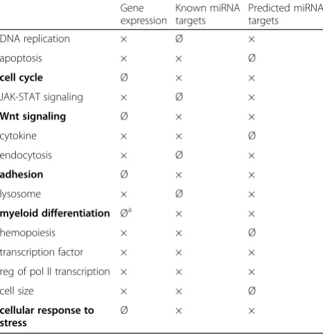
To compare the gene ontology results of MLL-AF9 de-pendently regulated genes [7] with those of known and predicted targets of MLL-AF9 dependently regulated miRNAs, we compiled a table with related gene ontology terms that were present in at least two of the three data sets (Table 1). This overview illustrates that genes dir-ectly involved in the cell cycle, Wnt signaling, adhesion, myeloid differentiation as well as in the cellular response to stress are preferentially present in the miRNA target gene data sets and thus these processes may be preferen-tially influenced by MLL-AF9 via miRNA expression.
Gene regulatory effects of miR-511 expression in THP1 monoblasts
While the preceding ontology analyses may suggest cer-tain functional roles of the set of MLL-AF9 differentially regulated miRNAs in the context of leukemia, one has to take into account that these functions are always con-text dependent and will have to be experimentally vali-dated. To this end, we decided to further experimentally investigate the functional role of miR-511, one of the most potently activated miRNAs after MLL-AF9 deple-tion. Concerning known targets, so far only TRIB2, TLR4 and CD80 have been validated for miR-511 in other cellular contexts [28, 29].
To identify targets and effects of miR-511 in monoblasts, we performed miR-511 up-regulation in THP1 cells by transfecting miRNA mimics. The subsequently analyzed gene expression profiles via whole genome arrays revealed 653 probes representing 627 genes to be differentially expressed between miR-511 and negative control mimic treatments (profiling data have been deposited in NCBI’s Gene Expression Omnibus (GEO) [18] and are available under the accession number GSE70490). Of these, 45 % were down-regulated. The previously described miR-511 target genes were either not expressed above background (TRIB2 and CD80) or not differentially regulated (TLR4) on transcript level in this setting.
interleukin 1 beta (IL1B) as well as the down-regulated genes cyclin D1 (CCND1) and platelet-derived growth fac-tor alpha polypeptide (PDGFA). For fibroblast growth fac-tor recepfac-tor 3 (FGFR3) a trend towards down-regulation was observed (Fig. 3).
MiR-511 directly regulates cyclin D1
For the three genes which were down-regulated on tran-scriptional level after miR-511 treatment (CCND1,FGFR3 andPDGFA), luciferase reporter assays were performed to test for direct miR-511 targeting. Full-length 3′UTRs of
Fig. 2MiRNA-target gene prediction associated functional annotation terms identified via DAVID. Gene ontology analysis was performed for all THP1-expressed, targets predicted via TargetScan with a minimum context + score of ±0.4 ofMLL-AF9knockdown associated miRNAs. Annotation terms were manually assorted to five higher-order terms according to the major role of the process in the biological setting under investigation. Annotation terms assigned to two higher-order terms are placed between these two and separated by dotted division lines. Some annotations were abbreviated as indicated by superscript numbers:1regulation of Wnt receptor signaling pathway,2positive regulation of Wnt receptor signaling pathway,
3
these transcripts were cloned into siCHECK2 vector. FGFR3- and PDGFA-3′UTRs were not affected by miR-511 in dual luciferase assays while CCND1-3′UTR was targeted by miR-511 as indicated by significantly reduced Renillaluciferase signals (Fig. 4).
To further validate the direct interaction and to deter-mine which binding sites for miR-511 are relevant for CCND1 regulation, seed sequences of predicted binding sites were deleted (Fig. 5a, b) and again dual luciferase assays were performed. Here, binding site 1 did contrib-ute significantly to CCND1-downregulation although only to a small extent, while binding site 2 deletion was able to completely abrogate regulation through miR-511 (Fig. 5c). Parallel deletion of both binding sites even led to an up-regulation of protein expression (Fig. 5c).
This phenomenon could be theoretically due to an off-target binding of the employed negative control mimic to the Renilla luciferase- or the subsequent 3′UTR-se-quence. However, using negative control mimic #2 and no mimic as further controls did yield similar results as with negative control mimic #1 and thus disagree with this hypothesis. Another possible explanation would be an off-target binding of miR-511 to the housekeeping fire-fly luciferase sequence, however, sequence comparisons do not support this possibility and, more importantly, the luciferase expression from the vector without 3′UTR is not affected by miR-511 treatment (compared to negative control mimics or no mimic). We thus currently cannot define the cause of the up-regulatory effect of miR-511 in luciferase assays when both binding sites were deleted fromCCND1-3′UTR. However, the employed controls in-dicated that this observation was not due to a miRNA-mimic off-target binding and that the effect is dependent on the presence ofCCND1-3′UTR.
Table 1Comparison between gene ontology results of MLL-AF9 dependently expressed genes and miRNA target genes
Gene expression
Known miRNA targets
Predicted miRNA targets
DNA replication × Ø ×
apoptosis × × Ø
cell cycle Ø × ×
JAK-STAT signaling × Ø ×
Wnt signaling Ø × ×
cytokine × × Ø
endocytosis × Ø ×
adhesion Ø × ×
lysosome × Ø ×
myeloid differentiation Øa × ×
hemopoiesis × × Ø
transcription factor × × ×
reg of pol II transcription × × ×
cell size × × Ø
cellular response to stress
Ø × ×
Compared were the gene ontology results obtained via DAVID software between MLL-AF9 dependently expressed genes [7] and known (miRTarBase) as well as predicted (TargetScan) target genes of MLL-AF9 dependently expressed miRNAs. × present in the data set, Ø not present in the data set
a
refers to gene ontology terms directly concerning myeloid differentiation (e.g. “regulation of myeloid cell differentiation”), whereas terms indirectly related to myeloid differentiation (e.g.“phagocytosis”) were also present within the gene expression profile. Terms in bold letters were preferentially present in the miRNA target gene data sets
Fig. 3MiR-511 dependently expressed gene transcripts. Subsequent to introduction of miR-511 mimics in THP1 cells, differential expression of a subset of potentially important transcripts identified in the gene expression screen was verified in three additional independent experiments via RT-qPCR on experimental days 3, 6 and 9. Columns indicate log2 fold change (log2FC) between miR-511 and negative control #1 mimic treatments.
MiR-511 reduces cyclin D1 protein level and percentage of cells in S-phase in THP1
In the next step we examined the effect of miR-511 on the protein expression of cyclin D1 (CCND1) via flow cyto-metric analysis of cyclin D1 stained THP1 cells transfected either with miR-511 or control #1 mimic. Time course ex-periments showed a trend towards reduced cyclin D1 pro-tein expression on day 6 and significant reduction on day 9 after miR-511 mimic treatment (Fig. 6a).
Due to the experimental design with reseeding of cells prior to mimic transfections, cell proliferation could only be assessed over a period of 3 days in a row, within
which no significant difference between miR-511 mimic and control mimic treatments was observed. Concerning an effect of miR-511 on the colony formation capacity of THP1 monoblasts, we observed a significant reduction of colony numbers after miR-511 mimic treatment when compared to negative control #2 mimic (p 0.02 for day 10 and 14), but only a trend towards reduced colony numbers when compared to negative control #1 mimic (p 0.15 and 0.11 for day 10 and 14 respectively, Fig. 6b).
Cyclin D1 is required for cell cycle G1/S transition [30]. To test for the corresponding functional effect of miR-511, we determined the cell cycle distribution of THP1 cells in miRNA mimic experiments via flow cy-tometry. Indeed, the percentage of cells in S-phase was significantly reduced in miR-511 mimic treatments on experimental days 6 and 9 (Fig. 6c).
MiR-511 is associated with differentiation in THP1 cells
Cyclin D1 has been described to inhibit ligand-induced PPAR-gamma function [31] while ligand activation of the PPAR-gamma/retinoid X receptor-alpha heterodimer in myelomonocytic cell lines is known to induce mono-cytic differentiation [32, 33]. Thus, expression of the miR-511 target cyclin D1 may indirectly influence monocytic differentiation. Additionally, miR-511 has been described to be upregulated after differentiating human blood monocytes into macrophages [29]. Con-cordantly, we observed miR-511 to be significantly up-regulated (10.8-fold) 24 h after inducing differentiation
Fig. 4Effect of miR-511 mimic on 3′UTRs of selected genes in a dual luciferase reporter assay. Columns represent mean over five independent experiment, bars indicate standard error of the mean. *p< 0.05 in all replicate experiments
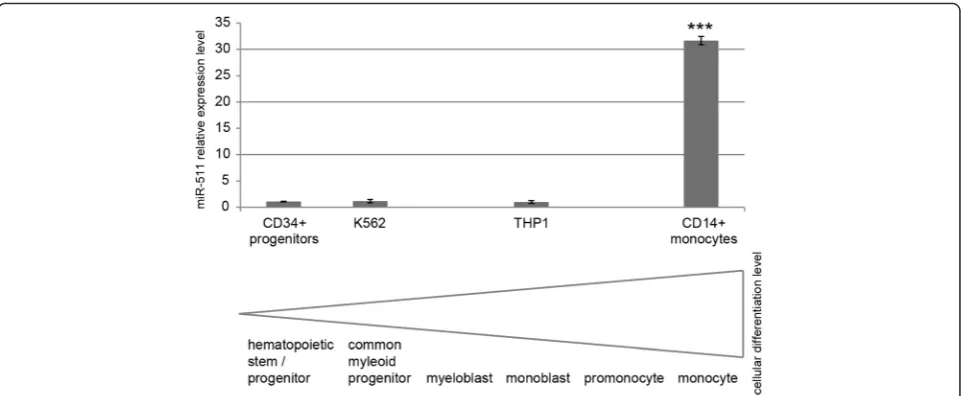
of THP1 cells via 5 ng/ml phorbol 12-myristate 13-acetate (PMA) in three independent experiments (p< 0.05). In this setting, cellular differentiation was confirmed via cell adherence, morphological changes (cell protrusions, fusi-form cells) and growth arrest. Additionally, although representing more differentiated cell types, we observed the leukemic cell lines THP1 and K562 to express similar
low levels of miR-511 as primary CD34+ hematopoietic stem/progenitor cells, while primary CD14+ monocytes expressed significant higher miR-511 levels (Fig. 7).
In this context, we asked if miR-511 is able to promote cellular differentiation in the AML cell line THP1. For this purpose, we treated THP1 cells with miR-511 mimic or negative control mimics and analysed the expression
of surface myeloid differentiation marker CD11b and CD38 [34] via flow cytometry. While CD11b was not expressed above background (isotype control) in any treatment (Additional file 2: Figure S4), CD38 signal to noise ratio was significantly higher in miR-511 treated compared to both negative control mimic treated THP1 cells and this effect intensified over the experimental time course (Fig. 6d). Compared to CD14+ mono-blasts, untreated THP1 cells expressed 4.8-fold higher levels of CD38.
MiR-511 may thus exert pro-differentiation roles in THP1 cells, as in addition to being a marker for myeloid differentiation, CD38 has been described to promote cel-lular differentiation of HL-60 cells in response to 1,25-dihydroxyvitamin D3 and retinoic acid [34]. As CD38 is not a predicted target of miR-511 via TargetScan, this ef-fect of miR-511 on monocytic differentiation is likely mediated via other target genes.
MiR-511 augments migratory capability of THP1 cells
Migratory capability of myeloid cells is closely linked to their differentiation status: during normal monocyte maturation, cells gain the capability of locomotion [35]. In contrast to mature monocytes, AML monoblasts ap-pear to lack the apparatus necessary for locomotion as neither chemotaxis nor random migration could be de-tected in this primary cell type [35].
We thus asked, if miR-511 expression is able to raise the migratory capability – as a sign of differentiation – of THP1 monoblasts. Indeed, we were able to observe that miR-511 mimic treatment significantly augments the capability of THP1 cells to migrate through 5 μm transwells (Fig. 6e).
Discussion
We employed a quantitative profiling technique (LDAs) to analyze differential expression of miRNAs after effi-cient and specific MLL-AF9 depletion in THP1 cells and validated the results by single assay RT-qPCR for a subset of deregulated miRNAs. We excluded off-target effects by employing two distinctMLL-AF9-targeting as well as two distinct non-targeting control siRNA treatments and de-tected 21 miRNAs to be differentially expressed in MLL-AF9knockdown versus control treatments.
A causal explanation is still lacking for the majority of aberrantly expressed miRNAs in acute leukemia and ab-errant expression of neighboring genes, genomic aberra-tions and specific regulatory factors can only explain the minority of dysregulated miRNAs [36]. Our study how-ever, focused on the role of the epigenetic regulator MLL-AF9 alone, so that the observed differential regula-tion of miRNAs may be attributed to this factor.
MiR-196b has been previously described to be upregu-lated by MLL-AF9 employing Mll knockout or exogen-ous Mll / Mll-Af9 transduced murine cells [37–39]. We, however, observed a divergent expression of miR-196b levels between the two non-targeting control siRNA treatments which led to the exclusion of this miRNA from our differential miRNA expression signature. This observation suggests that siRNAs may have dir-ect or indirdir-ect off-target effdir-ects not only on mRNA expression but also on miRNAs and corroborates the importance of using adequate controls for off-target effects in siRNA studies. The high stringency of our experimental design might thus explain the relatively low number of differentially expressed miRNAs we detected. Because miRNA processing is a specifically regulated process [12], it is not surprising that we did
not detect miRNA host-genes to be differentially expressed in our data set.
Recently, in a number ofin vivostudies, miRNAs have been found to be differentially expressed among distinct cytogenetic groups of AML. However, the specific signa-tures differed among studies, most likely due to the lack of standardized methods used by different groups [40]. This also includes the use of different miRNA profiling platforms and versions resulting in diverse coverages of quantified miRNAs. Nevertheless, some concordance was detected between our in vitro data set and previ-ously published in vivo data. MiR-219 was described to be upregulated in AML with MLL-AF6 and MLL-AF9 versus other AML cases [41], while miR-589 was ob-served to be upregulated in MLL-rearranged AML ver-sus normal controls [42].
To interpret possible functional and biological effects of the MLL-AF9 dependently expressed miRNAs, we employed in silico gene ontology analyses from 1) known direct as well as 2) predicted mRNA targets of these miRNAs. While the first analysis approach has the advantage to be based on bona fide MTIs instead of more or less reliable predictions, it is limited by the fact that only a small portion of actual MTIs have been ex-perimentally validated up to date and thus this analysis can only yield a partial view on the actual roles the in-volved miRNAs play. The analysis approach employing predicted MTIs on the other hand is able to represent all miRNAs and a much larger proportion of target interac-tions, but will be hindered by false positive as well as false negative MTIs. To obtain the best possible comprehensive and reliable view on the functional roles these miRNAs play, we employed both analysis approaches.
We detected the known direct targets of our set of MLL-AF9 dependently expressed miRNAs to be mainly involved in proliferation and apoptosis, in functions re-lated to monocyte / macrophage differentiation as well as in transcriptional regulation and cancer. Five of the miR-NAs (miR-137,−214-3p,−301a-3p,−330-3p and−383-5p) emerged to possess target genes affecting all of these four categories, which may imply their relevance for leukemia. The gene ontology analysis ofin silicopredicted direct tar-gets of MLL-AF9 dependently expressed miRNA implied cell cycle promoting and checkpoint decreasing roles as well as stem cell supporting and cellular stress response suppressive roles for these miRNAs.
To predict which biological functions may be predom-inantly influenced by MLL-AF9 via miRNA expression (as opposed to direct gene regulatory effects of MLL-AF9), we compared the gene ontology results from our previously published MLL-AF9 dependent gene expres-sion profile [7] with the gene ontology results from known and predicted target genes of MLL-AF9 dependently expressed miRNAs. This analysis indicated that the cell
cycle, Wnt signaling, adhesion, myeloid differentiation as well as the cellular response to stress may be preferentially influenced by MLL-AF9 via miRNA expression.
We observed enriched influence of MLL-AF9 on down-stream transcriptional regulators both, via gene expression targeting as well as via miRNA expression targeting of MLL-AF9. However, due to the relative difficulty to thera-peutically target transcription factors, e.g. via small mol-ecule drugs, manipulation of these miRNAs may present a promising future opportunity to therapeutically tar-get deregulated gene expression in MLL-AF9 positive leukemia.
Altogether, these gene ontology analyses show that the MLL-AF9 dependently expressed miRNAs are presum-ably involved in many leukemia relevant functions. Is has to be stressed however, that these analyses are not able to take into account the extent of MLL-AF9 dependent miRNA expression strength. Additionally, targeting of single gene transcripts by a miRNA is dependent on the cellular context and e.g. the present amount of transcripts from other direct target genes of that miRNA. Thus, while these analyses may suggest certain functional roles of the set of MLL-AF9 differen-tially regulated miRNAs in the context of leukemia, these functions will have to be experimentally validated.
To venture into this direction, we subsequently ana-lyzed targets and functional roles of miR-511, which was one of the most deregulated miRNAs by MLL-AF9. To this end, we employed miRNA mimic mediated up-regulation of miR-511 in THP1 cells and subsequently analyzed a comprehensive gene expression profile as well as effects on functional level.
We were able to validate significant and strong up-regulatory effects of miR-511 on the expression ofCCL2, MMP9 and IL1Band significant down-regulatory effects onPDGFAand cyclin D1. Possible anti-leukemic roles of miR-511 via anti-angiogenesis, anti-proliferative as well as pro-differentiation and pro-immunosurveillance effects is suggested by known roles of these downstream genes in chemotactic activity [43], AML immunosurveillance [43], hematopoietic progenitor cell mobilization [44], macro-phage migration [45], monocytic differentiation [46, 47] and tumor cell proliferation, angiogenesis and metastasis [48]. As such, miR-511 might be e.g. an alternative way to raise immunosurveillance against AML monoblasts via its effect to induceCCL2expression.
differentiation [32]. Concordantly, we found miR-511 ex-pression to be significantly higher in monocytes as com-pared to leukemic cell lines and hematopoietic stem/ progenitor cells.
As miRNAs primarily act via post-transcriptional gene repression, the observed up-regulatory effects of miR-511 on CCL2, MMP9 and IL1B may be indirect. Al-though this does not necessarily lessen their importance in miR-511 mediated cellular functions, we aimed at val-idating direct miR-511 targets. To this end we pre-formed dual luciferase assays with cloned full-length
3′-UTR sequences of CCND1, FRFG3 and PDGFA and
confirmed a significant repressive effect of miR-511 mimic on cyclin D1 3′UTR. We further succeeded to experimentally confirm miR-511 binding sites within the CCND1 3′UTR which validated cyclin D1 as a direct and specific target of miR-511. While cyclin D1 is not a direct target ofMLL orMLLfusion genes such as MLL-AF9 [51], this observation is in concordance with the likelihood of miR-511 targeting as predicted via TargetS-can, which yielded a context + score of −0.43 for cyclin D1, while indicating a lower probability for FGFR3 and PDGFAof being a miR-511 target (context + score−0.11 and not predicted, respectively).
Although the luciferase assays demonstrated cyclin D1 to be a direct target of miR-511, this experimental ap-proach presents a relatively artificial system with overex-pression of the target gene 3′UTR cloned into a luciferase vector and introduced into HEK cells. To ad-dress the regulation within the relevant cellular context, we additionally confirmed a suppressive effect of miR-511 on cyclin D1 protein expression in THP1 mono-blasts via flow cytometry.
We further aimed to define cellular and functional consequences of miR-511 expression in the context of AML monoblasts. To this end, we were able to provide evidence for miR-511 to reduce the proportion of cells in S-phase of the cell cycle and to increase the migratory capability of the cells. Even though we detected THP1 cells to express 4.8-fold higher levels of CD38 compared to human primary CD14+ monocytes, CD38 is an early myeloid differentiation marker in myeloblasts [52] and, importantly, a known differentiation promoting ectoen-zyme receptor [34]. Thus, miR-511 expression may pro-mote monoblast differentiation as indicated by the raised expression of CD38.
As we did not observe significant effects on cellular proliferation within the experimental time frame (up to three days), these observations primarily suggest a role of miR-511 in monoblast differentiation, as cell cycle, cyclin D1 as well as migratory capability are also linked to myeloid differentiation: cell cycle lengthening is im-portant for macrophage differentiation (e.g. via leading to an accumulation of the hematopoietic transcription
factor PU.1) [53], decrease of cyclin D1 may induce monocytic differentiation via raised PPAR-gamma/retin-oid X receptor-alpha function [31, 32] and migratory capability / locomotion is a trait gained during mono-blastic differentiation [35]. Thus, besides a potential therapeutic use in leukemia vaccination approaches via its strong up-regulatory effect on CCL2 expression which is known to raise immunosurveillance [43], miR-511 may prove useful in differentiation treatment strat-egies. The latter therapeutic approaches stimulate the leukemic cells to differentiate while often simultaneously inducing growth arrest and apoptosis [33]. Due to their usually low toxicity profile, these differentiation treat-ments may be especially useful in combination regimens with cytotoxic chemotherapies [33].
Conclusions
We here present aMLL-AF9 dependent miRNA expres-sion profile encompassing 21 miRNAs and interpret the signature’s consequences via gene ontology analyses to influence a wide array of cellular functions important for leukemia maintenance. A comparison to functional roles of MLL-AF9 dependent gene expression suggested that the cell cycle, Wnt signaling, the cellular response to stress, adhesion as well as myeloid differentiation may be preferentially influenced byMLL-AF9 via miRNA ex-pression. Furthermore, many transcriptional regulators are among the known and predicted miRNA target genes and thus therapeutic miRNA manipulation may present a possibility to target these hard-to-drug pro-teins in the future. We further present miR-511, which is strongly repressed byMLL-AF9, to upregulateCCL2, a chemokine ligand important for immunosurveillance, to directly target the key cell cycle regulator cyclin D1, to inhibit cell cycle progression, to increase cellular migra-tion and to promote monoblastic differentiamigra-tion. With these effects, miR-511 may have a potential therapeutic use as a pro-differentiation agent as well as in leukemia vaccination approaches.
Additional files
Additional file 1:All (non-filtered) gene ontology results of known miRNAs targets.DAVID gene ontology analysis was performed for all THP1-expressed, validated direct targets (extracted from miRTarBase) of
MLL-AF9knockdown associated miRNAs. This file includes all extracted MTIs extracted from miRTarBase as well as all gene ontology results from DAVID analysis (clustered and non-clustered terms). (XLSX 125 kb)
Additional file 2: Figure S1:Correlation between miRNA data from LDA and single assay RT-qPCR afterMLL-AF9knockdown in THP1 cells.
Additional file 3:All (non-filtered) gene ontology results of predicted miRNAs targets.DAVID gene ontology analysis was performed for all THP1-expressed targets predicted via TargetScan with a minimum context + score of 0.4 ofMLL-AF9knockdown associated miRNAs. This file includes all predicted MTIs as well as all gene ontology results from DAVID ana-lysis (clustered and non-clustered terms) with ap-value below 0.1 and a fold enrichment above 1.5. (XLSX 475 kb)
Additional file 4:Functional disease ontology (FunDO) analysis results of genes differentially regulated by miR-511 mimic treatment.
Analysis was used to extract leukemia and cancer related genes. (XLSX 22 kb)
Competing interests
The authors have no competing interests.
Authors’contributions
KKF conceived and designed the study, acquired, analyzed and interpreted the data and wrote the manuscript. PP performed statistical analyses of array data and critically revised the manuscript. JvF analyzed and interpreted data and critically revised the manuscript. TM, AAR and IR participated in the study conception and design and critically revised the manuscript. All authors read and approved the final manuscript.
Acknowledgements
This work was supported in part by the“Mehr LEBEN für krebskranke Kinder –Bettina Braeu”Foundation, the“Christina-Bergmann”Foundation and the German Research Foundation GRK 1202:“Oligonucleotides in Cell Biology and Therapy”. The authors would like to thank Katherine L.B. Borden from Université de Montréal, Canada, for generously providing
pcDNA3.1LacZ-CCND1-3′UTR vector and Kristin Haehnel and Carola Laudano for excellent technical assistance.
Author details
1
Division of Pediatric Hematology and Oncology, Children’s Research Center, Dr. von Hauner Children’s Hospital, Ludwig-Maximilians-Universität München, Lindwurmstrasse 2a, 80337 Munich, Germany.2Lehrstuhl für
Genomorientierte Bioinformatik, Technische Universität München, Maximus-von-Imhof-Forum 3, 85354 Freising, Germany.3Children’s Research Center, Dr. von Hauner Children’s Hospital, Ludwig-Maximilians-Universität München, Lindwurmstrasse 2a, 80337 Munich, Germany.
Received: 19 September 2015 Accepted: 3 January 2016
References
1. Dobson CL, Warren AJ, Pannell R, Forster A, Lavenir I, Corral J, et al. The mll-AF9 gene fusion in mice controls myeloproliferation and specifies acute myeloid leukaemogenesis. EMBO J. 1999;18(13):3564–74.
2. Chen W, Kumar AR, Hudson WA, Li Q, Wu B, Staggs RA, et al. Malignant transformation initiated by Mll-AF9: gene dosage and critical target cells. Cancer Cell. 2008;13(5):432–40.
3. Chowdhury T, Brady HJM. Insights from clinical studies into the role of the MLL gene in infant and childhood leukemia. Blood Cells Mol Dis. 2008;40(2):192–9. 4. Balgobind BV, Raimondi SC, Harbott J, Zimmermann M, Alonzo TA,
Auvrignon A, et al. Novel prognostic subgroups in childhood 11q23/ MLL-rearranged acute myeloid leukemia: results of an international retrospective study. Blood. 2009;114(12):2489–96.
5. Slany RK. The molecular biology of mixed lineage leukemia. Haematologica. 2009;94(7):984–93.
6. Mohan M, Lin C, Guest E, Shilatifard A. Licensed to elongate: a molecular mechanism for MLL-based leukaemogenesis. Nat Rev Cancer. 2010;10(10):721–8. 7. Fleischmann KK, Pagel P, Schmid I, Roscher AA. RNAi-mediated silencing of MLL-AF9 reveals leukemia-associated downstream targets and processes. Mol Cancer. 2014;13(1):27. doi:10.1186/1476-4598-13-27.
8. Bartel DP. MicroRNAs: target recognition and regulatory functions. Cell. 2009;136(2):215–33. doi:10.1016/j.cell.2009.01.002.
9. Butrym A, Rybka J, Baczynska D, Tukiendorf A, Kuliczkowski K, Mazur G. Low expression of microRNA-204 (miR-204) is associated with poor clinical outcome of acute myeloid leukemia (AML) patients. J Exp Clin Cancer Res: CR. 2015;34:68. doi:10.1186/s13046-015-0184-z.
10. Fan MQ, Huang CB, Gu Y, Xiao Y, Sheng JX, Zhong L. Decrease expression of microRNA-20a promotes cancer cell proliferation and predicts poor survival of hepatocellular carcinoma. J Exp Clin Cancer Res: CR. 2013;32(1):21. doi:10.1186/1756-9966-32-21.
11. Zhou Y, Huang Z, Wu S, Zang X, Liu M, Shi J. miR-33a is up-regulated in chemoresistant osteosarcoma and promotes osteosarcoma cell resistance to cisplatin by down-regulating TWIST. J Exp Clin Cancer Res: CR. 2014;33:12. doi:10.1186/1756-9966-33-12.
12. Naidu S, Magee P, Garofalo M. MiRNA-based therapeutic intervention of cancer. J Hematol Oncol. 2015;8(1):68. doi:10.1186/s13045-015-0162-0. 13. R_Development_Core_Team. R. A language and environment for statistical
computing. Vienna, Austria: R Foundation for Statistical Computing; 2008. 14. Mestdagh P, Van Vlierberghe P, De Weer A, Muth D, Westermann F,
Speleman F, et al. A novel and universal method for microRNA RT-qPCR data normalization. Genome Biol. 2009;10(6):R64.
15. Smyth GK. Linear models and empirical bayes methods for assessing differential expression in microarray experiments. Stat Appl Genet Mol Biol. 2004;3:Article3.
16. Benjamini Y, Hochberg Y. Controlling the false discovery rate: a practical and powerful approach to multiple testing. J Royal Stat SocietySeries B (Methodological). 1995;57(1):289–300.
17. Dorsam ST, Ferrell CM, Dorsam GP, Derynck MK, Vijapurkar U, Khodabakhsh D, et al. The transcriptome of the leukemogenic homeoprotein HOXA9 in human hematopoietic cells. Blood. 2004;103(5):1676–84.
18. Barrett T, Troup DB, Wilhite SE, Ledoux P, Evangelista C, Kim IF, et al. NCBI GEO: archive for functional genomics data sets - 10 years on. Nucleic Acids Res. 2011;39 suppl 1:D1005–10.
19. Vandesompele J, De Preter K, Pattyn F, Poppe B, Van Roy N, De Paepe A et al. Accurate normalization of real-time quantitative RT-PCR data by geometric averaging of multiple internal control genes. Genome Biology. 2002;3(7): research0034.1-research.12.
20. Smyth GK. Limma: linear models for microarray data. In: Gentleman R, Carey V, Dudoit S, Irizarry R, Huber W, editors. Bioinformatics and Computational Biology Solutions using R and Bioconductor. New York: Springer; 2005. p. 397–420.
21. Hsu SD, Tseng YT, Shrestha S, Lin YL, Khaleel A, Chou CH, et al. miRTarBase update 2014: an information resource for experimentally validated miRNA-target interactions. Nucleic Acids Res. 2014;42(Database issue):D78–85. doi:10.1093/nar/gkt1266.
22. Garcia DM, Baek D, Shin C, Bell GW, Grimson A, Bartel DP. Weak seed-pairing stability and high target-site abundance decrease the proficiency of lsy-6 and other microRNAs. Nat Struct Mol Biol. 2011;18(10):1139–46. doi:10.1038/nsmb.2115.
23. Huang DW, Sherman BT, Lempicki RA. Systematic and integrative analysis of large gene lists using DAVID bioinformatics resources. Nat Protoc. 2009;4(1):44–57.
24. Huang DW, Sherman BT, Lempicki RA. Bioinformatics enrichment tools: paths toward the comprehensive functional analysis of large gene lists. Nucleic Acids Res. 2009;37(1):1–13.
25. Osborne JD, Flatow J, Holko M, Lin SM, Kibbe WA, Zhu LJ, et al. Annotating the human genome with Disease Ontology. BMC Genomics. 2009;10 Suppl 1:S6. 26. Culjkovic B, Topisirovic I, Skrabanek L, Ruiz-Gutierrez M, Borden KL.
eIF4E promotes nuclear export of cyclin D1 mRNAs via an element in the 3′UTR. J Cell Biol. 2005;169(2):245–56. doi:10.1083/jcb.200501019. 27. Fan X, Kurgan L. Comprehensive overview and assessment of computational
prediction of microRNA targets in animals. Brief Bioinform. 2014. doi:10.1093/bib/bbu044.
28. Zhang C, Chi YL, Wang PY, Wang YQ, Zhang YX, Deng J, et al. miR-511 and miR-1297 inhibit human lung adenocarcinoma cell proliferation by targeting oncogene TRIB2. PLoS One. 2012;7(10):e46090. doi:10.1371/journal.pone.0046090.
29. Tserel L, Runnel T, Kisand K, Pihlap M, Bakhoff L, Kolde R, et al. MicroRNA expression profiles of human blood monocyte-derived dendritic cells and macrophages reveal miR-511 as putative positive regulator of toll-like receptor 4. J Biol Chem. 2011;286(30):26487–95.
30. Baldin V, Lukas J, Marcote MJ, Pagano M, Draetta G. Cyclin D1 is a nuclear protein required for cell cycle progression in G1. Genes Dev.
1993;7(5):812–21.
32. Tontonoz P, Nagy L, Alvarez JG, Thomazy VA, Evans RM. PPARgamma promotes monocyte/macrophage differentiation and uptake of oxidized LDL. Cell. 1998;93(2):241–52.
33. Nowak D, Stewart D, Koeffler HP. Differentiation therapy of leukemia: 3 decades of development. Blood. 2009;113(16):3655–65.
34. Kauss MA, Reiterer G, Bunaciu RP, Yen A. Human myeloblastic leukemia cells (HL-60) express a membrane receptor for estrogen that signals and modulates retinoic acid-induced cell differentiation. Exp Cell Res. 2008;314(16):2999–3006. 35. Glasser L. Functional differentiation in acute monoblastic leukemia. Am J
Clin Pathol. 1981;75(1):122–5.
36. Schotte D, Pieters R, Den Boer ML. MicroRNAs in acute leukemia: from biological players to clinical contributors. Leukemia. 2012;26(1):1–12. 37. Popovic R, Riesbeck LE, Velu CS, Chaubey A, Zhang J, Achille NJ, et al.
Regulation of mir-196b by MLL and its overexpression by MLL fusions contributes to immortalization. Blood. 2009;113(14):3314–22.
38. Cierpicki T, Risner LE, Grembecka J, Lukasik SM, Popovic R, Omonkowska M, et al. Structure of the MLL CXXC domain-DNA complex and its functional role in MLL-AF9 leukemia. Nat Struct Mol Biol. 2010;17(1):62–8.
39. Bernt KM, Zhu N, Sinha AU, Vempati S, Faber J, Krivtsov AV, et al. MLL-rearranged leukemia is dependent on aberrant H3K79 methylation by DOT1L. Cancer Cell. 2011;20(1):66–78.
40. Marcucci G, Mrozek K, Radmacher MD, Garzon R, Bloomfield CD. The prognostic and functional role of microRNAs in acute myeloid leukemia. Blood. 2011;117(4):1121–9.
41. Garzon R, Volinia S, Liu CG, Fernandez-Cymering C, Palumbo T, Pichiorri F, et al. MicroRNA signatures associated with cytogenetics and prognosis in acute myeloid leukemia. Blood. 2008;111(6):3183–9.
42. Jiang X, Huang H, Li Z, He C, Li Y, Chen P, et al. miR-495 is a tumor-suppressor microRNA down-regulated in MLL-rearranged leukemia. Proc Natl Acad Sci U S A. 2012;109(47):19397–402.
43. Legdeur MC, Broekhoven MG, Schuurhuis GJ, Beelen RH, Ossenkoppele GJ. Monocyte-chemoattractant-protein-1-mediated migration of human monocytes towards blasts from patients with acute myeloid leukemia. Cancer Immunol Immunother. 2001;50(1):16–22.
44. Pruijt JF, Fibbe WE, Laterveer L, Pieters RA, Lindley IJ, Paemen L, et al. Prevention of interleukin-8-induced mobilization of hematopoietic progenitor cells in rhesus monkeys by inhibitory antibodies against the metalloproteinase gelatinase B (MMP-9). Proc Natl Acad Sci U S A. 1999; 96(19):10863–8.
45. Gong Y, Hart E, Shchurin A, Hoover-Plow J. Inflammatory macrophage migration requires MMP-9 activation by plasminogen in mice. J Clin Invest. 2008;118(9):3012–24. doi:10.1172/JCI32750.
46. Reikvam H, Hatfield KJ, Oyan AM, Kalland KH, Kittang AO, Bruserud O. Primary human acute myelogenous leukemia cells release matrix metalloproteases and their inhibitors: release profile and pharmacological modulation. Eur J Haematol. 2010;84(3):239–51.
47. Chan S, Fung M, Mak N, Leung K. Involvement of interleukin-1 in the differentiation-inducing activity of tumor necrosis factor-alpha on a murine myeloid leukemia (WEHI-3B JCS). Int J Oncol. 1997;10(4):821–6.
48. Tripurani SK, Cook RW, Eldin KW, Pangas SA. BMP-specific SMADs function as novel repressors of PDGFA and modulate its expression in ovarian granulosa cells and tumors. Oncogene. 2013;32(33):3877–85. doi:10.1038/onc.2012.392. 49. Fu M, Wang C, Li Z, Sakamaki T, Pestell RG. Minireview: Cyclin D1: normal and
abnormal functions. Endocrinology. 2004;145(12):5439–47. doi:10.1210/en.2004-0959.
50. Guo Y, Yan K, Fang J, Qu Q, Zhou M, Chen F. Let-7b expression determines response to chemotherapy through the regulation of cyclin D1 in glioblastoma. J Exp Clin Cancer Res: CR. 2013;32:41. doi:10.1186/1756-9966-32-41.
51. Wang QF, Wu G, Mi S, He F, Wu J, Dong J, et al. MLL fusion proteins preferentially regulate a subset of wild-type MLL target genes in the leukemic genome. Blood. 2011;117(25):6895–905. doi:10.1182/blood-2010-12-324699.
52. Lamkin TJ, Chin V, Varvayanis S, Smith JL, Sramkoski RM, Jacobberger JW, et al. Retinoic acid-induced CD38 expression in HL-60 myeloblastic leukemia cells regulates cell differentiation or viability depending on expression levels. J Cell Biochem. 2006;97(6):1328–38. doi:10.1002/jcb.20745.
53. Kueh HY, Champhekar A, Nutt SL, Elowitz MB, Rothenberg EV. Positive feedback between PU.1 and the cell cycle controls myeloid differentiation. Science. 2013;341(6146):670–3. doi:10.1126/science.1240831.
• We accept pre-submission inquiries
• Our selector tool helps you to find the most relevant journal
• We provide round the clock customer support
• Convenient online submission
• Thorough peer review
• Inclusion in PubMed and all major indexing services
• Maximum visibility for your research
Submit your manuscript at www.biomedcentral.com/submit