R E S E A R C H
Open Access
Proteomic analysis of the effects of exogenous
calcium on hypoxic-responsive proteins in
cucumber roots
Lizhong He
1†, Xiaomin Lu
1,2†, Jing Tian
1, Yanjuan Yang
1, Bin Li
1, Jing Li
1and Shirong Guo
1*Abstract
Background:Hypoxia acts as a plant stress factor, particularly in cucumbers plants under hydroponic culture. Calcium is involved in stress signal transmission and in the growth of plants. To determine the effect of exogenous calcium on hypoxic-responsive proteins in cucumber (Cucumis sativusL. cv. Jinchun No.2) roots, proteomic analysis was performed using two-dimensional electrophoresis (2-DE) and mass spectrometry.
Results:Cucumber roots were used to analyze the influence of hypoxia on plants. The expressions of 38 protein spots corresponding to enzymes were shown to change in response to hypoxia. Of these, 30 spots were identified by matrix-assisted laser desorption/ionization-time of flight mass spectrometry (MALDI-TOF/TOF MS analysis). The proteins were categorized according to functional groups, including glycolysis, the tricarboxylic acid (TCA) cycle, fermentative metabolism, nitrogen metabolism, energy metabolism, protein synthesis and defense against stress. Exogenous calcium appeared to alleviate hypoxic stress via these metabolic and physiological systems. Western blotting was used to analyze the accumulation of alcohol dehydrogenase (ADH) and pyruvate decarboxylase (PDC); calcium further increased the expression of ADH and PDC under hypoxia. In addition, semi-quantitative reverse transcription-polymerase chain reaction (RT-PCR) was used to assess the transcript levels of differentially expressed proteins.
Conclusions:Exogenous calcium enhanced the expression of enzymes involved in glycolysis, the TCA cycle, fermentative metabolism, nitrogen metabolism, and reactive oxygen species (ROS) defense in plants under hypoxia. Calcium appears to induce hypoxic tolerance of cucumber seedlings. These phenomena have prompted us to further investigate the mechanisms by which cucumbers respond to exogenous calcium under hypoxia.
Keywords:Cucumber, Calcium, Hypoxic stress, Proteomics
Background
Cultivated plants that produce vegetables, crops and fruits are frequently subjected to submerged conditions (so-called hypoxia) caused by flooding [1], waterlogging [2], irrigation or hydroponic culture [3]. Plants subjected to hypoxia undergo dramatic metabolic changes and in-duce defensive mechanisms to cope with the potential damage caused. Hypoxia induces enhanced aerenchyma formation, stem elongation, gas film around submerged-leaves [4] and shoot biomass [5]. The first process to be
influenced by the metabolic change induced by a short-age of oxygen is respiration. Hypoxic stress interferes with the electron transport system causing a lack of suit-able electron acceptors, which appears to be linked to the saturated conditions of a redox state, accumulation of NAD(P)H and suppressed synthesis of ATP [6]. Energy from respiratory metabolism is necessary for the growth and yield of plants. Cucumber plants are sensitive to hyp-oxia, which frequently causes large reductions in yield [7].
Exogenous calcium can improve the suppression of growth/development of plants and help to maintain cell function by relieving gene repression caused during salt stress [8], anoxia [9], and chilling [10]. The involvement of calcium in oxygen debt responses is also observed in many plants. For example, the oxygen debt (anoxia) in cells of maize, rice and wheat plants causes a rise in * Correspondence:[email protected]
†Equal contributors
1College of Horticulture, Nanjing Agriculture University/Key Laboratory of
Southern Vegetable Crop Genetic Improvement, Ministry of Agriculture, Nanjing 210095, P. R. China
Full list of author information is available at the end of the article
© 2012 He et al.; licensee BioMed Central Ltd. This is an Open Access article distributed under the terms of the Creative Commons Attribution License (http://creativecommons.org/licenses/by/2.0), which permits unrestricted use, distribution, and reproduction in any medium, provided the original work is properly cited.
cytoplasmic Ca2+concentration [11,12]. In addition, ele-vated calcium levels significantly influence metabolic fluxes and substrate oxidation under hypoxic condition [13]. According to our previous research [14], exogenous calcium enhances the biomass and soluble protein con-tent of cucumber seedlings under hypoxia (Additional file 1: Table S1). Thus, calcium appears to act as a sig-naling component during anoxia signal transduction in
plants. The alteration in Ca2+ concentration seems to
decrease cytosolic pH, which probably represents a major signal in cells under suspension culture and in in-tact seedlings [15]. However, the mechanism of calcium’s involvement in resistance to hypoxia remains unclear.
Proteomic analysis, commonly using mass spectrom-etry (MS), is a powerful technique that facilitates the visualization and comparison of complex mixtures of proteins. Proteomic analysis provides a large amount of information on individual proteins involved in specific biological responses. Recently, there have been many proteomic studies of cucumber plants [7,16-22]; however, none of them investigated the effects of exogenous cal-cium on the proteome of cucumber seedlings under root-room hypoxia stress. The purpose of the present study is to use proteomics to clarify the relationship be-tween calcium and proteins in cucumber plants under hyp-oxic stress. We found that exogenous calcium could enhance both responsive metabolism and fermentative me-tabolism of cucumber seedlings, improving their tolerance to hypoxia. The results should provide a basis for future studies at both the physiological and molecular levels.
Results and discussion
Identification and functional classification of proteins by MALDI-TOF/TOF
To examine the effect of exogenous calcium on the proteome of cucumber seedlings under hypoxic stress in water culture, 2-DE analysis of total proteins in the roots was performed. Root proteins were extracted from cu-cumber seedlings exposed to normoxic, hypoxic and
hypoxic + 4 mM CaCl2 (hypoxic + Ca2+) conditions.
Pro-teins purified from roots were separated by 2-DE and
ana-lyzed by Imagemaster™2D Platinum software; the pIvalue
and molecular masses of these protein spots ranged from 4 to 7 and from 14.4 to 116.0 kDa, respectively. The charac-teristics of proteins in representative gels are shown in Figure 1 and described below.
Approximately 500 spots were detected in Coomassie blue (CBB)-stained gels. Thirty-eight of these spots showed significant changes in relative volume (>1.5-fold) and were excised from the gels for MALDI-TOF/TOF MS analyses.
Thirty spots were identified using the NCBI viridiplan-tae database (V.2010.12.10, 184045 sequences) and the NCBI EST viridiplantae database (V.2010.12.10, 1847412
sequences), giving an identification success rate approxi-mately 79%. The results are summarized in Table 1. Four proteins (spots 2, 11, 12 and 36) expressed under
nor-moxic and hypoxia + CaCl2conditions did not appear to
be expressed in hypoxia-treated plants. Spot 28 was present in plants under hypoxia and hypoxia + CaCl2, but was not present in plants under normoxic condition. Six-teen proteins (spots 4, 5, 8, 10, 13, 15, 16, 17, 18, 20, 22, 25, 27, 30, 32 and 37) were downregulated in hypoxia-treated plants, but upregulated in hypoxia + Ca2+treated plants. Four proteins (spots 24, 33, 34 and 38) were
downregulated under hypoxia and hypoxia + CaCl2
con-dition. The expressions of two proteins (spots 14 and 19) under hypoxia + CaCl2conditions were significantly greater than those under normoxic and hypoxic conditions. Two proteins (spot 26 and 31) were upregulated under hypoxic
conditions and further upregulated under hypoxia + CaCl2
conditions. Spot 1 was significantly accumulated under hyp-oxic and normhyp-oxic conditions.
Eight identified spots (spots 1, 2, 13, 14, 15, 24, 37 and 38) were annotated either as unnamed proteins or ESTs in the databases, and three spots (spots 25, 32, 34) were described in The Rice Annotation project Database (RAP-DB). We searched for their homologs using BLAST http://www.ncbi. nih.gov/BLAST/ and their protein or nucleotide sequences as queries. The six proteins showing the highest similarity are listed in Table 2. These similar proteins showed more than 85% positives at the amino acid level, indicating that they might have similar functions. The remaining 19 identi-fied proteins were involved in various biological processes and could be classified into three groups [23,24]. The first group consists of proteins involved carbon metabolism, ni-trogen metabolism, and energy metabolism. The second group consists of regulatory proteins involved in translation and synthesis. The third group consists of proteins partici-pating the stress response.
Structural proteins and enzymatic proteins involved in energy metabolism
Most of the identified proteins were structural proteins (non-enzymatic proteins) and enzymes involved in en-ergy metabolism. These enzymes seem to have particu-larly important roles in cucumber plants under hypoxic conditions. The expressions of cytoplasmic aconitate hydratase (spot 4), pyruvate dehydrogenase 2 (spot 20), cytoplasmic malate dehydrogenase (spot 22) and pyru-vate dehydrogenase e1 alpha subunit (spot 27) from the TCA cycle were downregulated under hypoxic
condi-tions, but upregulated under hypoxia + CaCl2conditions.
The TCA cycle is a key component of the metabolic pathway by which all aerobic organisms generate energy by oxidization of pyruvate into carbon dioxide and water. Pyruvate dehydrogenase 2 (PDH2), a pyruvate de-hydrogenase E1 beta isoform, and pyruvate dede-hydrogenase
Heet al. Proteome Science2012,10:42 Page 2 of 15
e1 alpha subunit are involved in the formation of cellular energy through the TCA cycle and in the synthesis of acetylcholine (acety1-CoA). Acetyl-CoA may then be used in the TCA cycle to carry out cellular respiration; thus, pyruvate dehydrogenase links the glycolytic pathway to the TCA cycle and releases energy via NADH. Calcium activates pyruvate dehydrogenase, isocitrate
dehydrogen-ase (IDH) andα-ketoglutarate dehydrogenase [25].
Aconi-tate hydratase (aconitase) catalyses the stereospecific isomerization of citrate to isocitrate via cis-aconitate in the TCA cycle [26,27]. Malate dehydrogenases (MDH), which is essential to the TCA cycle, catalyses the conver-sion of oxaloacetate to malate [28]. Increased malate levels in plants contributes to plant acid resistance and aluminum toxicity tolerance [29]. In apples and tomatoes, significant accumulation of malate dehydrogenases gene transcripts is related to plant and cell growth, as well as to tolerance of salt stress [30]. The activities of enzymes such as succinate dehydrogenase (SDH), isocitrate dehydrogen-ase (IDH) and malate dehydrogendehydrogen-ase (MDH) in the TCA
cycle are different between the hypoxia and hypoxia +
CaCl2conditions, where exogenous calcium promotes the
actives of SDH and IDH in cucumber [31]. Moreover, cal-cium acts to maintain higher activities of MDH and SDH and a certain level of aerobic respiration in pepper [32]. Thus, exogenous calcium seems to induce tolerance to hypoxia in cucumber plants through the activation of the enzymes involved in the TCA cycle.
Cytosolic enolase is expressed in many plant species in response to various environmental stresses, such as salt stress [33], cold [34,35] and drought [36]. However, the level of protein expression appears not to be correlated with the enzyme’s activity. Although enolase activity was increased by the stresses, the level of expression of the enolase protein showed no fluctuation [37] or was even observed to significantly decrease [33]. In the present study, this enolase (spots 10 and 11) appeared to be downregulated under hypoxia compared to hypoxia +
CaCl2 and control. These results imply that enolase
ac-tivity is regulated at the posttranscriptional level under Figure 1Two-dimensional electrophoresis gel of separated proteins at three treatments.The 2-DE protein profiles of cucumber seedling roots under normoxic (A), hypoxia (B) and hypoxia + CaCl2(C). The expression of numbered proteins was attended significantly and these
proteins were identified by MALDI-TOF/TOF MA (see Tables 1 and 2).
Heet al. Proteome Science2012,10:42 Page 3 of 15
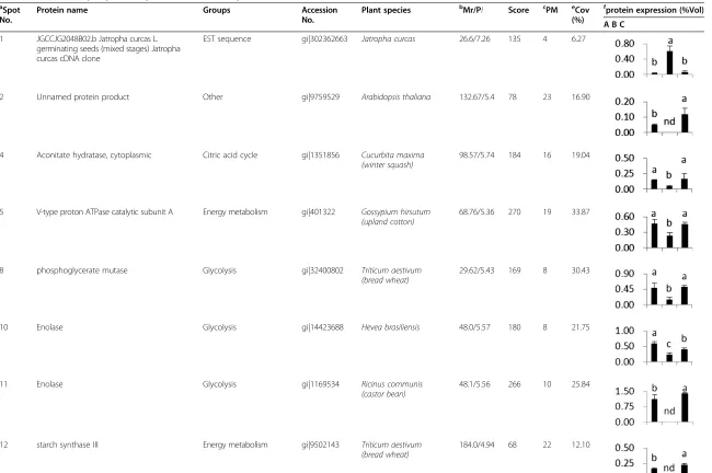
Table 1 Differentially expression proteins identified by MALDI-TOF/TOF MS
a Spot No.
Protein name Groups Accession
No.
Plant species bMr/PI Score cPM eCov (%)
f
protein expression (%Vol)
A B C
1 JGCCJG2048B02.b Jatropha curcas L. germinating seeds (mixed stages) Jatropha curcas cDNA clone
EST sequence gi|302362663 Jatropha curcas 26.6/7.26 135 4 6.27
2 Unnamed protein product Other gi|9759529 Arabidopsis thaliana 132.67/5.4 78 23 16.90
4 Aconitate hydratase, cytoplasmic Citric acid cycle gi|1351856 Cucurbita maxima (winter squash)
98.57/5.74 184 16 19.04
5 V-type proton ATPase catalytic subunit A Energy metabolism gi|401322 Gossypium hirsutum (upland cotton)
68.76/5.36 270 19 33.87
8 phosphoglycerate mutase Glycolysis gi|32400802 Triticum aestivum
(bread wheat)
29.62/5.43 169 8 30.43
10 Enolase Glycolysis gi|14423688 Hevea brasiliensis 48.0/5.57 180 8 21.75
11 Enolase Glycolysis gi|1169534 Ricinus communis
(castor bean)
48.1/5.56 266 10 25.84
12 starch synthase III Energy metabolism gi|9502143 Triticum aestivum
(bread wheat)
184.0/4.94 68 22 12.10
He
et
al.
Proteome
Science
2012,
10
:42
Page
4
of
15
http://ww
w.proteomesci
.com/cont
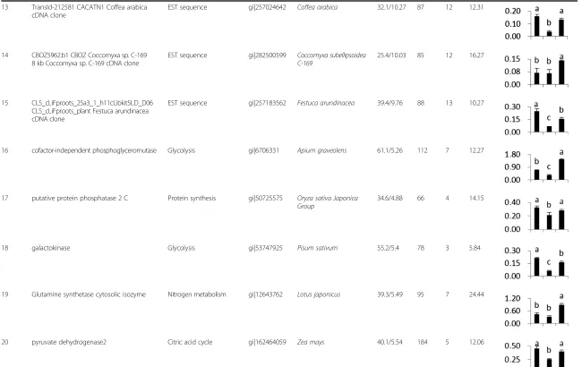
Table 1 Differentially expression proteins identified by MALDI-TOF/TOF MS(Continued)
13 TransId-212581 CACATN1 Coffea arabica cDNA clone
EST sequence gi|257024642 Coffea arabica 32.1/10.27 87 12 12.31
14 CBOZ5962.b1 CBOZ Coccomyxa sp. C-169 8 kb Coccomyxa sp. C-169 cDNA clone
EST sequence gi|282500599 Coccomyxa subellipsoidea C-169
25.4/10.03 85 12 16.27
15 CLS_cLiFproots_25a3_1_h11cLibkit5LD_D06 CLS_cLiFproots_plant Festuca arundinacea cDNA clone
EST sequence gi|257183562 Festuca arundinacea 39.4/9.76 88 13 10.27
16 cofactor-independent phosphoglyceromutase Glycolysis gi|6706331 Apium graveolens 61.1/5.26 112 7 12.27
17 putative protein phosphatase 2 C Protein synthesis gi|50725575 Oryza sativa Japonica Group
34.6/4.88 66 4 14.15
18 galactokinase Glycolysis gi|53747925 Pisum sativum 55.2/5.4 78 3 5.84
19 Glutamine synthetase cytosolic isozyme Nitrogen metabolism gi|12643762 Lotus japonicus 39.3/5.49 95 7 24.44
20 pyruvate dehydrogenase2 Citric acid cycle gi|162464059 Zea mays 40.1/5.54 184 5 12.06
He
et
al.
Proteome
Science
2012,
10
:42
Page
5
of
15
http://ww
w.proteomesci
.com/cont
Table 1 Differentially expression proteins identified by MALDI-TOF/TOF MS(Continued)
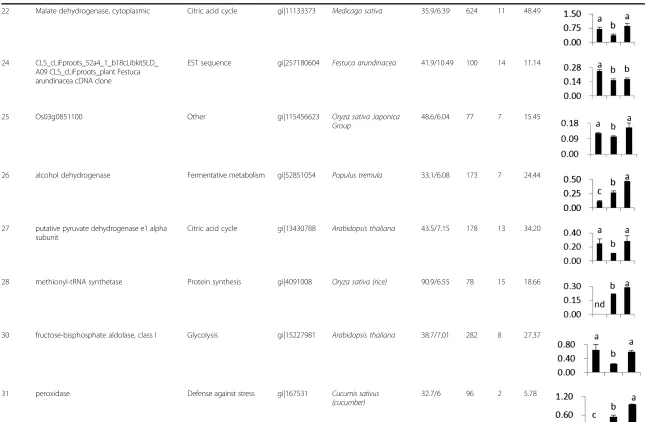
22 Malate dehydrogenase, cytoplasmic Citric acid cycle gi|11133373 Medicago sativa 35.9/6.39 624 11 48.49
24 CLS_cLiFproots_52a4_1_b18cLibkit5LD_ A09 CLS_cLiFproots_plant Festuca arundinacea cDNA clone
EST sequence gi|257180604 Festuca arundinacea 41.9/10.49 100 14 11.14
25 Os03g0851100 Other gi|115456623 Oryza sativa Japonica
Group
48.6/6.04 77 7 15.45
26 alcohol dehydrogenase Fermentative metabolism gi|52851054 Populus tremula 33.1/6.08 173 7 24.44
27 putative pyruvate dehydrogenase e1 alpha subunit
Citric acid cycle gi|13430788 Arabidopsis thaliana 43.5/7.15 178 13 34.20
28 methionyl-tRNA synthetase Protein synthesis gi|4091008 Oryza sativa (rice) 90.9/6.55 78 15 18.66
30 fructose-bisphosphate aldolase, class I Glycolysis gi|15227981 Arabidopsis thaliana 38.7/7.01 282 8 27.37
31 peroxidase Defense against stress gi|167531 Cucumis sativus
(cucumber)
32.7/6 96 2 5.78
He
et
al.
Proteome
Science
2012,
10
:42
Page
6
of
15
http://ww
w.proteomesci
.com/cont
Table 1 Differentially expression proteins identified by MALDI-TOF/TOF MS(Continued)
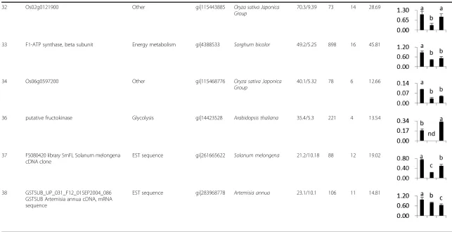
32 Os02g0121900 Other gi|115443885 Oryza sativa Japonica
Group
70.3/9.39 73 14 28.69
33 F1-ATP synthase, beta subunit Energy metabolism gi|4388533 Sorghum bicolor 49.2/5.25 898 16 45.81
34 Os06g0597200 Other gi|115468776 Oryza sativa Japonica
Group
40.1/5.32 78 6 12.66
36 putative fructokinase Glycolysis gi|14423528 Arabidopsis thaliana 35.4/5.3 221 4 13.54
37 FS080420 library SmFL Solanum melongena cDNA clone
EST sequence gi|261665622 Solanum melongena 21.2/10.18 88 12 19.02
38 GSTSUB_UP_031_F12_01SEP2004_086 GSTSUB Artemisia annua cDNA, mRNA sequence
EST sequence gi|283968778 Artemisia annua 23.1/10.1 106 11 14.81
a
Spot numbers are given in Figure1. b
Theoretical molecular mass (Mr) and isoelectric point (pI) of the identified proteins. e
The percent coverage of peptides. f
he relative levels of protein expression. A: normoxic; B: hypoxia; C hypoxia + CaCl2.
He
et
al.
Proteome
Science
2012,
10
:42
Page
7
of
15
http://ww
w.proteomesci
.com/cont
anaerobic conditions and its relative amount is increased by exogenous calcium during hypoxia.
The expression of phosphoglycerate mutase (PGAM) (spot 8) and cofactor-independent phosphoglyceromutase (iPGAM) (spot 16) markedly decreased under hypoxic stress, but increased on the addition of exogenous calcium. PGAM is a key enzyme in glycolysis, catalyzing the inter-conversion of the phosphate group from C-3 to C-2, which results in the conversion of 3-phosphoglycerate (3PGA) to 2-phosphoglycerate (2PGA). PGAMs are divided into two evolutionarily unrelated groups based on whether they re-quire 2, 3-biphosphoglycerate as a cofactor: cofactor dependent PGAMs (dPGAMs) and cofactor-independent PGAM (iPGAMs). The iPGAMs are commonly present in higher plants, some invertebrates, fungi, and bacteria [38]. PGAMs are important to stomatal movement, vegetative
biomass production, and reproduction inArabidopsis[39].
Transgenic potato plants with reduced iPGAM enzyme ac-tivity showed reduced growth because of a reduced photo-synthetic rate [40]. These phenomena suggest that the conversion of 3PGA to 2PGA may be inhibited under hyp-oxia, and that exogenous calcium may increase the abun-dance of the proteins.
Fructose-bisphosphate aldolase (FBP aldolase, spot 30) is also an essential enzyme involved in glycolysis. It catalyzes a reversible cleavage reaction of fructose-1, 6-bisphosphate (F-1, 6-BP) into two trioses: glyceraldehydes-3-phosphate and dihydroxyacetone phosphate (DHAP) [41]. Increased FBP aldolase activity stimulates the glycolytic pathway and plays an important role in gibberellin A (GA)-induced growth of rice roots [42] and in signal transduction [43]. In the present study, downregulation of FBP aldolase under the hypoxia altered the levels of glycolysis and inhibited the growth of cucumber roots. Exogenous calcium signifi-cantly elevated the quantity of the FBP aldolase, which may help alleviate the effects of hypoxic stress. This result is consistent with the expression profile of this protein in cucumber roots under salt stress [20].
The glycolytic pathway is the major source of energy when oxygen availability decreases below the level at which oxygen becomes limiting for oxidative phosphorylation [44]. Pyruvate produce from glycolysis is consumed by fer-mentative metabolism, which involves pyruvate decarb-oxylase (PDC) and alcohol dehydrogenase (ADH, spot 26). ADH catalyzes the reduction of pyruvate to ethanol and
results in continuous NAD+regeneration. ADH is
consid-ered essential for survival of plants during anaerobic condi-tions [45]. Ruthenium red, an organelle calcium channel blocker, dramatically reduced anoxia-induced ADH activity [46] and gene expression [47]. As expected, the quantity of ADH was increased under hypoxia and increased further under hypoxia + CaCl2.
Spot 12, spot 18 and spot 36 were identified as starch synthase III (SSIII), galactokinase and fructokinase, re-spectively. SS is involved in the elongation of the linear chains of starch [48]. SSIII specifically catalyzes the for-mation of chains with a degree of polymerization (DP) of 12 to 25. Other SS isoforms cannot fulfill this func-tion [49]. Galactokinase is involved in the conversion of stachyose to sucrose in the cucumber peduncle [50]. Fructokinase specifically catalyzes the transfer of a phos-phate group from ATP (the substrate) to fructose as the initial step in its utilization. Recent studies have sug-gested that sucrose and hexoses (mainly glucose and fructose) can act as sensing-molecules to elicit sugar responses in both source and sink organs when plants are under abiotic stress [51], and can control distinct aspects of plants’development [52]. In the present study, these enzymes were significantly downregulated under
hypoxia, but upregulated under hypoxia + CaCl2. A
de-crease of starch or carbon metabolic activity was also observed in other studies using various plant species under hypoxia [44,53]. These observations provide a convenient explanation of the adaptive response of plants to hypoxia, namely that plants limit their energy consumption by suppressing the synthesis of storage
Table 2 Homologs of unknown proteins
Spot No.
Accession
No.a Homologue
NCBI accession No.b Protein Name Plant species Identc Posd
B1 gi|302362663 CAI83772.1 glyceraldehyde-3-phosphate-dehydrogenase Lupinus albus 93% 97%
2 gi|9759529 NP_200612.2 FIP1 [V]-like protein Arabidopsis thaliana 99% 99%
13 gi|257024642 ACD03224.1 xyloglucan endotransglucosylase Actinidia deliciosa 77% 91%
25 gi|115456623 AAG32661.1 translational elongation factor EF-TuM Zea mays 89% 94%
32 gi|115443885 XP_003573599.1 pentatricopeptide repeat-containing protein At1g02060
Brachypodium distachyon 80% 90%
34 gi|115468776 BAD33043.1 putative protein phosphatase 2 C Oryza sativa Japonica Group 100% 100%
a
The gi number of the unknown proteins.b
The accession number of homologues.c
Identities.d
Positives.
Heet al. Proteome Science2012,10:42 Page 8 of 15
substances, such as starch and protein [54,55]. Calcium seems to enhance carbohydrate metabolism and induces sugar signaling to enhance tolerance of cucumber plants subjected to hypoxic stress.
According to recent studies using transgenic plants, over-expression or altered over-expression of glutamine synthetase (GS) promotes the development of plants [56] such as
wheat [57] and Lotus corniculatus[58]. The expression of
this protein (GS, spot 19) decreased under hypoxia, but was significantly enhanced under hypoxia + CaCl2. Thus, cal-cium appears to regulate nitrogen (N) metabolism through GS to relieve O2-deficient conditions in cucumber plants subjected to hypoxia.
ATP synthases (ATPases) are membrane-bound enzyme complexes/ion transporters that combine ATP synthesis and/or hydrolysis with the transport of protons through the membrane [20], playing a key role in biological energy metabolism. ATPases differ in respect to function (ATP synthesis and/or hydrolysis), structure (F-, V- and A-ATPases contain rotary motors) and in the type of ions they transport [59,60]. Two ATPases, i.e. V-type proton ATPase catalytic subunit A (spot 5) and F1-ATP synthase, beta subunit (spot 33) were remarkably decreased under
hypoxia. Under hypoxia + CaCl2, the level of the former
(spot 5) was restored and the latter (spot 33) showed a ten-dency to be somewhat restored. These restored levels did not reach the level of the control (normoxic conditions). V-type proton ATPases generate a proton electrochemical gradient, which is the driving force utilized by the tono-plast Na+/H+antiporter, to compartmentalize Na+into the vacuole [61]. F1-ATPases in mitochondria, chloroplasts and bacterial plasma membranes are the prime producers of ATP, using the proton gradient generated by oxidative phosphorylation (mitochondria) or photosynthesis (chloro-plasts). Mitochondrial Ca2+ accumulation triggers activa-tion of mitochondrial metabolism, which increases ATP synthesis in mitochondria and ATP levels in cytosol [62]. This phenomenon suggests that hypoxia dramatically inhi-bits energy metabolism in cucumber plants, and in the case of these two ATPases, calcium cannot completely restore them to normoxic levels.
Calcium is an essential element for cell growth and plays a role as a second messenger in signal transduction path-ways [63]. Therefore, it is not surprising that calcium is implicated in plant metabolism regulation signaling, par-ticularly in association with oxygen deprivation [64]. According to Gao et al., exogenous calcium induces the promotion of physiologically active factors and matters in muskmelon plants, as compared to the factors and matters observed in plants under hypoxic stress after 6 days [65].
CaCl2pretreatment increased the accumulation of amino
acids in rice roots under anaerobic stress, possibly via a Ca-Camodulin complex involved in the transduction of an anaerobic signal that inhibits proteolysis and solute release
[9]. In addition, downregulation of a suite of energy meta-bolic pathways, and therefore, oxygen-consumption, is a class of plant hypoxic responses [66]. In the present study, enzymes of carbon and nitrogen metabolism in the cytosol, mitochondria and chloroplasts were significantly induced by exogenous calcium. Thus, calcium enhances the toler-ance of cucumber plants under hypoxia by regulating metabolic systems in the glycolytic pathway and the TCA cycle, and the activity of enzymes, such as ADH and GS. Although exogenous calcium had only a slight effect on ATPases, this effect seems to be part of the global effect of calcium on metabolism in cucumber plants.
Regulatory proteins
Plant growth and productivity is suppressed by hypoxic stress or flooding [67]. As the cell metabolism adapts to hypoxia, increased protein degradation might control the levels of one or more regulators/enzymes [68]. Protein phosphatase 2C (PP2Cs, spot 17) decreased under hypoxia
and increased under hypoxia + CaCl2. This enzyme is a
negative regulator of stress signaling in plants and mam-mals [69] and acts predominantly through the signaling pathway of the stress hormone, abscisic acid (ABA) [70]. Thus, the increase in the amount of the enzyme under
hypoxia + CaCl2 may imply that the exogenous calcium
influences ABA signaling to relieve hypoxic stress. Spot 28 was identified as methionyl-tRNA synthetase (MetRS). This enzyme is a multi-domain protein that specifically
binds tRNAMet and catalyzes the synthesis of
methionyl-tRNAMet[71], giving it a vital role in protein biosynthesis. The MetRS gene has been described in the mitochondria and chloroplasts ofArabidopsis thaliana[72] and has been linked with plant cell anti-oxidant defense during oxidative stress [73]. Although MetRS was not detected under nor-moxic conditions, it was induced under hypoxia and fur-ther increased under hypoxia + CaCl2. This result suggests that the expression of MerRS under hypoxia represents a stress response of the cucumber seeding and calcium enhanced this response under hypoxic stress.
Proteins related to the stress response
Excessive generation of reactive oxygen species (ROS) or oxidative stress is an integral part of many stress situa-tions, including hypoxia [74]. Higher plants have active
oxygen-scavenging systems, consisting of multiple
defense enzymes that can modulate the steady-state level of ROS [75]. Peroxidase (POD, spot 31), a ubiquitous en-zyme [76] present in plants, microbes, fungi and verte-brates. This enzyme acts as a biological catalyst to scavenge H2O2[77]. The activities and gene expressions of superoxide dismutase (SOD) and ascorbate peroxidase (APX) were increased in pigeon pea [78], mung bean [74] and cucumber [79,80] under waterlogged condi-tions. These phenomena were consistent with the
Heet al. Proteome Science2012,10:42 Page 9 of 15
changes in the expression of POD in the present study. In addition, the expression of POD was further increased by treatment with calcium. Thus, exogenous calcium can enhance the activities of ROS-scavenging enzymes to defend against the damage caused by ROS, which would suppress the effects of hypoxic stress.
Validation of differentially expressed proteins by western blotting
To maintain ATP levels in plants under hypoxia, the
plants seem to continuously regenerate of NAD+ in the
cytosol (glycolysis) and mitochondria (TCA cycle). Etha-nol formed by alcohol fermentation diffuses rapidly out of the cells, inducing a considerable loss of carbon dur-ing hypoxia. In this regard, pyruvate decarboxylase (PDC) and alcohol dehydrogenase (ADH) are considered as important plant proteins for coping with hypoxia-induced cellular damage [81]. According to Kang et al. [82], 24-epibrassinolide (EBR) further enhances ADH ac-tivity in hypoxic cucumber roots. Therefore, PDC and ADH were analyzed by western blotting to verify the proteomic data. As shown in Figure 2, the expression of PDC and ADH seems to change among plants grown under the three culture conditions. The PDC protein levels were upregulated under hypoxia, as compared to normoxic (control) conditions, and was further
upregu-lated under hypoxia + CaCl2. Under hypoxia and
hyp-oxia + CaCl2, ADH showed a similar tendency that of
PDC, although the level of ADH under normoxic condi-tions was very low. The western blot results correspond well with the proteomic results and were consistent with previous research.
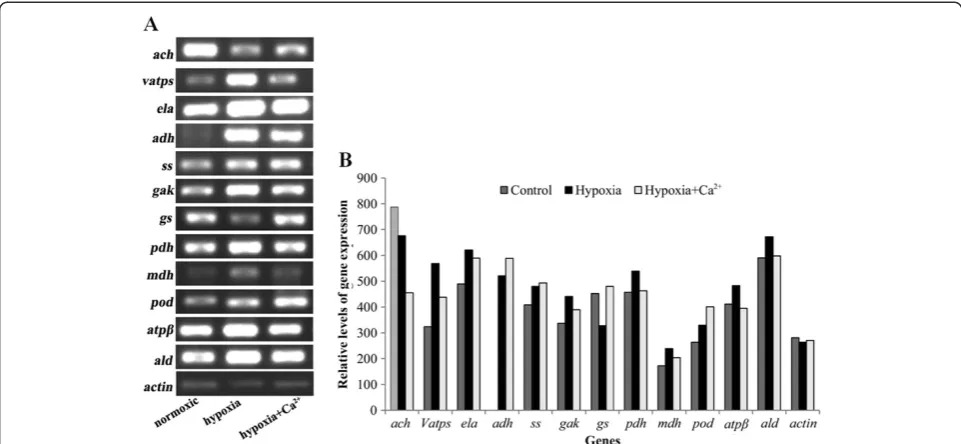
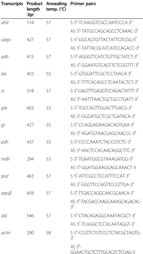
Transcript accumulation patterns for 12 candidate proteins
RT-PCR was used to analyze the changes in gene expres-sion at the mRNA level of 12 identified proteins involved in glycolysis, the TCA cycle, energy metabolism, nitro-gen metabolism, fermentative metabolism and defense against stress (Figure 3,A and B). Different peptide sequences obtained from protein spots were used to
design primers to compare mRNA accumulation under
control, hypoxia and hypoxia + CaCl2conditions, 3 days
after treatment. As shown in Figure 3A and B, the
mRNA levels of seven transcripts (vatps, ela, adh, gas,
pdh, mdh, atpβ, and ald) increased under hypoxia and
decreased under hypoxia + CaCl2; adh expression was
not detected under normoxic conditions. The gene
expressions ofssandpod increased under hypoxic stress
and further increased under hypoxia + CaCl2. The ach
gene showed an opposite tendency. Thus, the mRNA levels did not correspond with the protein levels. This is not surprising, because the final amount and activity of a protein represents an accumulation regulatory events at their transcriptional, post-transcriptional, transla-tional, and post-translational levels [83]. Therefore, the validity of estimating gene expression levels using pro-tein expression data requires further study.
Conclusions
Proteomic analysis is an effective means for clarifying pro-tein expression patterns and permits the identification of candidate proteins. In the present study, calcium was demonstrated to be involved in the short-term hypoxic tol-erance of cucumber plants. Exogenous calcium enhanced the expression of proteins involved in glycolysis, the TCA cycle, nitrogen metabolism, protein synthesis, fermentative metabolism and ROS defense. This phenomenon suggests that exogenous calcium could induce hypoxia tolerance by improving enzyme activity in systems related to respiratory metabolism and stress defense in cucumber plants. How-ever, western blotting and RT-PCR analyses showed differ-ent results for the candidate proteins. In general, exogenous calcium improves the hypoxia tolerance of plants via mul-tiple systems that are regulated by mulmul-tiple genes relating to various metabolic and signaling pathways. The present study provides evidence of the mitigating effect of exogenous cal-cium on the growth and metabolic activities of cucumber plants restrained under hypoxia. Further proteomic studies in this area are clearly warranted and are ongoing.
Methods
Plant materials and growth conditions
Cucumber (Cucumis sativusL. cv. Jinchun No.2, hypoxia
sensitive [84]) seeds were sterilized with 0.5% (W/V) so-dium hypochlorite solution for 10 min and then washed thoroughly with deionized water. The washed seeds were sown on two layers of wet filter paper and incubated in the dark at 28°C for 24 h. The germinated seedlings were transplanted to plastic trays (41 × 41 × 5 cm) containing
quartz sand and grown at 25–30°C (day) and 15–18°C
(night), with 60–75% relative humidity (RH), in a green-house of Nanjing Agriculture University in 2010. The seedlings were supplied with 1/2-strength Hoagland’s nu-trient solution (pH 6.5 ± 0.1, EC 2.0–2.2 dS m−1). At the Figure 2Western blot analysis of PDC and ADH expression
level under three treatments.
Heet al. Proteome Science2012,10:42 Page 10 of 15
2nd leaf development stage, relatively uniform seedlings were transferred to tanks containing full strength Hoag-land’s nutrient solution. The solution was renewed every
3 days. The solution in the tanks was kept at 20–25°C
and aerated with an air pump to keep the dissolved oxy-gen (DO) level at 8.0 ± 0.2 mg L−1(the optimum DO level for cucumber). At the 3rd leaf development stage, seedlings were subjected to one of three treatments. (1) Control: 1/2
Hoagland’s solution (containing 2 mM Ca2+) with DO of
8.0 ± 0.2 mg L−1. (2) Hypoxia treatment: 1/2 Hoagland’s
solution (containing 2 mM Ca2+) with DO of 1.0 ±
0.1 mg L−1, which was prepared by pumping N2-gas into
the nutrient solutions as the hypoxic treatment. The oxygen concentration in the nutrient solutions was moni-tored with an automatic DO control system (Quantum-25, Quantum Analytical Instruments Inc., USA). (3)
Hypoxia + CaCl2 treatment: 1/2 Hoagland’s solution +
4 mM CaCl2 with DO of 1.0 ± 0.1 mg L−1. The oxygen
concentration in the nutrient solutions was controlled as in the hypoxia treatment.
Protein extraction
For analysis of total protein, root samples were harvested 3 days after the end of hypoxic treatment. Protein extrac-tion was performed according to a modified version of the
method of Hurkman [85]. Root samples (1–2 g fresh
weight) were ground in a mortar with liquid nitrogen. The ground samples were suspended in 30 mM
2-amino-2-(hydroxymethyl)-1,3-propanediole (Tris)-HCl (pH 8.7) containing 1 mM ethylene glycol-bis(2-aminoethylether)-N,N,N’,N’-tetraacetic acid (EGTA), 1 mM dithiothreitol (DTT) and 1 mM phenylmethyl sulfonyl fluoride (PMSF), and then centrifuged at 15,000 g for 20 min. An aliquot (1 ml) of the resulting supernatant was placed into a tube and precipitated with acetone containing 10% TCA
and 0.07% β-mercaptoethanol. The resulting protein
sample was allowed to precipitate overnight at −20°C
and then centrifuged at 20,000 g for 25 min. The pellet was rinsed three times with cold acetone containing
0.07% β-mercaptoethanol and allowed to stand at −20°C
for 1 h. Finally, the protein pellet was air-dried and used for 2-DE.
2-DE
Isoelectric focusing (IEF) was performed according to the
methods of Duncan and Hershey [86] and O’Farrell [87].
The dried protein pellet was rehydrated in rehydration buf-fer: 7 M urea, 2 M thiourea, 4% 3-[(3-cholanidopropyl) dimethylammonio]-1-propanesulfonic acid (CHAPS) (w/v), 40 mM DTT, 0.5% (v/v) immobilized pH gradient (IPG)
buffer 4–7 and 0.01% (w/v) bromophenol blue. Protein
levels were quantified according to the Bradford method [88]. IPG strips of nonlinear pI 4–7 (13 cm) were loaded with 250μl of protein sample containing 800μg protein in a rehydration tray for 12–16 h at room temperature. Fol-lowing rehydration, the IPG strips were run on an Ettan Figure 3RT-PCR analysis of transcript levels of differentially expressed proteins under three treatments. ach: aconitate hydratase; vatps: V-type proton ATPase; ela: enolase; adh: alcohol dehydrogenase; ss: starch synthase; gak: galactokinase; gs: glutamine synthetase; pdh: pyruvate dehydrogenase; mdh: malate dehydrogenase; pod: peroxidase; atpβ: F1-ATP synthase, beta subunit; ald: aldolase. Transcript levels were measured three days after the treatments (A), and the relative abundance ratio of the genes was analyzed (B). A single concentration of cDNA was also used for amplification with ACTIN (AF171095, actin) primers. ACTIN was used as the internal standard to determine the extent of cDNA amplification.
Heet al. Proteome Science2012,10:42 Page 11 of 15
IPGphor 3 (GE Healthcare, USA). The voltage for IEF was set at 200 V for 1 h, followed by 500 V for 1 h, 1000 V for 1 h, 3000 V for 30 min, 5000 V for 30 min, gradient 8000 V for 30 min, and 8000 V rapid focus, reaching a total of 35,000 V h. The cell temperature was maintained at 20°C
with a maximum current of 50 μA per strip. After
run-ning the first dimension, IEF strips were equilibrated for 15 min with 10 ml DTT buffer containing 6 M urea, 30%
(v/v) glycerol, 2% SDS, 1% (w/v) DTT and 50 mM Tris–
HCl (pH8.8) and then with iodoacetamide buffer with 2.5% (w/v) iodoacetamide instead of DTT for 15 min.
The second dimensional SDS-polyacrylamide gel
electro-phoresis (SDS–PAGE) was carried out on running gels
(Hoefer SE600 Ruby Standard Vertical System, GE Health-care; 12.5% polyacrylamide) in the presence of SDS, as described by Laemmli [89]. The strips were embedded on the top of the SDS-gel and then sealed using a 1% molten agarose solution. Electrophoresis was carried out at 15 mA per gel until the bromophenol blue dye front reached about 1 cm from the bottom of the gel.
Image acquisition and analysis
For Coomassie brilliant blue (CBB) R-250 staining, the
gels were fixed overnight in a mixture of MeOH–H2O
(1:1, v/v) and AcOH:H2O (1:9, v/v) and then stained for
2 h in a mixture of AcOH:H2O(1:9, v/v) and 0.1% (w/v)
CBB R-250. The stained gels were destained in a mixture
of MeOH-H2O (1:1, v/v) and AcOH: H2O (1:9, v/v). The
CBB-stained 2-D gels were scanned using an Image scanner III (GE Healthcare). The digitized images were
analyzed with Imagemaster™ 2D Platinum version 5.0
(GE Healthcare). At least three gels from each treatment in three independent experiments were used for the analysis. The intensities of spots were quantified based on their rela-tive volume, which was determined by the ratio of the vol-ume of a single spot to the whole set of spots. Only spots with significant (at least 1.5-fold quantitative changes) and reproducible changes in three replicates were used for mass spectrometry. Student’st-test and a significance level of 95% were used for the statistical analysis of the gels. Only the spots showing a statistically significant difference in protein abundance between the treatments were consid-ered differentially expressed spots.
In-gel protein digestion, mass spectrometry and database search
Differentially expressed protein spots were excised from gels and transferred to sterilized 0.5 ml tubes. The excised protein spots were destained for 20 min with 100 mM
NH4HCO3in 30% acetonitrile (ACN) and then washed in
Milli-Q H2O. The spots were kept in 0.2 M NH4HCO3for 20 min and then lyophilized and rehydrated. Each spot was
digested overnight in 30μl of 50 mM NH4HCO3
contain-ing 50 ng trypsin (Promega, Madison, WI, USA). After
overnight digestion at 37°C, the peptides were extracted three times with a mixture of 50% ACN and 0.1%
CF3CO2H (TFA). Extracts were pooled together and
lyo-philized. The resulting lyophilized tryptic peptides were kept at−80°C until mass spectrometric analysis.
MALDI-TOF/TOF MS analysis and database searching
MS and MS/MS spectra were obtained using the ABI 4800 Proteomics Analyzer MALDI-TOF/TOF (Applied Biosystems, Foster City, CA, USA) operating in a result-dependent acquisition mode. Peptide mass maps were acquired in positive ion reflector mode (20 kV accelerat-ing voltage) with 1000 laser shots per spectrum. Monoi-sotopic peak masses were automatically determined
within the mass range 800–4000 Da, with a signal to
Table 3 Primer sequences used in RT-PCR
Transcripts Product length (bp)
Annealing temp. (°C)
Primer pairs
ahd 714 57 S 50-TCAAGGTCGCCAATCCCA-30
AS 50-TATGCCAGCAGCCTCAAAC-30
vatps 427 57 S 50-GGCAGTGTTACTATTGTCGG-30
AS 50-TATTACGCATCATCCAGACC-30
adh 415 57 S 50-AGGGTTCATCTGTTGCTATCT-30
AS 50-GGAATGTCAGTTCTCGGTTT-30
ela 455 55 S 50-GTGGATTCGCTCCTAACA-30
AS 50-TTTCACAGCCTCAATACTCT-30
ss 318 57 S 50-GAGTTTGAGGTCCAGACTATTT-30
AS 50-AATTTAACTGCTGCCTGATT-30
gsk 663 53 S 50-TGCCAGTTGGACTTGACG-30
AS 50-GGGATGCTCGCTGATACA-30
gs 427 55 S 50-CCAGGAGAAGACAGTGAA-30
AS 50-AGATGTAACGAGCAACCC-30
pdh 437 55 S 50-CCCCAAATCTACCGTCTC-30
AS 50-AACTCCACAACAGGCTTC-30
mdh 294 53 S 50-TGAATGGCGTAAAGATGG-30
AS 50-GGATGGAAGGAGCAAACT-3
pod 463 57 S 50-ATTCGCCTCCATTTCCAT-30
AS 50-GGCTTCCAGTTCCGTTGA-30
atpsβ 459 57 S 50-TTGACCAGGCAACGGAACA-30
AS 50 -TACGACCAAGCAAAGCAGACAC-30
ald 546 57 S 50-CTACAGAGGCAAATACGCT-30
AS 50-TCAGGCTCCACAATAGGT-30
actin 290 58 S 50 -CCGTTCTGTCCCTCTACGCTAGTG-30
AS 50
-GGAACTGCTCTTTGCAGTCTCGAG-3
S: Sense primer; AS: Anti-sense primer.
Heet al. Proteome Science2012,10:42 Page 12 of 15
noise ratio minimum set to 10 and a local noise window
width of m/z 250. The most intense ions were selected
as precursors for MS/MS acquisition, excluding com-mon trypsin autolysis peaks and matrix ion signals. In MS/MS-positive ion mode, spectra were averaged, colli-sion energy was 2 kV, and default calibration was set. Monoisotopic peak masses were automatically deter-mined with a signal to noise ratio minimum set to 5. The MS, together with MS/MS spectra were searched against the NCBI viridiplantae (V.2010.12.10, 184045 sequences) and NCBI EST viridiplantae databases (V.2010.12.10, 1847412 sequence) using the software
GPS Explorer™, version 3.6 (Applied Biosystems) and
MASCOT version 2.1 (Matrix Science, London, UK). The parameters used for searching were: trypsin cleav-age, one missed cleavage allowed; carbamidomethyl (C) set as a fixed modification; oxidation of methionines allowed as variable modification; peptide mass tolerance within 100 ppm; fragment tolerance set to ± 0.3 Da; and minimum ion score confidence interval for MS/MS data set to 95%.
RT-PCR analysis
Total RNA was extracted from roots as described in the TRI reagent protocol (Takara Bio Inc). For all samples,
total RNA (1μg) was converted to cDNA using a
Super-script first-strand synthesis system for RT-PCR accord-ing to the manufacturer’s instructions (Takara Bio Inc).
Primers were designed from the peptide sequences obtained after mass analysis according to NCBI and cu-cumber databases (cucu-cumber.genomics.org.cn). Gene-specific primers used for PCR are shown in Table 3. PCR conditions were optimized for each primer set. PCR was carried out by denaturing the cDNA at 94°C for 5 min; followed by 30 cycles of 94°C for 30 s, anneal-ing temperature (shown in Table 3) for 30 s, and exten-sion at 72°C for 35 s. The final PCR extenexten-sion step was at 72°C for 7 min. The amplified cDNA fragments were separated by 1% agarose gel electrophoresis.
Western blot analysis
The protein was extracted from roots using a mixture
con-taining 0.5 M Tris–HCl (pH 6.8), 20% (v/v) glycerol, 2%
(w/v) SDS, 5% (v/v) β-mercaptoethanol and 0.01% (w/v)
bromophenol blue. The extracted protein was quantified by
the Bradford method [88], denatured at 95°C for 3–5 min
and then stored at−20°C until analysis.
SDS-PAGE was performed according to the method of Laemmli [89]. After electrophoresis, protein bands were visualized with Coomassie blue R250. For western blot
analysis, proteins (15μg from each sample), separated by
SDS-PAGE as above, were transferred to a 0.45μm PVDF
membrane and detected with antibodies (produced in rabbit; Univ-bio, Shanghai, China) raised against ADH
(AS10_685), PDC (AS10_691), and SAM (positive con-trol). The membrane was blocked with 5% nonfat dry milk for 2 h and washed with TBST three times. The mem-brane was then probed with the appropriate rabbit
pri-mary antibody at a 1: 2000 dilution in TBST
supplemented with 5% nonfat dry milk. After an overnight incubation at 4°C, the membrane was washed with TBST and incubated at room temperature for 1 h with a Goat Anti-Rabbit IgG HRP-conjugate (1:1000 dilution with 5% dry milk) in TBST. The membrane was then washed with TBST three times and developed using diamino benzidene (DAB) and H2O2.
Additional file
Additional file 1:Table S1.Effect of Ca2+on biomass of cucumber
seedlings under hypoxia stress [14].
Competing interests
The authors declare that they have no competing interests.
Authors’contribution
HLZ carried out the 2-DE experiments and mass spectrometry analysis. LXM carried out the western blot experiments. LB and YYJ participated in the RT-PCR experiment. TJ and LJ participated in sample collection and protein extraction. GSR conceived, designed, and coordinated this study. All authors read and approved the final manuscript.
Acknowledgements
The research was support by the National Basic Research Program of China (973 Program) (No.2009CB119000), the National Nature Science Foundation of China (No.30871736, 30900995, 31071831, and 30571263), and partially supported by the earmarked foundation for Modern Agro-industry Technology Research System (No.CARS-25-C-03).
Author details
1
College of Horticulture, Nanjing Agriculture University/Key Laboratory of Southern Vegetable Crop Genetic Improvement, Ministry of Agriculture, Nanjing 210095, P. R. China.2Anhui Science and Technology University, Fengyang, 233100, An Hui, P. R. China.
Received: 19 January 2012 Accepted: 20 June 2012 Published: 12 July 2012
References
1. Bailey-Serres J, Voesenek LACJ:Flooding stress: acclimations and genetic diversity.Annu Rev Plant Biol2008,59:313–339.
2. Sairam R, Kumutha D, Ezhilmathi K, Deshmukh P, Srivastava G:Physiology and biochemistry of waterlogging tolerance in plants.Biologia Plantarum
2008,52:401–412.
3. Kang YY, Guo SR, Duan JJ:Effects of root zone hypoxia on respiratory metabolism of cucumber seedlings roots.Chin J Ecol2008,19:583–587. 4. Pedersen O, Rich SM, Colmer TD:Surviving floods: leaf gas films improve
O2and CO2exchange, root aeration, and growth of completely submerged rice.Plant J2009,58:147–156.
5. Mommer L, Visser EJW:Underwater photosynthesis in flooded terrestrial plants: a matter of leaf plasticity.Ann Bot2005,96:581–589.
6. Kennedy RA, Rumpho ME, Fox TC:Anaerobic metabolism in plants.
Plant Physiol1992,100:1.
7. Li J, Sun J, Yang Y, Guo S, Glick BR:Identification of hypoxic-responsive proteins in cucumber roots using a proteomic approach.Plant Physiol Biochem2012,51:74–80.
8. Henriksson E, Nordin Henriksson K:Salt-stress signalling and the role of calcium in the regulation of the Arabidopsis ATHB7 gene.Plant Cell Environ2005,28:202–210.
Heet al. Proteome Science2012,10:42 Page 13 of 15
9. Aurisano N, Bertani A, Reggiani R:Involvement of calcium and calmodulin in protein and amino acid metabolism in rice roots under anoxia.
Plant Cell Physiol1995,36:1525–1529.
10. Gao H, Chen G, Han L, Lin H:Calcium influence on chilling resistance of grafting eggplant seedlings.J Plant Nutr2005,27:1327–1339. 11. Yemelyanov V, Shishova M, Chirkova T, Lindberg S:Anoxia-induced
elevation of cytosolic Ca2+concentration depends on different Ca2+sources in rice and wheat protoplasts.Planta2011,234:271–280. 12. Subbaiah CC, Bush DS, Sachs MM:Mitochondrial contribution to the
Anoxic Ca2+signal in maize suspension-cultured cells.Plant Physiol1998,
118:759–771.
13. Gao H, Guo S:Influence of calcium on antioxidant system and nitrogen metabolism of muskmelon seedlings under nutrient solution hypoxia.
Acta Hort. (ISHS)2004,691:321–328.
14. He LZ, Guo SR, Lu XM, Wang LP, Yang YJ:Effects of calcium on soluble protein expression of cucumber seedlings under roo-zoon hypoxia stress.J Nanjing Agric Univ2012,35:21–25.
15. Evans NH, McAinsh MR, Hetherington AM, Knight MR:ROS perception in Arabidopsis thaliana: the ozone-induced calcium response.Plant J2005,
41:615–626.
16. Walz C, Giavalisco P, Schad M, Juenger M, Klose J, Kehr J:Proteomics of curcurbit phloem exudate reveals a network of defence proteins.
Phytochemistry2004,65:1795–1804.
17. Buhtz A, Kolasa A, Arlt K, Walz C, Kehr J:Xylem sap protein composition is conserved among different plant species.Planta2004,219:610–618. 18. Segarra G, Casanova E, Bellido D, Odena MA, Oliveira E, Trillas I:Proteome,
salicylic acid, and jasmonic acid changes in cucumber plants inoculated withTrichoderma asperellumstrain T34.Proteomics2007,7:3943–3952. 19. Donnini S, Prinsi B, Negri A, Vigani G, Espen L, Zocchi G:Proteomic
characterization of iron deficiency responses in Cucumis sativus L. roots.
BMC Plant Biol2010,10:268.
20. Du CX, Fan HF, Guo SR, Tezuka T, Li J:Proteomic analysis of cucumber seedling roots subjected to salt stress.Phytochemistry2010,71:1450–1459. 21. Todaka D, Kanekatsu M:Analytical method for detection of beta-amylase
isozymes in dehydrated cucumber cotyledons by using two-dimensional polyacrylamide gel electrophoresis.Anal Biochem2007,365:277. 22. Juszczuk IM, Rychter AM:BN-PAGE analysis of the respiratory chain
complexes in mitochondria of cucumber MSC16 mutant.Plant Physiol Biochem2009,47:397–406.
23. Li Q, Huang J, Liu S, Li J, Yang X, Liu Y, Liu Z:Proteomic analysis of young leaves at three developmental stages in an albino tea cultivar.
Proteome Sci2011,9:44.
24. Bevan M, Bancroft I, Bent E, Love K, Goodman H, Dean C, Bergkamp R, Dirkse W, Van Staveren M, Stiekema W:Analysis of 1.9 Mb of contiguous sequence from chromosome 4 of Arabidopsis thaliana.Nature1998,
391:485.
25. Denton RM, Randle PJ, Bridges BJ, Cooper RH, Kerbey AL, Pask HT, Severson DL, Stansbie D, Whitehouse S:Regulation of mammalian pyruvate dehydrogenase.Mol Cell Biochem1975,9:27–53.
26. Beinert H, Kennedy M:Aconitase, a two-faced protein: enzyme and iron regulatory factor.FASEB J1993,7:1442–1449.
27. Flint DH, Allen RM:Iron−sulfur proteins with nonredox functions.Chem Rev1996,96:2315–2334.
28. Yao YX, Li M, Zhai H, You CX, Hao YJ:Isolation and characterization of an apple cytosolic malate dehydrogenase gene reveal its function in malate synthesis.J Plant Physiol2011,168:474–480.
29. Tesfaye M, Temple SJ, Allan DL, Vance CP, Samac DA:Overexpression of Malate Dehydrogenase in Transgenic Alfalfa enhances organic acid synthesis and confers tolerance to aluminum.Plant Physiol2001,
127:1836–1844.
30. Yao YX, Dong QL, Zhai H, You CX, Hao YJ:The functions of an apple cytosolic malate dehydrogenase gene in growth and tolerance to cold and salt stresses.Plant Physiol Biochem2011,49:257–264.
31. Hu XH, Li J, Guo SR, Li Ji:Effects of Ca2+on respiratory metabolism in roots of cucumber seedlings under root-zone hypoxia stress.Acta Hortic Sin2007,33:1113–1116.
32. Zhang ER, Ren YY, Hu HQ, Liu YH, Chen SS:Effects of calcium on growth and respiratory metabolism of hot pepper seedling roots under flood stress.Acta Hortic Sin2009,36:1749–1754.
33. Yan S, Tang Z, Su W, Sun W:Proteomic analysis of salt stress-responsive proteins in rice root.Proteomics2005,5:235–244.
34. Lee DG, Ahsan N, Lee SH, Lee JJ, Bahk JD, Kang KY, Lee BH:Chilling stress-induced proteomic changes in rice roots.J Plant Physiol2009,166:1–11. 35. Zhao J, Zuo K, Tang K:cDNA Cloning and Characterization of Enolase
from Chinese Cabbage,Brassica campestrisssp. Pekinensis.Mitochondrial DNA2004,15:51–57.
36. Riccardi F, Gazeau P, de Vienne D, Zivy M:Protein changes in response to progressive water deficit in maize.Plant Physiol1998,117:1253–1263. 37. Forsthoefel NR, Cushman M, Cushman JC:Posttranscriptional and
Posttranslational Control of Enolase Expression in the Facultative Crassulacean Acid Metabolism Plant Mesembryanthemum crystallinum L.Plant Physiol1995,108:1185–1195.
38. Jedrzejas MJ:Structure, function, and evolution of phosphoglycerate mutases: comparison with fructose-2,6-bisphosphatase, acid phosphatase, and alkaline phosphatase.Prog Biophys Mol Biol2000,
73:263–287.
39. Zhao Z, Assmann SM:The glycolytic enzyme, phosphoglycerate mutase, has critical roles in stomatal movement, vegetative growth, and pollen production inArabidopsis thaliana.J Exp Bot2011,62:5179–5189. 40. Westram A, Lloyd JR, Roessner U, Riesmeier JW, Kossmann J:Increases of
3-phosphoglyceric acid in potato plants through antisense reduction of cytoplasmic phosphoglycerate mutase impairs photosynthesis and growth, but does not increase starch contents.Plant Cell Environ2002,
25:1133–1143.
41. Matsumoto M, Ogawa Ki:New insight into the Calvin Cycle Regulation Glutathionylation of Fructose Bisphosphate Aldolase in Response to Illumination. InPhotosynthesis Energy from the Sun. Edited by Allen JF, Gantt E, Golbeck JH, Osmond B. Netherlands: Springer; 2008:872–874.
42. Konishi H, Kitano H, Komatsu S:Identification of rice root proteins regulated by gibberellin using proteome analysis.Plant Cell Environ2005,
28:328–339.
43. Schaeffer GW, Sharpe FT, Sicher RC:Fructose 1,6-bisphosphate aldolase activity in leaves of a rice mutant selected for enhanced lysine.
Phytochemistry1997,46:1335–1338.
44. Rocha M, Licausi F, Araujo WL, Nunes-Nesi A, Sodek L, Fernie AR, van Dongen JT:Glycolysis and the Tricarboxylic Acid Cycle are linked by Alanine Aminotransferase during Hypoxia Induced by Waterlogging of Lotus japonicus.Plant Physiol2010,152:1501–1513.
45. Johnson JR, Cobb BG, Drew MC:Hypoxic Induction of Anoxia Tolerance in Roots of Adh1 Null Zea mays L.Plant Physiol1994,105:61–67.
46. Subbaiah CC, Zhang J, Sachs MM:Involvement of Intracellular Calcium in Anaerobic Gene Expression and Survival of Maize Seedlings.Plant Physiol
1994,105:369–376.
47. Nie X, Durnin D, Igamberdiev A, Hill R:Cytosolic calcium is involved in the regulation of barley hemoglobin gene expression.Planta2006,223:
542–549.
48. Szydlowski N, Ragel P, Raynaud S, Lucas MM, Roldán I, Montero M, Muñoz FJ, Ovecka M, Bahaji A, Planchot V,et al:Starch Granule Initiation in Arabidopsis Requires the Presence of Either Class IV or Class III Starch Synthases.The Plant Cell Online2009,21:2443–2457.
49. Zhang X, Szydlowski N, Delvallé D, D’Hulst C, James M, Myers A:
Overlapping functions of the starch synthases SSII and SSIII in amylopectin biosynthesis in Arabidopsis.BMC Plant Biol2008,8:96. 50. Miao M, Xu X, Chen X, Xue L, Cao B:Cucumber carbohydrate metabolism
and translocation under chilling night temperature.J Plant Physiol2007,
164:621–628.
51. Rosa M, Prado C, Podazza G, Interdonato R, González JA, Hilal M, Prado FE:
Soluble sugars—Metabolism, sensing and abiotic stress: A complex network in the life of plants.Plant Signal Behav2009,4:388. 52. Eveland AL, Jackson DP:Sugars, signalling, and plant development.
J Exp Bot2011,63(9):3367–3377.
53. Van Dongen JT, Roeb GW, Dautzenberg M, Froehlich A, Vigeolas H, Minchin PEH, Geigenberger P:Phloem Import and Storage Metabolism Are Highly Coordinated by the Low Oxygen Concentrations within Developing Wheat Seeds.Plant Physiol2004,
135:1809–1821.
54. Gupta KJ, Zabalza A, Van Dongen JT:Regulation of respiration when the oxygen availability changes.Physiol Plant2009,137:383–391.
55. Geigenberger P:Response of plant metabolism to too little oxygen.
Curr Opin Plant Biol2003,6:247–256.
56. Miflin BJ, Habash DZ:The role of glutamine synthetase and glutamate dehydrogenase in nitrogen assimilation and possibilities for
Heet al. Proteome Science2012,10:42 Page 14 of 15
improvement in the nitrogen utilization of crops.J Exp Bot2002,
53:979–987.
57. Habash DZ, Massiah AJ, Rong HL, Wallsgrove RM, Leigh RA:The role of cytosolic glutamine synthetase in wheat.Ann Appl Biol2001,138:83–89. 58. Vincent R, Fraisier V, Chaillou S, Limami MA, Deleens E, Phillipson B, Douat
C, Boutin JP, Hirel B:Overexpression of a soybean gene encoding cytosolic glutamine synthetase in shoots of transgenicLotus corniculatus L. plants triggers changes in ammonium assimilation and plant development.Planta1997,201:424–433.
59. Cross RL, Müller V:The evolution of A-, F-, and V-type ATP synthases and ATPases: reversals in function and changes in the H+/ATP coupling ratio.
FEBS Lett2004,576:1–4.
60. Rappas M, Niwa H, Zhang X:Mechanisms of ATPases-A multi-disciplinary approach.Curr Protein Pept Sci2004,5:89–105.
61. Chinnusamy V, Jagendorf A, Zhu JK:Understanding and improving salt tolerance in plants.Crop Sci2005,45:437–448.
62. Jouaville LS, Pinton P, Bastianutto C, Rutter GA, Rizzuto R:Regulation of mitochondrial ATP synthesis by calcium: evidence for a long-term metabolic priming.Proc Natl Acad Sci1999,96:13807–13812.
63. Chung HJ, Ferl RJ:Arabidopsis alcohol dehydrogenase expression in both shoots and roots Is conditioned by root growth environment.
Plant Physiol1999,121:429–436.
64. Rhoads DM:Plant mitochondrial retrograde regulation. InPlant Mitochondria. Volume 1. Edited by Kempken F. New York: Springer; 2011:411–437. Advances in Plant Biology.
65. Gao H, Jia Y, Guo S, Lv G, Wang T, Juan L:Exogenous calcium affects nitrogen metabolism in root-zone hypoxia-stressed muskmelon roots and enhances short-term hypoxia tolerance.J Plant Physiol2011,
168:1217–1225.
66. Peter G:Response of plant metabolism to too little oxygen.Curr Opin Plant Biol2003,6:247–256.
67. Drew MC, He CJ, Morgan PW:Programmed cell death and aerenchyma formation in roots.Trends Plant Sci2000,5:123–127.
68. Smalle J, Kurepa J, Yang P, Babiychuk E, Kushnir S, Durski A, Vierstra RD:
Cytokinin growth responses in Arabidopsis involve the 26S Proteasome subunit RPN12.The Plant Cell Online2002,14:17–32.
69. Vlad F, Rubio S, Rodrigues A, Sirichandra C, Belin C, Robert N, Leung J, Rodriguez PL, Laurière C, Merlot S:Protein phosphatases 2C regulate the activation of the Snf1-related kinase OST1 by Abscisic acid in Arabidopsis.The Plant Cell Online2009,21:3170–3184.
70. Lammers T, Lavi S:Role of Type 2C Protein Phosphatases in Growth Regulation and in Cellular Stress Signaling.Crit Rev Biochem Mol Biol2007,
42:437–461.
71. Havrylenko S, Legouis R, Negrutskii B, Mirande M:Methionyl-tRNA synthetase from Caenorhabditis elegans: A specific multidomain organization for convergent functional evolution.Protein Sci2010,
19:2475–2484.
72. Menand B, Maréchal-Drouard L, Sakamoto W, Dietrich A, Wintz H:A single gene of chloroplast origin codes for mitochondrial and chloroplastic methionyl–tRNA synthetase inArabidopsis thaliana.Proc Natl Acad Sci
1998,95:11014–11019.
73. Millar H, Considine MJ, Day DA, Whelan J:Unraveling the Role of Mitochondria During Oxidative Stress in Plants.IUBMB Life2001,
51:201–205.
74. Sairam RK, Dharmar K, Lekshmy S, Chinnusamy V:Expression of antioxidant defense genes in mung bean (Vigna radiata L.) roots under water-logging is associated with hypoxia tolerance.Acta Physiol Plant2010,
33:735–744.
75. Parida AK, Das AB:Salt tolerance and salinity effects on plants: a review.
Ecotoxicol Environ Saf2005,60:324–349.
76. Saraiva JA, Nunes CS, Coimbra MA:Purification and characterization of olive (Olea europaeaL.) peroxidase–evidence for the occurrence of a pectin binding peroxidase.Food Chem2007,101:1571–1579. 77. Ikehata K, Buchanan ID, Pickard MA, Smith DW:Purification,
characterization and evaluation of extracellular peroxidase from two Coprinus species for aqueous phenol treatment.Bioresour Technol2005,
96:1758–1770.
78. Sairam R, Kumutha D, Ezhilmathi K, Chinnusamy V, Meena R:Waterlogging induced oxidative stress and antioxidant enzyme activities in pigeon pea.Biol Plant2009,53:493–504.
79. Kang Yy, Guo Sr, Li J, Duan Jj:Effects of 24-Epibrassinolide on antioxidant system in cucumber seedling roots under hypoxia stress.Agric Sci China
2007,6:281–289.
80. Wang CY, Guo SR, Liu CJ:Effects of calcium on expression of defense enzymes isoenzymes in roots of cucumber seedlings under root-zone hypoxic stress.Acta Bot Boreali-Occidentalia Sin2009,9:1874–1880. 81. Mustroph A, Boamfa E, Laarhoven L, Harren F, Albrecht G, Grimm B:
Organ-specific analysis of the anaerobic primary metabolism in rice and wheat seedlings. I: dark ethanol production is dominated by the shoots.
Planta2006,225:103–114.
82. Kang YY, Guo SR, Li J, Duan JJ:Effect of root applied 24-epibrassinolide on carbohydrate status and fermentative enzyme activities in cucumber (Cucumis sativusL.) seedlings under hypoxia.Plant Growth Regul2009,
57:259–269.
83. Yan SP, Zhang QY, Tang ZC, Su WA, Sun WN:Comparative Proteomic Analysis Provides New Insights into Chilling Stress Responses in Rice.
Mol Cell Proteomics2006,5:484–496.
84. Ma YH, Guo SR:On different hypoxia tolerance of thirteen cucumber varieties.Jiangsu Agric Sci2004,5:68–70.
85. Hurkman WJ, Tanaka CK:Solubilization of plant membrane proteins for analysis by two-dimensional gel electrophoresis.Plant Physiol1986,
81:802.
86. Duncan R, Hershey JWB:Evaluation of isoelectric focusing running conditions during two-dimensional isoelectric focusing/sodium dodecyl sulfate-polyacrylamide gel electrophoresis: variation of gel patterns with changing conditions and optimized isoelectric focusing conditions.
Anal Biochem1984,138:144–155.
87. O’Farrell PH:High resolution two-dimensional electrophoresis of proteins.
J Biol Chem1975,250:4007.
88. Bradford MM:A rapid and sensitive method for the quantitation of microgram quantities of protein utilizing the principle of protein-dye binding.Anal Biochem1976,72:248–254.
89. Laemmli UK:Cleavage of structural proteins during the assembly of the head of bacteriophage T4.Nature1970,227:680–685.
doi:10.1186/1477-5956-10-42
Cite this article as:Heet al.:Proteomic analysis of the effects of exogenous calcium on hypoxic-responsive proteins in cucumber roots.
Proteome Science201210:42.
Submit your next manuscript to BioMed Central and take full advantage of:
• Convenient online submission
• Thorough peer review
• No space constraints or color figure charges
• Immediate publication on acceptance
• Inclusion in PubMed, CAS, Scopus and Google Scholar
• Research which is freely available for redistribution
Submit your manuscript at www.biomedcentral.com/submit
Heet al. Proteome Science2012,10:42 Page 15 of 15