METHODOLOGY
Protocol: an improved
and universal procedure for whole-mount
immunolocalization in plants
Taras Pasternak
1, Olaf Tietz
1, Katja Rapp
1, Maura Begheldo
4, Roland Nitschke
2,5, Benedetto Ruperti
4and Klaus Palme
1,2,3,5*Abstract
Rapid advances in microscopy have boosted research on cell biology. However sample preparation enabling excel-lent reproducible tissue preservation and cell labeling for in depth microscopic analysis of inner cell layers, tissues and organs still represents a major challenge for immunolocalization studies. Here we describe a protocol for whole-mount immunolocalization of proteins which is applicable to a wide range of plant species. The protocol is improved and robust for optimal sample fixation, tissue clearing and multi-protein staining procedures and can be used in combination with simultaneous detection of specific sequences of nucleic acids. In addition, cell wall and nucleus labelling can be implemented in the protocol, thereby allowing a detailed analysis of morphology and gene expres-sion patterns with single-cell resolution. Besides enabling accurate, high resolution and reproducible protein detec-tion in expression and localizadetec-tion studies, the procedure takes a single working day to complete without the need for robotic equipment.
Keywords: Immunolocalization, Tissue multi-protein expression, Whole-mount, 3D reconstruction, Protein–protein interaction
© 2015 Pasternak et al. This article is distributed under the terms of the Creative Commons Attribution 4.0 International License (http://creativecommons.org/licenses/by/4.0/), which permits unrestricted use, distribution, and reproduction in any medium, provided you give appropriate credit to the original author(s) and the source, provide a link to the Creative Commons license, and indicate if changes were made. The Creative Commons Public Domain Dedication waiver (http://creativecommons.org/ publicdomain/zero/1.0/) applies to the data made available in this article, unless otherwise stated.
Background
Multicolor immunolocalization and imaging approaches are increasingly used in plant biology for a variety of dif-ferent purposes including analysis of protein localization and protein–protein interactions in specific tissue con-texts [1], tracking of cell anatomy [2], visualizing tissue and cellular distribution of specific low molecular weight molecules (i.e. hormones such as auxin) [3] and record-ing signalrecord-ing events at the organelle subcellular level. In plants robust and reliable techniques are highly required for the accurate whole-mount visualization of subcellu-lar protein localization in relatively thick specimens, in a well preserved tissue structure to analyze patterns of gene expression in developmental studies. Current techniques for the whole-mount visualization of protein expression
and subsequent three-dimensional (3D) imaging include fluorescent protein localization [4] and immunolocaliza-tion with antibodies on Arabidopsis seedlings [5, 6]. These methods work relatively well on roots of very young Arabi-dopsis seedlings, where tissue penetration is facilitated but they are currently limited with respect to the depth of pen-etration within the tissue(s) and to the resolution that can be achieved. Confocal laser scanning microscopy of plant tissues allows analysis of relatively thin and semitranspar-ent organs, while penetration and optical sectioning for 3D reconstruction of relatively thick specimens is limited so that cellular and intracellular details are usually difficult to resolve also when two-photon confocal microscopes are used. Particularly, the simultaneous localization of nucleic acids (DNA, RNA) and of fluorescently labeled proteins (through translational fusions) are difficult to perform in depth on tissues, even if they have been cleared to reduce background fluorescence. Similarly, the use of antibod-ies labeled with fluorescent dyes for immunolocalization studies suffer from poor tissue penetration or bad tissue
Open Access
*Correspondence: [email protected]
5 Center for Biological Systems Analysis, University of Freiburg, Freiburg,
Germany
preservation after harsh chemical treatments which are necessarily performed to improve penetration of antibod-ies into deeper cell layers. In addition, currently available whole-mount protocols [5, 6] consist of a large number of steps and are sometimes poorly reproducible due to limitations with respect to antibody penetrance and tissue preservation [7, 8]. We have systematically analyzed criti-cal parameters for tissue fixation, improved cell permea-tion techniques and developed a protocol for reproducible visualization of internal tissue structures of different plant organs (e.g. siliques, ovules, roots) at all stages of develop-ment without requiredevelop-ment for sectioning.
Tissue fixation has been found as the most crucial step: effective and rapid penetration of the fixative in the inner cell layers has a primary importance for all further steps. Therefore an effective combination of vacuum with a detergent is crucial for successful fixation. The plant cuticle is an extracellular hydrophobic layer that covers the aerial epidermis of all land plants, providing protection against desiccation. In our protocol hot methanol (up to 60 °C) has been implemented as an effective way for permeabilization of the cuticle and increasing tissue permeabilization, espe-cially in dense organs in the inner cell layers.
We show that the protocol is fast, simple, suitable for automation, and presents a highly valuable, robust tool for the study of the cellular organization of a wide range of plant tissues. In addition the improved method allows simultaneous staining of nucleic acids and of pro-teins, and enables obtaining high resolution images of a quality suitable for 3D confocal reconstruction of cel-lular gene expression networks within a plant organ. We demonstrate the usefulness of this protocol for the characterization of auxin transport routes in a number of dicotyledonous and monocotyledonous plant spe-cies, during ovule reproductive organ development, and cytoskeleton labeling during mitosis. The reported proto-col allows robust immunolabeling of different tissues in a wide range of plant species at high penetration depth, independently from tissue transparency and density, ena-bling better resolution and 3-D reconstruction for digital atlas of whole plants organs (roots, leaf etc.) [9].
Methods
Reagents and solutions
Antifade mounting medium: Fluoromount G (refractive index 1.393; Southern Biotech, cat. no. 0100-01) or
Pro-longGold (refractive index 1.47; http://products.invitro-gen.com/ivgn/product/P36930);
Blocking solution: 2 % albumin fraction V BSA (Carl Roth, cat. no. 8076.2) in 1 × MTSB;
Calcofluor white (BR 28, Sigma, cat. no. F3543) (0.4 mg/l in 10 mM Tris-HCl pH 9.2) (dilute from 1 mg ml−1 stock in DMSO);
Cell wall digestion enzymes: 0.2 % Driselase (Sigma, cat. no. D9515), 0.15 % Macerozyme (Duchefa, cat. N M8002.0010) in 2 mM MES (Sigma, cat. no M3671), pH 5.0;
Nuclear stain: DAPI (4′,6-diamidino-2-phenylindole dihydrochloride; Sigma, cat. no. D9564) (0.2 mg/l) in water (dilute from 1 mg ml−1 stock in water). Note:
Dis-solve DAPI in water at a concentration of 1 mg/ml and dilute it before use to 2 μl in 10 ml. A 1 mg/ml solution is stable for at least 1 year at 4 °C;
Fixative solution: 2 % paraformaldehyde (Merck, cat. no. 1040051000) in 1× MTSB supplemented with 0.1 % Triton X-100 (Carl Roth, cat. no. 3051.2); Solution preparation: 2 g of Para-formaldehyde is dissolved in 20 ml of water (10 % stock solution) by stripping and slightly warming to 65-70 °C and addition of one drop of 1 M KOH. The stock solution can be stored in 2 ml aliquots at −20 °C. Prior to usage it is diluted to 2 % paraformaldehyde in using 2× MTSB and water to reach 1× MTSB (final concentrations);
Methanol (Carl Roth, cat. no. 4627.2) for tissue fixa-tion, clearing and cuticle solubilization;
MTSB (microtubule-stabilizing buffer): Preparation of stock solution (2× MTSB): 15 g PIPES (FW 302.4; Roth, cat. no. 9156.3), 1.90 g EGTA (FW 380.4; Roth, cat. no. 3054.2), 1.22 g MgSO4·7H2O (FW 246.48; Carl Roth, cat.
no. 8283.1) and 2.5 g KOH (FW 56.11; Carl Roth, cat. no. 6751.1) are dissolved in a total of 500 ml water at pH 7.0 (adjusted with 10 M KOH);
Permeabilization buffer: 3 % non-ionic detergent IGE-PAL CA-630 (Sigma, cat. no. I3021) (Similar to Nonidet P-40, which is no longer commercially available) plus 10 % dimethylsulfoxide (DMSO) (Carl Roth, cat. no. 4720.2) in 1× MTSB buffer;
Primary antibody solution: the primary antibody solu-tion is prepared in blocking solusolu-tion; the optimal anti-body concentration must be determined experimentally and can vary between 1:20 and 1:1000;
Propidium Iodine (PI, Sigma, cat. no. P4170) (1 mg/l) in 10 mM Tris–HCl, pH 7.5 diluted from 4 mg/ml stock in water;
RNAse solution (0.1 mg/ml) in 10 mM Tris–HCl, pH 7.5 (Sigma, cat. no. R5000) prepared from 1 mg/ml stock, diluted in water;
Secondary antibody solution (preparation in 1× block-ing buffer with 1:500 dilution immediately before use).
Equipment
Shaker for gentle shaking during fixation.
Agilent slides (G2534-60530 or G2534-60535, with 8 or 4 rubber frames (Additional file 1) for whole plant/organ labeling;
Conical tubes (Greiner) 15 and 50 ml;
Cover glasses: 0.17 mm thick; 24 × 40 mm (Carl-Roth, cat N. 1870.2); we recommend for high resolution microscopy always cover-glasses of defined thickness 0.17 mm ± 0.01 or 0.005 mm.
Incubator (37 °C);
Forceps (Carl Roth, cat. no. K341.1);
Humid chambers are prepared from 90 mm Petri dishes with wet absorbent paper inside;
Micropipettes;
Microscope slides for mounting of specimens after labeling;
Parafilm strips;
Poly-l-Lysine Coated Microscope Slides (e.g. from Pol-ysciences, cat. no. 22247-1) or home-made slides coated with 10 % Poly-l-Lysine solution were used when immu-nolocalization experiments were performed on proto-plasts or suspension cells;
Scalpel (Carl Roth, cat. no. 3607.1 or 3596.1); Stereo microscope;
Vacuum pump (water-jet type or comparable) with a desiccator;
Well Suspension Culture Plate 12 or 24 well (Greiner, cat. no. 665102 or 662102).
Protocol: procedure steps
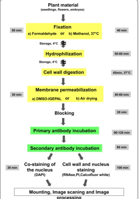
An overview of the main steps of the procedure is pre-sented in Fig. 1, with the indication of the time required to perform each step and of steps where the procedure can be stopped. The whole procedure is described step-by-step below by giving a detailed description followed by background notes with comments. The recommend volumes have been calculated for 24 wells plates and 8 rubber frames slides.
Step 1: Fixation
Fixation A (Formaldehyde) Timing: 50–60 min.
• Place explants in at least 1.5 ml of 2 % formaldehyde in 1× MTSB buffer supplemented with 0.1 % Triton, pH ~ 7, (ratio fixative/explants 10:1).
• Apply vacuum infiltration for 2–3 min and then (slowly) release vacuum. Repeat it once again. The fixation starts only after fixative penetrates (or air will be back to the desiccator).
• Check if explants have sunk at the bottom, and con-tinue fixation for 40 min under gentle shaking at 37 °C.
• Wash samples in 2 ml of distilled water ~10 min.
Alternative procedure
Fixation B (methanol) Timing: 40 min.
• Place explants in 1.5 ml of 100 % p.a. methanol for 20 min and incubate at 37 °C.
• replace with 0.8 ml of fresh 100 % p.a. methanol (60 °C), incubate vials for 3 min and gradually add water till final concentration of methanol reaches 20 % (ca 3.2 ml water). Thereafter transfer explants/ plants to a new vial with water. In our hands meth-anol preserved protein structure and has allowed combining successfully tissue clearing with cuticle solubilization.
Comments The goal of fixation is to maintain the cellular structure as intact as possible. Tissue fixation can be per-formed by two different ways (reported above as fixation A and B, respectively) depending on the proteins of interest. Fixation with formaldehyde (fixation A) crosslinks proteins
with cellular components which preserve tissue and cell morphology. Rapid penetration of the fixative into the cells is crucial for proper fixation. This is assured by vacuum infil-tration of the fixative, containing 0.1 % Triton (surfactant), into the tissue. Freshly prepared 2 % formaldehyde solution from para-formaldehyde powder is used for this purpose, giving best results. If commercially available 37 % formalde-hyde stock solution is used, the instability of formaldeformalde-hyde in solution and its polymerization during long term stor-age may hamper results and has to be taken into account. Specimens should be fixed in a multiwell culture plate with a large surface to enable efficient gas removal through vac-uum application during fixation procedure. In many cases methanol fixation (fixation B) alone is enough to preserve protein and cellular structure and has allowed in our hands combining successfully tissue clearing with cuticle solubili-zation, thus providing a good, faster and easier alternative to formaldehyde fixation. Absorbed methanol is oxidized within the plant cell to formaldehyde and formic acid [10].
In general, from our experience, methanol works well for membrane proteins. In addition, aerial parts of plants (leaves of certain species) have a highly hydrophobic cuti-cle to prevent water loss. In order to allow antibodies to penetrate inside cells, the cuticle needs to be solubi-lized. This can be achieved by treatment with methanol which solubilizes the majority of the cuticle and other waxes. We also experienced that methanol treatment also improved antibody penetration in the mature part of the root. Finally, chlorophyll, as a potential source of auto-fluorescence, is readily removed by methanol treatment as well. However, one should also consider that some epitopes are very sensitive to methanol and may be not accessible anymore for antibody binding so a comparison of the two fixative methods should be considered.
Step 2: Cuticle solubilization and tissue clearing— hydrophilisation
Timing: 50–60 min.
• Replace water (from fixation A) with ~0.8 ml of 100 % p.a. methanol (60 °C) and incubate for ~5–10 min or, from fixation B, directly proceed to the following step.
• Gradually decrease alcohol concentration by adding every 2 min 100–200 µl of water until the final alco-hol concentration reaches ~20 % (this corresponds to the addition of 3.2 ml of water).
• Wash twice for 5 min each in water.
• Transfer plants to the agilent slides pre-loaded with 60 µl of water.
Comments Gradual addition of water is important for preserving the structure of tissues/organs.
Step 3: Digestion of cell walls Timing: 45 min.
• Add 60 µl of the cell wall digestion solution into each well/frame (0.2 % Driselase and 0.15 % Macerozyme in 2 mM MES, pH 5.0).
• Incubate for 30–40 min. at 37 °C.
• Wash 1 × 4 min with 100 µl of the 1× MTSB pH 7.0.
Comments In contrast to animal cells, plant cells are sur-rounded by a rigid cell wall, which needs to be at least partially digested for efficient antibody penetration. Therefore tissues are incubated with cell wall degrad-ing enzymes. In addition, dense tissues specifically need to be macerated for effective antibody penetration into deeper layers. In the majority of published protocols Driselase is used dissolved in 1× MTSB buffer with pH of approximately 7.0 [3]. These conditions are suboptimal, because Driselase has quite low cell maceration activi-ties and its pectolytic and cellulolytic activiactivi-ties have an optimum pH ranging from 4.0 to 6.0 and from 3.0 to 5.0, respectively [11]. In order to improve the cell wall diges-tion and increase tissue maceradiges-tion a mixture of Drise-lase and Macerozyme R10 was used in MES buffer with pH 5.0. This treatment is gentler and results reproducibly in excellent preserved tissues.
Step 4: Membrane permeabilisation Timing: 30 min.
• Add 60 µl of the membrane permeabilisation solu-tion (3 % IGEPAL C630, 10 % DMSO in 1× MTSB) and incubate for 15–20 min at 37 °C.
• Wash 4 times with 1x MTSB for 3 min each.
Comments After partial digestion of cell walls, the cellu-lar membranes must be permeabilized. Membrane per-meabilization creates pores in membranes, which allow the antibody to penetrate. For this purpose treatment with a mixture of DMSO and the detergent IGEPAL CA-630 was applied. This treatment allows efficient and reproducible antibody penetration. As an alternative to treatment with IGEPAL/DMSO, one can completely dry the tissue on the slide. This option is favorable for cell monolayer cultures (see supplementary protocol for sus-pension cells), but also can help tissue permeabilization in the case of whole organs.
Step 5: Blocking Timing: 30 min.
Comments the goal of the blocking step is to minimize non-specific antibody binding. The minimal duration of the blocking is 20 min., however, in some cases (when background noise is high), it can be extended to up to 2 h.
Step 6: Primary antibody incubation Timing: 90–120 min.
• Replace blocking solution with 60 µl of the primary antibody solution and incubate for 1–2 h at 37 °C;
• Wash 2 × 5 min with 100 µl of the 1× MTSB.
Comments Do not mix solution during incubation with the primary antibody.
Antibodies used for immunostaining should be always affinity purified. According to our experience it is not advisable to use crude sera due to cross-reactivity with multiple proteins. Best results are obtained with antibod-ies against epitope tags (HA, Myc) or GFP, but this is only suitable for genetically modifiable species like Arabidop-sis. It is absolutely necessary to test antibody specificity in Western blots. A loss of function mutant where the protein of interest is absent, if available, should be ideally used as a negative control. As a negative control, samples should be also incubated in the presence of pre-immune serum.
Step 7: Secondary antibody incubation Timing: 60 min.
• Add 60 µl of the secondary antibody solution, and incubate for 1–2 h at 37 °C;
• Wash 3 × 5 min with 1x MTSB.
Comments Do not mix solution during incubation with the secondary antibody. The choice of fluorophore with which secondary antibodies are conjugated depends primarily on the task of investigation. Fluorophores are differing in terms of brightness, photobleaching and chemical stability. Many of the most popular secondary antibodies are Alexa conjugated (InVitrogen). However, recently new DyLight antibodies have been developed (InVitrogen, Agrisera, Abcam). DyLight ® conjugated secondary antibodies exhibit higher fluorescence inten-sity, photo stability and water solubility and remain fluorescent from pH 4 to pH 9. Additionally, the water solubility of the DyLight® fluorophores allows a high dye-to-antibody ratio to be achieved without causing precipi-tation of conjugates.
For protein co-localization studies up to four primary and secondary antibodies can be used simultaneously, but they should be raised in different animals to avoid cross-reactivity.
Step 8: Co‑staining of the nucleus Timing: 15 min.
• Add 100 µl of the DAPI containing solution (0.2 mg/l) and incubate for 10 min;
• Wash 3 × 5 min with 100 ml of distilled water.
Step 8 (alternative): Cell wall and nucleus staining Timing: 50 min.
• Incubate in 10 mM Tris–HCl, pH 7.5 for 10 min;
• Incubate in 100 µl of the RNAse solution in 10 mM Tris–HCl, pH 7.5 for 30 min at 37 °C;
• Wash 1 × 5 min with 100 µl of 10 mM Tris–HCl, pH 7.5;
• Incubate in 100 µl of the propidium iodine solution (0.4 mg/l) in 10 mM Tris–HCl, pH 7.5 for 10 min at 37 °C;
• Wash with water for 10 min;
• Incubate in 100 µl of the 10 mM Tris–HCl, pH 9.2 for 10 min;
• Incubate in 100 µl of the calcofluor white solution in 10 mM Tris, pH 9.2 for 20 min;
• Wash 2 × 5 min in the 10 mM Tris–HCl, pH 9.2.
Comments In order to show the proteins of interest in a cellular and organ continuity, additional staining of cell walls and of nuclei with calcofluor white and pro-pidium iodine, respectively, might be wishful. This procedure does not affect the detection of proteins. Calcofluor white requires an alkaline pH for binding to the cell wall. We recommend keeping pH at 8.5–9 also in the mounting solution by mixing 70 % of anti-fade medium with 30 % of 500 mM Tris–HCl, pH 9.2 (350 µl antifade medium + 150 µl 500 mM Tris–HCl, pH 9.2).
Step 9: Mounting
Transfer seedlings to microscopic slides with a jacket containing antifade medium, cover samples with a cover slip and store them in the fridge/cold-room (approxi-mately 5 °C).
(50 %), N-propyl gallate (15 mg/ml) (final concentration) and H2O (50 %). For long term storage of samples
addi-tion of 0.1 % sodium azide to the anti-bleaching soluaddi-tion
is mandatory. In order not to damage the samples we suggest to mount specimens after immunolocalization on microscopic slides with pre-inserted 120 µm spacer
Fig. 2 PIN1 protein localization in cotyledons and hypocotyls of Arabidopsis seedlings. Four days old seedlings were fixed for 20 min in methanol and subjected to the standard immunolocalization procedure as described. Anti-PIN1 mouse monoclonal primary antibody (clone 10A7), diluted 1: 50. ALEXA Fluor ® 488 conjugated goat anti-mouse IgG (Invitrogen) was used as secondary antibody diluted 1:800. Co-staining with DAPI visualized nuclei (red). a Cotyledon; b hypocotyl; Scale bar 20 µm
made from TVC isolation tape. The tape is cut in small stripes and pasted on the slide before samples insertion. Appropriate thickness of the jacket avoids tissue pressing and enables to reconstruct 3D images from the organs/
seedlings. For Arabidopsis whole mounted seedlings a 100 μm thick spacer is sufficient to keep the original 3D structure.
Comments and concluding remarks
The reported protocol for immunolocalization allows researchers to study metabolites, nucleic acids and pro-tein localization in virtually any plant species and organs in relatively thick specimens speeding up throughput and resolution of protein localization studies also in non-model plants. The presented methodologies significantly improve the accuracy and resolution of protein detection in expression and localization studies and do not have a limit regarding tissue type. Manual sectioning can be avoided and 3D reconstruction can be easily done. Its shortest version takes only 7 h to complete without the need for robotic equipment, as shown in Fig. 1. Addi-tional applications of the protocol are also provided for immunolocalisation on isolated plant cells and proto-plasts and for 3D reconstuction (Additional file 2).
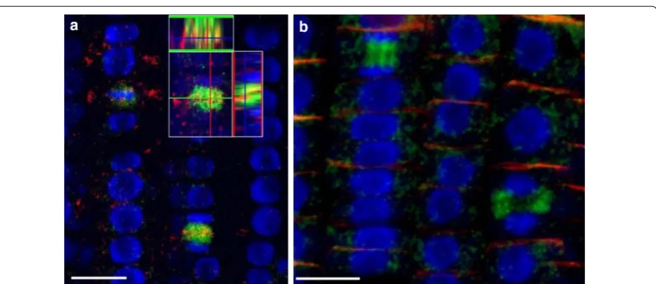
Previously published immunolocalization protocols [4] require at least two working days and cannot be applied to non-transparent tissues. These protocols have been applied for analysis of the root meristem of Arabidopsis thaliana, while for other plant species and for more dense tissues of Arabidopsis (e.g. hypocotyls or leaves) research-ers prefer to use paraplast sections which are labor and time consuming and do not allow 3D reconstruction. For example, Bustos-Sanmamed et al. [2] suggested to use paraplast sections for immunolocalization in Medicago plants, which are extremely time and labor consuming. In our hands Medicago can be subjected to whole-mount Fig. 4 Protein immunolocalization in Medicago sativa L. and Lycopersicum esculentum L. Plants were fixed for 30 min in formaldehyde. Anti-PIN1 mouse monoclonal primary antibody (clone 10A7) diluted 1:50 plus Alexa Fluor®488 goat anti-mouse IgG as secondary antibody diluted 1:800 (shown in green color) and H+-ATPase (AS07 260) rabbit primary antibody plus Alexa Fluor® 555 goat anti-rabbit IgG as secondary antibody diluted 1:800 (shown in red color) were used. Nuclei are visualized by co-staining with DAPI (blue). Scale bar 20 µm. White arrows show polar PIN1 localisa-tion. a Medicago sativa roots; b Medicago sativa leaf; c, d- Lycopersicum esculentum root
immunolocalization in any organ with further 3D recon-struction. Our whole-mount protocol is applicable to the analysis of any plant species and organ including non-transparent tissues. It is also easily applicable to suspen-sion cultures and can be completed for most specimens in 5–6 h. Detection of proteins deep inside tissues requires a fine balance between fixation, clearing of tissues, cell wall digestion and permeabilisation. Through improved tis-sue clearing combined with tistis-sue-specific combinations of cell wall degrading enzymes, proteins can be detected e.g. in ovules of intact pistils or xylem-parenchyma cells of
hypocotyls while keeping the outer cell structures intact (Figs. 2, 3). The excellent tissue preservation is demon-strated by labeling of microtubules and actin in the elon-gation zone of Arabidopsis roots (Fig. 4), which often appeared destroyed using previously published auto-mated whole-mount method [3]. Due to the use of small volumes in Microarray slides, the procedure described here reduces the amount of reagents and limits the use of particularly precious antibodies, but also allows handling of specimens up to 1 cm wide. The general applicability of the protocol was successfully tested for localization of
Fig. 6 Protein immunolocalization in different Triticum aestivum organs. Three days old wheat seedlings were fixed for 30 min in formaldehyde. Anti-PIN1 mouse monoclonal primary antibody (clone 10A7) diluted 1:50 and Alexa Fluor® 488 goat anti-mouse IgG as secondary antibody diluted 1:800 were used (shown in green color) (a–e); anti-PIN2 Guinea pig primary antibody plus Goat anti-Guinea pig IgG Alexa Fluor® 647 conjugate as secondary antibody diluted 1:800 (shown in red color) (e) and anti-BIP2 (AS09 615) rabbit primary antibody plus Goat anti-rabbit IgG DyLight® 549 conjugate (AS09 642) as secondary antibody diluted 1:3000 (shown in red color) (f) were used. Co-staining with DAPI visualizes nuclei (blue). a leaf; b meristem; c coleoptile; d–f roots. Arrows point polarly located PIN1 and PIN2 proteins. Scale bar 20 µm
PIN proteins in root and flower tissues from Medicago sativa, Triticum aestivum, Lycopersium esculentum, and
Hedera helix (Figs. 5, 6, 7). The fixation procedure using ethyldimethylaminopropyl carbodiimide (EDAC, carboxyl activating agent for hormones bonding with proteins) and formaldehyde was optimized for detection of low molecu-lar weight molecules (e.g. auxin) with antibodies (Fig. 8).
In addition, the protocol allows further appli-cations such as the detection of DNA replication events by using incorporation of the thymidine analogue BrdU/EdU into nuclear DNA followed by subsequent detection with an antibody recogniz-ing BrdU/EdU (Fig. 9) [12]. This approach opens the possibility to monitor the duration of the S and G2 phases of the cell cycle, as well as to detect cells
within tissues that undergo DNA reduplication. The protocol, being reasonably streamlined and simple, can be used for analysis of protein expression and localization in up to 30 samples simultaneously without the requirement of laboratory robots. As a concluding remark, our improved protocol, by keep-ing better intact organs structure, enables precise analysis of protein expression/localization in whole organs, thus performing a fundamental shift from two dimensional to three dimensional tissue atlases, required for our previously described automated organ analysis [9]. Examples of 3D reconstruction after immunolabelling with our protocol are shown on Additional files 3–6.
Fig. 8 Auxin immunolocalisation in Arabidopsis roots. Four days old Arabidopsis seedlings were treated with 1 µM 1-N-Naphthylphtha-lamic acid (NPA) for 24 h to enhance accumulation of auxin in roots. Seedlings were fixed for 20 min in 4 % EDAC in 1× MTSB, and next 30 min in 4 % EDAC+ 2 % Formaldehyde. Anti-indole 3 acetic acid (IAA) rabbit primary antibody (Agrisera, AS06 193) diluted 1:600 plus Goat anti-rabbit IgG (H&L), DyLight® 549 Conjugate (AS09 633) as secondary antibody diluted in 1:3000 (shown in red color) were used. Scale bar 20 µm
Authors’ contributions
TP carried out most of the experiments. OT and MB participated in the experiments on ovules and silique immunolocalization. BR, TP, OT, KP planned experiments. RN advised and helped in all microscopy related questions. TP, BR, OT, RN and KP interpreted results and wrote the manuscript. All authors read and approved the final manuscript.
Author details
1 Faculty of Biology, Institute of Biology II/Molecular Plant Physiology,
Uni-versity of Freiburg, Freiburg, Germany. 2 BIOSS Centre for Biological
Signal-ing Studies, University of Freiburg, Freiburg, Germany. 3 Freiburg Institute
for Advanced Studies (FRIAS), University of Freiburg, Freiburg, Germany.
4 Department of Agronomy, Food, Natural Resources, Animals and
Environ-ment, DAFNAE, University of Padova, Agripolis, Viale dell’Università, 35020 Leg-naro, Padova, Italy. 5 Center for Biological Systems Analysis, University
of Freiburg, Freiburg, Germany.
Acknowledgements
We thank the members of the Life Imaging Center (LIC) Freiburg for support in image acquisition and analysis. This work was supported by the Collaborative Research Center 746, the Excellence Initiative of the German Federal and State Governments (EXC 294), by the European Space Agency project ‘Highway’ (MAP Project 14341/00/NL/SH), the European Project ‘AUTOSCREEN’ (LSHG-CT-2007–037897), the German Aerospace Center, Space Administration on behalf of the Bundesministerium für Wirtschaft und Technologie (BMWi) and the Bundesministerium für Forschung und Technik (BMBF).
Additional files
Additional file 1. Agilent microarray slides suitable for immunolocalization.
Additional file 2. Supplementary protocols.
Additional file 3. 3D reconstruction of the Arabidopsis leaf after labelling with PIN1 antibody and co-staining with DAPI for cell visualization. Four days old Arabidopsis seedlings were fixed for 30 min in formaldehyde. Anti-PIN1 mouse monoclonal primary antibody (clone 10A7) diluted 1: 50 plus Alexa Fluor® 488 goat anti-mouse IgG as secondary antibody diluted 1: 800 (shown in green color) (panel A-E) were used; Co-staining with DAPI visualizes nuclei (shown as artificial color in white). Ortho-view is shown. Scale bar 50 µm.
Additional file 4. 3D reconstruction of Nicotaina tabacum roots after labelling with PIN1 antibody. Five days old Tobacco seedlings were fixed for 30 min in 2 % formaldehyde. Anti-PIN1 mouse monoclonal primary antibody (clone 10A7) diluted 1: 50 plus Alexa Fluor® 488 goat anti-mouse IgG as secondary antibody diluted 1: 800 (shown in green color) (panel A) were used. Co-staining with DAPI visualizes nuclei (shown as artificial color in white) (panel B). Ortho-view was shown. Scale bar 100 µm.
Additional file 5. 3D reconstruction of Arabidopsis leaf after labelling with calcofluor white (cell wall) and propiduim iodine (nucleus). Five days old seedlings have been fixed and stained with propidium iodine (nucleus is shown in red) and calcofluor white (cell wall, displayed in green). Ortho-view was shown. Scale bar 50 µm.
Additional file 6. Example of the automatic analysis of 3D images after EdU labelling. Five days old Arabidopsis seedlings have been incubated with EdU/colchicine for 90 min., fixed and cleared with hot methanol. Cell wall has been digested and membrane has been permeabilized. Seedlings have been incubated with EdU specific dye (C1037, Invitrogen) for 40 min., stained with DAPI and mounted on microscopic slides. Whole stacks have been scanned and 3D reconstruction has been performed using the iRoCS toolbox (http://lmb.informatik.uni-freiburg.de/lmbsoft/ iRoCS). Scale bar 50 µm. Nuclei are in red; EdU are in green. Axis is in yellow.
Competing interests
The authors declare that they have no competing interests.
Received: 16 July 2015 Accepted: 14 October 2015
References
1. Fukao Y. Protein–protein interactions in plants. Plant Cell Physiol. 2012;53(4):617–25.
2. Bustos-Sanmamed P, Laffont C, Frugier F, Lelandais-Brière C, Crespi M. Analyzing protein distribution in plant tissues using “Whole-Mount” immunolocalization. In: De Smet I, editor. Plant organogenesis. New York: Humana Press; 2013. p. 317–22.
3. Forestan C, Varotto S. Auxin immunolocalization in plant tissues. In: De Smet I, editor. Plant organogenesis. New York: Humana Press; 2013. p. 223–33.
4. Brandizzi F, Fricker M, Hawes C. A greener world: the revolution in plant bioimaging. Nat Rev Mol Cell Biol. 2002;3(7):520–30.
5. Friml J, Benkova E, Mayer U, Palme K, Muster G. Automated whole mount localisation techniques for plant seedlings. Plant J. 2003;34(1):115–24. 6. Sauer M, Paciorek T, Benková E, Friml J. Immunocytochemical
tech-niques for whole-mount in situ protein localization in plants. Nat Protoc. 2006;1(1):98–103.
7. Ditengou FA, Teale WD, Kochersperger P, Flittner KA, Kneuper I, van der Graaff E, Nziengui H, Pinosa F, Li X, Nitschke R, Laux T, Palme K. Mechanical induction of de novo lateral root initiation in Arabidopsis. Proc Natl Acad Sci USA. 2008;105(48):18818–23.
8. Tsugeki R, Ditengou FA, Sumi Y, Palme K, Okada K. NO VEIN mediates auxin-dependent specification and patterning in the Arabidopsis embryo, shoot and root. Plant Cell. 2009;21(10):3133–51.
9. Schmidt T, Pasternak T, Liu K, Blein T, Aubry-Hivet D, Dovzhenko A, Palme K. The iRoCS Toolbox–3D analysis of the plant root apical meristem at cellular resolution. Plant J. 2014;77(5):806–14.
10. Fall R, Benson A. A: leaf methanol—the simplest natural product from plants. Trends Plant Sci. 1996;1(9):296–301.
11. Noguchi S, Shimura G, Kawai M, Suga Y, Samejma H. Properties of partially purified cellulolytic and plant tissue macerating enzymes of Irpex lacteus Fr With special reference to their application. Agric Biol Chem. 1978;42(2):223–32.
12. Vogel W, Autenrieth M, Speit G. Detection of incorporation in mammalian chromosomes by a bromodeoxyuridine-antibody. Hum Genet. 1986;72(2):129–32.
Submit your next manuscript to BioMed Central and take full advantage of:
• Convenient online submission • Thorough peer review
• No space constraints or color figure charges • Immediate publication on acceptance
• Inclusion in PubMed, CAS, Scopus and Google Scholar • Research which is freely available for redistribution