R E S E A R C H A R T I C L E
Open Access
Outcomes after surgical resection of
pulmonary carcinoid tumors
Ikenna C. Okereke
1*, Angela M. Taber
2, Rogers C. Griffith
3and Thomas T. Ng
4Abstract
Background:Pulmonary carcinoid tumors make up approximately one percent of all pulmonary tumors, and controversy exists regarding management and prognosis. We undertook a retrospective analysis of all patients who underwent surgical resection of pulmonary carcinoid tumors at our institution.
Methods:From 1992 through 2014, 121 patients who underwent surgical resection of pulmonary carcinoid tumors were retrospectively reviewed. Patient demographics, pathologic data and long-term outcomes were recorded. Results:There were 96 patients with typical carcinoid tumors and 25 patients with atypical carcinoid tumors. All patients received complete resection of their tumors, with 90 % (109/121) of patients undergoing anatomic resection. There were no peri-operative mortalities. Eighty-one percent (98/121) of patients were female. Mean age was 60.7 years. Five and ten year survival rates were 96 % and 88 % respectively for typical carcinoid tumors, as compared to 87 % and 69 % respectively for atypical carcinoid tumors. Tumor size was not associated with survival (p= 0.98). Nodal metastases were evident in 8 % (8/96) of typical carcinoid tumors and 28 % (7/25) percent of atypical carcinoid tumors. Among typical carcinoid cases, the presence of nodal metastases were not associated with overall survival (p= 0.55). Among atypical carcinoid cases, the presence of nodal metastases also was not associated with survival (p= 0.53). No patients received neoadjuvant or adjuvant chemoradiation treatment.
Conclusions:Excellent long-term outcomes can be achieved following surgical resection of pulmonary carcinoid tumors. The presence of nodal metastases was not associated with overall survival. Tumor size was not associated with either recurrence rates or survival.
Keywords:Carcinoid, Lymphadenopathy, Mitosis
Background
Pulmonary carcinoid tumors make up approximately 1– 2 % of all pulmonary tumors [1]. They are derived from the Kulchitsky cell and generally have indolent growth patterns. Carcinoid tumors are categorized as typical or atypical, based on number of mitoses per high power field and presence of necrosis [2]. Surgical resection is the standard treatment for pulmonary carcinoid tumors [3]. There exists debate, however, about the use of adjuvant treatment in patients with locoregional metas-tases or patients with atypical tumors [4, 5]. We under-took a retrospective review to determine the outcomes
of patients with pulmonary carcinoid tumors who underwent surgical resection.
Methods
After approval by the institutional review board, a data-base search for pulmonary carcinoid tumors was per-formed from pathology records of two Brown University hospitals (The Rhode Island Hospital and The Miriam Hospital) over a 22 year period (1992–2014). Over this time span, 121 patients with pulmonary carcinoid tu-mors underwent surgical resection. Patients who were incidentally found to have a carcinoid tumorlet as part of their resection specimen were not included in the study. Pathologic confirmation of carcinoid tumor was established by a pathologist in all cases. Tumors were classified as typical carcinoid if there were less than 2 mitoses per 2 square millimeters (mm2) and no necrosis. * Correspondence:iokereke@usasurg.org
1Cardiothoracic Surgery, University of Texas Medical Branch, 301 University
Blvd, Galveston, TX 77555, USA
Full list of author information is available at the end of the article
Tumors were classified as atypical if there were between 2 and 10 mitoses per mm2or evidence of necrosis. Pa-tient demographics, use of chemotherapy or radiation, peri-operative variables, type of surgical resection, recur-rence rates and survival were analyzed retrospectively. Anatomic resection consisted of either segmentectomy, lobectomy, bi-lobectomy or pneumonectomy. Follow-up status was obtained from institutional records and veri-fied by the national social security database.
The Mann–Whitney test was used to compare differ-ences in continuous variables between different tumor groups. Fisher’s exact and Chi-squared tests were used to compare categorical variables. Univariate survival analysis was performed using Kaplan-Meier curves com-pared with log rank or Cox regression analysis. Multi-variate survival analysis was performed with Cox regression analysis, using variables with a p-value of less than 0.1 on univariate analysis.
Results
Patients
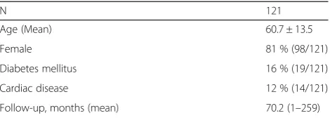
A list of patient demographics is shown in Table 1. Eighty-one percent (98/121) of the patients were female. Mean age was 60.7 years (11–81). Mean follow-up time was 70.2 months (1–259).
Surgical management
Anatomic resection was performed in 89 % (108/121) of patients. All patients had complete resection of their tumor. Eleven percent (13/121) of patients underwent resection using a minimally invasive, video-assisted ap-proach. There were no peri-operative mortalities. Mean length of stay was 4.9 days.
Pathologic analysis
Pathologic results are shown in Table 2. Mean size was 2.3 centimeters (cms). Twenty-one percent (25/121) of pa-tients had an atypical carcinoid tumor. Nodal metastases were present in 8 % (8/96) of patients with typical carcin-oid tumors and 28 % (7/25) of patients with atypical car-cinoid tumors (p< 0.01, Table 3). Patients with atypical carcinoid tumors had greater tumor size than patients with typical carcinoid tumors (2.0 cm vs. 3.2 cm,p< .01). Patients with atypical carcinoid tumors were also more likely to have increased number of lymph node metastases
than patients with typical carcinoid tumors (p< 0.01). In patients with atypical carcinoid tumors, the mean number of mitoses per high power field was 3.9.
Chemoradiation
No patient received neoadjuvant chemotherapy or radi-ation treatment. Similarly, no patient received adjuvant treatment in the early postoperative period. All patients who experienced recurrent disease received apy and/or radiation treatment. The usual chemother-apy regimen consisted of cisplatin-based treatment. The decision to use chemotherapy and/or radiation therapy was individualized in each of these patients and based on extent and location of recurrence and pres-ence of symptoms.
Disease-free survival and overall survival
At the time of last follow-up, 88 % (107/121) of patients were alive in the overall cohort. Five and ten year sur-vival overall were 93 % and 84 % respectively. Of the patients with typical carcinoid tumor, 89 % (85/96) were alive without disease, 1 % (1/96) was alive with distant recurrent disease, and 10 % (10/96) had died without disease. Five and ten year survival in patients with typ-ical carcinoid were 96 % and 88 % respectively.
Of the patients with atypical carcinoid tumor, 76 % (19/25) were alive without disease, 8 % (2/25) were alive with distant recurrent disease, 12 % (3/25) died with disease, and 4 % (1/25) died without disease. Two of the 3 patients who died with disease had distant recurrent disease, while the other patient had both distant and local recurrences. Five and ten year survival in patients with atypical carcinoid were 87 % and 69 % respectively.
Table 1Demographics
N 121
Age (Mean) 60.7 ± 13.5
Female 81 % (98/121)
Diabetes mellitus 16 % (19/121)
Cardiac disease 12 % (14/121)
Follow-up, months (mean) 70.2 (1–259)
Table 2Pathologic results
Typical histology 79 % (96/121)
Anatomic resection 89 % (108/121)
Size, cm (mean) 2.3 ± 1.4
Stage I 85 % (103/121)
II 7 % (8/121)
III 8 % (10/121)
Table 3Typical vs. atypical histology
Typical (N= 96)
Atypical (N= 25) p
-value
Age, years (mean) 60.0 ± 13.2 63.3 ± 9.8 0.24
Tumor size, cm (mean) 2.0 ± 1.3 3.2 ± 2.2 <0.01
Positive lymph node status 8 % (8/96) 28 % (7/25) <0.01
Number of lymph node metastases 0.1 0.6 <0.01
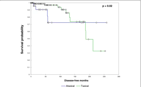
Disease-free survival 0.02
Overall survival 0.09
Disease-free survival was significantly worsened by in-creasing age (p= 0.03) and atypical histology (p= 0.02, Fig. 1). Disease-free survival was not associated with anatomic resection (p= 0.91), size (p= 0.11), positive lymph node status (p= 0.44) or number of lymph node metastases (p= 0.13).
Using univariate analysis, factors which were associ-ated with overall survival were age (p= 0.03) and num-ber of mitoses (p< 0.01). Overall survival was not affected, however, by typical/atypical histology (p= 0.09), positive lymph node status (p= 0.55) or anatomic resec-tion (p= 0.78). When multivariate analysis was used, in-cluding all variables with a p value less than .1, only the number of mitoses was significantly associated with sur-vival (p= 0.04).
Discussion
Overall survival for pulmonary carcinoid tumors is ex-cellent, and surgical resection remains the standard of care for these tumors [6]. There exist many different standards and practices for surgical management, however [7]. All of our patients underwent complete resection of their tumor. In our analysis both overall survival and disease-free survival were not affected by undergoing an anatomic resection. But in our study the overwhelming majority of patients had an anatomic
resection performed, and conclusions concerning ana-tomic resection vs. limited resection are difficult. We feel that an anatomic resection should be favored for carcinoid tumors, and limited resections should be considered only for patients who lack sufficient pul-monary reserve.
Similarly, presence or number of lymph node metasta-ses did not affect survival in our study. But given the relatively small number of patients in our study with nodal involvement, caution should be taken in determin-ing the prognostic impact of nodal status. And other studies have shown that nodal involvement does have an impact on outcome of patients with carcinoid tumors [8]. It does seem, however, that patients can live for ex-tended periods of time with microscopic disease, given the indolent nature of carcinoid tumors. Our surgical practice has been to perform a hilar and mediastinal lymph node dissection similar to what we perform for patients with non-small cell lung cancer.
Atypical carcinoid tumors do appear to have more aggressive biology, with statistically increased tumor size and propensity for lymph node metastases [9–12]. And while patients with atypical carcinoid tumors were more likely to develop recurrent disease, overall survival was not affected by typical/atypical histology. This trend occurred because the patients who developed recurrent
0 0.1 0.2 0.3 0.4 0.5 0.6 0.7 0.8 0.9 1
0 50 100 150 200 250 300
Disease-free months
Atypical Typical
p = 0.02
Sur
viv
a
l probab
ility
Fig. 1Relationship between typical/atypical histology and disease-free survival. Patients with atypical histology experienced a significantly
disease were able to survive for prolonged periods with-out significant symptomatology or complications [13, 14]. Though it appears that patients with atypical carcinoid tumors are more likely to recur, they can live with disease without considerable morbidity for prolonged periods.
Given the long time frame of the study, preoperative staging and postoperative surveillance strategy varied widely among the cohort [15]. In the early part of this study computed tomography (CT) scan only was used to stage the patient. Now we obtain positron emission tom-ography – computed tomography (PET-CT) scans dur-ing the preoperative workup of our patients. Our current postoperative surveillance strategy has been to perform CT scan evaluation every 6 months for the first 2 years, and then yearly thereafter until year 5. After that, CT scan is performed only if there is suspicion of recurrence based on symptomatology or clinical find-ings. In our study, only 1 patient recurred after 5 years from resection. That patient is currently alive with dis-ease but no symptoms.
There exist multiple different practice patterns re-garding the use of chemotherapy to treat pulmonary carcinoid tumors [16–19]. Chemotherapy is generally not recommended to treat a patient with typical carcinoid tumors who recurs, as typical carcinoid tu-mors are generally less responsive to chemotherapy [20]. The 1 patient in our study with typical carcinoid who recurred was, as such, treated with radiation therapy only. Patients with atypical carcinoid tumors who recur may experience a survival benefit with chemotherapy.
Along with the retrospective nature of the series, our study is limited by other factors. The overall number of patients in the series is relatively low, but the overall and disease-free survival rates were similar compared to other studies. And because disease recurrences were so infrequent, even in patients with atypical carcinoid tu-mors, it was difficult to determine a group of patients with a recurrence risk great enough to warrant chemora-diation therapy. A larger multi-center study would po-tentially identify patients who would benefit from adjuvant treatment, such as a patient with an atypical carcinoid tumor with a very high number of mitoses.
Conclusions
Patients with pulmonary carcinoid tumors can experi-ence excellent long term outcomes following complete resection of their tumors. Presence of lymph node me-tastases and tumor size do not appear to affect recur-rence rates or survival. Further multi-center studies may potentially identify a high-risk cohort that would benefit from neoadjuvant or adjuvant chemotherapy or radiation treatment.
Competing interests
The authors declare that they have no competing interests.
Authors’contributions
IO contributed to the data collection, statistical analysis and drafted the manuscript. AT contributed to the study design and the interpretation of the data. RG contributed to the data collection and study design. TN contributed to the statistical analysis and study design. All authors read and approved the final manuscript.
Author details
1Cardiothoracic Surgery, University of Texas Medical Branch, 301 University
Blvd, Galveston, TX 77555, USA.2Division of Oncology, Warren Alpert Medical
School of Brown University, Providence, RI, USA.3Department of Pathology
and Laboratory Medicine, Warren Alpert Medical School of Brown University, Providence, RI, USA.4Department of Surgery, Warren Alpert Medical School
of Brown University, Providence, RI, USA.
Received: 13 April 2015 Accepted: 5 February 2016
References
1. Naslund A, Rostad H, Strom E, Lund M, Strand T. Carcinoid lung tumors—incidence, treatment and outcomes: a population-based study. Eur J Cardiothorac Surg. 2011;39:565–9.
2. Tsuta K, Raso M, Kalhor N, Liu D, Wistuba I, Moran C. Histologic features of low and intermediate-grade neuroendocrine carcinoma (typical and atypical carcinoid tumors) of the lung. Lung Cancer. 2011;71:34–41.
3. Dahabreh J, Stathopoulos G, Koutantos J, Rigatos S. Lung carcinoid tumor biology: treatment and survival. Oncol Rep. 2009;21:757–60.
4. Hage R, de la Riviere A, Seldenrijk C, Van den Bosch J. Update in pulmonary carcinoid tumors: a review article. Ann Surg Oncol. 2003;10:697–704. 5. Mackley H, Videtic G. Primary carcinoid tumors of the lung: a role for
radiotherapy. Oncology. 2006;20:1537–43.
6. Kyriss T, Maier S, Veit S, Fritz P, Toomes H, Friedel G. Carcinoid lung tumors: long-term results from 111 resections. GMS Thorac Surg Sci. 2006;3:Doc03. 7. Filosso P, Rena O, Donati G, Casadio C, Ruffini E, Papalia E, et al. Bronchial
carcinoid tumors: surgical management and long-term outcome. J Thorac Cardiovasc Surg. 2002;123:303–9.
8. Fox M, Van Berkel V, Bousamra M, Sloan S, Martin R. Surgical management of pulmonary carcinoid tumors: sublobar resection versus lobectomy. Amer J Surg. 2013;205:200–8.
9. Thomas C, Tazelaar H, Jett J. Typical and atypical pulmonary carcinoids: outcome in patients presenting with regional lymph node involvement. Chest. 2001;119:1143–50.
10. Bini A, Brandolini J, Cassanelli N, Davolr F, Dolci G, Selltri F, et al. Typical and atypical pulmonary carcinoids: our institutional experience. Interact Cardiovasc Thorac Surg. 2008;7:415–18.
11. Garcia-Yuste M, Matilla J. The significance of histology: typical and atypical bronchial carcinoids. Thorac Surg Clin. 2014;24:293–7.
12. Wurtz A, Benhamed L, Conti M, Bouchindhomme B, Porte H. Results of systematic nodal dissection in typical and atypical carcinoid tumors of the lung. J Thorac Oncol. 2009;4:388–94.
13. Canizares M, Matilla J, Cueto A, Algar J, Muguruza I, Moreno-Mata N, et al. Atypical carcinoid tumours of the lung: prognostic factors and patterns of recurrence. Thorax. 2014;69:648–53.
14. Cardililo G, Sera F, Di Martino M, Graziano P, Giunti R, Carbone L, et al. Bronchial carcinoid tumors: nodal status and long-term survival after resection. Ann Thorac Surg. 2004;77:1781–5.
15. Lou F, Sarkaria I, Pietanza C, Travis W, Roh M, Sica G, et al. Recurrence of pulmonary carcinoid tumors after resection: implications for postoperative surveillance. Ann Thorac Surg. 2013;96:1156–62.
16. Wirth L, Carter M, Janne P, Johnson B. Outcome of patients with pulmonary carcinoid tumors receiving chemotherapy or chemoradiotherapy. Lung Cancer. 2004;44:213–20.
17. Ohta Y, Toda A, Ohta N, Oda M, Minato H, Nonomura A, et al. An atypical lung carcinoid tumor resected after induction therapy with involvement of the superior sulcus region: report of a case. Surg Today. 2002;32:632–4. 18. Granberg D, Eriksson B, Wilander E, Grimfjard P, Fjallskog M, Oberg K.
19. Carretta A, Ceresoli G, Arrigoni G, Canneto B, Reni M, Cigala C. Diagnostic and therapeutic management of neuroendocrine lung tumors: a clinical study of 44 cases. Lung Cancer. 2000;29:217–25.
20. Horsch D, Schmid K, Anlauf M, Darwiche K, Denecke T, Baum R, et al. Neuroendocrine tumors of the bronchopulmonary system (typical and atypical carcinoid tumors): current strategies in diagnosis and treatment. Conclusions of an expert meeting February 2011 in Weimar, Germany. Oncol Res Treat. 2014;37:266–76.
• We accept pre-submission inquiries
• Our selector tool helps you to find the most relevant journal • We provide round the clock customer support
• Convenient online submission • Thorough peer review
• Inclusion in PubMed and all major indexing services • Maximum visibility for your research
Submit your manuscript at www.biomedcentral.com/submit