Structural modification of lignan compoundS iSolated from
NectaNdra SpecieS (lauraceae)
Víctor Macías-VillaMizar
1,1*,§, luís cuca-suárez
11Departamento de Química, laboratorio de Productos Naturales, universidad Nacional de colombia. Av. Carrera 30 # 45-03. Cód. postal 111321; Edificio
476-Oficina 11; Bogotá D.C., Colombia. §Profesor Universidad del Magdalena, Colombia.
1-introduction
In the genus Nectandra, the presence of certain types of secondary metabolites has been determined, including sesquiterpenes, phytosterols, polyalcohols,
arylpropionic acid derivatives, flavonols, arylpropanoids, furofuran lignans, dihydrobenzofuran neolignans [1], and certain norlignans [2], alkaloids [3], tannins [4], diterpenes [5], and components of essential oils [6]. However, the chemotaxonomic characteristics are determined by the presence of lignan-type compounds [7]. The ultimate goal of structural modification of natural products is to obtain new drugs [8]. In that sense, there is a growing interest in lignans and their syn-thetic derivatives due to applications in cancer chemotherapy and various other pharmacological effects [9]. This work corresponds to the first report of this type
of structural modification of lignan compounds (7,7’-epoxylignans and diaryldimethylbutane lignans) isolated from Nectandra species. Therefore, this work can
be used as a starting point for structure-activity relationship studies.
eXperimental
materials and reagents
Benzyl bromide (Merck), acetone and toluene were freshly distilled before use. 2,3-Dichloro-5,6-dicyano-1,4-benzoquinone (DDQ) (Fluka purum ,97%), 1,4-dioxane (Aldrich, anhydrous 99.8%), pyridine (ACS reagent, ≥99.0%, Sigma-Aldrich) and acetic anhydride (ACS reagent, ≥98.0%, Sigma-Aldrich) were used. Purification of the products was carried out on a short silica gel column (100–200 mesh, Merck) using increasing percentage of ethyl acetate in hexane as elutant. NMR spectra: were recorded on a Bruker Avance 400
spectrometer (1H 400 MHz, 13C 100 MHz) using TMS as internal standard,
in deuterated chloroform (CDCl3) as solvent. The product ethers were
characterized by comparing spectral data of known compounds described in the literature and analysis of the spectral data.
figure 1.1H NMR (400 MHz, CDCl
3) Spectrum of Compound 1 (mixture
of veraguensin/galgravin)
Veraguensin: 1H NMR (400 MHz, CDCl
3), δ: 0.67 (3H, d, J = 7.0, H-9’),
1.07 (3H, d, J = 6.6, H-9), 1.79 (1H, m, H-8), 2.25 (1H, m, H-8’), 3.86 (s,
OCH3), 3.88 (s, OCH3), 3.89 (s, OCH3), 3.91 (s, OCH3), 4.42 (1H, d, J = 9.3,
H-7), 5.14 (1H, d, J = 8.6, H-7’), 6.86-7.08 (6H, m, H-2/5/6, H-2’/5’/6’). [v=veraguensin].
Galgravin:1H NMR (400 MHz, CDCl
3), δ: 1.05 (6H, d, J = 6.7, H-9/9’),
2.34 (2H, m, H-8/8’), 3.87 (s, OCH3), 3.88 (s, OCH3), 4.52 (2H, d, J = 6.4,
H-7/7’), 6.85-6.99 (6H, m, H-2/5/6, H-2’/5’/6’). [g=galgravin].
figure 2.13C NMR(100 MHz, CDCl
3) Spectrum of Compound 1 (mixture
of veraguensin/galgravin)
Veraguensin: 13C NMR (100 MHz, CDCl
3) δ: 149.1 (C), 148.7 (C), 148.7
(C), 148.2 (C), 133.9 (C), 133.6 (C), 119.3 (CH), 118.8 (CH), 111.1 (CH),
110.8 (CH), 110.5 (CH), 110.1 (CH), 87.4 (CH), 83.1 (CH), 56.1 (CH3), 56.0
(CH3) (x2), 55.9 (CH3), 48.0 (CH), 46.1 (CH), 15.2 (CH3), 15.1 (CH3).
Galgravin: 13C NMR (100 MHz) δ: 12.9 (CH
3), 44.3 (cHCH3), 55.8
(OCH3), 55.9 (OCH3), 87.2 (OCH(Ar)), 109.7 (Ar-C2), 110.9 (Ar-C5), 118.5
(Ar-C6), 134.8 (Ar-C1), 148.4 (Ar-C4), 148.9 (Ar-C3).
extraction of Secondary metabolites
Secondary metabolites were previously isolated from species of Nectandra
sp. in the Natural Products Laboratory of the National University of Colombia
and correspond to 7,7’-epoxilignan and diaryldimethylbutane lignans.
general procedure
aromatization of veraguensina and galgravin (1).
A solution of veraguensin and galgravin (90/10) 1, (5,0 mmol) and DDQ
(15,0 mmol) in toluene (20 mL) (the reaction mixture immediately turned deep green) was refluxed (100°C) for 6 h. The mixture was cooled, the precipitate
collected, the solvent evaporated under reduced pressure, and the resulting
residue purified by flash chromatography on silica gel (n-hexane/AcOEt= 7/3)
1H NMr (400 MHz, cDcl
3) spectral data ofVeraguensin: δ: 0.67 (3H, d, J = 7.0, H-9’), 1.07 (3H, d, J = 6.6, H-9), 1.79 (1H, m, H-8), 2.25 (1H, m, H-8’), 3.86 (s, OCH3), 3.88 (s, OCH3), 3.89 (s, OCH3), 3.91 (s, OCH3), 4.42 (1H, d, J
= 9.3, H-7), 5.14 (1H, d, J = 8.6, H-7’), 6.86-7.08 (6H, m, H-2/5/6, H-2’/5’/6’).
1H NMr (400 MHz, cDcl
3) spectral data ofGalgravin: δ: 1.05 (6H, d, J = 6.7, H-9/9’), 2.34 (2H, m, H-8/8’), 3.87 (s, OCH3), 3.88 (s, OCH3), 4.52 (2H, d,
J = 6.4, H-7/7’), 6.85-6.99 (6H, m, H-2/5/6, H-2’/5’/6’).
1H NMr (400 MHz, cDcl
3) spectral data oftl-1: δ 7.21 (4H, dd, J = 6.1, 1.9, H-2/2’ and H-6/6’), 6.94 (2H, d, J = 8.9, H-5/5’), 3.95 (3H, s, OCH3-3/3’), 3.92 (3H, s, OCH3-4/4’), 2.22 (s, 6H, H-9/9’) (see supporting information, Figure 3).
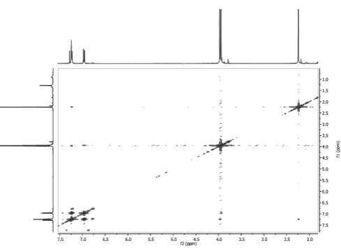
figure 5. COSY Spectrum of Compound 1 modified (tl-1) (mixture of veraguensin/galgravin modified)
figure 3.1H NMR (400 MHz, CDCl
3) Spectrum of Compound 1 modified
(tl-1) (mixture of veraguensin/galgravin modified)
1H NMR (400 MHz, CDCl
3) δ 7.21 (4H, dd, J = 6.1, 1.9, H-2/2’ and
H-6/6’), 6.94 (2H, d, J = 8.9, H-5/5’), 3.95 (6H, s, OCH3-3/3’), 3.92 (6H, s,
OCH3-4/4’), 2.22 (6H, s, H-9/9’).
figure 4.13C NMR (400 MHz, CDCl
3) Spectrum of Compound 1 modified
(tl-1) (mixture of veraguensin/galgravin modified)
13C NMR (100 MHz, CDCl
3) δ 149.2 (C) (x2), 148.3 (C) (x2), 147.1 (C)
(x2), 125.3 (C) (x2), 118.6 (CH) (x2), 117.9 (C) (x2), 111.5 (CH) (x2), 109.5 (CH) (x2), 56.2 (CH3), 56.1 (CH3), 10.0 (CH3) (x2).
figure 6. Expansion of the COSY Spectrum of Compound 1 modified (tl-1) (From 4.80 to 3.40 f1 and From 7.45 to 6.65 f2) (mixture of veraguensin/
galgravin modified)
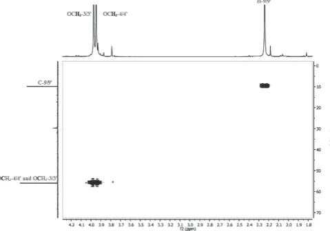
figure 8. HMQCSpectrum of Compound 1 modified (tl-1) (mixture of veraguensin/galgravin modified)
figure 9. Expansion of the HMQC Spectrum of Compound 1 modified (tl-1) (From 124.0 to 102.0 f1 and From 7.34 to 6.90 f2) (mixture of
veraguensin/galgravin modified)
figure 10. Expansion of the HMQC Spectrum of Compound 1 modified (tl-1) (From 70.0 to 0.0 f1 and From 4.20 to 1.80 f2) (mixture of veraguensin/
galgravin modified)
figure 11. DEPT-135° Spectrum of Compound 1 modified (tl-1) (mix-ture of veraguensin/galgravin modified)
figure 12.1H NMR (400 MHz, CDCl
3) Spectrum of Compound 2
(schi-neolignin B)
1H NMR (400 MHz, CDCl
3) δ 6.76 (2H, d, J = 8.1, ArH), 6.65 (1H, d, J
= 1.9, ArH), 6.63 (1H, dd, J = 8.1, 1.8, ArH), 6.58 (1H, d, J = 1.8, ArH), 2.56 (2H, dd, J = 13.5, 6.8, H-7/7’), 2.40 (2H, dd, J = 13.7, 7.8, 7/7’), 1.76 (2H, dd, J = 12.9, 6.5, H-8/8’), 0.83 (6H, d, J = 6.6, H-9/9’).
acetylation of schineolignin B (2).
Schineolignin B 2 (2,1 mmol), in a mixture of acetic anhydride and pyridine (5 mL/ 5 mL) was placed in a 50 mL pear-shaped flask. The mixture was stirred at 100 C for 15 h. Removal of the solvent under reduced pressure afforded a crude mixture, which was extracted with HCl solution followed by
extracted with NaHCO3 solution to give the products, which was purified by
column chromatography on silica gel (n-hexane/AcOEt= 8/2) sephadex
lH-20 in open column chromatography to give
5-(4-(3,4-dimethoxyphenyl)-2,3-dimethylbutyl)-2,3-dimethoxyphenyl acetate, 85% (tl-2) [11].
1H NMr (400 MHz, cDcl
3) spectral data of Schineolignin B: 1H NMR (400 MHz, CDCl3) δ: 6.76 (1H, d, J = 8.1, Ar-H), 6.65 (1H, d, J = 1.9, Ar-H),
6.63 (1H, dd, J = 8.1, 1.8, Ar-H), 6.58 (1H, d, J = 1.8, Ar-H), 2.56 (2H, dd, J =
13.5, 6.8, H-7/7’), 2.40 (2H, dd, J = 13.7, 7.8, H-7/7’), 1.76 (2H, dd, J = 12.9, 6.5, H-8/8’), 0.83 (6H, d, J = 6.6, H-9/9’).
1H NMr (400 MHz, cDcl
3) spectral data of tl-2: δ: 6.76– 6.57 (5H, m, Ar-H), 3.86–3.81 (12H, s, 3 x OCH3), 2.56 (2H, dd, J = 13.5, 6.7, H-7/7’), 2.40 (2H, dd, J = 13.5, 7.8, H-7/7’), 2.30 (3H, s, CH3-CO2-Ar), 1.79–1.73 (m, 2H,
Benzylation of meso-dihydroguaiaretic acid and threo -dihydroguai-aretic acid (3).
A mixture of 3(0.567 mmol) and sodium carbonate (11.4 mmol) in dry
acetone (36 ml) was heated to reflux for 1 h under nitrogen. Then, benzyl bromide (0.63 ml, 5.67 mmol) was added and the mixture was heated under reflux for an additional 3 h. After cooling to room temperature, the reaction mixture was filtered. The filtrate was concentrated and distilled under reduced
pressure in a rotary evaporatorto remove the excess unreacted benzyl bromide. The residue was chromatographed on silica gel (hexane/AcOEt= 8/2) and
sephadex lH-20 in open column chromatography to give
1-(benzyloxy)-4-(4-(3,4-dimethoxyphenyl)-2,3-dimethylbutyl)-2-methoxybenzene, 80% (tl-3)
[12, 13].
1H NMr (400 MHz, cDcl
3) spectral data of meso-dihydroguaiaretic acid
and threo-dihydroguaiaretic acid:δ: 6.82 (d, J = 8.0, 2H), 6.78 (dd, J = 8.2, 2.3, 2H), 6.67 (dd, J = 8.1, 1.9, 1H), 6.63 (d, J = 1.8, 1H), 6.60 (dd, J = 8.0, 1.8, 2H), 6.59 (d, J = 1.9, 1H), 6.54 (d, J = 1.8, 2H), 2.75 (dd, J = 13.5, 5.0, 2H), 2.54 (dd, J = 13.5, 7.1, 2H), 2.40 (dd, J = 13.6, 7.6, 2H), 2.30 (dd, J = 13.5, 9.2, 1H), 1.76 (dd, J = 13.3, 6.7, 2H), 1.75 (dd, J = 13.0, 6.6, 2H), 0.85 (dd, J = 6.6, 2.5, 6H).
1H NMr (400 MHz, cDcl
3) spectral data of tl-3: δ: 7.46 (2H, d, J = 7.3, H-2’’/2’’’ and 6’’/6’’’), 7.38 (2H, t, J = 7.4, H-3’’/3’’’ and H-5’’/5’’’), 7.32 (1H,
t, J = 7.2, H-4’’/4’’’), 6.81 (2H, d, J = 9.3, Ar-H), 6.79 (2H, d, J = 8.3, Ar-H), 6.69 (1H, d, J = 1.6, Ar-H), 6.64 (1H, dd, J = 9.4, 1.5, Ar-H), 6.57 (1H, dd, J
= 8.1, 1.6, Ar-H), 5.14 (4H, s, H-7’’/7’’’), 2.75 (2H, dd, J = 13.4, 4.9, H- 7/7’), 2.57 (2H, dd, J = 13.6, 6.7, H-7/7’), 2.40 (2H, dd, J = 13.6, 7.8, H-7/7’), 2.30 (2H, dd, J = 13.4, 9.3, H-7/7’), 0.86 (3H, d, J = 7.2, H-9/9’), 0.84 (3H, d, J =
6.8, H-9/9’) (see supporting information, Figure 15).
figure 13.1H NMR (400 MHz, CDCl
3) Spectrum of Compound 2
modified (tl-2) (schineolignin B modified)
1H NMR (400 MHz, CDCl
3) δ 6.76– 6.57 (5H, m, ArH), 3.86–3.81 (12H,
s, 3 x OCH3), 2.56 (2H, dd, J = 13.5, 6.7, H-7/7’), 2.40 (2H, dd, J = 13.5, 7.8,
H-7/7’), 2.30 (3H, s, CH3-CO), 1.79 – 1.73 (2H, m, H-8/8’), 0.83 (6H, d, J =
6.6, H-9/9’).
figure 14.1H NMR (400 MHz, CDCl
3) Spectrum of Compound 3 (
mix-ture of meso-dihydroguaiaretic acid and threo-dihydroguaiaretic acid)
1H NMR (400 MHz, CDCl
3) δ 6.82 (2H, d, J = 8.0), 6.78 (2H, dd, J = 8.2,
2.3), 6.67 (1H, dd, J = 8.1, 1.9), 6.63 (1H, d, J = 1.8), 6.60 (2H, dd, J = 8.0, 1.8), 6.59 (1H, d, J = 1.9), 6.54 (2H, d, J = 1.8), 5.44 (1H, OH), 2.75 (2H, dd, J = 13.5, 5.0), 2.54 (2H, dd, J = 13.5, 7.1), 2.40 (2H, dd, J = 13.6, 7.6), 2.30 (2H, dd, J = 13.5, 9.2), 1.76 (2H, dd, J = 13.3, 6.7), 1.75 (2H, dd, J = 13.0, 6.6), 0.85 (6H, dd, J = 6.6, 2.5).
figure 15.1H NMR (400 MHz, CDCl
3) Spectrum of Compound 3 modi
-fied (tl-3) (mixture of meso-dihydroguaiaretic acid and threo
-dihydroguai-aretic acid modified)
1H NMR (400 MHz, CDCl
3) 1H NMR (400 MHz, CDCl3) δ 7.46 (2H, d,
J = 7.3, H-2’’/2’’’ and 6’’/6’’’), 7.38 (2H, t, J = 7.4, H-3’’/3’’’ and H-5’’/5’’’), 7.32 (1H, t, J = 7.2, H-4’’/4’’’), 6.81 (2H, d, J = 9.3, Ar-H), 6.79 (2H, d, J =
8.3, Ar-H), 6.69 (1H, d, J = 1.6, Ar-H), 6.64 (1H, dd, J = 9.4, 1.5, Ar-H), 6.57 (1H, dd, J = 8.1, 1.6, Ar-H), 5.14 (4H, s, H-7’’/7’’’), 2.75 (2H, dd, J = 13.4, 4.9, H-7/7’), 2.57 (2H, dd, J = 13.6, 6.7, H-7/7’), 2.40 (2H, dd, J = 13.6, 7.8, H-7/7’), 2.30 (2H, dd, J = 13.4, 9.3, H-7/7’), 0.86 (3H, d, J = 7.2, H-9/9’), 0.84 (3H, d, J = 6.8, H-9/9’).
reSultS and diScuSSion
Three structural transformation process are presented in this article; and corresponds to the first report of this type of structural modification of
lignans isolated from Nectandra species. A direct method was developed
for the conversion of compound 1 to furan-type lignan. Additionally, the
and the transformed compound showed formation of derivatives compounds (see supporting information for details). Interestingly, few reports describe dehydrogenation, benzylation or acetylation of natural products isolates; to our knowledge the direct structural transformation of lignan compounds isolated
from Nectandra specieshas yet to be documented.
Comparison of spectroscopic data between the starting material
(veraguensin and galgravin) and the product (tl- 1) show the absence some
characteristics signals [such as: 4.42 (1H, d, J = 9.3, H-7), 5.14 (1H, d, J = 8.6, H-7’), and 4.52 (2H, d, J = 6.4, H-7/7’)], allow suggest the formation of tl-1.
The compound tl-2 has a signal 2.30 (3H, s, CH3-CO), among others;
which it is characteristic of the formation of the product.
The compound tl-3 has a signal 5.14 (4H, s, H-7’’/7’’’), among others;
which it is characteristic of the formation of the product. Additionally, the com-pound formed is absent the signal generated by the hydroxyl group [5.44 (1H,
s, OH)].
referenceS
1. J. M. Barbosa-Filho, M. Yoshida, O. R. Gottlieb, Phytochemistry. 28,
1991, (1989).
2. L. Chérigo, V. Polanco, E. Ortega-Barria, M. V. Heller, T. L. Capson, L. C. Rios, Nat. Prod. Res. 19, 373, (2005).
3. A. A. da Silva Filho, S. Albuquerque, M. L. e. Silva, M. N. Eberlin, D. M.
Tomazela, J. K. Bastos, J. Nat. Prod., 67, 42, (2004).
4. S. R. Farias-Moreno, A. Arnobio, J. José de Carvalho, A. L. Nascimento, M. O. Timoteo, B. Olej, E. K. Rocha, M. Pereira, M. Bernardo-Filho, L.
Querino de Araújo Caldas, Biol. Res. 40, 131, (2007).
5. J. C. Moro, J. B. Fernandes, P. C. Vieira, M. Yoshida, O. R. Gottlieb, H. E.
Gottlieb, Phytochemistry,26, 269, (1987).
6. B. Agius, M. Setzer, S. Stokes, T. Walker, W. Haber, W. Setzer, Int. J. Essen. Oil Ther. 1, 167, (2007).
7. J. G. Rohwer. Lauraceae: Nectandra. Flora Neotropica, Monograph 60,
in Flora Neotropica Monograph. vol. 60, T. N. Y. B. Garden, Ed., ed New
York, pp. 1-332, 1993.
8. J. Chen, W. Li, H. Yao, J. Xu, Fitoterapia. 103, 231, (2015).
9. M. Saleem, H. J. Kim, M. S. Ali, Y. S. Lee, Nat. Prod. Rep, 22, 696, (2005).
10. L. Dalla Via, E. Uriarte, E. Quezada, A. Dolmella, M. G. Ferlin, O. Gia, J.
Med. Chem. 46, 3800, (2003).
11. R. Nakamura, Y. Obora, Y. Ishii, Tetrahedron. 65, 3577, (2009).
12. H. S. P. Rao, S. Senthilkumar, J. Chem. Sci. 113, 191, (2001).
13. L. McMaster, W. Bruner, Ind. Eng. Chem. 28, 505, (1936).
14. M. Miyazawa, H. Kasahara, H. Kameoka, Phytochemistry. 46, 1173,
(1997).
15. Y. B. Xue, Y. L. Zhang, J. H. Yang, X. Du, J. X. Pu, W. Zhao, X. N. Li, W.