Effects of temperature on hematological and histopathological
changes and survival rate of juvenile
Fenneropenaeus
vannamei
experimentally challenged to White Spot Virus
Kakoolaki S.
1*; Sharifpour I.
1; Afsharnasab M.
1; Sepahdari A.
1;
Mehrabi M. R.
1; Ghaednia B.
2; Nezamabadi H.
1Received: January 2013 Accepted: April 2013
Abstract
Many shrimp farmers were suffering from White Spot Disease (WSD) onset in last decades. Oscillation of environmental factors could lead mortality in susceptible hosts. Our study was aimed to investigate the effect of different temperatures on juvenile
Fenneropenaeus vannamei experimentally exposed to White Spot Virus (WSV). Five hundred and forty juveniles were distributed among 3 treatments in triplicates, 22, 25 and 30°C and experimentally WSV were injected in the shrimps. Our results showed mortality started at 36 h post inoculation (hpi) in the treatment at 25°C (T25), meanwhile the mean
value of mortality percent at 54 hpi in T25 (71.10±17.35) showed the significant difference
(p=.045) with T22 (3.33±3.33) and T30 (Not Observed, NO.). Our results suggest that in site
selection, in primary stage of farm designing, water temperature at more than 29°C, should be considered as key environmental factor. This finding can lead us that why the White Spot Disease occurred with high mortality in some area when the days of shrimp culture were prolonged until mid autumn.
Keywords: White Spot Syndrom Virus, Temperature, Fenneropenaeus vannamei, Challenge
1- Iranian Fisheries Research Organization., P.O.Box: 13185-116, Tehran, Iran 2- Iran Shrimp Research Center, P.O.Box:1374, Bushehr, Iran
*Corresponding author's email:[email protected]
Introduction
Almost farmers were suffering from White Spot Virus invaded to the shrimp, Pennaeus indicus farms in Iran during 1992-2006 (Kakoolaki et al., 2010) and then Fenneropenaeus vannemei SPF (Specific Pathogen Free) were transported from Hawaii to Iran to control the disease (Kakoolaki et al., 2011). Shrimp culture has been a familiar industry and well extended throughout the world especially in south-east of Asia since 1990 (Sánchez-Martínez et al., 2007). Estimated losses due to WSV only in Asia have been approximately $ 6 billion (Lightner, 2011). Small size of the ponds, abrupt alteration in physico-chemical criteria specially salinity and temperature and prolonged the days of culture in the shrimp farms result in few epizootic of White Spot Diseases (WSD) in Iran in last decade (Kakoolaki, 2004; Kakoolaki et al., 2011; Peinado-Guevara and Lopez-Meyer, 2006; Soltani et al., 1998). So far, the effective way of temperature on WSV outbreak has not been well known. Rahman et al. (2007; 2006) showed that higher temperature with 33°C could be used to control the mortality of WSV infected shrimp in the field. Due to effects of temperature on metabolisms, growth, survival rate and immunological criteria, it becomes one of the most important environmental factor in shrimp farms (Wyban et al., 1995). Esparza-Leal et al. (2010) showed if days of shrimp culture extend to autumn, the susceptibility to WSV among the exposure shrimps could be increased (Esparza-Leal et al., 2010). The risk of WSV outbreak is reduced when the water temperature goes up and salinity fluctuation is
low (Tendencia et al., 2010b). In contrary to the results of Sahoo et al. (2005), the association between WSV outbreak and environmental risk factors such as temperature (Corsin et al., 2002; Peinado-Guevara and Lopez-Meyer, 2006), ammonia (Corsin et al., 2001) and salinity (Kakoolaki et al., 2011) Some scientist studied the effects of temperature on the expression of WSV gene (Granja et al., 2006; Reyes et al., 2007). Our study was aimed to investigate the effect of different temperature on mortality criteria in Fenneropenaeus vannemai exposed to WSV.
Materials and methods
Five hundred and forty juveniles of P. vannamei (7.98±0.54) were gathered from Quarantine research shrimp farm located in Bushehr province in south of Iran and then transferred to the Iran Shrimp Center for experimental and laboratory examinations. Shrimp were then acclimated to the determined conditions as treatments and corresponding controls in triplicates (T1= 22oC named 22,
T2=25ºC named 25 and T3=30 ºC named 30 as
treatments 1, 2 and 3). The shrimp were then distributed to 18 glass aquariums (50×50×60cm) which 60% of each was filled with well clean aerated seawater (42 ppt). The shrimp were kept, fed and siphoned in the current condition for 40 days before exposure to the virus, according to our previous article (Kakoolaki et al., 2011).
Virus with the titre of LD50= 1×105.4 and
code no. of WSV / irn / 1 / 2011 already had prepared in Motamedi lab in Iran were used in challenging with the treatments but control
groups were left untreated. The mixture was stored at -80 ºC until use (Motamedi Sedeh et al., 2011) . After acclimation, Shrimps were intramuscularly inoculated with 50 µl of virus solution (Kakoolaki et al., 2011).
After the mortality observation, 0.2ml of Hemolymph were withdrawn from the second leg's basement of ventral segments from 3 moribund shrimp of each triplicates in treatments and controls, using 1 ml syringe along with 26 gauge needle. Each syringe pre-filled with 0.8 ml Alsever solution as anticoagulant (Kakoolaki et al., 2010; Kondo, 2003). According to the method of Afsharnasab et al. (2009) the whole body of these three moribund shrimp were prepared to transfer to histopathology and PCR labs of Iran Shrimp Center. The appendages and gill tissues were cut, placed in Eppendorf micro tubes containing ethanol 70% for PCR technique using IQ2000 WSV commercial kit. Remaining tissues were placed in tubes containing Davidson's fixative. Davidson's fixative were discarded and replacing with ehanol 70% after 48-72 h in the lab and staining by H & E method(Lightner, 1996).
THC was carried out using the Neubauer type's Hemocytometer. A drop of hemolymph-anticuagulant mixture was then placed on the Hemocytometer and counting the cells under a light microscope ×10 as follows:
THC (Cells. Ml-1) =Average count of 5 cells×5×104×1/dilution (Kakoolaki et al., 2010).
0.2 ml of withdrawn Hemolymph was pre-filled with 0.1 ml fixative. Differential Hemocyte Count (DHC) was carried out using a slide, a drop of mixture solution was then
placed on it and staining with May-Grundwald Giemsa (MGG) method begun after then. The method for fixation and staining of the hemolymph was carried out based on the new methods given in previous article (Kakoolaki et al., 2011)
TPP (µg.dl-1) was examined using centrifuge (3000 rpm for 10 min. at 4oc) the mixture of distinct anticoagulant and collected hemolymph (n=6) of each triplicate of the treatments. The supernatants were remove and transferred after 24 h with freezing condition to the lab to examine TPP of the treatments using biochemical autoanalyzer (Technicon, RA 1000).
The PCR examinations of the treatments were assessed using IQ2000, WSV commercial kit after exposure the shrimp to the virus.
One Way-ANOVA and Bonferroni multi-comparison tests were carried out to compare the effects of different degrees of temperature on mortality rate, Epigastric hematopoietic spheroid values and THC, TPP and DHC values after challenging the virus against the treatments using SPSS 17. The mean values of criteria are given as Mean ± SE of Mean.
Results
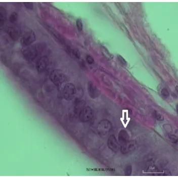
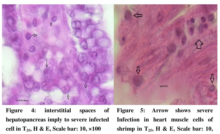
The results of the PCR of the primary stocking of shrimp were negative before the exposure to the virus was begun but based on the Figures 2-6 the hypertrophied cells and the inclusion bodies of the WSV observed through to the target tissues such as gills and epithelial cells of gut, interstitial spaces of hepatopancreas and heart muscle cells. Histopathological finding showed the severe
infection in T25 in comparison to T22 and T30, respectively. The PCR results of the treatments were positive (based on the IQ2000,
WSV commercial kit) after exposure the shrimp to the virus (Fig. 1).
Figure 1: The positive result of PCR for the WSV of the pooled samples from each treatment (IQ2000, WSV commercial kit)
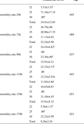
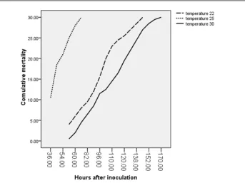
Cumulative mortality and mortality percent were shown in figure 7 and table 1. According to these, the mortality percent at 54
h post inoculation (hpi) in T25 (71.10±17.35)
had significant difference (p=.045) with T22
(3.33±3.33) and T30 (Not Observed, NO.).
Table 1: Mortality percent differences between treatments (α=0.05, n=3)
Mean±Std. Error Sig.
mortality.rate.54h
22 3.33±3.33a 71.10±17.35 .00a
24.81±12.65
.045 25
30 Total
mortality.rate.72h
22 26.70±.00 28.90±17.35 11.13±4.82 22.24±5.90
.089 25
30 Total
mortality.rate.96h
22 24.43±4.82a .00 23.30±.00a 15.91±4.21
.037 25
30 Total
mortality.rate.110h
22 23.33±3.33a .00
12.23±2.93a 11.85±3.60
.043 25
30 Total
mortality.rate.138h
22 16.63±8.81 .00 31.10±4.45 15.91±5.32
.051 25
30 Total
mortality.rate.170h
22 5.56±1.13a
.00a 22.23±3.99 9.26±3.54
.021 25
30 Total
*Different scripts show the significant differences at the level of .05
Based on the table 2 the quantity of Spheroids more developed (p< .05) in T25
(7.50±0.18)and T22 (6.37± 0.26) in comparison
to T30 (1.62± 0.53).
Table 2: Spheroid formation in different treatments
spheroids N Mean±Std. Error Minimum Maximum Sig.
22oc 8 6.37± 0.26a
7.50± 0.18a 1.62± 0.53 5.16± 0.56
6.00 8.00
25oc 8 7.00 8.00 .000
30oc 8 .00 4.00
Total 24 .00 8.00 *Different scripts show the significant differences at the level of .05
The mortality of T25 started in 36 hpi with
about 33.30 % of stock and completed after 60 h.
Figure 2: Infected epithelial cells in gill, H & E, Scale bar: 10, ×100
Figure 3: Infected epithelial cell in mid gut, H & E, Scale bar: 10, ×100
Figure 4: interstitial spaces of hepatopancreas imply to severe infected cell in T25, H & E, Scale bar: 10, ×100
Figure 5: Arrow shows severe Infection in heart muscle cells of shrimp in T25, H & E, Scale bar: 10,
×100
Figure 6: Shows mild Infection in heart muscle cells of shrimp in T30,
H & E, Scale bar: 10, ×100
The maximum mortality percent (71.10) was occurred in T25 at the primary hours (36
h) after inoculation but and mortality in T22
and T30 begun at 58 h. The mortality percents
of T25 became 100% at 60 h with 28.90% but in T22 and T30 were completed at 152 and 170
hpi, respectively. The mean value of total protein, total hemocte count (THC) , total
differentiate count containing large granular cell (LGC), semi granular cell (SGC) and hemocyte count (HC) were given in table 3. The mean value of total protein in T30 shrimp
hemolymph (157.82± 22.59) was significantly higher (p= .012) than that of in T22 (76.60±
3.57) and T25 (106.72± 17.80) but no
significant difference between T22 and T25 was
observed ( p> .05)..The mean value of THC in T30 (16.06± 2.48) was significantly higher than
those of T22 (8.96± 1.81)and T25 (7.52± 1.18).
Of course, there is a significant difference
between the two last (p<.05). *No mortality were observed in whole control groups (fig. 7).
Table 3: Hemolymph parameters in different treatments
N Mean ±Std. Error Minimum Maximum Sig.
TOTAL PROTEIN ug/dl
22oc 6 76.60± 3.57 ab 106.72± 17.80 ab 157.82± 22.59 b 113.71± 12.18
66.60 90.90
25oc 6 60.00 175.00 .012 30oc 6 81.80 220.00
Total 18 60.00 220.00
THC×105
22oc 6 8.96± 1.81a
7.52± 1.18ab 16.06± 2.48ac
10.85± 1.37
2.10 15.00
25oc 6 3.40 12.00 .014 30oc 6 9.60 22.60
Total 18 2.10 22.60
LGC×105
22oc 6 30.33± 1.68a 28.83± 1.01ab 34.16± 1.44ac 31.11± 0.93
26.00 35.00
25oc 6 26.00 33.00 .046 30oc 6 29.00 38.00
Total 18 26.00 38.00
SGC×105
22oc 6 46.83± 1.49 56.50± 2.17a 54.50± 0.92a 52.61± 1.33
43.00 52.00
25oc 6 48.00 63.00 .002 30oc 6 52.00 57.00
Total 18 43.00 63.00
HC×105
22oc 6 21.50± 1.38 14.66± 1.74a 10.33± 0.21a 15.50± 1.31
17.00 27.00
25oc 6 10.00 22.00 .000** 30oc 6 10.00 11.00
Total 18 10.00 27.00 *Different scripts show the significant differences at the level of .05
**Significant difference was observed at the level of .01
Figure 7: Cumulative mortality in different treatments after determined hpi.
Discussion
Oscillations of environmental factors could result in high mortality rate in shrimp farms. Due to the effect of these factors e.g. temperature increase susceptibility of the host as well the viral replication process(Moser et al., 2012). According to the figure 1, it seems that the load of the virus in T30 was lower than
that of the T22 and T25. In addition, the
mortality of T30 showed (Fig. 1 , Table 1) a
slight trend in contrary to T25. This result is
close to finding of Granja et al. (2006) that showed the load of White Spot Virus (WSV) is reduced when the shrimp maintain in higher temperature. Other scientists (Du et al., 2006; Guan et al., 2003) showed that increasing the temperature to around 30 and more could reduce the replication of WSV even stop it in Marsupenaeus japonicas and procambarus clarkia, respectively. Similar to the results of Granja et al. (Granja et al., 2006) and Vidal et
al. (2001) that showed the optimum temperature for loading the virus in P. vannamei is 25oC, our finding confirmed that the higher mortality in P. vannamei with water salinity of 42 ppt is 25OC and the temperature of 30OC (Figs 1, 7 ) is less predisposing degree for loading virus and. This findings are contradicted to the result of Moser et al. (2012) that showed the mortality rate is low and the replication of WSV is high in the shrimps kept in 29-30OC. But in similar to our finding, they showed the WSD onset occurred 36 hpi in shrimps kept in temperature at 25OC and contrarily, they found lower temperature allows no replication of the virus. The daily water-temperature fluctuations with hyperthermia (>32 C) could partially reduce WSV replication in some ponds (Rahman et al., 2007; Vidal et al., 2001) but this partial protection decreased when water temperature
declined from 28.0–33.4OC to 24.9OC while WSD onset was observed in almost cages (Esparza-Leal et al., 2010). Similarly, Tendencia, et al. (2010a) showed the higher prevalence of WSD in the sea was higher in November until January and confirmed WSD onset in earthen ponds is associated with lower degrees of temperature (Tendencia et al., 2010b). Results showed that hyperthermia reduces the expression of WSV genes on shrimp sub-cuticular epithelial cells (Reyes et al., 2007).
Following our results, the mean value of total protein and THC are higher in temperature 30OC than temperatures 22 OC and 25 OC. Based on the table 3 the percent value of LGC in T30 is significantly higher (P=.046) than the other treatments and followed by T22
and T25. Sudden mortality 36 hpi in T25 could
be due to the stressfulness of this degree for the shrimp and or optimum degree for replication of the virus. This finding can lead us that why the White Spot Disease occurred in Khuzestan in south of Iran when the days of shrimp culture were prolonged until mid autumn. White Spot Disease onset occurred in all area in south of Iran exception Hormozegan province where the pond water temperature was usually more than the 29-30OC during the summer. But in Khuzestan province the fluctuation of pond-water temperature within 24 h sometimes reach 5-7 degrees (Kakoolaki, 2004). Our results suggest that in site selection in primary stage of farm designing, water temperature more than 29OC, such as Hormozegan province in south of Iran, should be considered.
References
Afsharnasab, M., Mortezaei, R., Yegane, V. and Kazemi, B., 2009. Gross sign, histopathology and polymerase chain reaction observations of white spot syndrome virus in shrimp specific pathogen free Litopeneaus vannamei in iran.
Asian Journal of Animal and Veterinary
Advances, 4 (6), 297-305.
Corsin, F., Turnbull, J. F., Hao, N. V., Mohan, C. V., Phi, T. T., Phuoc, L. H., Tinh, N. T. N., et al., 2001. Risk factors associated with white spot syndrome virus infection in a Vietnamese rice-shrimp farming system. Disease of Aquatic Organisms 47, 1-12.
Corsin, F., Phi, T. T., Phuoc, L. H., Tinh, N. T. N., Hao, N. V., Mohan, C. V., Turnbull, J. F., et al., 2002. Problems and solutions with the design and execution of an epidemiological study of white spot disease in black tiger shrimp (Penaeus monodon) in Vietnam. Preventive Veterinary Medicine, 53 (1-2), 117-132.
Du, H. H., Li, W. F., Xu, Z. R. and Kil, Z. S., 2006. Effect of hyperthermia on the replication of white spot syndrome virus (wssv) in
Procambarus clarkii. Diseases of Aquatic Organisms, 71 (2), 175-178.
Esparza-Leal, H. M., Magallón-Barajas, F. J., Portillo-Clark, G., Perez-Enriquez, R., ÁLvarez-Ruíz, P., Escobedo-Bonilla, C. M., Méndez-Lozano, J., et al., 2010. Infection of wssv-negative shrimp, Litopenaeus vannamei, cultivated under fluctuating temperature conditions. Journal of the World Aquaculture Society, 41 (6), 912-922.
Granja, C. B., Vidal, O. M., Parra, G. and Salazar, M., 2006. Hyperthermia reduces viral
load of white spot syndrome virus in Penaeus vannamei. Diseases of Aquatic Organisms, 68 (2), 175-180.
Guan, Y., Yu, Z. and Li, C., 2003. The effects of temperature on white spot syndrome infections in Marsupenaeus japonicus. Journal of Invertebrate Pathology 83, 257-260.
Kakoolaki, S., 2004. Key points in shrimp health management, Tehran: Shams, 114.
Kakoolaki, S., Sharifpour, I., Soltani, M., Mousavi, H. A. E., Mirzargar, S. and Rostami, M., 2010. Selected morpho-chemical features of hemocytes in farmed shrimp,
Fenneropenaeus indicus in iran. Iranian Journal of Fisheries Sciences, 9 (2), 219-232.
Kakoolaki, S., Soltani, M., Ebrahimzadeh Mousavi, H. A., Sharifpour, I., Mirzargar, S., Afsharnasab, M. and Motalebi, A. A., 2011.
The effect of different salinities on mortality and histopathological changes of spf imported
Litopenaeus vannamei, experimentally exposed to white spot virus and a new differential hemocyte staining method. Iranian Journal of Fisheries Sciences, 10 (3), 447-460.
Kondo, M. 2003. Experiments of body defence mechanisms in crustacean. In: NFU (ed.). Shimonoseki: NFU.
Lightner, D., 1996. A handbook of pathology and diagnostic procedures for diseases of penaeid shrimp, LA, Baton Rouge: World Aquaculture Society.
Lightner, D., 2011. Virus diseases of farmed shrimp in the western hemisphere (theamericas): A review. Journal of Invertebrate Pathology,
106, 110-130.
Moser, J. R., Álvarez, D. A. G., Cano, F. M., Garcia, T. E., Molina, D. E. C., Clark, G. P., Marques, M. R. F., et al., 2012. Water temperature influences viral load and detection of white spot syndrome virus (wssv) in
Litopenaeus vannamei and wild crustaceans.
Aquaculture, 326–329, 9-14.
Motamedi Sedeh, F., Afsharnasab, M., Heidarieh, M., Shafaee, S. K., Rajabifar, S., Dashtiannasab, A. and Razavi, M. H., 2011.
Comparison virus titration of the iranian white spot syndrome virus isolate, on crayfish Astacus leptodactylus and penaeus semiculcatus. Iranian Journal of Fisheries Sciences, in press.
Peinado-Guevara, L. I. and Lopez-Meyer, M., 2006. Detailed monitoring of white spot syndrome virus (wssv) in shrimp commercial ponds in sinaloa, mexico by nested pcr.
Aquaculture, 251 (1), 33-45.
Rahman, M. M., Escobedo-Bonilla, C. M., Corteel, M., Dantas-Lima, J. J., Wille, M., Alday Sanz, V., Pensaert, M. B., et al., 2006.
Effect of high water temperature (33ºc) on the clinical and virological outcome of experimental infections with white spot syndrome virus (wssv) in specific pathogen-free (spf)
Litopenaeus vannamei. Aquaculture, 261, 842-849.
Rahman, M. M., Corteel, M., Dantas-Lima, J. J., Wille, M., Alday-Sanz, V., Pensaert, M. B., Sorgeloos, P., et al., 2007. Impact of daily fluctuations of optimum (27 degrees c) and high water temperature (33oc) on Penaeus vannamei
juveniles infected with white spot syndrome virus (wssv). Aquaculture, 269 (1-4), 107-113.
Reyes, A., Salazar, M. and Granja, C., 2007.
Temperature modifies gene expression in subcuticular epithelial cells of white spot
syndrome virus-infected Litopenaeus vannamei.
Developmental and Comparative Immunology,
31 (1), 23-29.
Sahoo, A. K., Patil, P. and Shankar, K. M., 2005.
White spots? A loaded question for shrimp farmers. Current Science, 88 (12), 1914-1917.
Sánchez-Martínez, J. G., Aguirre-Guzmán, G. and Mejía-Ruíz, H., 2007. White spot syndrome virus in cultured shrimp: A review.
Aquaculture Research, 38 (13), 1339-1354.
Soltani, M., Kakoolaki, S. and Keisami, M., 1998. Isolation and identification of dominant vibrio species in farmed prawn of health station, Bushehr. Journal of Veterinary Research, 55 (2), 28-32.
Tendencia, E. A., Bosma, R. H., Usero, R. C. and Verreth, J. A. J., 2010a. Effect of rainfall and atmospheric temperature on the prevalence of the whitespot syndrome virus in pond-cultured
penaeus monodon. Aquaculture Research, 41 (4), 594-597.
Tendencia, E. A., Bosma, R. H. and Verreth, J. A. J., 2010b. Wssv risk factors related to water physico-chemical properties and microflora in semi-intensive penaeus monodon culture ponds in the philippines. Aquaculture, 302 (3-4), 164-168.
Vidal, O. M., Granja, C. B., Aranguren, F., Brock, J. A. and Salazar, M., 2001. A profound effect of hyperthermia on survival of
Litopenaeus vannamei juveniles infected with white spot syndrome virus. Journal of the World Aquaculture Society, 32 (4), 364-372.
Wyban, J., Walsh, W. A. and Godin, D. M., 1995. Temperature effects on growth, feeding rate and feed conversion of the pacific white shrimp (Penaeus vannamei). Aquaculture, 138,
267-279.