Marcin Masalski
1, 2, A–F, Bartosz Banaś
2, B–D, Tomasz Kręcicki
1, E, FThe Influence of Eyeball Rotation on the Results
of Auditory Steady-State Responses
Wpływ pozycji gałek ocznych na wyniki badania słuchowych potencjałów
wywołanych stanu ustalonego
1 Department and Clinic of Otolaryngology, Head and Neck Surgery, Wroclaw Medical University, Wrocław, Poland
2 Institute of Biomedical Engineering and Instrumentation, Wroclaw University of Technology, Wrocław, Poland
A – research concept and design; B – collection and/or assembly of data; C – data analysis and interpretation;
D – writing the article; E – critical revision of the article; F – final approval of article; G – other
Abstract
Background. The testing of auditory steady-state responses consists in recording the electrophysiological response to an auditory stimulus. Due to this response, in addition to changes in electric potentials caused by neuron impuls-es in the auditory path, the sonomotor reflex can also be observed. The sonomotor reflex shows muscle rimpuls-esponsimpuls-es to auditory events, and in the case of auditory evoked potentials it mainly consists of post-auricular muscle responses. When the eyes are rolled to the side during testing, the post-auricular muscle response to an auditory stimulus is stronger, which in turn can contribute to improving response detection.
Objectives. The aim of this study was to test the influence of eyeball rotation on the results of auditory steady-state responses.
Material and Methods. Auditory evoked potentials were tested in a group of ten people with normal hearing. Each person was examined three times: (i) with eyes closed, (ii) with eyes open looking straight ahead and (iii) with eyes open and rolled to the side.
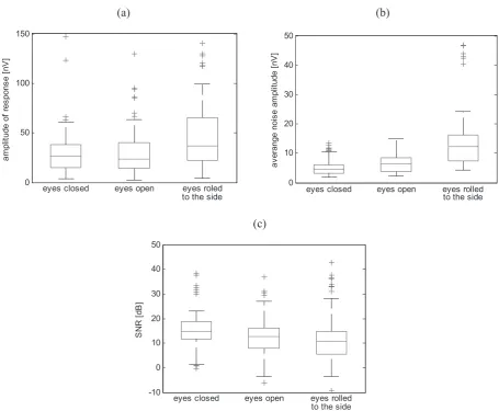
Results. The median electrophysiological response amplitude recorded when the eyes were rolled to the side was approximately 40% higher than the median response amplitude recorded in other positions (with eyes closed, and with eyes open looking straight ahead). At the same time, during tests with the eyes rolled to the side, a 170% increase in the noise median was observed, compared to the tests conducted with the eyes closed.
Conclusions. Rolling the eyes to the side does not improve the detection of response, as the observed increase in noise amplitude is much higher than the increase in the amplitude of the electrophysiological response (Adv Clin Exp Med 2013, 22, 4, 549–554).
Key words: auditory steady-state responses (ASSRs), postauricular muscle response.
Streszczenie
Wprowadzenie. Badanie słuchowych potencjałów wywołanych polega na rejestracji odpowiedzi elektrofizjologicz-nej na bodziec dźwiękowy. W odpowiedzi tej, oprócz zmian potencjałów elektrycznych wywołanych impulsacją neuronów drogi słuchowej, można wyróżnić także komponent sonomotoryczny. Komponent sonomotoryczny jest mięśniową reakcją organizmu na bodziec dźwiękowy i w przypadku słuchowych potencjałów wywołanych składa się głównie z reakcji mięśnia zausznego. Boczne zwrócenie gałek ocznych podczas badania wzmacnia reakcję mięś-nia zausznego na stymulacje dźwiękową i tym samym może przyczynić się do poprawy detekcji odpowiedzi.
Cel pracy. Określenie wpływu pozycji gałek ocznych na wyniki badania słuchu metodą potencjałów wywołanych stanu ustalonego.
Materiał i metody. Badania słuchowych potencjałów wywołanych przeprowadzono na grupie 10 osób z prawidło-wym słuchem. Każda osoba została zbadana trzykrotnie: (i) z zamkniętymi oczami, (ii) z otwartymi oczami, patrząc na wprost, oraz (iii) z otwartymi oczami przy bocznie zwróconych gałkach ocznych.
Adv Clin Exp Med 2013, 22, 4, 549–554
ISSN 1899–5276
ORIgINAl PAPERS
The testing of auditory evoked potentials con-sists in recording electrophysiological responses to auditory stimuli. The auditory evoked poten-tials make it possible to evaluate the transmission of nerve impulses in the auditory path. On the ba-sis of electrophysiological responses to audito-ry events the hearing thresholds of the examined person can be estimated. In addition to changes in electric potentials caused by neuron impulses in the auditory path, the electrophysiological re-sponse also shows the sonomotor reflex, which is the muscle response to auditory events. On the one hand, the existence of the sonomotor reflex in the recorded auditory steady-state responses can make the evaluation of transmission in the auditory path difficult due to overlapping muscle and auditory potentials, but on the other, it can make the detec-tion of responses to auditory stimuli easier, since the total muscle and auditory potential is easier to identify in the signal registered during testing. In the case of auditory evoked potentials the sonomo-tor reflex mainly consists of post-auricular muscle responses (PAMRs) [1, 2]. PAMRs are unstable re-sponses, varying from patient to patient [3, 4], but they can be strengthened when the eyes are rotat-ed to the side [5, 6].
Auditory evoked potentials can be divided into transient responses (electrophysiological respons-es to short auditory stimuli), and steady-state re-sponses (SSRs: electrophysiological rere-sponses to a series of stimuli presented at high repetition frequency).
Some previous researchers have proposed that the detection of transient responses may be im-proved when the eyeballs are rolled to the side [7, 8]. According to those authors, in most subjects it is possible to objectively reconstruct a pure tone au-diogram in this way.
The aim of the current study was to determine the influence of eyeball rotation on the results of auditory steady-state responses, especially on the possibility of improving the detection of responses recorded when the eyeballs are rolled to the side.
Auditory steady-state responses (SSRs) are a sinusoidal wave generated by the overlapping of transient responses due to a high stimulation frequency. The frequency of a SSR is equal to the
frequency of a sound stimulus. SSRs can be ob-tained by applying a short sound stimulus at a high repetition frequency or a continuous modulat-ed stimulus [9]. The detection of steady-state re-sponses can be made on the basis of EEg signals by comparing the signal’s amplitudes at the stimula-tion frequency with the amplitude of adjacent fre-quency bands. In this case PAMRs will be observed as an increase in the response amplitude [10].
The influence of eyeball rotation on SSR re-sults was tested for multifrequency steady-state responses (MF-SSRs). MF-SSRs are responses to stimuli presented simultaneously with a modula-tion frequency in the 70–100 Hz range [9]. They are often used to evaluate the hearing threshold.
Material and Methods
Auditory evoked potentials were tested in a group of 10 subjects (age from 25 to 33) with normal hearing. Each person was examined three times: (i) with eyes closed, (ii) with eyes open look-ing straight ahead and (iii) with eyes open and rolled to the side. During all the examinations the subjects were lying comfortably on their backs and were asked not to fall asleep. During the first ex-amination the patients’ eyes were closed, during the second they were looking straight ahead, and during the third their eyes were fixed on points lo-cated to the side, within the scope of binocular vi-sion. As looking to one side for a long time is un-comfortable, the subjects switched from looking toward one side to the other every minute, which was signalled by a touch of the hand. The exami-nation was conducted with the use of a diagnostic system for objective audiometry [11].
Stimulus
Auditory stimulation was performed binau-rally. Eight stimuli were presented at the same time, four on the right side and four on the left side. On both sides the fundamental frequencies fc of the stimuli were set to 500 Hz, 1 kHz, 2 kHz
and 4 kHz respectively. Each stimulus had unique modulation frequency fm; on the right side these
Wyniki. Mediana amplitudy odpowiedzi elektrofizjologicznej zarejestrowanej w warunkach bocznego zwrócenia gałek ocznych była wyższa o około 40% od mediany amplitudy odpowiedzi zarejestrowanej w pozostałych bada-niach, tj. przy oczach zamkniętych i oczach otwartych patrząc na wprost. Jednocześnie w badaniu przy gałkach ocznych zwróconych bocznie zaobserwowano wzrost mediany szumu aż o 170% w porównaniu do badania prze-prowadzonego przy oczach zamkniętych.
Wnioski. Boczne zwrócenie gałek ocznych nie przynosi poprawy detekcji odpowiedzi, ponieważ obserwowany wzrost amplitudy szumu jest dużo większy od wzrostu amplitudy odpowiedzi elektrofizjologicznej (Adv Clin Exp Med 2013, 22, 4, 549–554).
were (respectively) 73.37 Hz, 81.52 Hz, 89.67 Hz, and 97.83 Hz; and on the left: 77.45 Hz, 85.60 Hz, 93.75 Hz and 101.90 Hz. Each stimulus represent-ed an intensity of 50 dBHl determinrepresent-ed by ampli-tude A. All the stimuli s were generated according to the formulas below:
) 2 sin( 2 )
( f i
f f m i m m c
f π τ
ϕ = (1)
)), ( 2 sin( )) 2 sin( 1 ( )
(i A m f ti f ti i
s = + a πm πm +ϕ (2)
where:
mf is the frequency modulation index determined
at the level of 20%
ma is the amplitude modulation index determined
at the level of 100%
t is the signal sample time of 1/48 kHz
The sound stimulus was generated by the sound system of a personal computer and presented through audiometric earphones TDH-39. Calibra-tion of sound generaCalibra-tion system was performed us-ing a type 4153 artificial ear made by Brüel & Kjær.
Registration of the EEG Signal
The EEg signal was measured using a two-channel measurement system. In each two-channel changes in the electric potentials between an trode placed on the top of the head and an elec-trode placed behind the auricle were registered. The latter electrodes were placed on the post-au-ricular muscle, approximately 2 cm above their normal location on the mastoid process in order to increase the sonometric reflex [4–6]. The EEg signal registration was carried out using a Tucker-Davis Technologies system consisting of an HS4 pre-amplifier and a DB4 amplifier. The signal was recorded in the 70–200 Hz band, with a sampling frequency of 48 kHz, and was subject to x92 dec-imation. Epochs with a length of 0.49 s, in which the maximum amplitude exceeded the level of 8 µV, were regularly removed from the registered signal. The remaining epochs were combined into 7.85 second sweeps.Analysis of the EEG Signal
For each examination (eyes closed, eyes open looking straight ahead and eyes open with the eyeballs rolled to the side) a 10 minute EEg sig-nal consisting of 7.85 second sweeps was regis-tered. The sweeps were averaged with the use of weights which are the reciprocal of a variance of the averaged sweep [12]. Then, on the basis of the Fast Fourier Transform (FFT), the spectrum of the averaged signal was obtained. The amplitude of
response aresp to a stimulus is the amplitude of the
spectrum at the modulation frequency of the stim-ulus [13] and was calculated according to the for-mula below:
N c
aresp = resp , (3) where:
cresp is the FFT combined coefficient at resp index
representing the modulation frequency fm, and
N is number of samples in a sweep (N = 4096). For each stimulus the amplitude of the ipsilat-eral response (the response from the channel col-lecting the EEg signal on the same side as the stim-ulus) and of contralateral response (the response from the channel collecting the EEg signal on the opposite side from the stimulus) were calculated. Then, for each response, the average noise in the adjacent frequency bands was calculated according to the formula below:
∑
≠ − =
+
= /2
0 2 / 2 2 1 n i n i i resp noise N c n
a , (4)
where:
n is the number of adjacent fre-quency bands (n = 16)
Next, for each response/noise pair, the signal-to-noise ratio (SNR) was calculated according to the formula below:
= noise resp dB a a
SNR 20log10 (5)
The ratio 2 noise resp a a
represents the F statistic
used in the detection of steady-state responses [14].
Results
to the tests conducted with the eyes closed. Due to the significantly higher increase of noise than of the amplitude of response, the signal-to-noise ratio de-creased during tests conducted with the subjects’ eyeballs rolled to the side, compared to tests con-ducted with the subjects’ eyes closed (p = 0.001) and to the tests conducted with the subjects’ eyes open and looking straight ahead (p = 0.05).
Figure 2a shows the medians of response am-plitudes obtained from the examination series in relation to the fundamental frequency of the stim-uli. The biggest increase in response during tests with eyes rolled to the side occurred at high lev-els of fundamental frequencies of the stimulus, which is consistent with the observations of oth-er authors [6, 7]. For the frequencies of 2 kHz and 4 kHz, it is above 50% higher than in other test
positions (i.e., with the eyes closed and with the eyes open looking straight ahead). However, this increase is insufficient to compensate for the in-crease of noise and to improve SNR.
The amplitudes of ipsilateral and contralater-al responses obtained during tests when the sub-jects had their eyeballs rolled to the side are similar (Fig. 2b). The increase in the amplitude of the con-tralateral response tested with the eyeballs rolled to the side is slightly higher than the increase in the ipsilateral response; this difference results from smaller amplitudes of contralateral response in the other positions.
During the examinations it was noticed that the value of the response with the eyeballs rolled to the side is characterized by significant variabil-ity from subject to subject. Figure 3 presents the
Fig. 1. Amplitude of response (a), average noise (b) and signal-to-noise ratio (c) for examination conducted with eyes closed, with the eyes open and with the eyeballs rolled to the side (a cross – outlier, a square – lower quartile Q1 and upper quartile Q3, horizontal line – median, wavy line – the biggest and the smallest value within the range
〈Q1 – 1.5IQR, Q3 + 1.5IQR〉, where IQR = Q3 – Q1)
Ryc. 1. Amplituda odpowiedzi (a), średni szum (b) oraz stosunek sygnału do szumu (c) dla badań przy oczach zamkniętych, oczach otwartych i gałkach zwróconych bocznie (krzyżyk – pomiar odstający, prostokąt – dolny kwartyl Q1 i górny kwartyl Q3, linia pozioma – mediana, wąsy – najmniejsza i największa wartość w przedziale 〈Q1 – 1.5IQR, Q3 + 1.5IQR〉, gdzie IQR = Q3 – Q1)
) b ( )
a (
0 50 100 150
am
pl
itu
de
o
f r
es
po
ns
e
[n
V
]
eyes closed eyes open eyes roled to the side
0 10 20 30 40 50
av
er
an
ge
n
oi
se
a
m
pl
itu
de
[n
V
]
eyes closed eyes open eyes rolled to the side
(c)
-10 0 10 20 30 40 50
S
N
R
[d
B
]
amplitude of response in relation to the subjects tested. The subject with the initials MM demon-strated a significantly higher response than the other patients, and only in his case did the SNR in-crease during the examination conducted with his eyes rolled to the side.
Discussion
The median of the electrophysiological re-sponse amplitude recorded when the subjects’ eyes were rolled to the side was approximately 40% higher than the median of response amplitude re-corded in other cases (i.e., with the subjects’ eyes closed and with their eyes open looking straight ahead). However, this increase is insufficient to
compensate for the increase in noise amplitude, which is 170% higher than in the tests conducted with eyes closed. For this reason, moving the eyes to the side does not lead to improvement in the de-tection of evoked potential responses. The increase in noise amplitude is connected with stimulation of the optic centers [15] and activation of the eye movement system [16].
The examinations showed that the increase in the amplitude of the sonomotor reflex from the post-auricular muscle is characterized by high in-tersubject variability. Moreover, forced movement of the eyeballs to the sides was uncomfortable for the examined subjects and cannot be successfully applied in the case of young children.
The tests were performed while registering MF-SSRs with eight stimuli presented simultaneously
Fig. 2. The median of the response amplitudes in the examination series in relation to the fundamental frequency (a) and type of response (b)
Ryc. 2. Mediana amplitudy odpowiedzi w poszczególnych badaniach w zależności od częstotliwości podstawowej (a) oraz rodzaju odpowiedzi (b)
Fig. 3. Amplitudes of the responses with eyes closed, eyes open and eyes rolled to the side reordered in relation to the subjects tested
Ryc. 3. Amplituda odpowiedzi w badaniu przy oczach zamkniętych, otwartych i gałkach ocznych zwróconych bocznie w podziale na osoby badane
)
b
(
)
a
(
500 Hz 1000 Hz 2000 Hz 4000 Hz
0 10 20 30 40 50 60
the median of response amplitude [nV]
fundamental frequency of the stimulus ipsilateral contralateral 0
10 20 30 40 50 60
the median of response amplitude [nV]
type of response
eyes closed eyes open eyes rolled to the side
0 50 100 150 200 250
the median of response amplitude [nV]
eyes closed eyes open eyes rolled to the side
at modulation frequencies within the 70–100 Hz range. Steady-state responses can be also obtained by stimulation at frequencies of approximately 40 Hz. The 40 Hz responses have different prop-erties, which limits their application in hearing threshold detection; for example, their amplitude significantly decreases in the presence of other
stimuli or during sleep [17, 18]. However, the in-fluence of eyeball rotation on 40 Hz responses may turn out to be higher, because the PAMR ampli-tude increases with the decrease in the stimulation frequency [4]. Therefore, it seems reasonable to conduct analogical tests for a single stimulus mod-ulated with a frequency of about 40 Hz.
References
[1] Davis H, Lowell E, Goldstein R: Sonomotor reflexes: Myogenic evoked potentials. Acta Otolaryngol 1965, 206, 122–128.
[2] Bochenek W, Bochenek Z: Postauricular (12 ms latency) responses to acoustic stimuli in patients with peripheral, facial nerve palsy. Acta Otolaryngol 1976, 81, 264–269.
[3] Picton TW, Hillyard SA, Krausz HI, Galambos R: Human auditory evoked potentials. I: Evaluation of compo-nents. Electroencephalogr Clin Neurophysiol 1974, 36, 179–190.
[4] Jacobson GP, McCaslin DL: The Vestibular Evoked Myogenic Potential and Other Sonomotor Evoked Potentials. In: Auditory evoked potentials: basic principles and clinical application. Eds.: Burkard RF, Eggermont JJ, Don M, lippincott Williams & Wilkins, Baltimore 2007, 572–598.
[5] Patuzzi RB, O’Beirne GA: Effects of eye rotation on the sound-evoked post-auricular muscle response (PAMR). Hear Res 1999, 138, 133–146.
[6] O’Beirne GA, Patuzzi RB: Basic properties of the sound-evoked post-auricular muscle response (PAMR). Hear Res 1999, 138, 115–132.
[7] Patuzzi RB, Thomson SM: Auditory evoked response test strategies to reduce cost and increase efficiency: the postauricular muscle response revisited. Audiol Neurootol 2000, 5, 322–332.
[8] Purdy SC, Agung KB, Hartley D, Patuzzi RB, O’Beirne GA: The post-auricular muscle response: an objective electrophysiological method for evaluating hearing sensitivity. Int J Audiol 2005, 44, 625–630.
[9] Picton TW, John MS, Dimitrijevic A, Purcell D: Human auditory steady-state responses. Int J Audiol 2003, 42, 177–219.
[10] Picton TW, John MS, Purcell DW, Plourde G: Human auditory steady-state responses: Effects of recording tech-nique and state of arousal. Anesth Analog 2003, 97, 1396–1402.
[11] Masalski M, Giżewski S: System diagnostyczny do obiektywnego badania słuchu metodą słuchowych potencjałów wywołanych stanu ustalonego. Przegl Elektrotech 2009, 85, 45–48.
[12] John MS, Dimitrijevic A, Picton TW: Weighted averaging of steady-state responses. Clin Neurophysiol 2001, 112, 555–562.
[13] Lins OG, Picton TW: Auditory steady-state responsem to multiple simultaneous stimuli. Electroencephalogr Clin Neurophysiol 1995, 96, 420–432.
[14] Dobie RA, Wilson MJ: A comparison of t test, F test, and coherence methods of detecting steady-state audito-ry-evoked potentials, distortion-product otoacoustic emissions, or other sinusoids. J Acoust Soc Am 1996, 100, 2236–2246.
[15] Barry RJ, Clarke AR, Johnstone SJ, Magee CA, Rushby JA: EEg differences between eyes-closed and eyes-open resting conditions. Clin Neurophysiol 2007, 118, 2765–2773.
[16] Iwasaki M, Kellinghaus C, Alexopoulos AV, Burgess RC, Kumar AN, Han YH, Lüders HO, Leigh RJ: Effects of eyelid closure, blinks, and eye movements on the electroencephalogram. Clin Neurophysiol 2005, 116, 878–885.
[17] Van Maanen A, Stapells DR: Comparison of multiple auditory steady-state responses (80 versus 40 Hz) and slow cortical potentials for threshold estimation in hearing-impaired adults. Int J Audiol 2005, 44, 613–624.
[18] Van der Reijden CS, Mens LH, Snik AF: Frequency-specific objective audiometry: tone-evoked brainstem responses and steady-state responses to 40 Hz and 90 Hz amplitude modulated stimuli. Int J Audiol 2006, 45, 40–45.
Address for correspondence:
Marcin Masalski
Department and Clinic of Otolaryngology, Head and Neck Surgery Wroclaw Medical University
Borowska 213 50-556 Wrocław Poland
Tel.: +48 71 734 37 00 Mobile: +48 515 086 252
E-mail: [email protected]
Conflict of interest: None declared