Jowita Woźniak
1, A–F, Alicja Kędzia
1, A, C, D–F, Krzysztof Dudek
2, A–D, FAnatomical Variability of Median Nerve Formation
in Human Foetuses in Clinical Aspect
Zmienności anatomiczne powstawania nerwu pośrodkowego
u płodów ludzkich w aspekcie klinicznym
1 Department of Normal Anatomy, Wroclaw Medical University, Poland
2 Institute of Machines Design and Operation, Technical University of Wrocław, Poland
A – research concept and design; B – collection and/or assembly of data; C – data analysis and interpretation;
D – writing the article; E – critical revision of the article; F – final approval of article; G – other
Abstract
Background. The median nerve is an important nerve leaving the brachial plexus. Median nerve damages may result from tunnel syndromes or injuries. The nerve anatomical variants are of great clinical importance in hand surgery.
Objectives. Clinical evaluation of median nerve divergence from brachial plexus morphological variability in foetal period.
Material and Methods. The material consisted of 220 brachial plexus sections derived from 110 foetuses aged 14–32 weeks of foetal life (50 females and 60 males, in CRL:80–233 mm). The survey incorporated the following methods: dissection, anthropological, image digital acquisition, Image J computer transformation system, GIMP programme and statistical methods. Typology assessment was based on 0/1 system. Sexual dimorphism and sym-metry were examined.
Results. Median nerve left directly lateral cord in 5 cases. In 59 (26.81%) plexuses, anterior division of middle trunk co-created median nerves anomalies. The total of 9 types of anterior division of middle trunk as well as of median nerve were distinguished. Median nerve double root leaving lateral cord was observed in 10 (9.09%) cases, whereas triple lateral root was seen in one case. In 1/3 of the examined plexuses, median nerve roots combined to form the nerve beneath humeral bone head and even in ½ of the bone distal length (type II and III). Type II prevailed more often on the left side.
Conclusions. Median nerve roots as well as the median nerve itself are characteristic for significant morphologi-cal variability. Nerve roots low junction into median nerve is clinimorphologi-cally favourable as it can prevent nerve damage during injuries (Adv Clin Exp Med 2012, 21, 6, 735–742).
Key words: brachial plexus, anatomy, variations, median nerve, prenatal period.
Streszczenie
Wprowadzenie. Nerw pośrodkowy jest ważnym nerwem, odchodzącym od splotu ramiennego. Uszkodzenie nerwu może wystąpić w wyniku urazów oraz zespołów tunelowych. Warianty anatomiczne nerwu mają znaczenie kliniczne w chirurgii ręki.
Cel pracy. Ocena zmienności morfologicznej odejścia nerwu pośrodkowego od splotu ramiennego w okresie pre-natalnym, w aspekcie klinicznym.
Materiał i metody. Do badań włączono 220 preparatów splotów ramiennych pochodzących od 110 płodów między 14. a 32. tygodniem życia płodowego, w tym 50 płci żeńskiej oraz 60 płci męskiej, w przedziale v-tub: 80–233 mm. W pracy zastosowano metody: preparacyjną, antropologiczną, cyfrową akwizycję obrazów, komputerowy system przetwarzania obrazu Image J, program GIMP, a także metody statystyczne. Do oceny typologii zastosowano sys-tem 0/1. Zbadano symetrię i dymorfizm płciowy.
Wyniki. Nerw pośrodkowy odchodził bezpośrednio od pęczka bocznego w 5 przypadkach. W 59 (26,81%) splotach anomalie korzeni nerwu pośrodkowego współtworzyła gałąź przednia pnia środkowego. Wyróżniono 9 typów ano-malii gałęzi przedniej pnia środkowego i korzeni nerwu pośrodkowego. Podwójny korzeń boczny nerwu
pośrodko-Adv Clin Exp Med 2012, 21, 6, 735–742 ISSN 1899–5276
ORIGINAL PAPERS
(C6–Th1) undergoes damages due to ulnar joint
injuries. Clinical image reveals: impaired fingers flexion in interphalangeal joints, atrophy of thumb abduction, flexion and opposition, fingers flexion impairment in proximal interphalangeal joints, hand backtrack and abduction disorder. In hand bending action, only IV, V and partly III fingers bend (blessing hand) and thenar muscle atrophy begins. This nerve can also undergo impairment in the humeral segment.
Autopsical studies of peripheral nerves and brachial plexuses were carried out as early as in the 19th century. In 1896, Paterson described
periph-eral nerves segmental distribution in both limbs as well as particular muscles innervation [1]. In 1904, Harris examined mammals and vertebrates plex-uses morphological variants and distribution [2]. In turn, Kerr presented numerous morphological variants of brachial plexus elaborated on the ba-sis of autopsical examinations made in the period 1895–1910 which comprised 175 brachial plexuses originating from adult corpses [3]. Brachial plexus and terminal nerves variability were examined in adults by Matejcik [4], in newborns by Uzun and Bilgic [5] and foetuses by Uysal et al. [6]. Avail-able literature does not provide much information concerning brachial plexus surveys on the foetal material. The goal of this study was filling this subject gap namely the assessment of variability of median nerve divergence from brachial plexus in human foetuses in clinical aspect.
Material and Methods
The analysis comprised 220 brachial plexus sections from 110 foetuses aged 14–32 weeks of foetal life in CRLrange 80–233 mm, including 50 females and 60 males. The survey exploited the foetuses which did not reveal post mortal autolysis or any other external developmental deficiencies. The material came from the collection of Normal Anatomy Dept. of Wroclaw Medical University. In the survey, the following methods were incor-porated: anthropological method, dissection,
im-well as optic microscope were used to prepare and magnify the sections. Morphology assessment of median nerve divergence was made with the use of Image J v.1.41 and GIMP software. The height of lateral and medial roots combination into me-dian nerve in relation to humeral bone was evalu-ated with “0/1” system, where: 0 – stood for roots into nerve combination in axillary fossa, 1 – stood for combination in ½ of humeral bone proximal length and 2 – stood for combination in ½ of hu-meral bone distal length.
Results
The survey initial stage was based on defining the height of lateral and medial roots nerves com-bination into median nerve. The following 3 mor-phological types were distinguished: type I (regu-lar one) – in which the roots combined in axil(regu-lary fossa, type II – in which the roots combined into nerve beneath axillary fossa and type III – in which the combination took place at ½ of humeral bone distal length. Fig. 1 presents each type. Fig. 2 and table 1 present the percentage of male and female foetal nerves roots combinations. No sexual di-morphism was found in the frequency of nerves roots combinations into median nerve. However, type II asymmetry was detected on the left side – Fig. 3, Table 2.
with lateral cord, which is formed only by ante-rior division of upper trunk – found in 4 plexuses; 5F: similarly to E, but the division gives off acces-sory combination to medial root of median nerve – observed in 1 plexus; 5G: in 3 cases, the division joined lateral cord, giving off two accessory parts to medial root of median nerve and median nerve; 5H: in 10 plexuses, the ADMT bifurcated joining median nerve two roots. Type I was most often observed. It presented division combination with medial root of median nerve and was observed in 32 (14.54%) plexuses (Fig. 5I).
Besides, roots variants were observed in the form of: median nerve double lateral root leaving lateral bundle as observed in 20 (9.09%) cases (Fig. 6)
and triple lateral root observed in 1 plexus (Fig. 7). In turn, in one plexus, anastomosis between medi-an nerve medi-and musculocutmedi-aneous nerve took place (Fig. 8).
Discussion
Significant variability was characteristic for median nerve roots structure and median nerve itself. Median nerve leaved from lateral cord di-rectly without roots formation in 5 plexuses. Me-dian nerve roots combination height was observed and 3 types were distinguished. In 32.82% of cases, nerve roots combination took place in ½ of
bra-65,0 33,0
2,0
67,5 30,8
1,7
0 10 20 30 40 50 60 70 80 % I
II III
M F
Fig. 1. Median nerve roots combination types in relation to axillary fossa and humeral bone, type I – nerve roots com-bine in axillary fossa, type II – nerve roots comcom-bine in median nerve in ½ of humeral bone proximal length, type III – nerve roots combine into median nerve in ½ of humeral bone distal length
Ryc. 1. Typy połączenia korzeni nerwu pośrodkowego względem dołu pachowego i kości ramiennej, typ I – korzenie nerwu łączą się w nerw w dole pachowym, typ II – korzenie nerwu łączą się w nerw pośrodkowy w ½ proksymalnej długości kości ramiennej oraz typ III – korzenie nerwu łączą się w nerw pośrodkowy w ½ dystalnej długości kości ramiennej
Fig. 2. Nerve roots combinations per-centage in male and female foetuses (on both sides collectively): I – medial and lateral roots combination in axil-lary fossa or at the humeral head height; II – nerve roots combination in ½ of humeral bone proximal length; III – median nerve roots combination in ½ of humeral bone distal length
chium proximal length. Beneath axillary fossa and in ½ brachium distal length, the roots combined in 4 cases (1.82%). Type II (combination in ½ of hu-meral bone proximal length but beneath axillary fossa) prevailed more often on the left side.
The significant number of median nerve anomalies is strictly connected to the anterior di-vision of middle trunk variability. Besides, very often, a double and triple root of median nerve formed by lateral cord has been observed. These observations are of great clinical importance in reconstruction surgery, hand surgery and
trauma-tology. The prevalence of nerve roots into nerve low combination – types II and III may prevent nerve damage during injuries. Such variants ap-pear in every third plexus and they are regarded clinically favourable. In available literature, such a variation has been described but with smaller ob-servation percentage. In turn, only in one plexus, the junction between median and musculocutane-ous nerves was observed.
malnej długości kości ramiennej) (40%) (38%) (26%) (23%)
III – in ½ of distal humerus length (w ½ dystalnej
długości kości ramiennej) 0 (0%) 1 (2%) 2 (4%) 1 (2%)
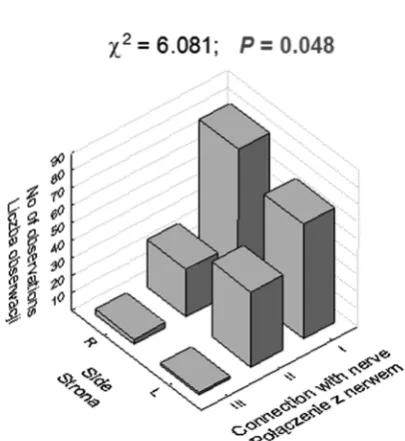
Fig. 3. Observations number in subgroups differenti-ated with reference to median nerve roots combina-tion height and posicombina-tion on the left or right and χ2 test
result
Ryc. 3. Liczba obserwacji w podgrupach różniących się typem określającym wysokość połączenia korzeni nerwu pośrodkowego i położeniem po stronie lewej i prawej oraz wynik testu χ2
Table 2. Percentage of observations diverse in respect of median nerve roots combination height in side subgroups, comparison result
Tabela 2. Liczności (odsetek) obserwacji różniących się typem określającym wysokość połączenia korzeni nerwu pośrodkowego w podgrupach różniących się stroną i wynik porównań
Median nerve roots combination height type (Typ wysokości połączenia korzeni nerwu pośrodkowym)
Body side (Strona ciała) L
N = 110 RN = 110 P
I – in axillary fossa (w dole
pachowym)
66 (60%) 80 (73%) 0.048
II – in ½ of proximal humerus length (w ½ prok-symalnej długości kości ramiennej)
43 (39%) 27 (25%)
III – in ½ of distal humerus length (w ½ dystalnej długości kości ramiennej)
Fig. 4. Median nerve (13) leaves directly lateral cord (20), where: C5 – Th1 – brachial plexus roots, 8 –
musculocutane-ous nerve, 9 – axillary nerve, 10 – radial nerve, 11 – lateral root of median nerve, 12 – medial root of median nerve, 13 – median nerve, 14 – ulnar nerve, 16/17 – brachium and antebrachium medial cutaneous nerves, 18 – medial cord, 19 – posterior cord, 20 – lateral cord, 21 – anterior division of upper trunk, 22 – posterior division of upper trunk, 23 – anterior division of middle trunk, 24 – posterior division of middle trunk, 25 – posterior division of lower trunk, 26 – anterior division of lower trunk, 27 – upper trunk, 28 – middle trunk, 29 – lower trunk
Ryc. 4. Nerw pośrodkowy (13) odchodzi bezpośrednio od pęczka bocznego (20), gdzie: C5 – Th1 – korzenie splotu
ramiennego, 8 – nerw mięśniowo-skórny, 9 – nerw pachowy, 10 – nerw promieniowy, 11 – korzeń boczny nerwu pośrodkowego, 12 – korzeń przyśrodkowy nerwu pośrodkowego, 13 – nerw pośrodkowy, 14 – nerw łokciowy, 16/17 – nerwy skórne przyśrodkowe ramienia i przedramienia, 18 – pęczek przyśrodkowy, 19 – pęczek tylny, 20 – pęczek boczny, 21 – gałąź przednia pnia górnego, 22 – gałąź tylna pnia górnego, 23 – gałąź przednia pnia środkowego, 24 – gałąź tylna pnia środkowego, 25 – gałąź tylna pnia dolnego, 26 – gałąź przednia pnia dolnego, 27 – pień górny, 28 – pień środkowy oraz 29 – pień dolny
Fig. 5. Median nerve roots variants concreated by anterior division of middle trunk where: C5 – Th1 – brachial plexus
roots, 8 – musculocutaneous nerve, 11 – lateral root of median nerve, 12 – medial root of median nerve, 13 – median nerve, 20 – lateral cord, 23 – anterior division of middle trunk
Ryc. 5. Warianty korzeni nerwu pośrodkowego współtworzone przez gałąź przednią pnia środkowego, gdzie: C5 – Th1
of median nerve, where: C5 – Th1 – brachial plexus roots, 8 – musculocutaneous nerve, 9 – axillary nerve, 10 – radial
nerve, 11 – lateral root of median nerve, 12 – medial root of median nerve, 13 – median nerve, 14 – ulnar nerve, 16/17 – brachium and antebrachium medial cutaneous nerves, 18 – medial cord, 19 – posterior cord, 20 – lateral cord, 21 – anterior division of upper trunk, 22 – posterior division of upper trunk, 23 – anterior division of middle trunk, 24 – posterior division of middle trunk, 25 – posterior division of lower trunk, 26 – anterior division of lower trunk, 27 – upper trunk, 28 – middle trunk, 29 – lower trunk
Ryc. 6. Podwójny korzeń boczny nerwu pośrodkowego – dodatkowe połączenie między pęczkiem bocznym (20) a korzeniem przyśrodkowym (12) nerwu pośrodkowego, gdzie: C5 – Th1 – korzenie splotu ramiennego, 8 – nerw
mię-śniowo-skórny, 9 – nerw pachowy, 10 – nerw promieniowy, 11 – korzeń boczny nerwu pośrodkowego, 12 – korzeń przyśrodkowy nerwu pośrodkowego, 13 – nerw pośrodkowy, 14 – nerw łokciowy, 16/17 – nerwy skórne przyśrodko-we ramienia i przedramienia, 18 – pęczek przyśrodkowy, 19 – pęczek tylny, 20 – pęczek boczny, 21 – gałąź przednia pnia górnego, 22 – gałąź tylna pnia górnego, 23 – gałąź przednia pnia środkowego, 24 – gałąź tylna pnia środkowego, 25 – gałąź tylna pnia dolnego, 26 – gałąź przednia pnia dolnego, 27 – pień górny, 28 – pień środkowy oraz 29 – pień dolny
Fig. 7. Median nerve lateral triple root – nerve double fibres (red stars) leaving lateral cord (20) and reaching medial root (12) of median nerve (13), where: C5 – Th1 – brachial plexus roots, 8 – musculocutaneous nerve, 9 – axillary
nerve, 10 – radial nerve, 11 – lateral root of median nerve, 12 – medial root of median nerve, 13 – median nerve, 14 – ulnar nerve, 16/17 – brachium and antebrachium medial cutaneous nerves, 18 – medial cord, 19 – posterior cord, 20 – lateral cord, 21 – anterior division of upper trunk, 22 – posterior division of upper trunk, 23 – anterior division of middle trunk, 24 – posterior division of middle trunk, 25 – posterior division of lower trunk, 26 – anterior division of lower trunk, 27 – upper trunk, 28 – middle trunk, 29 – lower trunk
Ryc. 7. Potrójny korzeń boczny nerwu pośrodkowego – podwójne włókna nerwowe (czerwone gwiazdki) odchodzące od pęczka bocznego (20) do korzenia przyśrodkowego (12) nerwu pośrodkowego (13), gdzie: C5 – Th1 – korzenie
Matejcik [4] observed terminal nerve anoma-lies in 60 cases. Median nerve roots combined in the brachium inferior part in 9 out of 110 exam-ined plexuses – 8.18%. In their surveys carried out on new-born babies, Uzun and Bigic [5] described the prevalence of median nerve triple root in 4 out of 130 examined plexuses. Terminal ramifi-cations anomalies prevailed quite often and they were described in the paper by Uysal et al. [6]. Median nerve roots combined in brachium distal part in 8.5% of cases. Kocabiyik et al. [7] discussed the case of median and musculocutaneous nerves accessory combination in brachium distal part. Also Pandey and Shukla [8], Kaus and Wójtowicz [9], Uzun et al. [10], Singhal et al. [11] as well as Badawoud [12] presented prevalence of combin-ing branch between median and ous nerves. The combination of
musculocutane-ous and median nerves was mentioned by Aktan et al. [13]. According to the authors, knowledge concerning brachial plexus anatomical variations is of great importance in collar surgery and axil-lary area surgery [14]. Such a big number of bra-chial plexus morphological variants may result in posttraumatic diverse clinical conditions. Hence, it seems to be reasonable to make brachial plexus visual examinations prior to surgical procedures.
The authors concluded that median nerve roots as well as median nerve itself are character-istic for morphological variability. Median nerve roots low combination in a nerve beneath axillary fossa (type II and III) seems clinically favorable as on brachium injury, the nerve part may stay free from damage. Type II – median nerve roots com-bining into the nerve in 1/2 of brachium proximal length appeared on the left.
Fig. 8. Musculocutaneous nerve and median nerve anastomosis, where: C5 – Th1 – brachial
plexus roots, 8 – musculocutaneous nerve, 13 – median nerve
Ryc. 8. Anastomoza między nerwem mięśnio-wo-skórnym a pośrodkowym, gdzie: C5 – Th1
– korzenie splotu ramiennego, 8 – nerw mię-śniowo-skórny, 13 – nerw pośrodkowy
References
[1] Paterson AM: A discussion of some points in the distribution of the spinal nerve. J Anat Physiol 1896, XXX(X), 530–538.
[2] Harris W: The true form of the brachial plexus, and its motor distribution. J Anat Physiol 1904, XXXVIII(XVIII), 399–422.
[3] Kerr AT: The brachial plexus of nerves in man, the variations in its formation and branches. Am J Anat 1918, 23, 285–395.
[4] Matejcik V: Aberrant formation and clinical picture of brachial plexus from the point of view of a neurosurgeon. Bratisl Lek Listy 2003, 104, 291–299.
[5] Uzun A, Bilgic S: Some variations in the formation of the brachial plexus in infants, Tr J Med Sci 1999, 2, 573–577.
[6] Uysal II, Seker M, Karabulut AK, Büyükmumcu M, Ziylan T: Brachial plexus variations in human foetuses. Neurosurgery 2003, 53, 676–684.
[8] Kocabiyik N, Palcin B, Yazar F, Ozan H: An accessory branch of musculocutaneus nerve joining median nerve. Neuroanatomy 2005, 4, 13–15.
[9] Pandey SK, Shukla VK: Anatomical variations of the cords of brachial plexus and median nerve. Clin Anat 2007, 20, 150–156.
[10] Kaus M, Wójtowicz Z: Communicating branch between the musculocutaneus and median nerves in human. Folia Morphol (Warsz) 1995, 54, 273–277.
[11] Uzun A, Seelig Jr LL: A variation in the formation of the median nerve: communication branch between the mus-culocutaneus and median nerves in man. Folia Morphol (Warsz) 2001, 60, 99–101.
Tel.: +48 71 784 00 80
E-mail: [email protected] Conflict of interest: None declared Received: 29.12.2011