Anna Boguszewska-Czubara
1, Anna Hordyjewska
1, Kazimierz Pasternak
1,
Małgorzata Kiełczykowska
1, Jadwiga Jaworska-Adamu
2, Radosław Szalak
2The Level of Calcium and Magnesium in Blood
of Rats Receiving Various Doses of Silicon
Stężenie wapnia i magnezu we krwi szczurów
otrzymujących różne dawki krzemu
1 Department of Medical Chemistry, Medical University of Lublin, Poland
2 Department of Animal Anatomy and Histology, University of Life Sciences in Lublin, Poland
Abstract
Background. Silicon, the third most abundant trace element of the human body, is listed as an essential one. It is especially associated with connective tissues as it has been found to take part in bone development, collagen formation and mineralization of bone matrix. Silicon is also implicated in mammalian hormonal control and in protecting against heart disease in humans.
Objectives. The influence of different doses of orally-administered silicon on calcium and magnesium concentra-tions in the blood of experimental animals was evaluated.
Material and Methods. The experiment was carried out on male Wistar rats. A control group was given distilled water to drink. The rats in group 0 were given a solution of sodium hydroxide (0.001 mol/L), whereas animals in groups 1, 2 and 3 received solutions of orthosilicic acid of three different concentrations (0.05%, 0.5% and 1%) as the only drinking fluids. Blood was collected after 4 and then 8 weeks of the experiment. Determination of calcium and magnesium concentrations in the blood was performed by the ICP-AES method.
Results. Silicon administration caused an increase in blood calcium concentration after 4 as well as after 8 weeks of the experiment. Four-week-long silicon intoxication caused a decrease in blood magnesium concentrations, whereas an increase in blood magnesium level in groups 0, 1 and 3 and a decrease in group 2 after 8 weeks of the experiment were noted.
Conclusions. Silicon was found to significantly influence metabolism of calcium and magnesium. Its interac-tion with calcium during the process of bones mineralizainterac-tion suggests that Si supplementainterac-tion may be helpful in preventing osteoporosis in postmenopausal women whose calcium intake is insufficient. Homeostasis in min-eral metabolism and balance between elements are very important matters, therefore silicon metabolism and its interactions with other elements and nutrients should be further investigated (Adv Clin Exp Med 2011, 20, 6, 677–682).
Key words: calcium, magnesium, silicon, rats, chronic toxicity.
Streszczenie
Wprowadzenie. Krzem jest niezbędny do prawidłowego funkcjonowania organizmów żywych. Jest pierwiastkiem występującym w śladowych ilościach i zajmuje wśród nich trzecie miejsce co do rozpowszechnienia. Szczególnie istotną rolę odgrywa w funkcjonowaniu tkanek łącznych – rozwoju kości i tworzeniu kolagenu. Badania wykazują, że krzem odgrywa również pewną funkcję w prawidłowej gospodarce hormonalnej organizmu oraz może działać ochronnie w przypadkach chorób kardiologicznych u ludzi.
Cel pracy. Badanie wpływu doustnego podawania różnych dawek krzemu na stężenia wapnia i magnezu we krwi szczurów.
Materiał i metody. Badanie przeprowadzono na szczurach samcach rasy Wistar. Grupa kontrolna otrzymywała do picia wodę redestylowaną. Zwierzętom z grupy 0 podawano wodny roztwór wodorotlenku sodu (0,001 mol/l), a szczurom z grup 1, 2 i 3 roztwory kwasu ortokrzemowego o różnych stężeniach (0,05%, 0,5% i 1%) jako jedyne płyny do picia. Krew do badań pobierano po 4 i 8 tygodniach trwania doświadczenia. W pobranych próbkach krwi oznaczano stężenia wapnia i magnezu metodą ICP-AES.
Wyniki. Podawanie krzemu spowodowało zwiększenie stężenia wapnia we krwi zarówno po 4, jak i 8 tygodniach
Adv Clin Exp Med 2011, 20, 6, 677–682 ISSN 1230-025X
oRIGINAL PAPERS
trwania eksperymentu. Stężenie magnezu zmniejszyło się po 4-tygodniowej intoksykacji, a po 8 tygodniach nastąpił wzrost stężenia tego pierwiastka we krwi zwierząt grup 0, 1 i 3 oraz spadek w grupie 2.
Wnioski. Badania wykazały, że krzem znacząco wpływa na metabolizm wapnia i magnezu. Interakcje między wap-niem i krzemem, zachodzące w procesie mineralizacji kości, sugerują, że suplementacja krzemu może zapobiegać osteoporozie u kobiet po menopauzie, u których pobranie wapnia nie jest dostateczne. Zachowanie homeostazy metabolizmu biopierwiastków i równowaga między nimi jest niezwykle istotną kwestią, dlatego metabolizm krze-mu i jego interakcje z innymi pierwiastkami i składnikami pożywienia powinny być przedmiotem dalszych badań
(Adv Clin Exp Med 2011, 20, 6, 677–682).
Słowa kluczowe: wapń, magnez, krzem, szczury, toksyczność przewlekła.
Trace elements are very important factors con-ditioning the proper functions of living organisms. Although silicon is the second most abundant el-ement in the biosphere after oxygen, its very low bioavailability for the human body means that the influence of silicon on metabolic processes is only fragmentarily known and poorly understood [1].
Silicon is necessary for growth and bone cal-cification and as a biological cross-linking agent of connective-tissue-based membrane structures [2, 3]. This element is considered to have benefi-cial effects on several human disorders including osteoporosis [4], ageing of skin, hair and nails [5] as well as atherosclerosis [6]. It has also been sug-gested that silicon and silicic acid may decrease the bioavailability of aluminum by blocking uptake through the gastrointestinal tract and by imped-ing reabsorption in the kidneys, thus protectimped-ing the body in that way against the toxic (especially neurotoxic) effect of aluminum [7]. Anticance-rous, antiatherosclerotic and antidiabetic effects of silicon have also been suggested [8].
The average daily dietary intake of silicon is about 20–50 mg/person/day, with higher intakes for men than women [9]. The bioavailable form of silicon is silicic acid or orthosilicic acid that is mainly found in food rich in fiber and whole grains, vegetables, fruit and drinking water. Various alco-holic beverages such as beer or wine also contain silicon in reasonable amounts [10, 11]. Silicon pro-vided with food is digested in the gastrointestinal tract to silicic acid, which is then absorbed [12]. It is distributed with the blood into various tissues and organs where it can exert its effects. The greatest amounts of silicon are accumulated in the kidney, liver, bones, skin, spleen and lungs [13] and free orthosilicic acid, not bonded to proteins, occurs in the blood [14]. The total body content of silicon for a subject with a body weight of 70 kg is in the range of 140 to 700 mg. The amount of silicon in human tissues decreases with age and the develop-ment of some diseases, e.g.: atherosclerosis [13].
Calcium, which accounts to 1–2% of the to-tal body weight of an adult man, is a major com-ponent of mineralized tissues (teeth and bones), which contain around 99% of the total body
con-tent of that element. Calcium exerts important roles in intracellular as well as extracellular pro-cesses, such as muscle contraction, neuronal con-ductivity or hormone release [15]. It is a second messenger transmitting signals between the plas-ma membrane and the interior of the cell. Calcium participates in blood clotting and it is a cofactor of adhesion molecules [16]. It is essential for the proper formation of bone, where it provides the bones’ structural strength and serves as a calcium reservoir for the body to maintain its homeostasis in states of short-term calcium depletion [17].
Calcium homeostasis is tightly regulated by processes such as absorption, excretion, secretion and storage in bone, being involved in maintain-ing the concentration of Ca in the plasma within a narrow range, usually 8.5 to 10.5 mg/dL (2.1 to 2.6 mmol/L) [18]. The level of calcium in the blood is regulated primarily by hormones: parathyroid hormone, 1,25 dihydroxycholecalciferol (1,25 (oH)2D3) and calcitonin [19].
To maintain a normal level of calcium in the blood without weakening the bones, people need to consume at least 1000 to 1500 milligrams of cal-cium a day. Hypercalcemia occurs when the level of serum-ionized calcium increases. It is mainly caused by malignancy, hyperparathyroidism, acute kidney diseases, total parenteral nutrition and chronic therapy with some drugs (e.g. diuretics). The most common symptoms of hypercalcemia are: gastrointestinal symptoms, fatigue and muscle weakness, nephrogenic diabetes insipidus, cardio-vascular effects (e.g. hypertension and shortening of the QT interval and cardiac arrhythmias) and even acute renal failure. on the opposite side, hy-pocalcemia is due to a low ionized calcium con-centration in blood serum. It manifests itself with perioral numbness and carpopedal spasms of the hands and feet and it can be caused by vitamin D deficiency, hypoparathyroidism, pseudohypopara-thyroidism and a high rate of tissue consumption of calcium [20].
almost all processes in the body. Magnesium par-ticipates in numerous metabolic pathways occur-ring in a cell. It is an activator for about 300 en-zymes, therefore it takes part in the metabolisms of carbohydrates, nucleic acids and proteins [22]. It is also necessary in energy metabolism as a criti-cal cofactor in any reactions powered by ATP and cell proliferation [23]. Magnesium stabilizes DNA structure, influences RNA transcription and takes part in nucleic acid and protein synthesis as well as protecting biological membranes [21]. It is also a calcium channel antagonist and therefore mag-nesium plays an important role in the modulation of activity governed by intracellular calcium con-centration fluxes such as muscle contraction or insulin release [24].
Maintaining magnesium homeostasis is a very important task. Changes in magnesium status mainly concern the extracellular pool of the mac-roelement, that is blood serum, while intracellular magnesium concentration is well-regulated and conserved [25]. Therefore, any disturbances in magnesium homeostasis can be observed by its serum content. The normal concentration of se-rum magnesium is 1.8 to 2.3 mg/dL (0.75 to 0.95 mmol/l) [26]. The appropriate concentration of magnesium in blood serum is mainly regulated by the kidneys, where reabsorption takes place, and bowels, where magnesium is absorbed [25]. Mag-nesium deficiency is usually caused by its insuf-ficient intake with diet, disturbances of its absorp-tion or excreabsorp-tion (e.g. renal failure), drugs used (e.g. diuretics) or stress [27]. Dietary magnesium deficiency plays an important role in the patho-genesis of several cardiovascular diseases includ-ing sudden cardiac death and cardiac arrhythmias, vascular implications of diabetes mellitus and hy-pertension [26].
The aim of the study was to evaluate the influ-ence of three different doses of orally administered silicon on calcium and magnesium concentrations in the blood of experimental animals.
Material and Methods
The experiment was carried out on five groups of adolescent male Wistar rats (ten animals each). The control group was given distilled water to drink. The rats of group 0 were given a solution of sodium hydroxide at a concentration of 0.001 mol/L, whereas the animals of groups 1, 2 and 3 received solutions of orthosilicic acid (H4Sio4) as
the only drinking fluids. As a source of orthosilicic acid, the preparation “Compendium krzemowe” was used. Group 1 received 0.05% solution of the preparation in a solution of NaoH (0.001 mol/L),
group 2 – a 0.5% solution of the preparation in 0.001 mol/L NaoH and group 3 was given a 1% solution of the preparation in 0.001 mol/L NaoH. “Compendium krzemowe” is an orthosilicic acid in the form of a gel. The weights of the animals at the beginning of the experiment were within the range of 180–230 g. The rats had free access to standard feed LSM and drinking solutions. Half of the experimental animals from each group were sacrificed under pentothal narcosis after 4 weeks and the rest of the animals after 8 weeks of the experiment. Each time, whole blood was col-lected and then stored at a temperature of –20oC
until analysis. The determination of calcium and magnesium in the blood was performed using in-ductively coupled plasma emission spectrometry (ICP-AES, Liberty II AX, Varian) with set-up and conditions according to the method accredited by the local Voivodship Inspectorate of Environment Protection. The method is based on the measure-ment of emitted radiation intensity at characteris-tic wave lengths for the element (Mg – 277.983 nm, Ca – 315.887 nm) and it is characterized by a lin-ear relationship between emission intensity and the concentration of the determined element.
Comparisons between the control and tested groups as well as between silicon supplemented groups were made using the c-Cochran-Cox test. Values of p < 0.05 were considered significant.
The study was performed according to statu-tory bioethical standards and approved by the I Local Ethical Commission of the Medical Uni-versity of Lublin, acceptance no. 550/AM/2005.
Results
The results of calcium concentrations in the blood of rats receiving various doses of silicon are presented in Fig. 1.
A significant increase in the blood calcium con-centration of animals of all the experimental groups versus the control group was stated after a 4-week-long period of silicon administration. After 8 weeks of the experiment, an increase in blood calcium concentration was also observed in all of the exam-ined rats in comparison with the control group, but statistical significance was stated only in the case of group 2. The period of silicon intoxication duration influenced calcium concentration in the blood only in the case of animals from the control group, where a statistically significant increase after 8 weeks was noted in comparison with the value obtained after 4 weeks of the experiment duration.
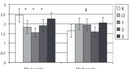
In the blood of the animals submitted to 4-week-long silicon intoxication, a statistically significant decrease in magnesium concentration was found in the case of groups 0, 1 and 2 and only a slight de-crease in the case of group 3 in comparison to the control group. A small increase in magnesium con-centration in the blood of the animals from groups 0, 1 and 3 and a slight decrease in the case of rats from group 2 versus the control group were stated after 8 weeks of the experiment duration. A statisti-cally significant influence of the silicon
administra-tion period was noted only for animals receiving the lowest silicon dose (group 1). There was an increase of magnesium concentration in the blood after 8 weeks in comparison to the value obtained after 4 weeks of the experiment.
Discussion
Silicon is co-located with calcium in osteoid tissue, thus some interaction between these ele-ments have been suspected to occur in the process-es of bone growth and mineralization [28]. In the earliest stage of calcification in active calcification sites in young bones, when the calcium content of the preosseous tissue is very low, there is a direct relationship between silicon and calcium. There-fore, it has been suggested that silicon is associated with calcium in the early stages of bone formation. It has been demonstrated that dietary silicon in-creased the rate of mineralization, especially in the case of calcium-deficient rats [29]. other findings have revealed that, in rats fed low calcium diets, bone composition was affected by silicon depriva-tion: the deprivation depressed the tibia and skull concentrations of calcium, magnesium, and phos-phorus [30]. These facts can be interpreted as the promotion of bone mineralization by silicon un-der conditions of low levels of calcium in the diet, but on the other hand, it may also indicate calcium – silicon interactions in gut lumen that could re-duce the gastrointestinal absorption of silicon [3]. Mineral metabolism and tissue mineral com-position as a response to the administration of a supplement with a high content of silicon and aluminum (sodium zeolite A) was investigated in calves. The contents of silicon, aluminum and nu-merous other mineral elements including calcium and magnesium were determined in blood plasma, urine and numerous organs. Correlations between silicon and calcium and magnesium were found in that experiment [31]. Another experiment car-ried out on calves supplemented with stabilized orthosilicic acid concerned the effect of Si on Ca, Mg, and P concentrations in serum and collagen concentrations in skin and cartilage. The posi-tive correlation between the serum Si concentra-tion and the collagen concentraconcentra-tion in cartilage as well as the serum Ca concentration, respectively, stated in that the study suggested the involvement of Si both in the formation of extracellular matrix components and in Ca metabolism [32]. Similarly, mineral balance was investigated on horses fed two different supplemental silicon sources. The results of that study were in accordance with the data obtained for calves. Both supplements were able to alter Ca retention [33].
Fig. 1. The influence of silicon administration on blood concentrations of calcium in rats, * – statistically significant differences vs. control at p ≤ 0.05; # –
statis-tically significant differences vs. values obtained after 4 weeks of the experiment, p ≤ 0.05
Ryc. 1. Wpływ podawania krzemu na stężenia wapnia we krwi szczurów, * – różnice statystycznie istotne w porównaniu z grupą kontrolną przy p ≤ 0.05; # różnice statystycznie istotne w porównaniu z wyni- kami uzyskanymi po 4 tygodniach trwania doświadczenia, p ≤ 0,05
0 1 2 3 4 5 6 7
Ca 4 weeks Ca 8 weeks
mmol/L
K O 1 2 3
* * * * # *
Fig. 2. The influence of silicon administration on blood concentrations of magnesium in rats, * – statistically significant differences vs. control at p ≤ 0.05; # – statistically significant differences vs. values obtained after 4 weeks of the experiment, p ≤ 0.05
Ryc. 2. Wpływ podawania krzemu na stężenia ma- gnezu w krwi szczurów, * – różnice statystycznie istotne w porównaniu z grupą kontrolną przy p ≤ 0,05; # – różnice statystycznie istotne w porównaniu z wyni- kami uzyskanymi po 4 tygodniach trwania doświad-czenia, p ≤ 0,05
0 0,5 1 1,5 2 2,5 3
Mg 4 weeks Mg 8 weeks
mmol/L
K O 1 2 3
Although the authors cannot directly com-pare their results with those presented above, be-cause the literature data presents values obtained in blood serum or blood plasma, not in whole blood, but their results confirm the occurrence of interactions between dietary silicon and calcium and magnesium levels in the blood of experimen-tal animals. Silicon supplementation caused an increase in blood calcium concentrations after 4 weeks and 8 weeks, but in the case of a shorter pe-riod of intoxication, the changes were significant. The smaller differences after 8 weeks between the control group and supplemented groups may be a result of compensative mechanism effects, which were activated after prolonged silicon administra-tion. Magnesium concentration in the blood de-creased after 4 weeks of silicon administration, while at 8 weeks an increase was reported. The ob-served changes in magnesium concentration can be explained not only by adaptive mechanism of the body but also by antagonism between calcium and magnesium ions.
Considering the question of sodium influence on the examined element concentrations, one study indicated that short-term intakes of salt sup-plements (50 g/kg of diet) significantly increased urinary Ca and Mg excretion in rats [34]. The ef-fect of salt on urinary mineral excretion was most distinct in the case of Ca, with a three-fold increase in animals given salt-supplemented compared with non-supplemented diets. The published literature on sodium and calcium metabolism indicates that the average loss of calcium is 1 mmol Ca (40 mg) per 100 mmol (2290 mg) of sodium, and without any adaptive compensatory mechanisms, a daily loss of 40 mg calcium would deplete 10% of the skeleton within ten years [36]. However, adaptive mechanisms which are present in the body prevent
excessive calcium loss and maintain Ca homeostasis [35]. After calcium liberating from bones its serum concentration rises, and then after its filtration by the kidneys hypercalcuria occurs. In present study, calcium concentration in the blood increased af-ter sodium hydroxide administration, especially after 4 weeks, while after 8 weeks, a decrease in calcium level was observed, probably as a result of the adaptive mechanism effect. In the case of mag-nesium, the changes were not so well-marked, and after 8 weeks the magnesium concentration in the group of animals receiving sodium hydroxide and the lowest silicon dose were similar.
There are studies showing that supplemen-tal silicon exerts a beneficial effect on bone and its mineral composition as well as on important bioelement content in the blood and tissues of experimental animals. However further evidence is needed to state whether silicon deprivation can lead to a decrease in bioelement content in the body and if it can be prevented by nutritional sup-plementation or physiological intake of silicon.
The role of silicon in human biology is poorly understood, therefore studies on its interactions with mineral elements as well as with other bio-logically important molecules are necessary. The interaction between silicon and calcium during the process of bone mineralization suggests that Si supplementation may be helpful in preventing osteoporosis in postmenopausal women whose calcium intake is insufficient. Homeostasis in min-eral metabolism and balance between elements is a very important matter. Silicon was found to significantly influence the metabolism of calcium and magnesium, thus the question whether its ex-cess would disturb the balance of other elements should be further investigated.
References
[1] Birchall JD, Bellia JP, Roberts NB: on the mechanisms underlying the essentiality of silicon – interactions with aluminium and copper. Coord Chem Rev 1996, 149, 231–240.
[2] Jugdaohsingh R: Silicon and bone health. J Nutr Health Aging 2007, 11, 99–110.
[3] Sripanyakorn S, Jugdaohsingh R, Thompson RPH, Powell JJ: Dietary silicon and bone health. Nutr Bull 2005, 30, 222–230.
[4] Jugdaohsingh R, Tucker KL, Qiao N, Cupples LA, Kiel DP, Powell JJ: Dietary silicon intake is positively associ-ated with bone mineral density in men and premenopausal women of the Framingham offspring cohort. J Bone Miner Res 2004, 19, 297–307.
[5] Barel A, Calomme M, Timchenko A, De Paepe K, Demeester N, Rogiers V, Clarys P, Vanden Berghe D: Effect of oral intake of choline-stabilized orthosilicic acid on skin, nails and hair in women with photodamaged skin. Arch Dermatol Res 2005, 297, 147–153.
[6] Peluso MR, Schneeman BO: A food-grade silicon dioxide is hypocholesterolemic in the diet of cholesterol-fed rats. J Nutr 1994, 124, 853–860
[7] Reffitt DM, Jugdaohsingh R, Thompson RP, Powell JJ: Silicic acid: its gastrointestinal uptake and urinary excre-tion in man and effects on aluminium excreexcre-tion. J Inorg Biochem 1999, 76, 141–147.
[9] Jugdaohsingh R, Anderson SH, Tucker KL, Elliott H, Kiel DP, Thompson RP, Powell JJ: Dietary silicon intake and absorption. Am J Clin Nutr 2002, 75, 887–893.
[10] González-Muñoz MJ, Peña A, Meseguer I: Role of beer as a possible protective factor in preventing Alzheimer’s disease. Food Chem Toxicol 2008, 46, 49–56.
[11] Thiel G, Geisler G, Blechschmidt I, Danzer K: Determination of trace elements in wines and classification according to their provenance. Anal Bioanal Chem 2004, 378, 1630–1636.
[12] Popplewell JF, King SJ, Day JP, Ackrill P, Fifield LK, Cresswell RG, di Tada ML, Liu K: Kinetics of uptake and elimination of silicic acid by a human subject: a novel application of 32Si and accelerator mass spectrometry. J Inorg Biochem 1998, 69, 177–180.
[13] Bissé E, Epting T, Beil A, Lindinger G, Lang H, Wieland H: Reference values for serum silicon in adults. Anal Biochem 2005, 337, 130–135.
[14] D’Haese PC, Shaheen FA, Huraib SO, Djukanovic L, Polenakovic MH, Spasovski G, Shikole A, Schurgers ML, Daneels RF, Lamberts LV: Increased silicon levels in dialysis patients due to high silicon content in the drinking water, inadequate water treatment procedures, and concentrate contamination: a multicentre study. Nephrol Dial Transplant 1995, 10, 1838–1844.
[15] Pegoraro AA, Rutecki GW, Lohr JW: Hypocalcemia. E-Med J 2002, 3, 1–2.
[16] Power ML, Heaney RP, Kalkwarf HJ, Pitkin RM, Repke JT, Tsang RC, Schulkin J: The role of calcium in health and disease. Am J obstet Gynecol 1999, 181, 1560–1569.
[17] Flynn A: The role of dietary calcium in bone health. Proc Nutr Soc 2003, 62, 851–858.
[18] Cashman KD: Calcium intake, calcium bioavailability and bone health. Br J Nutr 2002, 87(2), S169–177.
[19] Passeri G, Vescovini R, Sansoni P, Galli C, Franceschi C, Passeri M: Italian Multicentric Study on Centenarians (IMUSCE): Calcium metabolism and vitamin D in the extreme longevity. Exp Gerontol 2008, 43, 79–87.
[20] Moe SM: Disorders involving calcium, phosphorus, and magnesium. Prim Care 2008, 35, 215–237.
[21] Saris NE, Mervaala E, Karppanen H, Khawaja JA, Lewenstam A: Magnesium. An update on physiological, clini-cal and analyticlini-cal aspects. Clin Chim Acta 2000, 294, 1–26.
[22] Rubin H: The membrane, magnesium, mitosis (MMM) model of cell proliferation control. Magnes Res 2005, 18, 268–274.
[23] Wolf FI, Cittadini A: Magnesium in cell proliferation and differentiation. Front Biosci 1999, 4, D607–D617.
[24] Sanders GT, Huijgen HJ, Sanders R: Magnesium in disease: A review with special emphasis on the serum ionized magnesium. Clin Chem Lab Med 1999, 37, 1011–1033.
[25] Vormann J: Magnesium: nutrition and metabolism. Mol Aspects Med 2003, 24, 27–37.
[26] Topf JM, Murray PT: Hypomagnesemia and hypermagnesemia. Rev Endocr Metab Disord 2003, 4, 195–206.
[27] Błach J, Nowacki W, Mazur A: Magnesium in skin allergy. Postepy Hig Med Dosw 2007, 61, 548–554.
[28] Perry CC, Keeling-Tucker T: Aspects of the bioinorganic chemistry of silicon in conjunction with the biometals calcium, iron and aluminium. J Inorg Biochem 1998, 69, 181–191.
[29] Kim MH, Bae YJ, Choi MK, Chung YS: Silicon supplementation improves the bone mineral density of calcium-deficient ovariectomized rats by reducing bone resorption. Biol Trace Elem Res 2009, 128(3), 239–247.
[30] Carlisle EM: Biochemical and morphological changes associated with long bone abnormalities in silicon defi-ciency. J Nutr 1980, 110, 1046–1056.
[31] Turner KK, Nielsen BD, O’Connor-Robison CI, Nielsen FH, Orth MW: Tissue response to a supplement high in aluminum and silicon. Biol Trace Elem Res 2008, 121, 134–148.
[32] Calomme MR, Vanden Berghe DA: Supplementation of calves with stabilized orthosilicic acid. Effect on the Si, Ca, Mg, and P concentrations in serum and the collagen concentration in skin and cartilage. Biol Trace Elem Res 1997, 56, 153–165.
[33] O’Connor CI, Nielsen BD, Woodward AD, Spooner HS, Ventura BA, Turner KK: Mineral balance in horses fed two supplemental silicon sources. J Anim Physiol Anim Nutr 2008, 92, 173–181.
[34] Creedon A, Cashman KD: The effect of high salt and high protein intake on calcium metabolism, bone composi-tion and bone resorpcomposi-tion in the rat. Br J Nutr 2000, 84, 49–56.
[35] Teucher B, Fairweather-Tait S: Dietary sodium as a risk factor for osteoporosis: where is the evidence? Proc Nutr Soc 2003, 62, 859–866.
[36] Teucher B, Dainty JR, Spinks CA, Majsak-Newman G, Berry DJ, Hoogewerff JA, Foxall RJ, Jakobsen J, Cashman KD, Flynn A, Fairweather-Tait SJ: Sodium and bone health: impact of moderately high and low salt intakes on calcium metabolism in postmenopausal women. J Bone Miner Res 2008, 23, 1477–1485.
Address for correspondence:
Anna Boguszewska-Czubara Department of Medical Chemistry Medical University of Lublin Chodźki 4a
20-093 Lublin Poland
E-mail: [email protected] Conflict of inerest: None declared