R E S E A R C H
Open Access
Telomere shortening and telomere position effect
in mild ring 17 syndrome
Cecilia Surace
1*†, Francesco Berardinelli
2†, Andrea Masotti
3, Maria Cristina Roberti
1, Letizia Da Sacco
3,
Gemma D
’
Elia
1, Pietro Sirleto
1, Maria Cristina Digilio
4, Raffaella Cusmai
5, Simona Grotta
1, Stefano Petrocchi
1,
May El Hachem
6, Elisa Pisaneschi
1, Laura Ciocca
1, Serena Russo
1, Francesca Romana Lepri
1, Antonella Sgura
2and Adriano Angioni
1Abstract
Background:Ring chromosome 17 syndrome is a rare disease that arises from the breakage and reunion of the short and long arms of chromosome 17. Usually this abnormality results in deletion of genetic material, which explains the clinical features of the syndrome. Moreover, similar phenotypic features have been observed in cases with complete or partial loss of the telomeric repeats and conservation of the euchromatic regions. We studied two different cases of ring 17 syndrome, firstly, to clarify, by analyzing gene expression analysis using real-time qPCR, the role of the telomere absence in relationship with the clinical symptoms, and secondly, to look for a new model of the mechanism of ring chromosome transmission in a rare case of familial mosaicism, through cytomolecular and quantitative fluorescencein-situhybridization (Q-FISH) investigations.
Results:The results for the first case showed that the expression levels of genes selected, which were located close to the p and q ends of chromosome 17, were significantly downregulated in comparison with controls. Moreover, for the second case, we demonstrated that the telomeres were conserved, but were significantly shorter than those of age-matched controls; data from segregation analysis showed that the ring chromosome was transmitted only to the affected subjects of the family.
Conclusions:Subtelomeric gene regulation is responsible for the phenotypic aspects of ring 17 syndrome; telomere shortening influences the phenotypic spectrum of this disease and strongly contributes to the familial transmission of the mosaic ring. Together, these results provide new insights into the genotype-phenotype relationships in mild ring 17 syndrome.
Keywords:Genetic syndrome, Telomere position effect, Ring 17 chromosome, Telomere shortening
Background
Ring chromosomes are unusual circularized chromosomes, the vast majority of which arise sporadically [1]. Two types of ring chromosome can be related to different clinical pre-sentations: the non-supernumerary ring, which replaces one of the normal homologs, with the karyotype 46,(r) and the supernumerary ring, which is often very small, with the karyotype 47,+(r). The non-supernumerary ring chro-mosomes are usually associated with abnormal phenotypes because of loss of genomic material at one or both ends. In
some cases, no deletion has been detected and the pheno-typic features have been attributed to mitotic ring instability [2].
The classic mode of ring chromosome formation consists of two terminal breaks in both chromosome arms, followed by a reconnection of the broken ends, leading to a loss of genetic material. Alternatively, they can be formed by fu-sion of subtelomeric sequences or telomere-telomere fufu-sion with no deletion, resulting in complete ring chromosomes.
Telomeres, the nucleoprotein complexes that cap eukaryotic chromosomes, play two major roles in cell physiology. First, they are critical factors in determining the lifespan of mammalian cells. Second, they are essential for genome integrity maintenance. In addition to their role in * Correspondence:cecilia.surace@opbg.net
†Equal contributors 1
Cytogenetics and Molecular Genetics Unit,‘Bambino Gesù’Children’s Hospital, IRCCS, Piazza S. Onofrio 4, 00165 Rome, Italy
Full list of author information is available at the end of the article
protecting the ends of chromosomes, telomeres can influ-ence the expression of nearby genes, an event called the telomere position effect. Previous reports demonstrated that the telomere position effect is an epigenetic process that involves changes in chromatin conformation and de-pends on both the distance from the telomere and the telo-mere’s length [3]. The telomere position effect also occurs in mammalian cells, and may play a role in human genetic disease, as a result of the repositioning of active genes near telomeres or subtelomeric sequences following chromo-some rearrangements [4-8], such as those seen in individ-uals with ring chromosomes [9,10].
Rings of chromosome 17 are relatively rare and only 17 cases have been reported in the literature to date. Among these 17 cases, 11 showed no deletions of the Miller-Dieker critical region with mild phenotypic features [11] consisting of growth delay, intellectual disability, seizures,café au lait skin spots and minor facial dysmorphism.
The ring chromosomes 17 of the majority of these pa-tients were described at the banding level, and only two cases pointed at the molecular level [11-13]. Cytomolecular investigation of the telomeric and subtelomeric regions of ring 17 is essential for the disclosure of candidate genes or gene expression regulating regions, which are potentially responsible for the phenotypic characteristics.
We report here the results of new investigations of a previously described patient with mild ring 17 syndrome and studies of an unusual case of ring chromosome 17 syndrome with a mosaic familial transmission.
Results Case 1
Clinical presentation
The clinical features of patient 1 have been previously reported [11]. We evaluated the patient at four years of age. Clinical examination showed prominent eyes, epi-canthal folds, anteverted nares, thick lips, small teeth, prognathism, generalizedcafé au laitskin spots, and hy-pochromic cutaneous lesions. Developmental milestones were moderately delayed and language was absent. The patient developed epilepsy when he was two years old: this was characterized by tonic seizures that were diffi-cult to control by medical treatment. The patient’s ductus arteriosus was closed by cardiac catheterization at five years of age. Cytogenetic investigations from per-ipheral blood cells revealed the following karyotype: 46, XY,r(17)(p13q25)[27]/45,XY,−17[3]. Chromosome ana-lysis of fibroblasts obtained by dyschromic skin spot bi-opsy showed the sole ring 17 pattern without the monosomic cell line. Fluorescence in-situ hybridization (FISH) investigations revealed the loss of r(17) telomeres and conservation of the whole adjacent, euchromatic, subtelomeric regions.
Gene expression analysis
The expression levels of 24 selected genes within a 1.5 Mb region starting from each telomere and one housekeeping gene (GAPDH) of the ring 17 patient were measured in each sample by real-time qPCR and compared with con-trols. Two other genes (NF1andLIS1) that are located out-side the 1.5 Mb region (17q11.2 and 17p13.3, respectively), were also considered. Of a total of 26 genes analyzed, 14 genes were statistically significant (P< 0.05), and 12 of these were located within the 1.5 Mb region (Table 1). Of these latter genes, seven were found to be deregulated by a fold change (FC) greater than 1.5:RPH3AL,FAM57A,GEMIN4,
NXN in the sub-telomere region of 17p and ARHGDIA,
NARF, andFN3Klocated in the 17q arm. TheFOXK2gene displayed a downregulation that was below the FC
thresh-old of 1.5 (FC =−1.44). The modulation of YWHAE,
MYOC1,SIRT7, andRNMTL1is negligible, suggesting that these genes are not sensitive to the telomere influence. Interestingly, theNF1gene displayed a significant upregula-tion of 2.56, whereas LIS1 is downregulated −1.76 fold. Taken together, these results emphasize the downregulation of the majority of genes included within the investigated sub-telomere regions.
Case 2
Clinical presentation
encephalopathy, and the child experienced cognitive deterioration.
The mother’s child had multiple café-au-lait spots on her trunk without facial anomalies or cognitive deficit.
Classical and molecular cytogenetic investigations
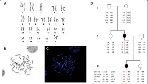
A study of the lymphocytes revealed a mosaic kar-yotype: 46,XX,r(17)(p13q25)[36]/46,XX[14] (Figure 1A). G-banding analysis of the fibroblasts showed two cell lines, the most abundant clone having a normal karyo-type (98%) and the other having a double ring 17 (2%). Chromosome analysis of the patient’s parents displayed a normal paternal karyotype, and a mosaic ring 17 ma-ternal karyotype: 46,XX,r(17)(p13q25)[22]/46,XX[28]. All the FISH pan-telomeric (Figures 1B,C) and subtelomeric probes showed clear signals in the patient and her mother, both on the normal chromosome 17 and on the ring. This confirmed that no terminal deletion had oc-curred. An array-CGH study of both cases did not detect any submicroscopic deletions or duplications. Segrega-tion analysis of the family group revealed that the ring of the patient and her mother derived from the
chromo-some 17 of the patient’s maternal grandmother
(Figure 1D). The same study also excluded uniparental disomy because all of the informative markers were found on both the normal and the rearranged chromo-somes. Additional microsatellite investigation, performed on fibroblasts of normal skin and acaféau lait skin spot biopsy of the patient, displayed the presence of a biallelic pattern of transmission related to each parent (Table 2).
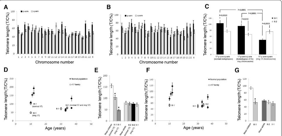
Quantitative fluorescence in-situ hybridization (Q-FISH) analysis
Chromosome-specific telomere length was assessed in either normal (Figure 2A) or ring metaphases (Figure 2B) obtained from the patient. Data showed that the means of all telomere lengths in normal and ring metaphases (61 and 53 T/C% respectively) did not differ signifi-cantly. Moreover, the telomere length patterns in the two metaphase classes were very similar, and in both the shortest telomeres were located at 20q, 16p, 21q, 22q, 19p, 17p, and 17q whereas the longest were located at Xp, 4p, 21p, 12q, 8p, 14p, 1p, and 6q.
Telomere length analysis of chromosome 17 in the pa-tient showed that telomere length in the ring chromo-some (48 T/C%) was significantly shorter than the sum of the telomeres at the p-ter and the q-ter in normal chromosome 17, if scored in normal (104 T/C%) or ring metaphases (95 T/C%) (Figure 2C). Conversely, the
telo-meric DNA in the ring chromosome of the patient’s
mother (76 T/C%) was comparable in length to that of the normal chromosome 17 in normal metaphases (77 T/C%) and in ring metaphases (67 T/C%) (Figure 2C). As expected, telomeres in the normal chromosome 17 were longer in the patient than in her mother, while telomeres in the ring chromosome were significantly shorter in the patient than in her mother (48 T/C% and 76 T/C% respectively) (Figure 2C).
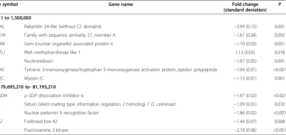
To compare the ring 17 of the family to those of nor-mal healthy individuals, lymphocytes from nine (four children and five adults) normal donors were also ana-lyzed for telomere length (Table 3). Considering the Table 1 Expression levels of selected sub-telomere genes of the ring17 patient, significantly deregulated compared with controls
Gene symbol Gene name Fold change
(standard deviation)
P
17p: 1 to 1,500,000
RPH3AL Rabphilin 3A-like (without C2 domains) −2.99 (0.15) 0.001
FAM57A Family with sequence similarity 57, member A −1.61 (0.04) 0.002
GEMIN4 Gem (nuclear organelle) associated protein 4 −1.70 (0.03) 0.001
RNMTL1 RNA methyltransferase like 1 1.13 (0.03) 0.018
NXN Nucleoredoxin −1.87 (0.05) 0.001
YWHAE Tyrosine 3-monooxygenase/tryptophan 5-monooxygenase activation protein, epsilon polypeptide −1.04 (0.01) <0.001
MYO1C Myosin IC −1.15 (0.01) 0.001
17q: 79,695,210 to- 81,195,210
ARHGDIA ρGDP dissociation inhibitorα −1.67 (0.02) <0.001
SIRT7 Sirtuin (silent mating type information regulation 2 homolog) 7 (S. cerevisiae) −1.09 (0.01) 0.010
NARF Nuclear prelamin A recognition factor −1.86 (0.02) <0.001
FOXK2 Forkhead box K2 −1.44 (0.07) 0.008
FN3K Fructosamine 3 kinase −2.18 (0.06) <0.001
mean cellular telomere length, we found an age-related telomere erosion in the control individuals, with the mean telomere lengths ranging from 94 T/C% in children to 58 T/C% in adults (Figure 2F,G). The values
for the patient’s mother and aunt (54 and 50 T/C%
respectively) were comparable to the age-matched con-trols (58 T/C%), indicating a normal telomere length in both family members. Interestingly, the patient showed reduced telomeric lengths with telomeres much shorter (53 T/C%) than the other age-matched children (94 T/C%) (Figure 2F,G).
When we turned our attention to chromosome 17 telomere lengths (ring and normal), we observed the same trend in mean cellular telomere length. Thus, the
patient displays telomeres on chromosome 17 (both ring and normal) shorter than those of normal healthy chil-dren, whereas the other members of the family were not significantly different in telomere length, as compared with age-matched controls (Figure 2D,E).
Discussion
Ring chromosomes are rare cytogenetic abnormalities that usually occur as a consequence of breaks and dele-tions at both ends of the chromosome, with subsequent fusion of the remaining portions. The breakpoints may be located within euchromatic regions, with partial monosomy of the distal ends of the short and long arms of the chromosome, but sometimes they may involve only one or both the telomere repeats. Though uncom-mon, the last mechanism has been described in the lit-erature as a telomere-telomere fusion with no detectable loss of euchromatin [14,15].
In a previous report [11], we have described the case of a ring 17 chromosome in a young boy, providing an accurate characterization of the defect, which resulted in complete loss of the telomeric ends with the conserva-tion of the whole euchromatic regions. The clinical pic-ture was reported as a mild ring 17 syndrome with moderate intellectual disability, seizures, dysmorphic traits, andcafé au lait and hypochromic skin spots. We Table 2 Segregation analysis of the fibroblasts of the
patient
D17S1839 D17S1301 D17S752 D17S788
III-1 normal andcafé au laitskin spot biopsy
234/242 148/153 162/171 195/193
II-2 maternal peripheral blood
242/245 148 162 183/195
II-3 paternal peripheral blood
234/252 153/157 158/171 189/193
These data clearly show that the proband III-1 inherited one chromosome 17 from her mother (II-2) and the other one from her father (II-3).
compared this to the most severe lissencephaly syn-drome, which involves functional loss of theLIS1gene.
Several hypotheses have been proposed to explain the relevant phenotype of the mild ring chromosome 17 syndrome, such as‘dynamic mosaicism’or the‘ring syn-drome’. However, they have never been supported by clear experimental evidence. It has been suggested that the epigenetic silencing of gene expression through a telomere position effect is one of the alternative mecha-nisms that occur in ring chromosome syndrome [9]. This silencing effect contributes to an alteration of the function of genes located near the ring fusion point by modulating (mainly downregulating) their expression in a telomere-dependent fashion [10,16-18].
Here, we note that, in the absence of detectable telo-mere repeats in the first patient [11], a group of non-contiguous genes that were within an interval of 1.5 Mb from the telomeres of p and q arms were significantly downregulated compared with the controls. Although the biological functions of these genes are manifold, most of the latter play a key role in cellular and tissue functions (that is,RPH3AL, FN3K, andNARF), cell cycle
(that is, ARHGDIA), DNA processing and regulation
(that is,FOXK2andGEMIN4) and cell growth and early development (that is, NXNandFAM57A). Our data sug-gest that the concomitant downregulation of these genes
may lead to an imbalance of cell activities and hamper proper physiological interactions, ultimately producing the phenotypic aspects related to ring 17 syndrome.
Moreover, the upmodulation ofNF1and the
downregu-lation ofLIS1, located outside the studied 1.5 Mb region, reveal that the observed effects are not linearly corre-lated to the distance from the telomeres.
The second case we studied regards a young girl with a cytogenetic finding of mosaic ring 17 chromosome. The clinical phenotype presents, as in the previous case, the classical features of mild ring 17 syndrome, with the key symptoms being seizures, intellectual disability, and
café au lait skin spots. Nevertheless, chromosome and molecular cytogenetic investigations demonstrated that the telomere ends were preserved. Familial studies re-vealed that the mother was the carrier of a mosaic ring 17 chromosome with the sole phenotypic feature ofcafé
analysis that clearly showed that the ring chromosome derived from the maternal grandmother and was trans-mitted only to the affected subjects. We also excluded uniparental disomy, since all of the informative markers were found on both the normal and the rearranged chromosomes. Moreover, we tested a set of microsatel-lites on normal skin and acafé au lait skin spot biopsy of the patient, and this showed the presence of a biallelic pattern of transmission related to each parent.
Analysis of telomere lengths in the patient’s lymphocytes revealed that telomeres of chromosome 17 were the short-est compared with the other chromosomes of the cell in both normal and ring metaphases (Figure 2A,B). This evi-dence raises the hypothesis that chromosome 17 could be prone to reaching critical telomere lengths, leading to an increased risk of ring formation.
When we compared the telomere lengths of the ring chromosome of the patient and her mother with the mean telomere lengths of chromosome 17 in age-matched con-trols, we found that telomeres of the ring chromosome of the patient were at least three times shorter (48 T/C% vs. 155 T/C%) (Figure 2D,E), whereas no remarkable differ-ences were found for the patient’s mother (76 T/C% vs. 80 T/C%). Therefore, assuming that the ring chromosome is generated during prenatal life and that ring chromosome telomeres do not shorten upon cell divisions (as telomeres usually do), we can hypothesize that the generation of the ring chromosome is the result of an extensive loss of telo-mere repeats at an early stage of development.
The mosaicism rate is another important aspect to consider in determining the phenotypic features of the syndrome. Values less than 10% do not usually present
clinical manifestation, whereas higher rates are associ-ated strictly with different degrees of disease expression. Conversely, this rate tends to decline through one’s life-span. In fact, the rearranged chromosome is preferen-tially lost during cell divisions, leading to apoptosis and premature cell death. In our case, the patient and her mother showed respectively 72% and 44% of mosaic cells in the peripheral blood. Nevertheless, only the patient presented relevant symptoms. The telomere shortening observed in the patient with respect to the mother ap-pears as an additional factor that might have influenced the generation of the ring chromosome and the occur-rence of this peculiar phenotype. These hypotheses are also confirmed by the first case that we investigated, which completely lacked the telomeres, as well as previ-ous reports describing ring chromosome 17 with subte-lomeric deletions, but conserving theLIS1gene.
Conclusions
In summary, our studies clearly outline that telomere length reduction plays a determining role in at least two important aspects of the ring 17 syndrome.
First, the cases we presented show a strong genotype-phenotype correlation regarding clinical symptoms and telomeric shortening. Patient 1, with the most severe phenotype, lost the telomeric ends completely. Patient 2, with an intermediate picture, displayed a high shortening of the telomeric repeats, while her mother, affected by a lower reduction of telomere length, presented with only skin spots without any other symptom.
Second, telomere shortening acts as a novel recog-nized mechanism responsible for ring formation and Table 3 Telomere lengths in members of the ring 17 family and in normal age-matched individuals
Age (years)
Cell scored
Mean cellular telomere length
Normal metaphases
Ring metaphases
17p arm +17q arm 17p arm +17q arm (homolog of ring
chromosome)
17p arm +17q arm (ring chromosome)
III-1 10 51* 53.0 ± 4.6 103.7 ± 9.2 94.5 ± 6.3 48.1 ± 5.6
cntr1 10 20 75.1 ± 6.5 108.9 ± 8.9
cntr2 11 20 91.4 ± 7.4 153.9 ± 11.8
cntr3 12 20 117.8 ± 11.3 193.3 ± 16.5
cntr4 12 20 95.5 ± 8.4 163.7 ± 14.1
II-1 30 19 49.9 ± 5.6 71.9 ± 7.7
II-2 34 68** 54.0 ± 4.8 77.0 ± 4.2 66.8 ± 4.6 75.9 ± 5.6
cntr5 33 20 50.5 ± 3.6 68.7 ± 6.0
cntr6 33 20 63.8 ± 6.0 62.7 ± 6.1
cntr7 33 20 49.1 ± 4.3 60.7 ± 3.6
cntr8 37 20 45.7 ± 3.6 76.0 ± 4.2
cntr9 37 20 80.9 ± 7.7 132.7 ± 14.9
mosaicism assessment. In fact, we demonstrated that the ring chromosome was not selectively lost during the cell cycle, being the autologous maternal linear chromosome present on all cells in the skin biopsy. Moreover, the ring chromosome of the patient’s mother, being unable to change its structural characteristic during gametogen-esis, has telomeres longer than that of the patient. These observations led us to hypothesize that the chromosome transmitted is the normal autologous one and that, as a consequence of the telomere shortening during embryo-genesis, it underwent a circularization process, ultim-ately leading to the formation of a ring chromosome. The stage in which this occurrence takes place is respon-sible for the different mosaicism rate.
The influence of telomere shortening and the telomere position effect should also be investigated in other pa-tients carrying the ring chromosome 17; however, since the ring chromosome 17 syndrome is very rare, a big ef-fort by all the scientific community will be necessary to recruit a larger number of patients.
Methods
Gene expression analysis
All the samples were collected according to the current ver-sion of the Declaration of Helsinki and written informed consent for publication of this manuscript and accompany-ing images was obtained from the patients’parents and rel-atives. All experiments were ethically approved by the ‘Bambino Gesù’Children’s Hospital Scientific Board.
Skin fibroblasts of the patient were obtained from a bi-opsy of a hypochromic cutaneous lesion using standard methods. Skin fibroblasts of two healthy children were also collected and used as calibrator samples (reference). Total RNA was isolated from fibroblasts with TRIzol (Ambion, Life Technologies, Paisley, UK) according to the manufacturer’s protocol. For qPCR analysis, 1μg of total RNA was reverse-transcribed to cDNA using a high-capacity cDNA archive kit (Applied Biosystems, Life Technologies, Paisley, UK). The expression levels of 26 selected genes and one housekeeping gene (GAPDH) of the ring17 patient were measured in each sample by real-time qPCR using pre-designed TaqMan gene ex-pression assays on an ABI PRISM 7900HT Sequence Detection System (Applied Biosystems) according to the manufacturer’s protocol, and compared with controls.
The investigations involved 15 genes (ACTG1, GCGR,
ARHGDIA, NPB, SIRT7, SECTM1, UTS2R, NARF, FOXK2, RAB40B, FN3KRP, FN3K, TBCD, ZNF750, METRNL) and
9 genes (DOC2B, RPH3AL, FAM57A, GEMIN4, RNMTL1,
NXN, TUSC5, YWHAE, MYO1C) located on the sub-telomere region of the long and short arms, respectively, of chromosome 17 encompassing a 1.5 Mb region, starting from each telomere. Two other genes (NF1 and LIS1)
placed outside the 1.5 Mb region (17q11.2 and 17p13.3, re-spectively), were also investigated.
For each sample, three replicates were run for each gene in a 96-well plate. Real-time qPCR reactions were carried out following the manufacturer’s instructions.
Gene expression values were determined as ΔCt (Ct
(GAPDH)−Ct(gene)) and relative quantities between
different samples were determined as ΔΔCt (Δ Ct(pa-tient)−ΔCt(calibrator sample)) [19]. All data are expressed as mean ± standard deviation. The statistical comparison between the ring 17 patient and control in-dividuals was performed using Student’s ttest at a level of significance of 0.05 (P< 0.05).
Classical and molecular cytogenetic investigations Karyotyping with G-banding was performed using stand-ard methods on lymphocytes obtained from peripheral blood and fibroblasts from a biopsy of skin with or with-out hypochromic cutaneous lesions. Chromosomes were also examined on lymphocytes of both of the patient’s parents, as well as her sister, her maternal grandparents and her aunt. The karyotypes were described according to the International System for Human Cytogenetic No-menclature (ISCN 2009).
To search for possible deletions at telomeric regions, FISH was performed with human pan-telomeric probes, P1 artificial chromosome (CTD-2348K1, 17p13.3) and fosmid (WI-837H17, 17q25.3) clones. The probes were directly labeled with Cy3-dUTP and fluorescein-dUTP (Perkin Elmer Life Sciences, Boston, MA, USA).
Array-CGH was performed using Agilent Technolo-gies Array-CGH Kits (Santa Clara, CA, USA), as de-scribed previously [20]. The platform is a 60-mer oligonucleotide-based microarray with an overall median probe spatial resolution of 13 kb. To evaluate whether the copy number variations, detected by array-CGH, were potentially correlated with the clinical phenotype of our patient, bioinformatic analysis was carried out, consulting the Database of Genomic Variants BioXRT (http://projects.tcag.ca/variation/).
Segregation analysis
USA) following the standard protocol. One primer from each pair was fluorescently labeled and PCR products were run on an ABI Prism 310 (Applied Biosystems), using GeneMapper v 3.0 software.
Additional studies were performed also on café au lait skin spots and a normal skin biopsy of the patient to inves-tigate the parental transmission of the chromosome 17.
Q-FISH analysis
Q-FISH staining was performed as previously described [21] with minor modifications. Briefly, 48 hours after the seeding, slides were rinsed with PBS at pH 7.5, and fixed in 4% formaldehyde for 2 min. After two rinses in PBS, the slides were incubated in acidified pepsin solution for 10 min, rinsed, and dehydrated through graded alcohols. Slides and probes (Cy3 linked telomeric and chromosome 2 centromeric PNA probe, DAKO Cytomatation, Denmark) were co-denatured at 80°C for 3 min and hybridized for 2 hours at room temperature in a humidified chamber. After hybridization, slides were washed twice for 15 min in 70% formamide, 10 mM Tris at pH 7.2, and 0.1% BSA, followed by three 5-minute washes in 0.1 M Tris at pH 7.5, 0.15 M NaCl, and 0.08% Tween 20. Slides were then dehydrated with an ethanol series and air dried. Finally, slides were counterstained with 4,6-diamidino-2-phenylindole (Sigma Aldrich, St. Louis, MO) in Vectashield (Vector Laborator-ies, Burlingame, CA). Images were captured at 63× magnifi-cation with an Axio Imager M1 (Carl Zeiss, Germany) equipped with a charge-coupled device camera, and the telomere size was analyzed with ISIS software (Meta-Systems, Germany). The software calculates telomere lengths as the ratio between the fluorescence of each telo-mere signal and the fluorescence of the centrotelo-mere of chromosome 2, used as the internal reference in each meta-phase analyzed. The centromere 2 DNA sequence, which the probe recognizes, has a stable length and can be used as a reference. Data were expressed as a percentage (T/C%) [22]. For each individual, at least 20 metaphases have been analyzed.
Abbreviations
BSA:Bovine serum albumin; EEG: Electroencephalogram; FC: Fold change; FISH: Fluorescencein-situhybridization; PBS: Phosphate-buffered saline; PCR: Polymerase chain reaction; Q-FISH: Quantitative fluorescencein-situ hybridization; qPCR: Quantitative polymerase chain reaction; STR: Short tandem repeat.
Competing interests
The authors declare that they have no competing interests.
Authors’contributions
CS, MCR, PS, LC, and SR performed biological sample processing and cytomolecular investigations. FB and AS carried out Q-FISH analysis. AM and LDS performed gene expression studies. GD, SG, SP, and EP carried out segregation analysis and interpreted the data. MCD, RC, and MEH recruited patients, collected biological samples, and performed clinical evaluations. CS, FB, AM, MCR, LDS, AS, and AA analyzed the data and drafted the manuscript. FRL critically reviewed the manuscript. CS, MCR, and AA conceived and designed the study. All authors read and approved the final manuscript.
Author details 1
Cytogenetics and Molecular Genetics Unit,‘Bambino Gesù’Children’s Hospital, IRCCS, Piazza S. Onofrio 4, 00165 Rome, Italy.2Department of Biology, University‘Roma Tre’, Rome, Italy.3Gene Expression-Microarrays Laboratory,‘Bambino Gesù’Children’s Hospital, IRCCS, Rome, Italy.4Medical Genetics Unit,‘Bambino Gesù’Children’s Hospital, IRCCS, Rome, Italy. 5Neurology Unit,‘Bambino Gesù’Children’s Hospital, IRCCS, Rome, Italy. 6
Dermatology Unit,‘Bambino Gesù’Children’s Hospital, IRCCS, Rome, Italy.
Received: 23 September 2013 Accepted: 13 December 2013 Published: 7 January 2014
References
1. Kosztolanyi G, Méhes K, Hook EB:Inherited ring chromosomes: an analysis of published cases.Hum Genet1991,87:320–324.
2. Kosztolanyi G:Does ring syndrome exist? An analysis of 207 case reports on patients with a ring autosome.Hum Genet1987,75:175–179. 3. Tham WH, Zakian VA:Transcriptional silencing atSaccharomyces
telomeres: implications for other organisms.Oncogene2002,21:512–521. 4. Fisher J, Upadhyaya M:Molecular genetics of facioscapulohumeral
muscular dystrophy (FSHD).Neuromuscul Disord1997,7:55–62. 5. Baur JA, Zou Y, Shay JW, Wright WE:Telomere position effect in human
cells.Science2001,292:2075–2077.
6. van Geel M, Dickson MC, Beck AF, Bolland DJ, Frants RR, van der Maarel SM, de Jong PJ, Hewitt JE:Genomic analysis of human chromosome 10q and 4q telomeres suggests a common origin.Genomics2002,79:210–217. 7. Inoue K, Osaka H, Thurston VC, Clarke JT, Yoneyama A, Rosenbarker L, Bird
TD, Hodes ME, Shaffer LG, Lupski JR:Genomic rearrangements resulting in PLP1 deletion occur by nonhomologous end joining and cause different dysmyelinating phenotypes in males and females.Am J Hum Genet2002, 71:838–853.
8. Clapp J, Bolland JD, Hewitt JE:Genomic analysis of facioscapulohumeral muscular dystrophy.Brief Funct Genomic Proteomic2003,2:213–223. 9. van Karnebeek CD, Quik S, Sluijter S, Hulsbeek MM, Hoovers JM, Hennekam
RC:Further delineation of the chromosome 14q terminal deletion syndrome.Am J Med Genet2002,110:65–72.
10. Jalal SM, Harwood AR, Sekhon GS, Pham Lorentz C, Ketterling RP, Babovic-Vuksanovic D, Meyer RG, Ensenauer R, Anderson MH Jr, Michels VV:Utility of subtelomeric fluorescent DNA probes for detection of chromosome anomalies in 425 patients.Genet Med2003,5:28–34.
11. Surace C, Piazzolla S, Sirleto P, Digilio MC, Roberti MC, Lombardo A, D'Elia G, Tomaiuolo AC, Petrocchi S, Capolino R, El Hachem M, Claps Sepulveda D, Sgura A, Angioni A:Mild ring 17 syndrome shares common phenotypic features irrespective of the chromosomal breakpoints location.Clin Genet 2009,76:256–262.
12. Havlovicova M, Novotna D, Kocarek E, Novotna K, Bendova S, Petrak B, Hrdlicka M, Sedlacek Z:A girl with neurofibromatosis type 1, atypical autism and mosaic ring chromosome 17.Am J Med Genet Part A2007, 143:76–81.
13. Vazna A, Havlovicova M, Sedlacek Z:Molecular cloning and analysis of breakpoints on ring chromosome 17 in a patient with autism.Gene2008, 407:186–192.
14. Vermeesch JR, Baten E, Fryns JP, Devriendt K:Ring syndrome caused by ring chromosome 7 without loss of subtelomeric sequences.Clin Genet 2002,62:415–417.
15. Le Caignec C, Boceno M, Jacquemont S, Nguyen The Tich S, Rival JM, David A:Inherited ring chromosome 8 without loss of subtelomeric sequences.
Ann Genet2004,47:289–296.
16. Perrod S, Gasser SM:Long range silencing ad position effects at telomeres and centromeres: parallels and differences.Cell Mol Life Sci 2003,60:2303–2318.
17. Bickmore WA, van der Maarel SM:Perturbations of chromatin structure in human genetic disease: recent advances.Hum Mol Genet2003, 12:R207–R213.
18. Pedram M, Sprung CN, Gao Q, Lo AW, Reynolds GE, Murnane JP:Telomere position effect and silencing of transgenes near telomeres in the mouse.
Mol Cell Biol2006,26:1865–1878.
20. Roberti MC, Surace C, Digilio MC, D’Elia G, Sirleto P, Capolino R, Lombardo A, Tomaiuolo AC, Petrocchi S, Angioni A:Complex chromosome rearrangements related 15q14 microdeletion plays a relevant role in phenotype expression and delineates a novel recurrent syndrome.
Orphanet J Rare Dis2011,19:1750–1172.
21. Berardinelli F, Antoccia A, Cherubini R, De Nadal V, Gerardi S, Cirrone GA, Tanzarella C, Sgura A:Transient activation of the ALT pathway in human primary fibroblasts exposed to high-LET radiation.Radiat Res2010, 174:539–549.
22. Perner S, Brüderlein S, Hasel C, Waibel I, Holdenried A, Ciloglu N, Chopurian H, Nielsen KV, Plesch A, Högel J, Möller P:Quantifying telomere lengths of human individual chromosome arms by centromere-calibrated fluorescence in situ hybridization and digital imaging.Am J Pathol2003, 163:1751–1756.
doi:10.1186/1756-8935-7-1
Cite this article as:Suraceet al.:Telomere shortening and telomere position effect in mild ring 17 syndrome.Epigenetics & Chromatin 20147:1.
Submit your next manuscript to BioMed Central and take full advantage of:
• Convenient online submission
• Thorough peer review
• No space constraints or color figure charges
• Immediate publication on acceptance
• Inclusion in PubMed, CAS, Scopus and Google Scholar
• Research which is freely available for redistribution