©2014, Kent Holtorf, MD Journal Compilation ©2014, AARM DOI 10.14200/jrm.2014.3.0103
ABSTRACT
There have been recent advances in understanding of the local control of thyroid
activity and metabolism, including deiodinase activity and thyroid hormone
membrane transport. The goal of this review is to increase the understanding of
the clinical relevance of cellular deiodinase activity. The physiologic significance
of types 1, 2 and 3 deiodinase (D1, D2 and D3, respectively) on the intracellular
production of T3 are discussed along with the importance and significance of the
production of reverse T3. The difference in the pituitary and peripheral activity of
these deidoidinases under a wide range of common physiologic conditions results
in different intracellular T3 levels in the pituitary and peripheral tissues, resulting
in the inability to detect low tissue levels of thyroid hormone in peripheral tissues
with TSH testing. This review demonstrates that extreme caution should be used in
relying on TSH or serum thyroid levels to rule out hypothyroidism in the presence of
a wide range of conditions, including physiologic and emotional stress, depression,
dieting, obesity, leptin insulin resistance, diabetes, chronic fatigue syndrome,
fibromyalgia, inflammation, autoimmune disease, or systemic illness, as TSH levels
will often be normal despite the presence of significant hypothyroidism. The review
discusses the significant clinical benefits of thyroid replacement in such conditions
despite having normal TSH levels and the superiority of T3 replacement instead of
standard T4 therapy.
Keywords: Deiodinase; T3; Reverse T3 (RT3); D1; D2; D3
Metabolic Activity
InTRODuCTIOn
To accurately assess thyroid function, it must be
understood that deiodinase enzymes are
essen-tial control points of cellular thyroid activity that
determine intracellular activation and deactivation
of thyroid hormones. This local control of cellular
thyroid levels is mediated through three different
deiodinase enzymes present in different tissues
in the body; type I deiodinase (D1) and type II
deiodinase (D2) increase cellular thyroid activity
by converting inactive thyroxine (T4) to the active
triiodothyronine (T3) while type III deiodinase (D3)
reduces cellular thyroid activity by converting T4
to the anti-thyroid reverse T3 (reverse T3)
1–9(see Figure 1).
The activity of each type of deiodinase enzyme
changes in response to differing physiologic
condi-tions, and this local control of intracellular T4 and
T3 levels results in different tissue levels of T4
and T3 under different conditions. Because it is
the activity of these deiodinases and transport of
T4 and T3 into the cell that determines tissue and
cellular thyroid levels and not serum thyroid levels,
serum thyroid hormone levels may not
necessar-ily predict tissue thyroid levels under a variety of
physiologic conditions.
DeIODInASe Type I (D1)
D1 converts inactive T4 to active T3 throughout
the body, but D1 is not a significant determinant
of pituitary T4 to T3 conversion, which is
con-trolled by D2.
1, 7, 10D1 but not D2 is suppressed and
down-regulated (decreasing T4 to T3 conversion)
in response to physiologic and emotional stress;
11–22depression;
23–45dieting;
46–51weight gain and leptin
resistance;
47–91insulin resistance, obesity and
diabe-tes;
91–99inflammation from autoimmune disease or
Pituitary Cell
Conditions that cause low cellular T3 (hypothyroidism) not detected by TSH levels
Cells in the rest of the Body
TSH
Condition: TSH decreased (TSH fails to demonstrate hypothyroidism with normal TSH)
Condition: Cellular Hypothyroidism & worsening of symptoms/condition TSH
Increased
T3 levels Increased
rT3 levels Reduced
T3 levels
Reduced Reduced
Increased
Increased
Decreased
Type II deiodinase (D2)
(stimulated)
Type III deiodinase (D3)
(stimulated)
Blocks
Blocks
Type I deiodinase (D1)
(suppressed)
T3
T4 T4
T3 rT3
Diabetes Depression Caloric Reduction
(Dieting) Inflammation
Stress Obesity
PMS Leptin Resistance
Aging Autoimmune Disease
Insulin Resistance Fibromyalgia
CFS
Cause: The conditions listed above activate type II deiodinase in the pituitary (D2), causing an increased T4 to T3 conversion in the pituitary. This causes an increase in pituitary T3 levels and a subsequent decrease in TSH levels (there is no type III deiodinase in the pituitary so no reverse T3 is produced).
Cause: The conditions listed above suppress type I deiodinase (D1), which cause a decrease in T4 to T3 conversion in the rest of the body. This results in low intracellular T3 levels with subsequent hypothyroid symptoms. Additionally, the conditions listed above also stimulate type III deiodinase (DIII), which results in an increased conversion of T4 to reverse T3. This increase in reverse T3 further suppresses T4 to T3 conversion and blocks the T3 receptor, worsening hypothyroid symptoms.
Normal or Low TSH Generalized Cellular Hypothyroidism
©2009 Kent Holtorf, MD All Rights Reserved
systemic illness;
11, 100–113chronic fatigue syndrome
and fibromyalgia;
114–118chronic pain;
119–123and
expo-sure to toxins and plastics.
124–132In the presences of
such conditions there are reduced tissue levels of
active thyroid in all tissues except the pituitary. The
reduced thyroid tissue levels with these conditions
is often quoted as a beneficial response that lowers
metabolism and thus does not require treatment, but
there is no evidence to support such a stance, while
there is significant evidence demonstrating it is a
detrimental response.
133–140In addition, D1 activity is also lower in
females,
141, 142making women more prone to tissue
hypothyroidism, with resultant depression, fatigue,
fibromyalgia, chronic fatigue syndrome, and
obe-sity despite having normal TSH levels.
DeIODInASe Type II (D2)
Thyroid stimulating hormone (TSH) is produced
in the pituitary and is regulated by intra-pituitary
T3 levels, which often do not correlate or provide
an accurate indicator of T3 levels in the rest of the
body. Using the TSH as a indicator for the body’s
overall thyroid status assumes that the T3 levels
in the pituitary directly correlate with that of other
tissues in the body and that changes directly
cor-relate with that of T3 in other tissue of the body
under a wide range of physiologic conditions. This,
however, is shown not to be the case; the pituitary
is different than every other tissue in the body.
Due to a unique make-up of deiodinases in the
pitu-itary, it will respond differently and often opposite
to that of every other tissue in the body. Numerous
conditions result in an increase in pituitary T3
levels while simultaneously suppressing cellular T3
levels in the rest of the body, making the pituitary,
and thus the TSH, a poor indicator for tissue thyroid
levels in the rest of the body under numerous
physi-ologic conditions.
In addition to having a unique make-up of
deiodin-ases, the pituitary also contains unique membrane
thyroid transporters and thyroid receptors. As
opposed to the rest of the body that is regulated by
both D1 and D3, the pituitary contains little D1 and
no D3;
134so pituitary T3 levels are determined by
D2 activity,
1, 7, 10which is 1000 times more efficient
at converting T4 to T3 than the D1 enzyme
pres-ent in the rest of the body
1, 10, 46, 143, 144and is much
less sensitive to suppression by toxins and
medica-tions.
145Though D2 activity is present in human
skeletal muscle (unexpected from studies in rats),
there is less D1 and D3 present in the pituitary than
in the other tissues of the body.
274In the pituitary,
80–90% of T4 is converted to T3
2, 4, 146while only
about 30–50% of T4 in the peripheral tissue is
converted to active T3.
2, 147This is due to the
inef-ficiency of D1 and the presence of D3 in all tissues
of the body except the pituitary that competes with
D1 and converts T4 to reverse T3.
7Additionally, D2 also has an opposite response
from that of D1 to physiologic and emotional stress,
depression, both dieting and weight gain, PMS,
diabetes, leptin resistance, chronic fatigue syndrome,
fibromyalgia, inflammation, autoimmune disease, and
systemic illness. D2 is stimulated and up-regulated
(increased activity) in response to such conditions,
increasing intra-pituitary T4 to T3 conversion while
the rest of body suffers from diminished levels of
active T3. This causes the TSH to remain normal
despite the fact that there is significant cellular
hypo-thyroidism present in the rest of the body.
Thus, the pituitary levels are under completely
differ-ent physiologic control and T3 levels will always
be significantly higher than anywhere else in the
body.
2, 4, 148–154Consequently, if the TSH is elevated,
even mildly, it is clear that many tissues of the body
will be deficient in T3; but due to the different
physi-ology, a normal TSH cannot be used as a reliable
indicator for normal T3 levels in the rest of the body.
Different thyroid levels and conditions will have
dif-ferent effects on the T3 levels in the pituitary than in
the rest of the body, resulting in different T3 levels
in the pituitary and the rest of the body, making the
TSH unreliable under numerous circumstances. For
instance, as the levels of T4 decline, as in
hypothy-roidism, the activity of the D2 increases and is able
to partially compensate for the reduction in serum
T4.
3, 155–163On the other hand, with reduced T4
lev-els, the activity and efficiency of D1 decreases
164–169resulting in a reduction in cellular T3 levels while
the TSH remains unchanged due to the ability of the
pituitary D2 to compensate for the diminished T4.
worsened in numerous conditions. These include
chronic emotional or physical stress, chronic
ill-ness, diabetes, insulin resistance, obesity, leptin
resistance, depression, chronic fatigue syndrome,
fibromyalgia, PMS, and both dieting and weight
gain. In such conditions, tissue levels of T3 are
shown to drop dramatically out of proportion with
serum T3 levels.
8, 9, 100–102, 105, 106, 111, 170While serum
T3 levels may drop by 30%, which is significant but
still may be in the so-called “normal range,” tissue
T3 may drop by 70–80%, resulting in profound
cel-lular hypothyroidism with normal serum TSH, T4,
and T3 levels.
8, 11, 100–102, 111, 144, 170Consequently, in
the presence of such conditions, the TSH is a poor
indicator for peripheral thyroid levels and a normal
TSH should not be considered a reliable indicator
for an individual being euthyroid (normal thyroid),
especially in the presence of symptoms consistent
with thyroid deficiency.
Doctors in the thyroid division of the Department
of Medicine at Brigham and Women’s Hospital
and Harvard Medical School investigated how the
pituitary’s unique deiodinase makeup responds
dif-ferently than the tissues of the rest of body and how
the pituitary is a poor indicator for thyroid levels
in the rest of the body. In their review published in
Endocrine Reviews
, the authors state, “The
approxi-mately 1000-fold lower Km of D2 than D1 [D2 is
1000 times more efficient] may give this enzyme
a major advantage in terms of extrathyroidal T3
production... The free T3 concentration in different
tissues varies according to the amounts of hormone
transported and the activity of the tissue
deiode-nases. As a result, the impact of the plasma thyroid
hormones on target tissues is not the same in every
tissue.”
1In the journal
Endocrinology
, Lim
et al.
mea-sured peripheral (liver) and pituitary levels of T3
in response to induced chronic illness.
208They
found that pituitary T3 and TSH levels remained
unchanged while the peripheral tissues were
sig-nificantly reduced. The authors summarize their
findings by stating:
“The reduction in hepatic nuclear T3 content
and T3-Cmax in the Nx2 rats is consistent
with the presence of selective tissue deficiency
of thyroid hormones. The pituitary, however,
had normal T3 content, suggesting a
dissocia-tion in thyroid hormone-dependent metabolic
status between peripheral tissue (liver) and the
pituitary. This explains the failure to observe
an increase in serum TSH level, a
manifesta-tion of reduced intracellular rather than serum
T3 concentration…Most interesting, we found
that, in contrast to the liver, the pituitary of the
Nx rats was not deprived of thyroid hormone.
This finding offers a convincing explanation
of the failure to observe an increase of serum
TSH when illness or stress-induced reduction of
hepatic T4 5
′
-monodeiodination causes a fall in
serum t3 concentration.”
208In the
New England Journal of Medicine
, Larsen
et al.
summarize the fact that the pituitary has
a unique composition of deiodinases that is not
present in any other tissue in the body, making the
pituitary T3 levels, and thus the TSH, a poor
indica-tor for tissue T3 in the rest of the body – stating
that the TSH cannot be reliably used as a marker of
thyroid status in the rest of the body.
146“Changes in pituitary conversion of T4 to T3
are often opposite of those that occur in the
liver and kidney under similar circumstances.
The presence of this pathway of T3 production
indicates that the pituitary can respond
inde-pendently to changes in plasma levels of T4
and T3…Given these results, it is not
surpris-ing that a complete definition of thyroid status
requires more than the measurement of the
serum concentrations of thyroid hormones. For
some tissues, the intracellular T3 concentration
may only partially reflect those in the serum.
Recognition that the intracellular T3
concen-tration in each tissue may be subject to local
regulation and an understanding of the
impor-tance of this process to the regulation of TSH
production should permit a better appreciation
of the limitations of the measurements of serum
thyroid hormone and TSH levels.”
146DeIODInASe Type III (D3)
The pituitary is the only tissue that does not contain
D3,
7which converts T4 to reverse T3 and competes
with D1 that converts T4 to T3.
8, 9, 11, 12, 23, 24, 92, 103, 171–175D1 and T4 to T3 conversion,
145, 177, 179, 182–184and
blocks T4 and T3 uptake into the cell,
175, 185all
reducing intracellular T3 levels and thyroid activity.
Because many tissues may have abundant D3
levels while the pituitary is uniquely void of D3,
7the inhibitory effects on the peripheral tissues
caus-ing hypothyroidism are not reflected by TSH testcaus-ing
(see Figures 2 and 3).
Reverse T3 is present in varying concentrations
in different tissues and with different
individu-als.
1, 9, 12, 61, 62, 148, 171–175, 186, 187It is up-regulated with
chronic physiologic stress and illness
1, 9, 187and
is an indicator for reduced T4 to T3 conversion
and low intracellular T3 levels even if the TSH is
normal.
9, 12, 103, 171, 174, 176, 185, 187, 188Because increased serum and tissue level of reverse
T3 will result in a blocking of the thyroid
recep-tors, even small increases in reverse T3 can result
in a significant decrease in thyroid action and result
in severe hypothyroidism not detected by
stan-dard blood tests.
176–181Because any T4 given will
contribute to more reverse T3, T4-only
prepara-tions should not be considered optimal thyroid
replacement in the presence of high or high-normal
reverse T3 levels
189–193while T3 can be significantly
beneficial.
52, 53, 114–117, 193–207STReSS
Chronic physiologic stress results in decreased D1
activity
11–17, 208and an increase in D3 activity,
1, 9, 187decreasing thyroid activity by converting T4 into
reverse T3 instead of T3.
1, 9, 187, 208, 209Conversely,
D2 is stimulated, which results in increased T4
to T3 conversion in the pituitary and reduced
pro-duction of TSH.
11, 16, 18–22, 208The increased cortisol
levels seen with stress also contribute to
physi-ologic disconnect between the TSH and peripheral
tissue T3 levels.
16, 18–20This stress induced reduced
tissue T3 level and increased reverse T3 results in
tissue hypothyroidism and potential weight
gain, fatigue, and depression.
12, 13, 186, 210–212This
vicious cycle of weight gain, fatigue, and
depres-sion that is associated with stress can be prevented
with supplementation with timed-released
T3
25, 26, 52, 114–117, 191, 193–207, 213, 214but not
T4.
52, 189–191, 193, 215, 216The reduced immunity from chronic stress has been
thought to be due to excess cortisol production, but the
associated reduction in tissue thyroid levels are shown
to play a larger role in the decreased immunity seen
with stress, and thyroid supplementation is shown to
reverse the stress induced reduction in immunity.
210TSH
Thyroid Pituitary
T4
(Inactive Hormone)
Reverse T3
(Anti-Thyroid)
T3
(Active Thyroid Hormone)
©2009 Kent Holtorf, MD All Rights Reserved
Th
y
r
oid
Physio
l
o
gy w
ithout
P
h
ysi
o
logi
c
al
S
tres
s
(ThyroidReleasing Hormone) TRH
(Th yroidStim
ulati ng Hor
mo ne) Hypothalamus
Feed
back b y thyr
oid hor
mones o
npituitary
As with stress, treatment with prednisone or other
glucocorticoid will suppress D1 and stimulate D3,
reducing T4 to T3 conversion and increasing T4 to
reverse T3, causing a relative tissue hypothyroidism
that is not detected by TSH testing.
12, 13, 18–21, 186, 211This low cellular thyroid level certainly contributes
to the weight gain and other associated side-effects
with such treatment. Thus, in stressed patients or
those treated with corticosteroids, there are reduced
tissue T3 levels that are not reflected by the TSH
level, making the TSH an inappropriate marker for
tissue levels of T3.
DepReSSIOn
Many depressed and bipolar patients have
undi-agnosed thyroid dysfunction as the underlying
cause or major contributor to their depression.
23–38The dysfunction present with these conditions
includes down regulation of D1 (reduced T4 to T3
conversion) and reduced uptake of T4 into the cell,
resulting in increased serum T4 levels with low
intracellular T3 levels
24–26, 30, 31, 35, 39–45and
upregu-lated D3, resulting in elevated reverse T3,
23, 24, 30, 31which blocks the thyroid effect
145, 176–186and is
an indicator of reduced transport of T4 into the
cell.
175, 185Additionally, studies show that depressed
patients have reduced T4 transport across the blood
brain barrier due to a defective transport protein,
transthyretin, resulting in significantly reduced
thyroid levels in the brains of depressed patients
despite “normal” serum levels and standard thyroid
tests
23, 39, 40as well as a reduced TSH response to
TRH.
28–31, 43–50It is not surprising that T4 and T4/T3 combinations
may have some benefit in depression; but due to the
suppressed T4 to T3 conversion from suppressed
D1
24–26, 30and reduced uptake of T4 into the cell and
brain,
25, 31, 39, 40timed-released T3 is significantly
more beneficial than T4 or T4/T3 combination
supplementation.
25, 41, 194, 213, 217–219TSH
Thyroid Pituitary
T4
(Inactive Hormone)
Normally checked by most doctors
Reverse T3
(Anti-Thyroid)
T3
(Active Thyroid Hormone)
©2009 Kent Holtorf, MD All Rights Reserved (ThyroidReleasing Hormone)
TRH
(Thyroid Stimu
lating Hormo
ne) Hypothalamus
Feed
back b y thyr
oid hor
mones o
npituitary
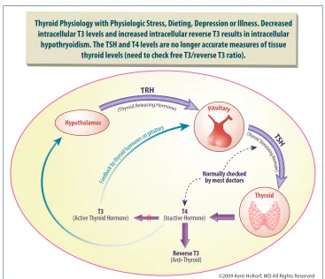
Thyroid Physiology with Physiologic Stress, Dieting, Depression or Illness. Decreased
intracellular T3 levels and increased intracellular reverse T3 results in intracellular
hypothryoidism. The TSH and T4 levels are no longer accurate measures of tissue
thyroid levels (need to check free T3/reverse T3 ratio).
In the
International Journal of Neruopsycho
pharmacology
, Posternak
et al.
published a double
blind placebo control trial of 50 patients with
normal thyroid function as defined by a normal
TSH (1.5±0.8). The patients were randomized
to receive 25 μg of T3 or placebo in addition to
antidepressant therapy.
214The study found almost a
two-fold increase in response rate with T3 and a 4.5
times greater likelihood of experiencing a positive
response at any point over a six-week period with
the addition of T3. Side effects were higher in
pla-cebo group on 10/11 criteria including a significant
increase in nervousness with the placebo group.
Kelly
et al.
investigated the effectiveness of T3 for
the treatment of biopolar disorder in patients who
had failed to adequately respond to an average of 14
medications used to treat their bipolar disorder. The
average dose of T3 used was 90.4 μg (range 13–188
μg). The medication was found to be well tolerated
and 84% experienced significant improvement and
33% had a full remission. Again, this is in patients
who had not previously responded to numerous
medications. One patient who was switched to T4
for cost reasons experienced a return of symptoms,
which resolved with the reintroduction of T3. The
authors concluded, “Augmentation with
supraphys-iologic doses of T3 should be considered in cases
of treatment resistant bipolar depression….”
219The authors thanked several doctors who
encour-aged them to go beyond the traditional 50 μg of T3
because it has helped so many of their patients.
With over 4000 patients, The Star*D Report is the
largest trial comparing antidepressant
effective-ness for depression. It found that 66% of patients
fail to respond to antidepressants or have
side-effects severe enough to discontinue use. Of those
who do respond, over half will relapse within
one year.
220The trial found that T3 was effective
even when other medications – such as
citalo-pram (Celexa), bupropion (Wellbutrin), sertraline
(Zolft), venlafaxine (Effexor), or cognitive therapy
– were not. It was shown to be 50% more
effec-tive, even with the less than optimal dose of 50
μg, under direct comparison with significantly
less side effects than commonly used therapeutic
approaches with standard antidepressants. The
authors included a case study to exemplify the
effectiveness of T3, especially when other
medica-tions are not:
“Ms. ‘B,’ a 44-year-old divorced white woman,
became depressed after losing her job as a
secre-tary in a law firm. She initially sought treatment
from her primary care physician and then entered
the STAR*D study. Ms. B met criteria for major
depressive disorder and generalized anxiety
disorder. Her baseline QIDS-SR score was 16.
After 12 weeks on citalopram, her QIDS-SR
score was 10 [minimal response]. She was
then randomly assigned to augmentation with
buspirone; she soon experienced gastrointestinal
distress, and she stopped taking buspirone after 6
weeks. She elected to try one more augmentation
agent and was randomly assigned to T3
augmen-tation. When she started T3 augmentation, her
QIDS-SR score was 12. After 4 weeks, she felt
that her mood and energy had lifted substantially.
She felt better able to make decisions, organize,
and prioritize and felt that she was able and ready
to look for another job. ‘I felt as if my brain
suddenly had oxygen,’ she said, ‘and everything
became clearer.’ After 12 weeks, Ms. B felt
back to normal, and her QIDS-SR score was 0
[complete resolution of symptoms].”
220With an understanding of thyroid physiology and
associated dysfunction that is present in depressed
patients, it is clear that
timed-released T3
supple-mentation should be considered in all depressed and
bipolar patients despite “normal” serum thyroid
lev-els. Additionally, straight T4 should be considered
inappropriate and suboptimal therapy for
replace-ment in such patients.
pAIn
Chronic pain will significantly suppress D1 and
upregulate D2, resulting in a reduction in tissue
T3 without a change in TSH. Thus, the significant
cellular hypothyroidism is not detected by serum
TSH and T4 testing.
119–122This cellular
hypothyroid-ism, which again is undiagnosed by standard blood
tests, increases the risk of the associated fatigue and
depression seen with chronic pain.
119–221is ineffective at reversing the suppressed tissue
T3 levels.
119–121, 221Thus, for those with significant
chronic pain or using significant amounts of narcotic
pain medicine, it must be understood that such a
condition is associated with low tissue thyroid levels
not detected by standard blood tests. Tolerance to
the inhibitory effect of narcotics on TSH secretion
and T4 to T3 conversion does not occur.
119, 122Expert
pain specialists understand this and recommend T3
supplementation to patients with significant pain or
on narcotic pain medications.
221DIeTIng
Acute or chronic dieting can result in a significant
decrease in intracellular and circulating T3 levels
by up to 50%,
46, 47, 51, 90which significantly reduces
basal metabolic rate (number of calories burned per
day) by 15–40%.
48, 222, 223With chronic dieting, the
thyroid levels and metabolism often do not return to
normal levels; the body stays in starvation mode for
years with significantly reduced metabolism despite
the resumption of normal food intake, making it
very difficult to lose or maintain lost weight.
48A study by Araujo
et al.
published in
American
Journal of Physiology, Endocrinology and
Metabolism
found that 25 days of calorie
restric-tion (dieting) significantly reduced D1, resulting in
reduced T4 to T3 conversion with a 50% reduction
in T3. This dramatic reduction in T3 was associated
with an increase in D2, so there was no increase in
TSH but rather a decrease from an average of 1.20
ng/mL to 0.7 ng/mL, demonstrating the fact that the
TSH is a poor marker for tissue T3 levels,
espe-cially in a chronically dieting patient.
47Fontana
et al.
found that T3 levels were
signifi-cantly decreased by 25% in chronically dieting
individuals compared to non-dieting individuals
with no difference in TSH and T4 (thus undetected
by TSH and T4 testing). This clinically significant
reduction in T3 levels, potentially causing inability
to lose weight or regaining of lost weight, fatigue,
and depression, remained in the normal range
despite the significant decline, demonstrating the
weakness and unreliability of the common use of
population reference ranges that consider 95% of
the population as “normal”.
49A study by Leibel
et al.
published in the journal
Metabolism
found that individuals who had
lost weight in the past had a significantly lower
metabolism than those of same weight who had
not gained or lost significant weight in the past.
48The metabolism in the weight reduced patients was
25% less than an equal weight person who did not
lose or gain significant weight in the past and equal
to someone who weighed 60% less than they did.
Additionally, the reduction was shown to be present
years later.
This 25% percent reduction in metabolism equates to
an approximate deficit of 500–600 cal per day. Thus,
if the previously overweight persons are to maintain
the reduced weight they lost, they must either eat
600 cal per day less compared to a person of the
same weight who has not had a weight problem or
must jog about 1 ½ hours per day to maintain the lost
weight. This equates to approximately a pound per
week of weight gain, explaining why weight is so
quickly gained without continued very strict dieting.
So many people who have difficulty keeping weight
off don’t eat excessively but are continually told they
are eating too much or they need to exercise more
by people who have never had a weight problem.
They are made to feel it is a character issue and that
nobody believes how little food they actually
con-sume. Unless the physiologic thyroid dysfunction is
corrected, any diet and exercise strategy is doomed.
Croxson
et al.
in
Journal of Endocrinology and
Metabolism
found that individuals with a history of
intense dieting had dramatic reductions in T4 to T3
conversion with an intracellular deficiency of T3.
The inadequacy and inaccuracy of standard TSH
and T4 testing was demonstrated, as such testing
failed to detect the dramatic reduction in tissue
levels of T3 in all of the patients.
50InSulIn ReSISTAnCe/DIABeTeS/
MeTABOlIC SynDROMe/OBeSITy
T4 to reverse T3, further reducing intracellular T3
levels.
91, 92, 94, 100, 145, 176–185, 224Additionally, the
ele-vated insulin will increase D2 activity and suppress
TSH levels, further decreasing thyroid levels and
making it inappropriate to use the TSH as a reliable
marker for tissue thyroid levels in the presence of
elevated insulin levels as occurs with obesity, insulin
resistance, or type II diabetes.
91–99, 225Pittman
et al.
found that normal individuals had a
77% conversion of T4 to T3, while diabetic
individu-als had a 45% conversion of T4 to T3 and increased
T4 to reverse T3. Improvement in glucose levels
only slightly increased T4 to T3 conversion to 46%.
93Islam
et al.
investigated the T4 to T3 conversion in
50 diabetic patients compared to 50 non-diabetic
controls. There was no difference in TSH and
free T4 levels, but the diabetic individuals had
significantly decreased free T3 levels (
P
=0.0001)
that averaged 46% less than controls. The FT3/
FT4 ratio was 50% less in diabetic patients
ver-sus controls. The TSH failed to elevate despite
the fact that serum T3 was approximately half of
normal.
92Saunders
et al.
also found that diabetics
had approximately a 50% reduction in T3 levels
and significantly increased reverse T3 levels and
decreased T3/reverse T3 ratios.
94In the
International Journal of Obesity
,
Krotkiewski
et al.
published the results of their
investigation of the impact of supplemental T3 on
cardiovascular risk in obese patients to partially
reverse the reduced T4 to T3 conversion seen with
obesity.
53Seventy obese patients with “normal”
standard thyroid function tests were treated with 20
μg of straight T3 for six weeks. While the dose was
not high enough to completely reverse the reduced
T4 to T3 conversion seen with obesity, there was a
significant reduction in a number of cardiovascular
risk factors, including cholesterol and markers for
insulin resistance. There were no side-effects in any
of the patients. The authors conclude, “T3 may be
considered to ameliorate some of the risk factors
associated with abdominal obesity, particularly in
some subgroups of obese women with a relative
resistance to thyroid hormones possibly dependent
on decreased peripheral deiodination of thyroxine
(T4).”
53Thus, replacement with timed-released T3
prepa-rations to normalize the reduced intracellular
T3 levels is appropriate in such patients despite
so-called “normal” levels while, on the contrary,
T4-only preparations do not address the
physi-ologic abnormalities of such patients and should
be considered inappropriate replacement for obese
patients or those with insulin resistance, leptin
resistance, or diabetes, as they do not address the
physiologic abnormalities in this group.
lepTIn
The hormone leptin has been found to be a major
regulator of body weight and metabolism. The
body secretes leptin as weight is gained to signal
the brain (specifically the hypothalamus) that there
are adequate energy (fat) stores. The hypothalamus
should then stimulate metabolic processes that
result in weight loss, including a reduction in
hun-ger, an increased satiety with eating, an increase in
resting metabolism, and an increase in lipolysis (fat
breakdown). New research has found that this leptin
signaling is dysfunctional in the majority of people
who have difficultly losing weight or are unable to
lose weight.
54–58The problem is not in the production of leptin;
studies show that the majority of overweight
individuals who are having difficulty losing weight
have a leptin resistance, where the leptin is unable
to produce its normal effects to stimulate weight
loss.
54–58This leptin resistance is sensed as
star-vation, so multiple mechanisms are activated to
increase fat stores, rather than burn excess fat
stores.
54–83Leptin resistance is shown to suppress
D1 and stimulate D2, resulting in reduced cellular
T3 but a reduction in serum TSH.
47, 84–89A study by
Cettour-Rose
et al.
published in
American Journal
of Physiology, Endocrinology and Metabolism
demonstrated that physiologic reversal of leptin
resistance restored deiodinase activity except in
the presence of elevated reverse T3.
86Thus, in the
combined with a high normal or elevated reverse
T3 (above 150).
exeRCISe
It has been shown that women or men who perform
more than moderate exercise, especially when
associated with dieting, have reduced T4 to T3
conversion and increase reverse T3, counteracting
many of the positive effects of exercise in women
including weight loss.
226, 227Consequently, T3 and
reverse T3 levels should be evaluated in individuals
who exercise and/or diet to better determine cellular
thyroid levels, as TSH and T4 would not
necessar-ily reflect tissue levels in such patients.
IROn DefICIenCy
Iron deficiency is shown to significantly reduce
T4 to T3 conversion, increase reverse T3 levels,
and block the thermogenic (metabolism boosting)
properties of thyroid hormone.
228–232Thus, iron
deficiency, as indicated by an iron saturation below
25 or a ferritin below 70, will result in diminished
intracellular T3 levels. Additionally, T4 should not
be considered adequate thyroid replacement if iron
deficiency is present.
228, 229, 231, 232InflAMMATIOn ASSOCIATeD
wITH COMMOn COnDITIOnS
The inflammatory cytokines IL-1, Il-6, C-reactive
protein (CRP), and TNF-alpha will significantly
decrease D1 activity and reduce tissue T3
levels.
102, 104–111Any person with an
inflamma-tory condition
–
including physical or emotional
stress,
233–238obesity,
238–242diabetes,
238, 239, 243depression,
244–246menopause (surgical or
natu-ral),
247heart disease,
238, 248, 249autoimmune disease
(lupus, Hashimoto’s, multiple sclerosis, arthritis,
etc.),
112, 113, 160, 250injury,
251chronic infection
252, 253or cancer
254–256–
will have a decreased T4 to
T3 conversion in the body and a relative tissue
hypothyroidism. The inflammatory cytokines will,
however, increase the activity of D2 and suppress
the TSH despite reduced peripheral T3 levels;
again, making a normal TSH an unreliable indicator
of normal tissue thyroid levels.
102, 104–111There is a direct inverse correlation between CRP
and reduced tissue T3,
110, 257so individuals with
elevated CRP (greater than 3 mg/L) or other
inflam-matory cytokines will have a significant reduction
in cellular T3 levels. The suppression of
intracellu-lar T3 levels correlates with the degree of elevation
of CRP, despite serum thyroid tests being
“nor-mal”.
110, 257Thus, if any inflammation is present,
which is found in numerous clinical and subclinical
conditions (as above), the body will have lower
cel-lular T3 levels that are often inadequate for optimal
functioning; but the pituitary will have increased
levels of T3, resulting in a lowering of the TSH that
would potentially be inappropriately interpreted as
an indication of “normal” thyroid levels.
Thus, any person with an inflammatory condition
will have diminished tissue levels of T3 potentially
severe enough to cause symptoms, but these
symp-toms will not be detected by standard thyroid testing.
Additionally, due to the reduced T4 to T3 conversion
induced by the inflammation in these conditions,
effective treatment must include T3 (combination
or, ideally, timed-released T3). Also, due to the
inflammatory suppression of TSH, not only is a
normal TSH necessarily an indication of
euthyroid-ism (normal thyroid), but also a suppressed TSH is
not necessarily an indication of excessive thyroid
with treatment. Rather, free T3 and reverse T3 levels
along with clinical parameters should be used to
determine optimal replacement doses of thyroid.
Additionally, inflammation will stimulate D3,
producing more reverse T3, further causing cellular
hypothyroidism not detected by TSH testing by
suppressing intracellular T4 to T3 conversion and
blocking the T3 receptor inside the cell.
258envIROnMenTAl TOxInS
1000 times more efficient at converting T4 to T3,
1, 143D2 is 100- to 1000-fold less sensitive to suppression
by toxins or by mineral or hormonal
deficien-cies.
1–5, 143, 215, 260, 261Thus, the D1 in the body is
suppressed by toxins, pesticides, and plastics at
lev-els that are hundreds to thousands times lower than
required to suppress the D2 in the pituitary. This is
proving to be a major problem for the population in
general; levels of plastics and other toxins commonly
found in individuals (toxins that are considered
“nor-mal” exposure) result in reduced levels of T3 in all
tissues with the exception of the pituitary, which is
resistant to the effect of toxins. Because the pituitary
is relatively unaffected, the reduced tissue thyroid
levels are not detected by standard TSH testing.
For instance, bisphenol-A, which is ubiquitous
in the environment and large amounts of which
can leach into food and liquids from plastic water
bottles and the lining of aluminum cans, is shown
to significantly block thyroid activity in all tissues
except the pituitary, potentially contributing to or
causing weight gain, fatigue, and depression but
not detected by TSH testing.
126, 127, 130, 131, 262Levels
of a number of thyroid blocking toxins, including
bisphenol-A and PBDEs, are significantly higher
in individuals in the United States (PBDEs being
especially high in California),
262, 263resulting in
reduced T3 effect in all tissues in almost all
indi-viduals in the United States compared to the rest of
the world that is not detected by standard thyroid
testing. This is potentially a significant contributor
to the epidemic of obesity and depression in the US.
TeSTOSTeROne
Low testosterone in men will result in a lowering of
D1 activity without changing pituitary D2.
141Thus, a
drop in testosterone will result in lower tissue levels
of T3 without producing an elevation of TSH.
141, 142Environmental factors, including pesticides, plastics,
4000
PBDE 47 PBDE 49 PBDE 100
3000
2000
PBDE dust concentration (par
ts per billion)
1000
UK
Germ any
Canada Cape
Cod
Boston Te
xas
Calif ornia
Washington ,
DC
Figure 4:
Reprinted (adapted) with permission from Zota AR, Rudel RA, Morello-Frosch RA, Brody JG. Elevated house dust and serum concentrations of PBDEs in California: unintended consequences of furniture flammability standards? Environ. Sci. Technol. 2008;42:8158–64.
100
r=0.076 p=NS
400 700 900 1,200
FT4 (pg/dl)
1,400 1,700
100 200 300 400
500 r=0.643
p<0.0001
FT3 (pg/dl)
0 1 2 3 4 5
0 20 40 60
r=0.377 p<0.0001
80 100 120
TSH (mU/I)
Age (years)
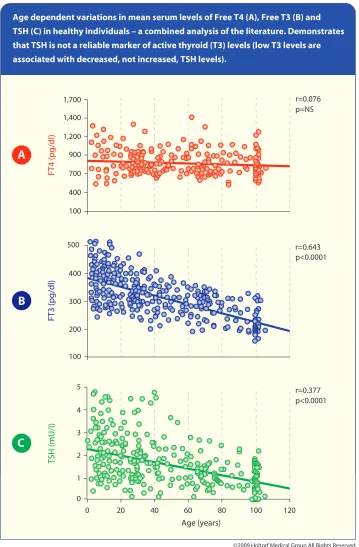
Age dependent variations in mean serum levels of Free T4 (A), Free T3 (B) and TSH (C) in healthy individuals – a combined analysis of the literature. Demonstrates that TSH is not a reliable marker of active thyroid (T3) levels (low T3 levels are associated with decreased, not increased, TSH levels).
A
B
C
©2009 Holtorf Medical Group All Rights Reserved
Figure 5:
1. Schwartz E, Holtorf, K. Hormones in Wellness and Disease Prevention: Common Practices, Current State of the Evidence, and Questions for the Future. Prim Care Clin Office Pract. 2008;35:669–705.
2. Mariotti S, Barbesino G, Caturegli P, et al. Complex alteration of thyroid function in healthy centenarians. J Clin Endocrinol Met. 1993;77(5):1132. 3. Verheecke, P. Free triiodothyronine concentration in serum of 1050 euthyroid children is inverselyrelated to their age. Clin Chem. 1997;43(6):963–7. 4. Quest Diagnostics assay validation free T3, free T4 and TSH, San Juan Capistrano, CA.
and other pollutants, have resulted in a significant
decrease in the average testosterone levels for
men, so most men will have, at least, a relative
deficiency of testosterone.
264Major laboratories
have, unfortunately, reduced the “normal” range of
free testosterone to maintain the 95th percentile as
normal, the result being that many abnormally low
levels will now be considered normal.
In particular, the majority of male diabetics and
those with insulin resistance will have suppressed
testosterone level that is in the low or low-normal
range, which further suppresses D1 and tissue T3
levels and perpetuates the weight gain or inability
to lose weight
–
worsening these conditions.
265–267gROwTH HORMOne
Growth hormone deficiency reduces T4 to T3
conversion and increases reverse T3 while
supple-mentation with growth hormone improves T4 to T3
conversion and reduces reverse T3.
186, 225, 268, 269The
age-associated decline in growth hormone certainly
contributes to the reduced T3 levels with age not
detected by TSH and T4 testing (see Figure 5).
InDIvIDuAl vARIATIOnS In
DeIODInASe
The relative amounts of D1, D2, and D3 vary in
different tissues among different individuals
270and
under varying conditions,
8, 11–21, 23–26, 28–45, 100–102, 105, 106, 111, 119–127, 144, 170, 209, 221resulting in hundreds of
possible symptoms with hypothyroidism; some
people have one symptom, some have a few, and
some people have many, depending on the relative
level of T3 in each tissue. Unfortunately, serum
thyroid levels often do not accurately reflect
intra-cellular tissue levels or levels in a particular tissue.
SuMMARy
With an improved understanding of thyroid
physiol-ogy that includes the local control of intracellular
activation and deactivation of thyroid hormones by
deiodinases, it becomes clear that standard thyroid
tests often do not reflect the thyroid status in the
tissues of the body, other than the pituitary. This is
especially true with physiologic and emotional stress,
depression, dieting, obesity, leptin insulin resistance,
diabetes, chronic fatigue syndrome and
fibromyal-gia, inflammation, autoimmune disease, or systemic
illness. Consequently, it is inappropriate to rely on
a normal or low TSH as an adequate or sensitive
indicator of normal or low tissue levels of T3 in the
presence of any such conditions, making the TSH a
poor marker for the body’s overall thyroid level.
In order to be appropriately and thoroughly evaluated
for thyroid dysfunction and obtain optimal treatment,
it is important that patients find a thyroidologist who
understands the limitations of standard thyroid
test-ing and can clinically evaluate patients by taktest-ing an
extensive inventory of potential signs and symptoms
that may be due to low tissue thyroid levels despite
normal standard thyroid tests.
DISClOSuRe Of InTeReSTS
Dr. Holtorf supplies supplements from
Holtraceuticals to support thyroid function to his
own patients, outside the submitted work.
RefeRenCeS
1. Bianco AC, Salvatore D, Gereben B, Berry MJ, Larsen PR. biochemistry, cellular and molecular biology, and physiological roles of the iodothyronine selenodeio-dinases. Endocrine Rev. 2002;23(1):38–89.
2. Silva JE, Larsen PR. Pituitary nuclear 3,5,3′ -tri-iodothyronine and thyrotropin secretion: an explanation for the effect of thyroxine. Science. 1977;198(4317):617–20.
3. Koenig RJ, Leonard JL, Senator D, Rappaport N. Regulation of thyroxine 5′-deiodinase activity by 3,5,3′-triiodothyronine in cultured rat anterior pituitary cells. Endocrinology. 1984;115(1):324–9.
5. Visser TJ, Kaplau MM, Leonard JL, Larsen PR. Evidence for two pathways of iodothyroinine 5′-deiodination in rat pituitary that differ I kinetics, pro-pylthiouracil sensitivity, and response to hypothyroidism. J Clin Invest. 1983;71:992.
6. Larsen PR, Silva JE, Kaplan MM. Relationship between circulation and intracellular thyroid haomrones: physio-logical and clinical implications Endcor Rev. 1981;2:87.
7. Kaplan MM. The role of thyroid hormone deiodination in the regulation of hypothalamo-pituitary function progress in neuroendocrinology. Neuroendocrinology. 1984;38:254–60.
8. Peeters RP, Geyten SV, Wouters PJ, et al. Tissue thyroid hormone levels in critical illness. J Clin Endocrinol Metab. 2005;12:6498–507.
9. Peeters RP, Wouters PJ, Toor HV, et al. Serum 3,3′,5′ -tri-iodothyronine (rT3) and 3,5,3′-triiodothyronine/rT3 are prognostic markers in critically ill patients and are associated with postmortem tissue deiodinase activities. J Clin Endocrinol Metab. 2005;90(8):4559–65.
10. Campos-Barros A, Hoell T, Musa A, Sampaolo S, et al. Phenolic and tyrosyl ring iodothyronine deiodination and thyroid hormone concentrations in the human central ner-vous system. J Clin Endocrinol Metab. 1996; 81:2179–85.
11. Chopra IJ, Chopra U, Smith SR, et al. Reciprocal changes in serum concentrations of 3,3′ ,5-triiodothyro-nine (T3) in systemic illnesses. J Clin Endocrinol Metab. 1975;41:1043–9.
12. Chopra IJ, Williams DE, Orgiazzi J, Solomon DH. Opposite effects of dexamethasone on serum concen-trations of 3,3′,5′- triiodothyronine (reverse T3) and 3,3′,5-triiodothyronine (T3). J Clin Endocrinol Metab. 1975;41:911–20.
13. Duick DS, Warren DW, Nicoloff JT, Otis CL, Croxson MS. Effect of single dose dexamethasone on the concentration of serum triiodothyronine in man. J Clin Endocrinol Metab. 1974;39(6):1151–4.
14. Cavalieri RR, Castle JN, McMahon FA. Effects of dexamethasone on kinetics and distribution of triiodo-thyronine in the rat. Endocrinology. 1984;114: 215–21.
15. Bianco AC, Nunes MT, Hell NS, Maciel RMB. The role of glucocorticoids in the stress-induced reduction of extrathyroidal 3,5,3’-triiodothyronine generation in rats. Endocrinology. 1987;120:1033–8.
16. DeGroot LJ. Non-thyroidal illness syndrome is func-tional central hypothyroidism, and if severe, hormone replacement is appropriate in light of present knowledge. J Endocrinol Invest. 2003;26:1163–70.
17. Reed HL, Brice D, Shakir KM, Burman KD, et al. Decreased free fraction of thyroid hormones after prolonged Antarctic residence. J Applied Physiol. 1990;69:1467–72.
18. Forhead AJ, Curtis K, Kaptein E, Visser TJ, Fowden Al. Developmental control of iodothyronine deiodinases by cortisol in the ovine fetus and placenta near term. Endocrinology. 2006;147:5988–94.
19. Nicoloff JT, Fisher DA, Appleman MD. The role of glucocorticoids in the regulation of thyroid function in man. J Clin Invest. 1970;49(10):1922–9.
20. Brabant G, Brabant A, Ranft U, Ocran K, et al. Circadian and pulsatile thyrotropin secretion in euthyroid man under the influence of thyroid hormone and gluco-corticoid administration. J Clin Endocrinol Metab. 1987;65:83–8.
21. Benker G, Raida M, Olbricht T, et al. TSH secretion in Cushing’s Syndrome: relation to glucocorticoid excess, diabetes, goitre, and the ‘Sick Euthyroid Syndrome’. Clin Endocrinol. 1990;33(6):777–86.
22. Mebis L, Langouche L, Visser TJ, Van den Berghe G. The type II iodothyronine is up-regulated in skeletal muscle during prolonged critical illness. J Endocrinol Metab. 2007;92(8):3330–3.
23. Linnoila M, Lamberg BA, Potter WZ, Gold PW, Goodwin FK. High reverse T3 levels in manic and uni-polar depressed women. Psychiatry Res. 1982;6:271–6.
24. Kjellman BF, Ljunggren JG, Beck-Friis J, Wetterberg L. Reverse T3 levels in affective disorders. Psychiatry Res. 1983;10:1–9.
25. Jackson I. The thyroid axis and depression. Thyroid. 1998;8(10):951–6.
26. Gitlin M, Altshuler LL, Frye MA, Suri M, Huynh EL, et al. Peripheral thyroid hormones and response to selec-tive serotonin reuptake inhibitors. J Psychiatry Neurosci. 2004;29(5):383–6.
27. Clausen P, Mersebach H, Nielsen B, et al.
Hypothyroidism is associated with signs of endothelial dysfunction despite 1-year replacement therapy with levothyroxine. Clin Endocrinol. 2009;70:932–7.
28. Duval F, Mokrani MC, Bailey P, Correa H, et al. Thyroid axis activity and serotonin function major depressive episode. Psychoneuroendocrinology. 1999;24:695–712.
29. Unden F, Ljunggren JG, Kjellman BF, Beck-Friis J, Wetterberg L. Twenty-four-hour serum levels of T4 and T3 in relation to decreased TSH serum levels and decreased TSH response to TRH in affective disorders. Acta Psychiatr Scand. 1986;73:358–65.
30. Linnoila M, Lamberg BA, Rosberg G, Karonen SL, Welin MG. Thyroid hormones and TSH, prolactin and LH responses to repeated TRH and LRH injec-tions in depressed patients. Acta Psychiatr Scand. 1979;59:536–44.
31. Kirkegaard C, Faber J. Altered serum levels of thyroxine, triiodothyronines and diiodothyronines in endogenous depression. Acta Endocrinol. 1981;96:199–207.
32. Sintzel F, Mallaret M, Bougerol T. Potentializing of tricyclics and serotoninergics by thyroid hor-mones in resistant depressive disorders. Encephale. 2004;30(3):267–75.
depression and thyroid function in subjects on and not on T4: findings from the Hunt study. Clin Endocrinol. 2009;71:574–80.
34. Thompson FK. Is there a thyroid-cortisol-depression axis? Thyroid Sci. 2007;2(10):1.
35. Forman-Hoffman V, Philibert RA. Lower TSH and higher T4 levels are associated with current depres-sive syndrome in young adults. Acta Psychiatr Scand. 2006;114:132–9.
36. Cole DP, Thase ME, Mallinger AG, et al. Slower treat-ment response in biolar depression predicted by lower pretreatment thyroid function. Am J Psychiatry. 2002; 159:116–21.
37. Premachandra1 BN, Kabir MA, Williams IK. Low T3 syndrome in psychiatric depression. J Endocrinol Invest. 2006;29:568–72.
38. Isogawa K, Haruo Nagayama H, Tsutsumi T, et al. Simultaneous use of thyrotropin-releasing hormone test and combined dexamethasone/corticotropine-releasing hormone test for severity evaluation and outcome prediction in patients with major depressive disorder. J Psychiatr Res. 2005;39:467–73.
39. Sullivan GM, Hatterer JA, Herbert J, Chen X, Rosse SP. Low levels of transthyretin in CSF of depressed patients. Am J Psych. 1999;156:710–5.
40. Hatterer JA, Herbert J, Hidaka C, Roose SP, Gorman JM. CSF transthyretin in patients with depression. Am J Psychiatry. 1993;150:813–5.
41. Whybrow PC, Coppen A, Prange AJ, Noguera R, Bailey JE. Thyroid function and the response to liothyronine in depression. Arch Gen Psychiatry. 1972;26:242–5.
42. Kirkegaard C, Faber J. Free thyroxine and 3,3′,5′ -triio-dothyroidnine levels in cerebralspinal fluid in patetns with endogenous depression. Acta Endocrinol. 1991;124:166–72.
43. Kirkegaard C. The thyrotropin response to thyrotro-pin-releasing hormone in endogenous depression. Psychoneuroendocrinology. 1981;6:189–212.
44. Baumgartner A, Graf KJ, Kurten I, Meinhold H. The hypothalamic-pituitary-thyroid axis in psychi-atric patients and healthy subjects Psychiatr Res. 12988;24:271–332.
45. Stipcevic T, Pivac N, Kozarie-Kovacic D, Muck-Seler D. Thyroid activity in patients with major depression. Coll Antropol. 2008;32(3):973–6.
46. Cheron RG, Kaplan MM, Larsen PR. Physiological and pharmacological influences on thyroxine to 3,5,3′-triiodothyronine conversion and nuclear 3,5,3′ -tri-iodthyroidne binding in rat anterior pituitary. J Clin Invest. 1979;64:1402–14.
47. Araujo RL, Andrade BM, da Silva ML, et al. Tissue-specific deiodinase regulation during food restriction and low replacement dose of leptin in rats. Am J Physiol Endocrinol Metab. 2009;296:E1157–63.
48. Leibel RL, Jirsch J. Diminished energy requirements in reduced-obese patients. Metabolism. 1984; 33(2):164–70.
49. Fontana L, Klein S, Holloszy JO, Premachandra BN. Effect of long-term calorie restriction with adequate protein and micronutrients on thyroid hormones. J Clin Endocrinol Metab. 2006;91(8):3232–5.
50. Croxson MS, Ibbertson HK. Low serum triiodothyronine (T3) and hypothyroidism. J Clin Endocrinol Metab. 1977;44:167–74.
51. Silva JE, Larsen PR. Hormonal regulation of iodothy-ronine 5-deiodinase in rat brown adipose tissue. Am J Physiol. 1986;251:E639–43.
52. Krotkiewski M, Holm G, Shono N. Small doses of triiodothyronine can change some risk factors associated with abdominal obesity. Int J Obesity. 1997;21:922–9.
53. Krotkiewski M. Thyroid hormones and treatment of obesity. Int J Obesity. 2000;24(2):S116–9.
54. Dagogo-Jack S. Human leptin regulation and promis in pharmacotherapy. Current Drug Targets. 2001;2:181–95.
55. Considine RV, Sinha MK, Heiman ML, Kriauciunas A, et al. Serum immunoreactive-leptin. Concentrations in normal-weight and obese humans. N Engl J Med. 1996;334:292–5.
56. Dagogo-Jack S, Tanellis C, Paramore D, Brother SJ, Land TM. Plasma leptin and insulin relationships in obese and nonobese human. Diabetes. 1996;45:695–8.
57. Maffei M, et al. Leptin levels in human and rodent: measurement of plasma leptin and ob NAN in obese and weight-reduced subjects. Nat Med. 1995;1;1155–61.
58. Hassink SG, Sheslow DV, de Lancey E, Opentanova I, Considine RV, Caro JF. Serum leptin in children with obesity. Relationship to gender and development. Pediatrics. 1996;98:201–3.
59. Kozlowska L, Rosolowska-Huszcz. Leptin, thyrotro-pin, and thyroid hormones in obese/overweight women before and after two levels of energy deficit. Endocrine. 2004;24(2):147–53.
60. Fekete C, et al. differential effects of central leptin, insulin, or glucose administration during fasting on the hypothalamic-pituitary-thyroid axis and feeding-related neurons in the arcuate nucleus. Endocrinology. 2006;147(1):520–9.
61. Ahima RS, Prabakaran D, Mantzoros C, Qu D, Lowell B, Maratos-Flier E, Flier JS. Role of leptin in the neuro-endocrine response to fasting. Nature. 1996;382:250–2.
62. Legradi G, Emerson CH, Ahima RS, Flier JS, Lechan RM. Leptin prevents fasting-induced suppression of prothyrotropin-releasing hormone messenger ribonucleic acid in neurons of the hypothalamic paraventricular nucleus. Endocrinology. 1997;138:2569–76.
64. Schwartz MW, Woods SC, Porte D, Seeley RJ, Baskin DG. Central nervous system control of food intake. Nature. 2000;404;661–71.
65. Mantzoros CS, Moschos SJ. Leptin: in search of role(s) in human physiology and path physiology. Clin Endocrinol. 1998;49:551–67.
66. Fruhbeck G, Jebb SA, Prentice AM. Leptin: physiology and pathophysiology. Clin Physiol. 1998;18:399–419.
67. Flier JS, Harris M, Hollenber A. Leptin, nutrition and the thyroid: the why, the wherefore and the wiring. J Clinical Invest. 2000;105(7):859–61.
68. Gon DW, He Y, Karas M, Reitman M. Uncoupling protein-3 is a mediator of thermogenesis regulated by thyroid hormone, beta 3-adernergic agonists and leptin. J Biol Chem. 1997;272:24129–32.
69. Cusin I, Rouru J, Visser T, Burger AG, Rohner-Jeanrenaud F. Involvement of thyroid hormones in the effect of intracerebroventricular leptin infusion on uncoupling protein-3 expression in rat muscle. Diabetes. 2000;49:1101–5.
70. Rosenbaum M, Godmsith R, et al. Low-dose leptin reverses skeletal muscle, autonomic, and neuroendocrine adaptations to maintenance of reduced weight. J Clin Invest. 2005;115:3579–86.
71. Rosenbaum M, Murphy, et al. Low dose leptin admin-istration reverses effects of sustained weight-reduction on energy expenditure and circulation concentra-tion of thyroid hormones. J Clin Endocrinol Metab. 2002;87(5):2391–4.
72. Leibel RL, et al. Changes in energy expenditure resulting from altered body weight. N Engl J Med. 1995;332:621–8.
73. Rosenbaum M, et al. The effects of changes in body and thyroid function. Am J Clin Nutr. 2000;71:1421–32.
74. Ahima R, et al. Role of leptin in the neuroendocrine response to fasting. Nature. 1996;382:250–2.
75. Rosenbaum M, et al. Effects of weight change on plasma leptin concentrations and energy expenditure. J Clin Endocrinol Metab. 1997;82:3647–54.
76. Légrádi G, Emerson CH, Ahima RS, Flier JS, Lechan RM. Leptin prevents fasting-induced suppression of prothyrotropin-releasing hormone messenger ribonucleic acid in neurons of the hypothalamic paraventricular nucleus. Endocrinology. 1998;138:2569–76.
77. Boozer CN, Leibel RL, Love RJ, Cha MC, Aronne LJ. Synergy of leptin and sibutramine in treatment of diet-induced obesity in rats. Metabolism. 2001;50:889–93.
78. Campfield LA, et al. Recombinant mouse OB protein: evidence for a peripheral signal linking adiposity and central neural networks. Science. 1995;269:546–8.
79. Farooqi I, et al. Effects of recombinant leptin therapy in a child with congenital leptin deficiency. N Engl J Med. 1999;341:879–84.
80. Chehab F. Leptin as a regulator of adipose tis-sue mass and reproduction. Trends Pharmacol Sci. 200;21:309–14.
81. Rosenbaum K, et al. The role of leptin in human physiol-ogy. N Engl J Med. 1999;341:913–5.
82. Naslund E, et al. Associations of leptin, insulin resistance and thyroid function with long-term weight loss in diet-ing reduced-obese men. J Int Med. 2000;248:299–308.
83. Doucette E, et al. Appetite after weight-loss by energy restriction and a low-fat diet-exercise follow up. Int J Obesity. 2000;24:906–14.
84. Lisboa PC, Oliveira KJ, Cabanelas A, Ortiga-Carvalho TM, Pazos-Moura CC. Acute cold exposure, leptin, and somatostatin analog (octreotide) modulate thyroid 5’-deiodinase activity. Am J Physiol Endocrinol Metab. 2003;284:E1172–6.
85. Cabanelas A, Lisboa PC, Moura EG, Pazos-Moura CC. Leptin acute modulation of the 5’-deiodinase activities in hypothalamus, pituitary and brown adipose tissue of fed rats. Hormone Metab Res. 2006;38(8):481–5.
86. Cettour-Rose P, Burger AG, Meier CA, Visser TJ, et al. Central stimulatory effect of leptin on T3 production is mediated by brown adipose tissue type II deiodinase. Am J Physiol Endocrinol Metab. 2002;283(5):E980–7.
87. Fekete C, Kelly J, Mihaly E, Sarkar S, Rand WM, Legradi G, et al. Neuropeptide Y has a central inhibi-tory action on the hypothalamic–pituitary–thyroid axis. Endocrinology. 2001;142:2606–13.
88. Fekete C, Legradi G, Mihaly E, Huang QH, Tatro JB, Rand WM, et al. A-Melanocyte-stimulating hormone is contained in nerve terminals innervating thyrotro-pin-releasing hormone-synthesizing neurons in the hypothalamic paraventricular nucleus and prevents fasting-induced suppression of prothyrotropin-releasing hormone gene expression. J Neurosci. 2000;20: 1550–8.
89. Legradi G, Emerson CH, Ahima RS, et al. Arcuate nucleus ablation prevents fasting-induced suppression of ProTRH mRNA in the hypothalamic paraventricular nucleus. Neuroendocrinology. 1998;68:89–7.
90. Vignati L, Finley RJ, Hagg S, Aoki TT. Protein conserva-tion during prolonged fast: a funcconserva-tion of triiodothyronine levels. Trans Assoc Am Physicians. 1978;91:169–79.
91. Katzeff HL, Selgrad C. Impaired peripheral thyroid hormone metabolism in genetic obesity. Endocrinology. 1993;132(3):989–95.
92. Islam S, Yesmine S, Khan SA, Alam NH, Islam S. A comparative study of thyroid hormone levels in diabetic and non-diabetic patients. SE Asian J Trop Med Public Health. 2008;39(5):913–6.