Research Article
Telomerase Expression Increased the Risk of Borderline Ovarian Tumors
Ekspresi Telomerase Meningkatkan Risiko Tumor Ovarium Borderline
Mila Maidarti1, Andrijono1, Primaria Dewi2, Joedo Prihartono3
1Department of Obstetrics and Gynecology 2Department of Pathology Anatomy
3Department of Public Health
Faculty of Medicine University of Indonesia/ Dr. Cipto Mangunkusumo Hospital
Jakarta
INTRODUCTION
One in 70 women will be suffering from ovarian cancer during her life, and borderline ovarian mors present in 15% of all epithelial ovarian tu-mors. Borderline ovarian tumors are neoplasms with changes in cell proliferation greater than that seen in benign ovarian tumors of the same type, but showed no destruction or invasion of the ovar-ium stroma.1 Histologic type of borderline ovarian tumors mostly found are serous or mucinous type
that show the feature of epithelial proliferation, ab-normal nucleus, and mitotic activity in the inter-mediate level, without any invasion into stroma.1,2 Compared with malignant epithelial ovarian mors which are invasive, borderline ovarian tu-mors are found in young women and at an early stage, with a good prognosis. The incidence rate of borderline ovarian tumors was lower than invasive ovarian cancer.2 Borderline ovarian tumors are often called ’ovarian tumors of low malignant po-tential’ because of the low histological differentia-Abstract
Objective: To compare the expression of telomerase in benign ovar-ian tumors and borderline ovarovar-ian tumors.
Methods: Thirteen samples of paraffin blocks of ovarian borderline tumors and benign ovarian tumors were taken from patients who underwent surgery from January 2006 to December 2011. In all samples, we performed immunohistochemical staining to the paraf-fin blocks. Semi quantitative determination of the expression of telomerase is done by an Anatomic Pathology specialist and Gyne-cologist, which already had the same perception about assessing the standardization sample. We used Fisher’s test to analyze the data.
Results: There were significant relationship in the moderate expres-sion of telomerase in the nucleus and cytoplasm between benign and borderline ovarian tumors, with an odds ratio of moderate telomerase expression in the cytoplasm and nucleus of 19.3 (95% CI: 1.4 - 943) and 26 (95% CI : 2.3 - 1211). This means that the risk of borderline ovarian tumors in the expression of moderate telom-erase in the cytoplasm was 3.19 times compared to the negative ex-pression, whereas in the nucleus it became 26-fold. There is no sig-nificant relationship among menopause, age, and telomerase ex-pression in the nucleus and cytoplasm.
Conclusion: There are significant differences in the expression of moderate strength telomerase in nucleus and cytoplasm between benign and borderline ovarian tumors.
[Indones J Obstet Gynecol 2013; 37-1: 32-40]
Keywords: benign ovarian tumor, borderline ovarian tumor, telo-merase expression
Abstrak
Tujuan: Membandingkan ekspresi telomerase pada tumor ovarium ji-nak dan tumor ovarium borderline.
Metode: Tiga belas sampel blok parafin tumor ovarium borderline dan tumor jinak ovarium diambil dari pasien yang menjalani operasi dari bulan Januari 2006 sampai bulan Desember 2011. Pada semua sampel parafin blok dilakukan pewarnaan imunohistokimia. Penen-tuan ekspresi telomerase secara semikuantitatif dikerjakan oleh dok-ter spesialis Patologi Anatomi dan dokdok-ter spesialis Obstetri Ginekologi, yang sebelumnya telah menyamakan persepsi mengenai penilaian standarisasi sampel. Analisis statistik menggunakan uji Fisher. Hasil: Terdapat hubungan bermakna ekspresi telomerase sedang dan negatif di inti dan sitoplasma dengan tumor jinak ovarium dan tumor ovarium borderline, dengan odd ratio ekspresi telomerase sedang di sitoplasma dan inti sebesar 19,3 ( IK 95%: 1,4 - 943) dan 26 (IK 95%: 2,3 - 1211). Ini berarti bahwa risiko terjadinya tumor ovarium border-line pada ekspresi telomerase sedang di sitoplasma adalah 3,19 kali dibandingkan ekspresi negatif, sedangkan di inti 26 kali lipat. Tidak terdapat hubungan bermakna menopause dan usia dengan ekspresi telomerase di inti dan sitoplasma.
Kesimpulan: Terdapat perbedaan bermakna ekspresi telomerase kekuatan sedang di inti dan sitoplasma tumor jinak dan tumor ovarium borderline.
[Maj Obstet Ginekol Indones 2013; 37-1: 32-40]
Kata kunci: ekspresi telomerase, tumor jinak ovarium, tumor ovarium borderline.
tion degree and the good prognosis.3 Pathogenesis of borderline ovarian tumors remain unclear, the clinical management is still controversial, espe-cially for borderline ovarian tumors serous type, which is a type of borderline ovarian tumors mostly found.4
Ovarian cancer pathophysiology has not been clear until now. One of the interesting things to ex-amine is the role of telomerase in cell aging proc-ess. Telomerase is a complex ribonucleoprotein that adds base sequences at the 3 ’end of DNA telomere. Telomere length is maintained by telo-merase, which is a specific DNA polymerase. Telomere in humans will be shortened progres-sively along with cell division.5 Malignant cells have high telomerase activity so referred as the survival cells that will continue to survive.6,7 Telomerase ac-tivity has been widely found in various kinds of malignant ovarian tumors, and rarely found in be-nign tumors or normal ovarian tissue.8 Telomerase can be used as a prognostic indicator in several types of cancer, in which telomerase activity in-creased in line with tumor progression.9-11
Borderline ovarian tumors are tumors that are interesting to study because it is not benign nor malignant tumors. Telomerase activities in border-line ovarian tumors vary in several studies. Several studies had shown that telomerase activity ranged between 60-100%, while other studies showed the number to be 17-20%.7 Telomerase is active in most tumor cells, but not in normal somatic cells, and can prevent progressive telomere shortening in the presence of cell division, causing tumor cells grew faster than normal cells.9,10 Stabilization of telomeres by telomerase can lead to unlimited cell proliferation.12 In malignant tumors such as leuke-mia, breast cancer, and meningioma, telomerase activity associated with a poor prognosis. Up until now there is no sufficient data to determine the role of telomerase as a factor in ovarian cancer prognosis.7 So we need a study to determine the effect of highly telomerase activity in malignant ovarian in relation to carcinogenesis of ovarian cancer so that it can be used as the basis for sub-sequent studies as a prognostic approach and treatment of malignancy, especially in malignant epithelial ovarian tumors. The purpose of this study is to determine the expression of telomerase in benign ovarian tumors, borderline ovarian tu-mors and assess the comparative telomerase ex-pression in borderline and benign ovarian tumors.
METHOD
This study was a case-control study. Research was conducted at the Department of Obstetrics and Gy-necology and Department of Pathology Anatomy Faculty of Medicine/RSUPNCM, started in July until November 2011.
Research sample was all paraffin blocks at the Department of Pathology Anatomy Faculty of Medicine/RSUPNCM of benign ovarian tumors and borderline ovarian tumors patients treated in on-cology ward of Department of Obstetrics and Gy-necology Faculty of Medicine/RSUPNCM from January 2006 to December 2011 and meet the in-clusion criteria were specimens collected from January 2006, from patients who did not suffer from malignancies in other organs, and had a com-plete medical records. The exclusion criteria were the paraffin blocks of non-epithelial ovarian tu-mors. The sampling will be done in simple random sampling, where all the paraffin blocks of ovarian tumors that meet the criteria will be numbered, and some of them will be selected with the help of random tables. Based on the sample calculation, we obtained 13 samples of benign ovarian tumors and 13 borderline ovarian tumors.
The paraffin blocks of borderline ovarian tumors and benign ovarian tumors were stained immuno-histochemically. The intensity of staining was evaluated subjectively using a scale ranging from 0 = no stain, 1 = weak staining, 2 = moderate stain-ing, 3 = strong staining.36 In every staining, we in-cluded a negative control by using the same ova-rian tissue and each one time staining process, we made as much as 13 sample for case-control group with a positive control of endometrial cancer.
Telomerase expression in cytoplasm and nu-cleus, were assessed by immunohistochemical staining, using Burstman score. Assessment cate-gories: number of positive cells: 0% = 0; < 10% = 1; 10-50% = 2; 51-80% = 3; and > 80% = 4. The intensity of color: Weak = 1+; Moderate = 2+; Strong = 3 +. Then multiplied and the result: nega-tive = score 0; weak = score 1; moderate = score 2-6; strong = score 8-12.
ver-sion 17.0. The relationship between telomerase ex-pression in borderline ovarian tumors and benign ovarian tumors was assessed by chi square tests or Fisher. The predictive value is considered sig-nificant if p value < 0.05. This research had re-ceived approval from the Faculty of Medicine Re-search Ethics Committee.
RESULT
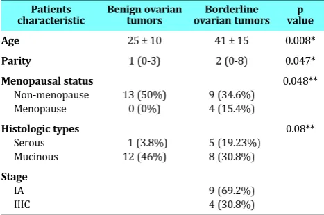
Data were obtained from immunohistochemical examination of 26 paraffin block samples which were still in a good condition. Samples were de-rived from borderline ovarian tumors and benign ovarian tumor patients treated at the oncology ward of Department of Obstetrics and Gynecology Faculty of Medicine/RSUPNCM Jakarta in January 2006 to December 2011. Immunohistochemical examination of samples from both groups was done by assessing the number of positive cells and intensity in the nucleus and cytoplasm. Assessment of telomerase expression was carried out using Burstman scores, then the samples divided into groups of strong, moderate, weak and negative. The data obtained from the examination was then processed using SPSS version 17.0 for windows. The number of cases, of patients with borderline ovarian tumors were 13 people, and control group consisted of 13 people. Fifteen patients (57.7%) aged over 30 years and 11 (42.3%) aged less than 30 years. The average age of patients with border-line ovarian tumors was older, and statistically sig-nificant (41 ± 15 year) compared to benign ovarian tumor group (25 ± 10 year).
To determine differences in age and parity groups of benign ovarian tumors and borderline ovarian tumors, we used the Mann Whitney test because the data is abnormally distributed. To find out the difference proportion of women who had experienced menopause and have not experienced menopause, and the proportion of histologic types in each group, we used Chi square or Fisher test. There are significant differences in the number of patients who had experienced menopause in the group of benign ovarian tumors and borderline ovarian tumors. Histopathology types of benign ovarian tumors are mostly mucinous, but there were no significant differences in the proportion of serous and mucinous types in both groups.
Table 1. The baseline characteristic of patients
Patients
characteristic Benign ovariantumors ovarian tumorsBorderline valuep
Age 25 ± 10 41 ± 15 0.008*
Parity 1 (0-3) 2 (0-8) 0.047*
Menopausal status
Non-menopause Menopause
13 (50%) 0 (0%)
9 (34.6%) 4 (15.4%)
0.048**
Histologic types
Serous Mucinous
1 (3.8%) 12 (46%)
5 (19.23%) 8 (30.8%)
0.08**
Stage
IA IIIC
9 (69.2%) 4 (30.8%)
*Mann Whitney test **Fisher test
We classified the samples into 4 different groups, namely the negative cases (score 0), weak expression (score 1), moderate expression (score 2-6), and strong expression (8-12). To determine the relationship between borderline and benign ovarian tumors and the telomerase expression in nucleus and cytoplasm, we used Fisher’s test. The results of Fisher’s test can be seen in the following table.
Table 2. Bivariate analysis of weak and moderate telo merase expression in the cytoplasm for case and control groups
Telomerase expression
Group
OR
(CI 95%) p* Borderline
ovarian tumorsBenign ovariantumors
Weak 1 2 3.5 (0.03-313) 0.42
Negative 1 7 Reff
Moderate 11 4 19.3 (1.4-943) 0.005
*The predictive value (p) obtained via two-way Fisher’s test
Table 3. Bivariate analysis of weak and moderate merase expression in the nucleus for case and control groups
Telomerase expression
Group
OR
(CI 95%) p* Borderline
ovarian tumorsBenign ovariantumors
Weak 0 1 1.3 (0.02-30)** 1.0
Negative 4 12 Reff
Moderate 9 0 26 (2.3-1211)** 0.00
*The predictive value (p) obtained via two-way Fisher’s test
**Odds ratio is obtained by adding 1 to each cell, because there is a cell whose value is 0
To determine the relationship between the age and telomerase expression in the nucleus or cyto-plasm, we used the two-way Fisher’s test. The age was classified into two groups, which were = 30 years old and < 30 years old. According to two-way Fisher’s test, there is no significant relationship
be-tween age and telomerase expression in the nu-cleus (p value = 0.1) nor the cytoplasm (p value = 0.2). Based on two-way Fisher’s test, we found no significant relationship between menopausal status and telomerase expression in the nucleus (p value = 0.11) nor the cytoplasm (p value = 0.3). Telom-erase expression in both the nucleus and cyto-plasm in borderline ovarian tumors with serous and mucinous types showed no significant differ-ences (p value > 0.05). We analysed the relation-ship between borderline ovarian tumor stage and telomerase expression, and there is no significant relationship between borderline ovarian tumor stage and telomerase expression in the nucleus and cytoplasm (p value > 0.05).
These following pictures are immunohisto-chemical examinations of benign ovarian tumors and borderline ovarian tumors.
Benign ovarian tumor
Positive control
Borderline ovarian tumor
Negative control
Weak cytoplasm expression
DISCUSSION
Borderline ovarian tumors accounts for 10-15% of all epithelial ovarian tumors, more than 96% of its histopathology type is mucinosum or serous. Other types can be either endometrioid, clear cell or tran-sitional cell tumors (Brenner). This study included 26 samples that are divided into two groups: case group, which is a sample derived from borderline ovarian tumors, a total of 13 samples, and the con-trol group, which is originated from benign ovarian tumors, a total of 13 samples. In the case group, mean age of patients is higher (41 ± 15) than the
control group (25 ± 10) and statistically significant.
These results are consistent with research con-ducted by Ayhan et al, in which the average age of patients with borderline ovarian tumors was 41.77
± 13.8844.13 The same conclusion is also reported by Berek, et al in 2007, who stated that borderline ovarian tumors mostly occur at age 30 - 50 years, while ovarian cancer generally occurs between the
ages of 50 - 70 years. Incidence of borderline ovar-ian tumors or invasive ovarovar-ian tumor is 1 in 10 patients aged 40 years, and after age 40 years, the incidence will increase to 1 in 3 women.10 It is be-cause borderline ovarian tumor is neoplasm with changes in cell proliferation greater than that seen in benign ovarian tumors of the same type, but showed no destruction or invasion of the ovarium stromal.1 Levi, et al, 1999, stated that the border-line ovarian tumors are epithelial ovarian tumor whose invasion is limited, have a good prognosis and occurs at the younger age.14
In this study, multiparity patients are more widely available in groups of borderline ovarian tu-mors compared to benign ovarian tutu-mors, and these differences were statistically significant (p = 0.047). This is contrasting to the research con-ducted by Riman et al, who reported that increas-ing parity reduces the risk of borderline ovarian tumor incidence.15 Parity is the most important Positive control Weak telomerase nucleus/cytoplasm activity
nongenetic factor. Nagell and his colleagues ana-lyzed the 12 case-control study in the United States and they reported a significantly decreased risk of ovarian cancer incidence in the presence of term pregnancies (OR = 0.47). The risk of ovarian cancer decreased progressively with increasing number of pregnancy.16 Ovarian cancer is associated with low parity and infertility. The relationship between parity and infertility on the risk of developing ovar-ian cancer have led to the hypothesis that suppres-sion of ovulation is probably an important factor. Theoretically, the epithelium surface having a de-cay and repair process repeatedly. This process is thought to cause a high probability of occurrence of mutations that can lead to oncogenic pheno-type.10
The most commonly found histologic type of borderline ovarian tumors was serous, meanwhile the most commonly found histologic type of benign ovarian tumors was mucinous type. However, the difference was not statistically significant (p = 0.08). In some literature it is said that the most common histopathologic type of borderline ovar-ian tumors is serous. Research conducted by Ayhan A, 2005, reported that the most commonly found histopathologic type of borderline ovarian tumors was serous (54%) followed by mucinous (39%) and other types of 7%.13 Another study conducted by Levi, et al, stated the same thing, with the num-ber of borderline ovarian tumors was 58% serous, 41% mucinosum, and 1% of other types.14 Most borderline ovarian tumors found is on stage IA which accounts for 61.5%. In accordance with re-search conducted by Andrijono, most borderline ovarian tumors were diagnosed at stage I (50% -90%), so the conservative surgery can be easily performed.17
All patients diagnosed as having benign ovarian tumors in this study have not been through meno-pause, while in the group of borderline ovarian tu-mors, there were four (15.4%) patients who had experienced menopause. This is related to age of onset of borderline ovarian tumors, which gener-ally occurs in older age compared to benign ovar-ian tumors. In this study the average age of pa-tients in the benign ovarian tumors were younger, and statistically significant.
Measurement of telomerase activity varies widely, as a diagnostic and prognostic modalities for some cancers. In this study, we evaluated the expression of telomerase in 26 patients with
be-nign ovarian tumors and borderline ovarian tu-mors. Expression of telomerase is not only found in the nucleus, but also in the cytoplasm. hTERT is the catalytic subunit of telomerase which is over-expressed in most tumor cells, and is required to maintain telomere length. Therefore, hTERT usu-ally work in the nucleus cell. But hTERT is also found in the mitochondria, to maintain catalytic ac-tivity. Some studies suggest that hTERT transloca-tion to mitochondria is in oxidative stress condi-tions. Phosphorylation at Tyr-707 in the presence of oxidative stress, causing translocation of hTERT from the nucleus to the cytoplasm and reduced an-tiapoptosis activity. Phosphorylation by AKT path-way led to hTERT location which is in the core. Transfer of hTERT from the nucleus to the cyto-plasm is highly dependent on the cell cycle, phos-phorylation status, transformation and DNA de-struction.18
The only benign ovarian tumors that showed weak expression in the nucleus is the mucinosum type. There are five patients with mucinosum be-nign tumors that showed moderate expression in the cytoplasm. In the serous type benign tumor, telomerase expression was not found either in the nucleus nor the cytoplasm. However, the distribu-tion of histopathology in benign tumors is uneven, where there is only one serous ovarian tumor. Burstman, 2005 conducted a study to determine the use of immunohistochemical expression of telomerase in benign tumors, borderline and ma-lignant ovarian tumors. In this study, he reported that there is no telomerase expression in both the nucleus nor cytoplasm in benign ovarian tumors. Whereas in borderline ovarian tumors, 7 of 10 cases (70%) showed strong expression of hTERT in the nucleus.9 Another study conducted by Kyo et al 1998, stated that telomerase activity most commonly found in malignant ovarian tumors, and very rarely found in benign tumors ovarium.19 Telomerase activity is positive in some benign ovarian tumors. Some of the regenerated tissue and proliferated cells showed that telomerase ac-tivity is lower.18
showed no telomerase activity or hTERT gene ex-pression. This likely occurs because the tumor has not yet demonstrated the telomerase regulation ac-tivity. However, the interesting thing is the exist-ence of alternative mechanisms of telomere elon-gation in immortal cells and some tumors, even though there is no telomerase activity.20 We ana-lyzed the data with chi-square with a 2x2 table and obtained statistically significant differences be-tween the moderate telomerase expression in the cytoplasm. Research conducted by Kyo et al, Burtsman et al, on immunohistochemical examina-tion to determine the expression of hTERT in ovar-ian tumors, reported that hTERT expression is not only happened in the nucleus, but also in the cyto-plasm. This statement is supported by Western blot examination of the nucleus and cytoplasm ex-tracts, where the nucleus and the cytoplasm showed a significant expression of hTERT. Biologi-cal function of hTERT cytoplasm remains specula-tive, the possibility of hTERT is located in the cy-toplasm is prior to translocation to the nucleus. It is said that cytoplasmic hTERT is not phosphory-lated and inactive and that the phosphorylation of hTERT associated with translocation from the cy-toplasm to the nucleus.21
Chi-square analysis was used to determine the relationship between histologic type of tumor and telomerase expression in the nucleus, and signifi-cant results obtained on the relationship between the moderate expression in the nucleus (p = 0.000). However, greater odds ratio is obtained for expres-sion in the nucleus (26) compared to expresexpres-sion in the cytoplasm (19.3). Another study conducted by Sun, et al, to evaluate telomerase activity and hTERT expression in clinical pathology of ovarian tumors concluded that from 10 borderline ovarian tumors cases, telomerase activity assay is per-formed by RT-PCR analysis of hTERT mRNA and 2 of 10 (20.0%) who showed telomerase activity. Telomerase activity in borderline ovarian tumors increased significantly compared to benign ovarian tumors and ovarian normal.22
Telomerase is believed to be critical for the con-tinued growth of cancer cells, in the absence of this enzyme, telomere length in cells that are dividing will be too short for the next chromosome replica-tion. This idea is supported by the presence of telomerase activity during progression of cancer cells that led to the interesting idea that the inter-ference function of telomerase can be an efficient approach to cancer therapy. The truth that
telom-erase is expressed in most, though not all, ovarian cancer, has potential applications for the detection of these tumors in clinical specimens. Malignant ovarian tumors usually spread to pelvic and ab-dominal cavity, unlike most other malignant tu-mors that rarely metastasize outside the cavity. Cy-tological examination of ascites fluid or pelvic or abdominal cavity fluid can be used to determine the activity of telomerase as a marker of tumor-specific, to complete the conventional approach for monitoring disease progression and theraupetic ef-fects.23
In this study, we found no significant relation-ship between age and the degree of telomerase ex-pression in the nucleus and cytoplasm. Similar re-sults were reported by Kyo, 1999 with a different inspection techniques ie no significant relationship between hTERT expression and age also clinical stage.24 Research conducted by Murakami, et al, 1997, which analyzes the length of telomeres in normal ovaries of women with varying ages to de-termine whether telomere shortening occurs in line with increasing age. It was found that, TRFs size range between 8-13 kbp and the length does not vary with increasing age.25 This is because there are other factors that affect the expression of telomerase. In a study conducted by Widschwend-ter et al, 2004 they found that hTERT methylation found more frequently in older patients. hTERT methylation never been found in ovarian cancer or in normal ovary patients aged less than 40 or 60 years. In that study, they also reported an in-creased frequency of experiencing hTERT methy-lation along with increasing age, while the expres-sion of hTERT mRNA is not affected by age.26
We also analyze the relationship between meno-pause and telomerase expression, and found no statistically significant association between meno-pausal status with telomerase expression in the nu-cleus nor the cytoplasm. In contrast to research Burstman, 2005, that found increased expression of telomerase in the group of women who had ex-perienced menopause compared to women who have not experienced menopause.9 Murakami, 1997 conducted a study of normal ovarian pre-menopausal women, and get a weak telomerase ex-pression in the cortex 3 of 4 (75%) premenopausal women (detected only 6 μg protein extracts), but
is expressed by a very weak strength in normal ovaries. hTERT expression of ovarian cancer and normal ovarian significantly different. However, no statistically significant relationship found between age and telomerase expression.26
Statistical test is performed to determine the re-lationship between tumor stage and telomerase ex-pression only for borderline ovarian tumors. Based on Fisher’s statistical test, there was no statistically significant relationship between tumor stage and telomerase expression. This is consistent with re-search conducted Burstman, 2005, which reported no significant relationship exists between the stage and telomerase expression. In Burstman studies, they also used immunohistochemical staining, but the samples derived from borderline ovarian tu-mors and malignant ovarian tutu-mors. Research con-ducted by Sun, et al reported an average increase of telomerase activity happened in advanced ovar-ian carcinoma (FIGO stage III / IV) than early-stage (FIGO stage I / II), but the level of telomerase ac-tivity between the two sub-groups did not differ significantly.13 The same is expressed by Wid-schwendter et al, 2004, who found no significant relationship between tumor stage, histopathology, and telomerase expression degree.26 Different re-sults were reported by Satoru Kyo, et al who con-ducted a study to determine the relationship of telomerase activity and clinical pathology of ovar-ian tumors . Average telomerase activity in tumor stage Ia+, Ib, Ic, II, III, and stage IV was 30 ± 10, 38 ± 9, 47 ± 23, 54 ± 17 and 11 ± 12 units. Telom-erase activity in stage IV was significantly higher than the stage of Ia+, Ib, Ic, and stage II. This sug-gests that telomerase activity increases with ovar-ian cancer progression.20
Although some literature said that hTERT ex-pression is regulated differently in various tissues, it is strongly influenced by several control mecha-nisms, such as a tumor suppressor, inhibition of the cell cycle, hormone receptors and protein virus.20 All serous papilyferum types of borderline ovarian tumors express telomerase with moderate strength in the nucleus and cytoplasm, however there is no statistically significant relationship between the histologic types of ovarian borderline tumors with telomerase expression. Research conducted by Tantbirojn et al, reported that the serous type of borderline ovarian tumors highest level of hTERT expression. Overall there were no significant dif-ferences between immunoreactivity of borderline ovarian tumors and their histology type. hTERT
immunoreactivity in that study were not found in benign tumors ovarium.27 Sehdev et al, 2003 re-ported that serosum papiliferum type of borderline ovarian tumor tend to bilateral (59-82%), ovarian surface involvement (50-65%), found in an ad-vanced stage (43-84%), microinvasive focus and invasive implants (16-91%), so that the recurrence incidence is high.28
This study has several drawbacks, including the limited number of cases. Despite the limitations, this study can be counted as a preliminary study, and it is necessary to conduct further studies with more samples.
CONCLUSION
Benign ovarian tumors mostly express telomerase with the moderate strength in the cytoplasm. Bor-derline ovarian tumors express telomerase with the moderate strength in the nucleus. There is a significant relationship between borderline ovar-ian tumors and benign ovarovar-ian tumors with mod-erate and negative telomerase expression in the nucleus and the cytoplasm.
Further research with larger sample size is needed to determine the role of telomerase expres-sion as a diagnostic and prognostic modalities for ovarian tumors and comparison of quantitative and semiquantitative examination to assess the ex-pression of telomerase in each type of ovarian tu-mors.
REFERENCES
1. Riman T, Dickman PW, Nilsson S, Correia N, Nordlinder H, Magnusson CM, Persson IR. Risk Factors for Epithelial Bor-derline Ovarian Tumors: Results of a Swedish Case-Control Study. J Gynecol Oncol. 2001; 83: 575-85.
2. Bulleti C, Albonetti A, Talamo T, Reggiani LB, Giacomucci E, Alfieri S, Flamigni C. Treatment Strategies of The Borderline Ovarian Tumors. 2. 1996: 24 - 8.
3. Boy Busmar. Kanker Ovarium: Farid Aziz, Andrijono, Sai-fuddin AB, editor. Buku Acuan Nasional Onkologi Ginek-ologi. Edisi I. Jakarta, 2006; 468-512.
4. Shih LM, Kurman RJ. Molecular Pathogenesis of Ovarian Borderline Tumors: New Insights and Old Challenges. Clin Cancer Res 2005; 11.
5. Avilion AA, Piatyszek MA, Gupta J, Shay JW, Bacchetti S, Greider CW. Human Telomerase RNA and Telomerase Ac-tivity in Immortal Cell Lines and Tumor Tissues. Cancer re-search 56. 1996: 645-50.
7. Wisman GB, Helder MN, De jong S, De vries GE, Van der zee GJ. Telomerase In Ovarian Cancer: A Possible Diagnostic Tool And Therapeutic Target? In: Jacobs IJ, Shepard JH, Oram DH, Blackett AD, Luesley DM, Berchuck A, Hudson CN. Ovarian Cancer. Oxford University Press, 2001: 275-8. 8. Cvetkovic D. Early Events in Ovarian Oncogenesis: A review.
Reprod Biol Endocrinol; 2003: 1-7.
9. Brustmann H. Immunohistochemical Detection of Human Telomerase Reverse Transcriptase (hTERT) and c-kit in Serous Ovarian Carcinoma: A Clinicopathologic Study. Gyne-col OnGyne-col. 2005; 98: 396-402.
10. Berek JS. Epithelial Ovarian Cancer. In: Berek JS, Hacker NF. Practical Gynecologic Oncology, 4th Ed. Philadelphia:
Lippin-cott Williams & Wilkins; 2005: 443-509.
11. Terasawa K, Sagae S, Takeda T, Ishioka S, Kobayashi K, Kudo R. Telomerase Activity In Malignant Ovarian Tumors with Deregulation of Cell Cycle Regulatory Proteins. Cancer Letters. 1999; 142: 207-17.
12. Akheshima R, Kigawa J, Oishi T. Telomerase Activity and p53-dependent apoptosis in Ovarian Cancer Cells. Bri J Cancer. 2001; 11:1551-5.
13. Ayhan A, Guven ES, Guven S. Recurrence and prognostic factors in borderline ovarian tumors. Gynecol Oncol. 2005; 98: 439-45.
14. Levi F, Vecchia CL. Borderline ovarian tumours in Vaud, Switzerland: incidence, survival and second neoplasms. Bri J Cancer. 1999; 79: 4-6.
15. Riman T, Dickman PW, Nilsson S, Correia N, Nordlinder H, Magnusson CM, et al. Risk factors for epithelial borderline ovarian tumors: results of a Swedish case-control study. Gynecol Oncol 2001; 83: 575-85.
16. Nagell V, R John. Ovarian Cancer: Etiology, Screening and Surgery. In: Rock JA, Jones HW. Te Linde’s Operative Gyne-cology 9th ed. Philadelphia: Lippincott Williams & Wilkins
2003: 1487-95.
17. Andrijono. Sinopsis Kanker Ginekologi. Jakarta. 2004: 93-102.
18. Galluzzi L, Morselli E, Kepp O, Vitale I. Mitochondrial gate-ways to cancer. Molecular Aspects Med. 2010; 31: 1-20.
19. Kyo S, Takakura MT, Murakami K, Saitoh R, Hirano H, Inoue M. Quantitative Differences in Telomerase Activity among Malignant, Premalignant, and Benign Ovarian Lesions. Clini Cancer Research. 1998; 4: 399-405.
20. Park TW, Riethdorf S, Riethdorf L. Differential telomerase activity, expression of the telomerase catalytic sub-unit and telomerase-rna in ovarian tumors. Int J Cancer 1999; (84): 426-31.
21. Kyo S, Masutomi K, Maida Y, Kanaya T, Yatabe N, Nakamura M, et al. Significance of immunological detection of human telomerase reverse transcriptase. Am J Pathol 2003; 163: 859-67.
22. Sun L, Wei M, Luo G, Liu J, Wang A, Mustea D, Könsgen W, Lichtenegger J, Sehouli. The telomerase activity and expres-sion of hTERT gene can serve as indicators in the anti-can-cer treatment of human ovarian cananti-can-cer. Euro J Obstet Gyne-col Reprod Biol. 2003; 130: 249-57.
23. Wan M, Zhi Li W. Telomerase Activity in Benign and Malig-nant Epithelial Ovarian Tumors. J the National Cancer Insti-tute. 1997; 89: 437-41.
24. Kyo S, Kanaya T, Takakura M, Tanaka M, Yamashita A. Ex-pression of human telomerase subunits in ovarian malig-nant, borderline and benign tumors. Int J Cancer. 1999; (80): 804-9.
25. Murakami J, Nagai N, Ohama K. Telomerase Activity in Ovar-ian Tumor. Am Cancer Society. 1997; 80: 1085-92. 26. Widschwendter A, Muller HM, Hubalek MM, Wiedemair A,
Goebel HF, Holzner EM. Methylation status and expression of human telomerase reverse transcriptase in ovarian and cervical cancer. Gynecol Oncol. 2004; 93: 407-16.
27. Tantbirojn P, Triratanachat S, Trivijitsilp P, Niruthisard S. Human Telomerase Reverse Transcriptase (hTERT) Ex-pression in Borderline Ovarian Tumors: An Immunohisto-chemical Study. J Med Assoc Thai. 2009; 92(3): 308-14. 28. Smith Sehdev AE, Sehdev PS, Kurman RJ. Noninvasive and