Ajmera. World Journal of Pharmaceutical and Life Sciences
STUDY OF ANTICANCER AND ANTIOXIDANT ACTIVITY OF WRIGHTIA
TINCTORIA ROXB
Ajmera Shanthipriya*
Department of Microbiology, Palamuru University, Mahabubnagar-509001, Telangana, India.
Article Received on 30/05/2017 Article Revised on 21/06/2017 Article Accepted on 12/07/2017
INTRODUCTION
Cancer is a generic term that has been used to describe diseases in which cells behave abnormally and divide uncontrollably with potential of invading various tissues. It is one of the most dreaded diseases of the 20th century and spreading further with continuance and increasing incidence in 21st century.[1] The relative roles of genetic makeup and environmental exposure in the causation of cancer have been a matter of debate.[2] Geographic differences and trends over time in the risk of cancer implicate environmental exposures as major causal factors and often identify the responsible carcinogens (e.g., Tobacco, alcohol, radiation, occupational toxins, infections, diet, drugs).[3] The disease affects various organs of the body and is a major cause of mortality worldwide. World Health Organization reported global mortality figures of 7.4 million, for which cancers of lung, stomach, liver, colon and breast were the major contributors. Studies done in developed countries like the United States have shown deaths attributable to cancer to be in decline since the 1990s.[4] The global mortality rates though are still expected to climb up to 12 million by the end of 2030, 30% of which would have been preventable. India has also seen an increase in the burden of the disease in recent years and age standardized.
Mortality rates have been reported to be 100 per 100,000 of the population.
The reported cancer rates in India are lower than western countries, but the figure is likely to be underestimated due to lack of reporting by almost 70% of India’s rural population.[5] The most common forms of cancer in Indian males have been cancers of the lung, pharynx, esophagus, tongue and stomach while cancers of the cervix, breast, ovary, esophagus and mouth dominate in the females.[6] While these are the most common causes, not all of these cause mortality at the same rate. The 5-survival rates for various cancers vary in Indian population and are higher for larynx, breast, colon and oral cavity cancers as compared to more fatal cancers of stomach, lung and pancreas.[7]
The treatment of cancer is generally based on histological grade, respectability and the presence or absence of metastasis. Interventions designed to be effective after the tumor metastasizes are ineffective for most cancers, therefore, great effort is being made in the development of targeted therapies to eradicate the development of metastatic disease.
World Journal of Pharmaceutical and Life Sciences
WJPLS
www.wjpls.org SJIF Impact Factor: 4.223
*Corresponding Author: Dr. Ajmera Shanthipriya
Department of Microbiology, Palamuru University, Mahabubnagar-509001, Telangana, India.
ABSTRACT
The aim of this study was to evaluate the anticancer and antioxidant activity of the ethanolic extract of Wrightia tinctoria roxb. (Apocynaceae) in rat models. The ethanolic extract of Wrightia tinctoria roxb was prepared using 90% ethanol by (1:4) ratio, for 20 hrs. The extract was distilled and concentrated. A I.P.dose of 50, 100 mg/kg of the extract was evaluated for anti-Cancer & antioxidant activity in CMC induced Cancer rats for 14th days. The body weight, MST, AST, ALT, Ascities fluid volume and urine parameters calcium, urea, uric acid, creatinine were analyzed. Then the histopathological studies were done for determination of the kidney stones. A dosage of 200 and 400 mg/kg of the extract significantly decreased (p < 0.05) serum and urine levels components in urolithiatic rats. The histopathological study has shown low kidney stones at 200mg/kg, 400mg/kg of the extract. The obtained results have shown that the Desmostachya bipinnata ethanolic extract has anti-urolithiatic activity and detailed investigation are required by using the purified compound from the ethanolic extract of Desmostachya bipinnata.
KEYWORDS: Desmostachya bipinnata, anti Urolithiatic activity, ammonium chloride and ethylene glycol
Approximately five decades of systemic drug discovery and development have established a respectable armamentarium of chemotherapeutic agents.[8] However, the need for more effective anticancer agents’ remains. Current chemotherapy consists of cytotoxic (cell killing) agents and anti-hormonal drugs which inhibit cellular proliferation. The most common tumors affecting humans are resistant to available drugs and the majority of these agents has limited activity. The lack of selectivity between effects on cancerous and normal tissue, severe side-effects and the dependence on cell cycle are some major issues hindering the development of novel anticancer agents.[9]
Plants have a long history of use in the treatment of cancer. Hartwell, in his review of plants used against cancer, lists more than 3000 plant species that have reportedly been used in the treatment of cancer.[10] Nature has provided effective anticancer agents in current use, which include drugs of microbial origin such as doxorubicin, dactomycin, bleomycin and a number of plant-derived drugs like taxol (paclitaxel), taxotere (docetaxel), vincristine, etoposide, topotecan and irinotecan.[9] Indeed, molecules derived from these natural sources have played, and continue to play, a dominant role in the discovery of leads for the development of conventional drugs for the treatment of cancer.[10]
Epidemiological studies have shown that an increased intake of antioxidants in the diet is inversely related to the incidence of cancer.[11] Antioxidants are substances that may protect cells from damage caused by free radicals that may lead to cancer. These entities interact with and stabilize free radicals and may prevent some of the damage; free radicals might otherwise cause. Radiation and chemotherapeutic agents induce oxidative stress in cells, which continues to increase as the disease progresses. Antioxidants have been found to selectively induce apoptosis in cancer cells and not normal cells. They have also been found to prevent angiogenesis and metastatic spread; one of the core concerns of cancer.
Antioxidants like Vitamin A, Vitamin C and selenium selectively induce apoptosis in cancer cells sparing normal cells. Therefore, antioxidants can act as effective adjuvants for cancer therapy.[11]
Plants have been a prime source of highly effective conventional drugs for the treatment of many forms of cancer. They are also known to possess excellent antioxidant activities. Many plants-lore are being explored for their medicinal value and an attempt has been made to assess the anticancer and antioxidant activity of the plant, Wrightiatinctoria Roxb.
It has been used in traditional medicine for hundreds of years for various conditions like jaundice, psoriasis and tooth decay.[12] It is known to possess antibacterial, anti-nociceptive and immunomodulatory properties.[13] The
chemical constituents of this plant include saponins, tannins, alkaloids, phenols and steroids which are suggestive of underlying anticancer and antioxidant activity. Hence, various in vitro and in vivo screening assays were performed on this plant to determine its potential to develop into a potent anticancer and antioxidant agent.
MATERIALS
Swiss albino mice (25-30g) and maintained under standard environmental laboratory conditions and fed with laboratory diet water add libitum. The study had done at Ra chem pharma Ltd, Balanagar, Hyderabad, Telangana, India. Cisplatin, Methanol, Boric acid, EDTA, Pencillin, streptomycin in From S.D Fine Chemicals Private Limited and all Respective reagents are prepared from Laboratory.
METHODS
Screening methods for in vitro antioxidant activity
DPPH radical scavenging assay: To the 1 mL of
various concentrations of extracts, 1 mL of solution of DPPH 0.1 mM (0.39 mg in 10 mL methanol) was added to the test tube. An equal amount of methanol and DPPH was added to the control. After 20 minutes of incubation in the dark, absorbance was recorded at 517 nm. An experiment was performed in triplicate. The percent scavenging was calculated using the formula.[14]
Percent Scavenging = [(Abs Control – Abs Test)/Abs Control] X 100
ABTS radical scavenging: In a 96-welled microtitre
plate, 40 μL of the plant extract, 200 μL of methanol and 30 μL of ABTS solution were added. This was performed in triplicate. The plate was then incubated at 37⁰C for 20 min, after which the absorbance was measured at 690 nm using an ELISA plate reader. Sample blank and control were also taken. The percent scavenging was calculated using the formula.[15]
Percent Scavenging = [(Abs Control – Abs Test)/Abs Control] x100
In vitro anticancer screening
Cell culture assays: Assessment of cytotoxicity
Maintenance of cell lines: HELA (human epithelial
cervical carcinoma) and MCF-7 (human breast adeno carcinoma) cells were procured from NCCS Pune.These cells were grown in 25 cm2 tissue culture flasks containing suitable media. These cells were maintained by using Dulbecco’s Minimum Essential Medium (DMEM) supplemented with 10% FBS, 1% Penicillin Streptomycin at 37ºC in CO2.incubator (NUAIRE, DHD Auto flow automatic CO2 incubator; NU-5501/E/G) in an atmosphere of humidified 5% CO2 and 95% air. The cells were maintained by routine sub culturing in 25cm2 tissue culture flasks.[15]
aspirated and washed with sterile phosphate buffered saline (PBS) .To the flasks, 2 mL of 0.1% trypsin-EDTA solution was added and after few seconds it was aspirated and flask was kept in incubator 2-3 min for detachment The flasks were removed from the incubator and the cell detachment was confirmed by observing under an inverted microscope (Nikon Eclipse TE 2000-5, Japan). Once the cells were completely detached from the flasks, 3 mL of DMEM media containing 10% FBS was added and mixed well Cell viability was checked with a small sample of the suspension by try pan blue dye exclusion test. From the stock cell suspension, 1 x 10⁴ viable cells/mL suspended in the media were seeded in 25cm2 tissue culture flask containing about 4mL of fresh media and incubated until the flasks attained 60-70% confluence.[15]
Preservation of the tumor cells: Tumor cells from the first and second passage of transplantation were stored in liquid nitrogen in cry vials containing media supplemented with 20% serum and 10% DMSO as preservative at a concentration of 1 X 10⁶ cells/ml. This constituted the tumor bank. After every 10 passages, that tumor cell line was discarded and new passage was started using the original tumor cells from the tumor bank.[15]
Trypsinization:To obtain a single cell suspension from a
monolayer culture, cells were dislodged from the culture flasks by trypsinization.From a 60-70% confluent flask, the culture media was aspirated out using amicropipette. Cells were washed with 3 mL of PBS to remove trace amounts of media. To each culture flask 2 mL of trypsin-EDTA was added and after a few seconds it was aspirated and the flask was kept in the incubator for 3-4 min for cell detachment Culture flasks were observed under an inverted microscope (Nikon Eclipse, Japan) to ensure that cells were completely dislodged. Trypsin activity was stopped by adding 2-3mL media containing 10% FBS.[15]
MTT assay method: Exponentially growing HeLa cells
were harvested from 25cm2 Tissue culture flasks and a stock cell suspension (1X10⁵cell/mL) was prepared with media at 96-well flat bottom tissue culture plate was seeded with 1x10⁴ cells in 0.1 mL of suitable media supplemented with 10% serum and allowed to attach for 24 hr. Test compounds were prepared just prior to the experiment in 0.1 % DMSO and serially diluted with suitable medium to get the different concentrations of 12.5, 25,50, 100 and 200 μg/mL After 24 hrs of incubation, cells were treated with 100 μL of test compounds from respective concentrations and the plates were again incubated for 48 hour The cells in the control group received only the medium containing the 0.1 % DMSO(vehicle Each treatment was performed in triplicates After the treatment, drug containing media was removed and washed with 200 μL of PBS .To each well of the 96 well plate, 20 μL of MTT reagent (Stock: 5 mg/mL in PBS) wasadded and incubated for 4h at 37ºC
After 4hrs of incubation the plate was inverted on tissue paper to remove the MTT reagent. The optical density (O.D) was measured by an Enzyme Linked Immunosorbent Assay (ELISA) plate reader at 540 nm.[16]
Percent cytotoxicity = [(Abs Control-Abs Blank)-(Abs Test-Abs Blank)/(Abs Control-Abs Blank)] X100
DNA fragmentation assay:MCF-7 cells (1X10⁶) were
seeded in T-25 flask in 5 mL of DMEM mediumsupplemented with 10% FBS and allowed to attach for 24h.The petroleum ether, ethyl acetate, butanolic and ethanolic extracts were added at theirIC50 (determined from SRB assay) to the flasks and the flasks were kept in Incubator for 48 hrs at 37°c Media containing floating cells was removed into centrifuge tubes and centrifuged at1000 rpm for 5 min 300 μl of lysis buffer was added to flask containing attached cells monolayer The cells were scraped and added to the cell pellet in the tubes obtained After centrifugation The cells pellet in lysis buffer was incubated at 50°C for 1 h, followed by addition of RNAse solution. After that cells were again kept at 50°C for 1 h. A brief exposure at 50°C for 2 min was followed to destroy the RNA. The processed cells were then cooled at room temperature, diluted with 30% glycerol in the ratio1:1 and loaded into the wells of agarose gel (1.5% in TBE buffer) Electrophoresis was carried by using Bio-Rad electrophoresis unit out at 60 V, 400 mA for 180 min using TBE buffer.[17]
In vivo anticancer screening in mice
a. % Increase in weight as compared to day
“0”weight: Upon weighing the animals on the day of
inoculation and after once in 3 days in the post inoculation period the % increase in weight was calculated using the formula % Increase in body weight = {(Animal Weight on respective day/Animal Weight on day 0)- 1} X 100[17]
b. Mean Survival time and % increase in life time:
The number of days the animal survived after inoculation was counted to give the Mean Survival Time. The % increase in life span was calculated using the formula –% ILS = ((MST of treated group – MST of control group)/MST of control group) X 100An enhancement of life span by 25% or more over that of control was considered as an effective antitumor response.[17]
c. Hematological Parameters: The following
hematological parameters were assessed in the treated mice to determine the effect of extracts/fractions.
WBC, RBC Hemoglobin. The hematological parameters were estimated using Blood cell counter.[17]
d. Ascites Fluid Volume: On the fourteenth day, the
animals were sacrificed, a small incision was made on the abdomen of all and the ascetic fluid was collected. The collected ascetic fluid volume was measured and compared in all the treated and EAC control groups.[17]
e. Tumor Angiogenesis: On the fourteenth day of tumor
tumor was dissected out and the formation of blood vessels was observed. New blood vessels that are formed within the peritoneal skin layer were observed using a magnifying glass and compared between the treated groups.[18-19]
f. Spleen weight:Spleen is the “graveyard of RBC”.
When increased numbers of RBCs die, the spleen enlarges in size. On the fourteenth day of tumor inoculation, animals were sacrificed and spleen was removed carefully and weight of the spleen was recorded and compared between the treated groups.[19]
g. Estimation of Plasma enzymes: On the fourteenth
day, animals were sacrificed and blood was collected into themicrocentrifuge tubes having anticoagulant (sodium salt of EDTA). The tubes were centrifuged and plasma separated out and stored at -70⁰C in deep freeze refrigerator. The liver enzymes AST and ALT were assessed by using the fully automated autoanalyser which will indicate the degree of hepatotoxicity.[19]
Solid tumor model
Tumor volume: Radius of developing tumor in mice
was measured using screw gauge at 3 days interval for 1 month and tumor volume was calculated using the formula=Tumor Volume, V= 4/3πab2Where, a and b represent the major and minor diameters respectively.[19]
Tumor weight: At the end of the fourth week, animals were sacrificed under anaesthesia using diethyl ether, the tumor was extirpated and weighed. The percentage inhibition was calculated by the formula=% Inhibition =
(1-B/A) × 100. Where, A and B represent the average tumor weight of control and treated grouprespectively.[19]
Statistical analysis: Data represent the mean + SEM of
the experimental values. Graphs were prepared by GraphPad Prism 5 software. Statistical analysis of the data was carried out by one way ANOVA followed by multiple comparison using Dunnett’s’t’ test with level of significance was set at p<0.05.[19]
RESULTS AND DISCOUSION
In vitro antioxidant activity
The ethanol, ethyl acetate, butanol and aqueous extracts were assayed for antioxidant activity.
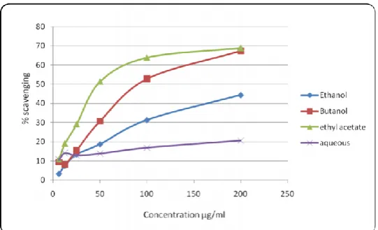
Scavenging of DPPH radicals by different concentrations of various extracts/fractions was assayed. The extracts/fractions showed concentration-dependant.
Effect. The ethyl acetate fraction showed IC50 of 98. 5μg/ml. The ethanol extract and butanol fraction showed IC50 of 213.5 and 126.2 μg/ml (Table 1, 9; Figure 1). Scavenging of ABTS radicals by different concentrations of extracts/fractions was assayed. The extracts/fractions showed concentration-dependant effect. The ethyl acetate and butanol fractions showed scavenging of the ABTS radical with IC50 of 35.5 and 35. 8 μg/ml. The ethanolic extract had IC50 values of 118.7 μg/ml and shown in both table and figures 1-3.
Table 1: Effect of different extracts of W. tinctoria on DPPH free radical scavenging activity.
Concentration (μg/ml) Percent scavenging
Ethanol Ethyl cetate Butanol Aqueous
6.25 3.24 ±1.12 10.8 ± 0.50 9.39 ± 1.95 10.64 ± 0.51 12.5 7.48 ± 0.28 19.0 ± 2.04 8.14 ± 2.15 14.13 ± 1.67 25 13.38 ± 1.42 29.17 ± 1.41 15.46 ±0.65 12.89 ±1.42 50 18.8 ± 1.08 51.45 ± 1.39 30.75 ± 1.45 13.89 ± 0.50 100 31.3 ± 2.46 63.92 ± 1.83 52.78 ± 0.21 16.87 ±1.82 200 44.3 ± 1.46 68.9 ± 0.71 67.41 ± 0.81 20.61 ±1.67
*All values are mean ± SEM of three values. a indicates p<0.05, b indicates p<0.01, c indicates p<0.001when compared to control
Table 2: Effect of different extracts of W.tinctoria on ABTS radical scavenging activity.
Concentration (μg/ml) Percent scavenging
Ethanol Ethyl acetate Butanol Aqueous
6.25 42.12 ± 1.06 45.45 ± 0.26 44.54 ±1.14 37.57 ± 0.54 12.5 41.51 ±1.30 47.87 ± 0.66 47.57 ±0.54 38.63 ± 0.26 25 45.23± 1.13 48.63 ± 0.69 49.24 ±0.66 40.3 ±0.40 50 45.9 ±0.26 53.03 ± 0.54 52.87 ± 1.06 41.81 ± 0.78 100 49.69 ± 1.06 57.42 ± 2.03 58.18 ±1.72 42.87 ± 0.15 200 54.69 ± 1.51 65.30 ± 0.75 67.87 ± 0.54 s45.9 ± 0.52
*All values are mean ± SEM of three values a indicates p<0.05, b indicates p<0.01, c indicates p<0.001when compared to control.
Figure 2: Effect of different extracts of W. tinctoria on ABTS radical scavenging activity.
In vitro anticancer screening
Table 3: In vitro cytotoxic effect of different extracts of W. tinctoria in HeLa (Human epithelial cervical
carcinoma) cells by MTT assay after 48 hours of exposure.
Extracts Concentration (μg/ml)
12.5 25 50 100 200
Control 0.99 ± 0.81 0.99 ± 0.81 0.99 ± 0.81 0.99 ± 0.81 0.99 ± 0.81 Ethanol 36.80 ± 0.32c 37.90 ± 2.54c 38.40 ± 6.90c 54.30 ± 2.12c 81.18± 3.55c Petroleum ether 8.29 ± 2.15 17.60 ± 5.79a 47.0 ± 4.70c 89.20 ± 3.18c 101.50 ± 0.33c Ethyl acetate 22.40 ± 7.60b 32.72 ± 8.90c 48.40 ± 3.59c 82.60 ± 3.59c 99.250.04c *All values are mean + SEM of three samples a indicates p<0.05, b indicates p<0.01, c indicates p<0.001when compared to control
Lane 1 2 3 4 5 6 7
1: Control Lane, 2: DMSO Control Lane, 3: Standard (Methotrexate) Lane, 4: Ethanol Lane, 5: Petroleum ether Lane, 6: Ethyl acetate Lane, 7: Butanol.
In vivo anticancer activity
MTT assay was performed in HeLa cells and after 48 hours of incubation it was found that all the extracts/fractions possessed concentration-dependent cytotoxicity. The ethanolic extract, petroleum ether and ethyl acetate fractions were found to possess 81, 100 and 99 % cytotoxicity respectively at the highest concentration of 200 μg/ml. The IC50 value of the ethanol extract was 91.4 and petroleum ether and ethyl acetate fractions were found to be 53.5 and 54.0 μg/ml respectively (Table 5.7, 5.10; 5.2.2. In vitro cytotoxicity in HeLa and MCF-7 cells by SRB assay.
The extracts/fractions were assessed for DNA fragmentation activity on MCF-7 cells. After 48 hours of incubation, cells were further processed and the isolated DNA was subjected to agarose gel electrophoresis. It was observed that the extracts/fractions at their IC50 values showed ladder-like pattern, whereas such a pattern was not observed in control. The control DNA was found to be intact, indicating that the extracts/fractions were found to be causing cell death by apoptosis and shown in tables 4-20.
Table 4: Effect of different extracts of W. tinctoria on % increase in body weight in EAC inoculated mice.
Group Dose(mg/kg) Day 3 Day 5 Day 7
Normal - 1.13 ± 0.99 2.42 ± 0.98 3.56 ± 1.98c EAC control 0.25 % CMC 9.39 ± 4.78 12.70 ± 2.27 21.70 ± 2.29 Standard 3.5 0.004 ± 1.99 0.89 ± 2.05a 1.21 ± 2.29c Petroleum ether 50 4.71 ± 2.35 5.70 ± 2.13b 13.06 ± 3.44 Petroleum ether 100 2.91 ± 2.81 7.57 ± 2.62 14.61 ± 4.41 Ethyl acetate 50 0.85 ± 1.66 3.51 ± 1.56 9.54 ± 1.17b Ethyl acetate 100 1.99 ± 1.84 5.75 ± 2.78 12.90 ± 5.88 *All values are Mean ± SEM of 8 mice, ap < 0.05, bp < 0.01, cp< 0.001 compared to EAC control
Table 5: Effect of different extracts of W. tinctoria on % increase in body weight in EAC inoculated mice.
Group Dose(mg/kg) Day 9 Day 11 Day 13
Normal - 4.26 ± 2.59c 5.75±2.79c 5.99 ± 2.02c EAC control 0.25 %CMC 25.53 ±2.52 28.82±2.30 32.18 ± 5.31 Standard 3.5 1.37 ± 3.24c 1.76±3.00c 1.73 ± 3.39c Petroleum ether 50 18.57 ±3.30 23.65±2.48 25.65 ± 3.01 Petroleum ether 100 18.64 ±4.41 19.83±5.39 22.91 ± 5.52 Ethyl acetate 50 14.57 ±1.59 15.13±1.22a 18.11 ± 2.42a Ethyl acetate 100 14.23 ±3.40 16.98 ± 3.60a 17.63 ± 4.45b
*All values are Mean ± SEM of 8 mice, a indicates p < 0.05, b indicates p < 0.01, indicates p < 0.001 when compared to EAC controls
Table 6: Effect of different extracts of W. tinctoria on Mean Survival Time in EAC inoculated mice.
Group Dose (mg/kg) MST
EAC control 0.25 % CMC 16.87 ± 0.93 Standard 3.5 39.87 ± 1.23c Petroleum ether 50 23.5 ± 1.21b Petroleum ether 100 20.87 ± 1.41 Ethyl acetate 50 27.75 ± 1.22c Ethyl acetate 100 30.37 ± 1.23c
* All values are mean + SEM of 8 mice, a indicates p<0.05, b indicates p<0.01, c indicates p<0.001when compared to EAC control
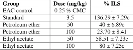
Table 7: Effect of different extracts on of W. tinctoria increase in life span in EAC inoculated mice.
Group Dose (mg/kg) % ILS
EAC control 0.25 % CMC - Standard 3.5 136.29 ± 7.29c Petroleum ether 50 40 ± 6.89c Petroleum ether 100 23.70 ± 8.41 Ethyl acetate 50 58.51 ± 7.23c Ethyl acetate 100 80 ± 7.25c
* All values are mean + SEM of 8 mice, a indicates p<0.05, b indicates p<0.01, c indicates p<0.001 when compared to EAC control.
Table 8: Effect of different extracts of W. tinctoria on WBC in EAC inoculated mice.
Group Dose (mg/kg) WBC (1X10³
cells/mm3)
Table 9: Effect of different extracts of W. tinctoria on RBC changes in EAC inoculated mice.
Group Dose
(mg/kg)
RBC(1x10⁶ cells/mm3)
Sham Control - 9.57 ± 0.49b EAC control 0.25 % CMC 7.41 ± 0.22 Standard 3.5 8.81 ± 0.23b Petroleum ether 50 8.66 ± 0.13 Petroleum ether 100 9.25 ± 0.39b Ethyl acetate 50 8.80 ± 0.19a Ethyl acetate 100 8.98 ± 0.45a *All values are mean + SEM of 3 mice, a indicates p<0.05, b indicates p<0.01, c indicates p<0.001 when compared to EAC control.
Table 10: Effect of different extracts of W. tinctoria on Haemoglobin level in EAC inoculated mice.
Group Dose (mg/kg) Haemoglobin
levels(g%)
Sham Control - 13.50± 0.67b EAC control 0.25 % CMC 9.26 ± 0.12 Standard 3.5 12.30 ± 0.10a Petroleum ether 50 13.41 ± 0.63b Petroleum ether 100 12.87 ± 1.02b Ethyl acetate 50 11.16 ± 0.23 Ethyl acetate 100 11.56 ± 0.45 *All values are mean + SEM of 3 mice, a indicates p<0.05, b indicates p<0.01, c indicates p<0.001when compared to EAC control.
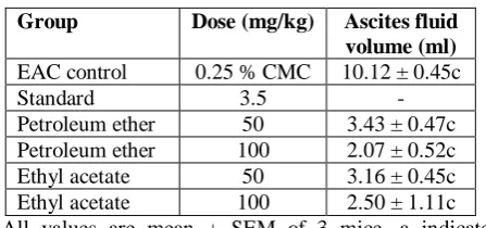
Table 11: Effect of different extracts of W. tinctoria on Ascites fluid volume in EAC inoculated mice.
Group Dose (mg/kg) Ascites fluid
volume (ml)
EAC control 0.25 % CMC 10.12 ± 0.45c Standard 3.5 - Petroleum ether 50 3.43 ± 0.47c Petroleum ether 100 2.07 ± 0.52c Ethyl acetate 50 3.16 ± 0.45c Ethyl acetate 100 2.50 ± 1.11c *All values are mean + SEM of 3 mice, a indicate p<0.05, b indicate p<0.01, c indicate p<0.001 when compared to EAC control.
Table 12: Effect of different extracts of W. tinctoria on Angiogenesis in EAC inoculated mice.
Group Dose (mg/kg) Number of
blood vessels
Sham Control - 17.40 ± 1.76c EAC control 0.25 % CMC 31.70 ± 1.85 Standard 3.5 16.30 ± 1.85c Petroleum ether 50 17.0 ± 1.53c Petroleum ether 100 19.30 ± 0.34c Ethyl acetate 50 14.30 ± 1.85c Ethyl acetate 100 12.60 ± 0.67c
All values are mean + SEM of 3 mice, a indicates p<0.05, b indicates p<0.01, c indicates p<0.001 when compared to EAC control.
Table 13: Effect of different extracts of W. tinctoria on spleen weight of EAC inoculated mice.
Group Dose (mg/kg) Spleen weight (mg)
Sham Control - 92.50 ± 5.78c EAC control 0.25 % CMC 215.90 ± 6.18 Standard 3.5 179.70 ± 9.30c Petroleum ether 50 93.70 ± 4.64c Petroleum ether 100 108.40 ± 5.47c Ethyl acetate 50 131.30 ± 5.39c Ethyl acetate 100 129.60 ± 9.49c *All values are mean + SEM of 3 mice, a indicates p<0.05, b indicates p<0.01, c indicates p<0.001when compared to EAC control.
Table 14: Effect of different extracts of W.tinctoria on AST level in liver of EAC inoculated mice.
Group Dose (mg/kg) AST (U/L)
Sham Control - 155.0 ± 5.19 EAC control 0.25 % CMC 205.0 ± 24.80 Standard 3.5 122.53 ± 32.60 Petroleum ether 50 178.67 ± 28.14 Petroleum ether 100 214.55 ± 10.11 Ethyl acetate 50 336.74 ± 27.50b Ethyl acetate 100 204.50 ± 9.52 *All values are mean + SEM of 3 mice, a indicates p<0.05, b indicates p<0.01, c indicates p<0.001when compared to EAC control.
Table 15: Effect of different extracts of W.tinctoria on ALT level in liver of EAC inoculated mice.
Group Dose (mg/kg) AST (U/L)
Table 16: Effect of different extracts of W.tinctoria on the tumor volume of DLA inoculated mice.
Group Dose
(mg/kg)
Tumor Volume on respective day (cm3)
Day 5 Day 10 Day 15
DLA control 0.25 % CMC 0.84 ± 0.02 0.91 ±0.04 1.26 ± 0.06 Standard(n=8) 3.5 0.24 ± 0.06c 0.30 ±0.06 0.350.07c Ethanol(n=8) 50 0.24 ± 0.05c 0.64 ±0.12 0.72 ± 0.18c Ethanol (n=8) 100 0.52 ± 0.07c 0.68 ±0.11 0.71 ± 0.05c Ethylacetate(n=8) 50 0.29 ± 0.06c 0.42+0.06c 0.32 ± 0.04c Ethylacetate(n=8) 100 0.26 ± 0.04c 0.37±0.09c 0.39 ± 0.09c Petroleumether(n=8) 50 0.25 ± 0.03c 0.36±0.10c 0.58 ± 0.08c Petroleumether(n=7) 100 0.29 ± 0.05c 0.39±0.09c 0.74 ± 0.16c
*All values are mean + SEM of indicated number of mice, a indicates p<0.05, b indicates p<0.01, c indicates p<0.001when compared to DLA control
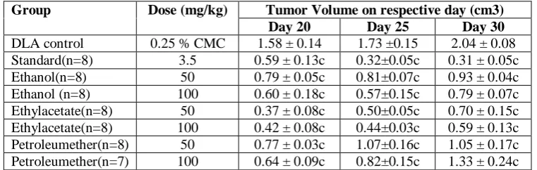
Table 17: Effect of different extracts of W. tinctoria on the tumor volume of DLA inoculated mice.
Group Dose (mg/kg) Tumor Volume on respective day (cm3)
Day 20 Day 25 Day 30
DLA control 0.25 % CMC 1.58 ± 0.14 1.73 ±0.15 2.04 ± 0.08 Standard(n=8) 3.5 0.59 ± 0.13c 0.32±0.05c 0.31 ± 0.05c Ethanol(n=8) 50 0.79 ± 0.05c 0.81±0.07c 0.93 ± 0.04c Ethanol (n=8) 100 0.60 ± 0.18c 0.57±0.15c 0.79 ± 0.07c Ethylacetate(n=8) 50 0.37 ± 0.08c 0.50±0.05c 0.70 ± 0.15c Ethylacetate(n=8) 100 0.42 ± 0.08c 0.44±0.03c 0.59 ± 0.13c Petroleumether(n=8) 50 0.77 ± 0.03c 1.07±0.16c 1.05 ± 0.17c Petroleumether(n=7) 100 0.64 ± 0.09c 0.82±0.15c 1.33 ± 0.24c
*All values are mean + SEM of indicated number of mice, a indicates p<0.05, b indicates p<0.01, c indicates p<0.001when compared to DLA control.
Table 18: Effect of different extracts of W. tinctoria on the percentage inhibition of tumor volume of DLA inoculated mice.
Group Dose
(mg/kg)
% inhibition of tumor
volume
DLA control 0.25 % CMC - Standard(n=8) 3.5 84.54 ± 2.58c Ethanol(n=8) 50 54.40 ± 2.23c Ethanol (n=8) 100 61.15 ± 3.79c Ethylacetate(n=8) 50 65.67 ± 7.50c Ethylacetate(n=8) 100 71.10 ± 6.81c Petroleumether(n=8) 50 38.11 ± 6.48c Petroleumether(n=7) 100 37.20 ± 10.70c *All values are mean + SEM of indicated number of mice, a indicates p<0.05, b indicates p<0.01, c indicates p<0.001when compared to DLA control.
Table 19: Effect of different extracts of W.tinctoria on the tumor weight of DLA inoculated mice.
Group Dose
(mg/kg)
Tumor weight (mg)
DLA control 0.25 % CMC
2.71 ± 0.09
Standard(n=8) 3.5 1.15 ± 0.05c Ethanol(n=8) 50 1.73 ± 0.06c Ethanol (n=8) 100 1.41 ± 0.02c Ethylacetate(n=8) 50 1.62 ± 0.14c Ethylacetate(n=8) 100 1.32 ± 0.04c Petroleumether(n=8) 50 1.67 ± 0.15c Petroleumether(n=7) 100 1.79 ± 0.15c *All values are mean + SEM of indicated number of mice, a indicates p<0.05, b indicates p<0.01, c indicates p<0.001 when compared to DLA control.
Table 20: Effect of different extracts of W.tinctoria on the % inhibition of tumor weight of DLA inoculated mice.
Group Dose
(mg/kg)
Tumor weight (mg)
*All values are mean + SEM of indicated number of mice, a indicates p<0.05, b indicates p<0.01, c indicates p<0.001 when compared to DLA control.
CONCLUSION
It can be concluded that the ethanolic extract of wrightia tinctoria barks exhibited signs of anticancer and antioxidant activity of tumor inoculated EAC mice decrease in the values of biochemical parameters and antioxidant activity. In the present study, the ethanolic extracts of wrightia tinctoria significantly reduced the elevated levels of angiogenesis, ascites fluid volume; RBC indicates inhibitory local effect on liquid tumor and the formation of new blood vessels. Hence, at this point it is concluded that the ethanolic extract of wrightia tinctoria possess anticancer activity. In case of antioxidant treated groups there will be decreasing DPPH scavenging enzyme activities such as inhibited the oxidant activity of enzymes and possibly could reduce the generation of free radicals and cancer. Finally, based on improvement biochemical parameters, antioxidant activity studies it is concluded that the ethanolic extracts of wrightia tinctoria bark part possess anti-cancer and antioxidant activity and thus supports the traditional application of the same under the light of modern science.
REFERENCES
1. Balachandran. P and Govindarajan.R; Cancer-an ayurvedic perspective; Pharmacol res, 2005; 51: 19-30.
2. Toh. C.K and Lim. W. T; Lung cancer in never-smokers; j clin pathol, 2007; 60: 337- 40.
3. Huang. S. Y, Yao. M, Tang. J. L, Lee. W. C, Tsay. W, Cheng. A. L, Wang. C. H, Chen. Y. C, Shen. M. C and Tien. H. F; epidemiology of multiple myeloma in taiwan: increasing incidence for the past 25 years and higher prevalence of extra medullary myeloma in patients younger than 55 years; cancer, 110; 896-905.
4. Edward. J. K. P, G. Mathews, J.Patterson, R. Ramkumar, D. Wilhelmsson, J. Tamelander and O. Linden; Status of coral reefs of the Gulf of Mannar, Southeastern India; In: Ten years after bleaching -facing the consequences of climate change in the Indian Ocean. Oceans Research and Development in the Indian Ocean (CORDIO) Status Report (edsD.O. Obura, J.Tamelander and Mombasa, 2008; 45-60. 5. Sinha. R, Anderson. D. E, Mcdonald. S. S and
Greenwald. P; Cancer risk and diet in India; j postgrad med, 2003; 49; 222-8.
6. Bobba. R, K. Y; Cancer in India – an overview; Journal for the medical, pharmaceutical and biotechnology industries, 2008; 5: 4-10.
7. Parkin. D. M, Pisani. P and Ferla. Y.J; global cancer statistics; ca cancer j clin, 1999; 49: 33-64.
8. L. A. Yarbro and R. Barber; Relationship of sediment sulfide to mortality of Thalassia
testudinum in Florida Bay. Bull. Mar. Sci, 1994; 54: 733–746.
9. Mukherjee. A. K, Basu.s, Sarkar.N and Ghosh. A. C; Advances in cancer therapy with plant based natural products; curr med chem., 2001; 8: 1467-86. 10. Newman. D. J and Cragg.G. M; Natural products as
sources of new drugs over the last 25 years; J nat prod, 2007; 70: 461-77.
11. Borek. C; dietary antioxidants and human cancer; Integr cancer ther, 2004; 3: 333- 41. 12.
12. Khyade. M. S and Vaikos. N. P; Pharmacological and Phyto-s standardization of leaves of wrightia tinctoria R.Br. ijprd, 2009.
13. Bigoniya. P, Shukla. A. Agrawal. G. P and Rana A. C; Pharmacological screening of wrigtia tinctoria bark hydro-alcoholic extract; Asian J. Exp. Sci, 2008; 22(3): 235- 244.
14. Sreejayan. N and Rao. M. N; Free radical scavenging activity of curcuminoids. Arzneimittelforschung, 1996; 46: 169-71.
15. Vaijanathappa. J and badami. S; Antiedematogenic and free radical scavenging activity of swertiamarin isolated from enicostemma axillare; planta med, 2009; 75: 12-17.
16. Mosmann. T, Rapid colorimetric assay for cellular growth and survival: application to proliferation and cytotoxicity assays; J Immunol methods, 1983; 65: 55-63.
17. Singh. N. P, Mccoy. M. T, Tice. R. R and Schneider. E. L; A simple technique for quantitation of low levels of dna damage in individual cells. exp. Cellres, 1998; 179: 184-191.
18. Gude. R. P, Binda. M. M, Boquete. A. L and bonfil. R. D; Inhibition of endothelial cell proliferation and tumor-induced angiogenesis by pentoxifylline; j cancer res clin oncol, 2001; 127: 625-30.
19. Leyon. P. Vand Kuttan. G; effect of tinospora cordifolia on the cytokine profile of angiogenesis-induced animals. int immunopharmacol, 2004; 4: 1569-75.