www.fm.viamedica.pl
Address for correspondence: Franciszek Burdan MD, PhD, Human Anatomy Department, Medical University School of Lublin, ul. Spokojna 1, 20–074 Lublin, Poland, tel: +48 81 532 21 18, fax: +48 81 532 89 03, e-mail: [email protected]
Visualisation of diverticula of the upper part
of the alimentary tract; comparison
of roentgenologic and endoscopic techniques
Franciszek Burdan
1, 2, Ingrid Różyło-Kalinowska
2, Wit Juśkiewicz
1, 3,
Ryszard Maciejewski
1, 3, Grzegorz Wallner
3, Witold Zgodziński
3,
Krzysztof Zinkiewicz
3, Janusz Złomaniec
2, Andrzej Dąbrowski
31Department of Human Anatomy, Medical University of Lublin, Poland 22nd Department of Radiology, Medical University of Lublin, Poland 32nd Department of General Surgery, Medical University of Lublin, Poland [Received 12 September 2001; Revised 20 October 2001; Accepted 24 October 2001]
Diverticula of the upper part of the alimentary tract, irrespective of their etiolo-gy, are frequently observed benign changes of the pharynx, oesophagus, sto-mach and duodenum. In the present work, patients of the II General Surgery Department of the Medical University of Lublin, with radiologically or endosco-pically proved diverticula of the upper part of the alimentary tract, were exa-mined. The presence of diverticula of such localisation was an indication for supplementary endoscopic or radiological examination. The localisation, size, diameter of the opening, mucosal relief of diverticula and its contiguity were checked and analysed. Our data suggest that both medical procedures are com-plementary to each other. All previously observed changes in diverticula of the thoracic part of the oesophagus and the infradiaphragm part of the alimentary tract were fully proved. The radiological examination gave a better view of Zen-ker’s diverticulum, especially in short and obese patients. Sampling and better visualisation of the diverticula opening testify to the unquestionable superiority of endoscopy. However, precise evaluation by radiological process fully completes the diagnostic protocol. Both diagnostic procedures are usually supplemented by manometric examination of the oesophagus and superior and inferior oesopha-geal sphincters. This enables the accurate diverticula etiology to be stated.
key words: diverticula, alimentary tract, pharynx, oesophagus, stomach, duodenum, radiology, endoscopy
INTRODUCTION
Diverticula of the upper part of the alimentary tract are frequently observed benign changes of the pha-rynx, oesophagus, stomach and duodenum. Most of them are acquired and develop at the age of 40 years or later. Congenital and early postnatal diver-ticula are rare but also seen in children and
adoles-cents. Some of the investigators do not distinguish the infantile type of diverticula and classify them as congenital ones [1, 10, 15, 29, 37].
true type formed by herniation of all layers of the alimentary tract wall through muscular defect or in places where arteries enter the wall. The pseudodi-verticula are only acquired and formed by mucosal and submucosal layers with some scattered muscle fibres herniated through the muscular fasciculi of the muscular layer of the wall. However, both types can co-exist in one patient. In such cases the pseudodi-verticula are usually smaller and surround the true ones, which are more prominent [10, 15, 37].
Diverticula, due to the different pathogenesis, are also divided into pulsion-type and the less frequent traction-type [10, 15, 31].
Anatomical classification divides diverticula de-pending on their localisation and number. In the pharynx, Zenker’s and the lower situated Killian--Jamieson’s diverticula are observed. The oesophageal diverticula are seen mostly in the middle (midthorac-ic divert(midthorac-icula) and distal third of the oesophagus (epi-phrenic diverticula). The postero-medial wall of the stomach, near the oesophagogastric junction, is the most common localisation of gastric diverticula. Un-like other localisations, duodenal diverticula occur more often as multiple lesions than single ones, lo-cated in the vicinity of the great duodenal papilla (pa-pilla of Vater) followed by transversal and ascending part of the duodenum [1, 10, 15, 20, 22, 29, 37].
The clinical symptoms of diverticula depend on their localisation, number, size and other concomi-tant diseases. There are no typical manifestations of diverticula. Usually they simulate signs of other dis-eases, e.g. symptoms of cholecystitis or pancreatitis are characteristic for duodenal diverticula, while dys-phagia, regurgitation and pain are common com-plaints in oesophageal ones. Single, especially small, lesions may be asymptomatic and such diverticula usually are an incidental finding. However, like the bigger ones, they can also produce different com-plications such as haemorrhage, perforation, ulce-ration and neoplastic transformation [10, 15, 22, 29]. The aim of our study was to compare the useful-ness and repeatibility of the conventional contrast fluoroscopy and endoscopic examinations. Both dia-gnostic methods are commonly and routinely used in medical procedures to examine the upper part of the alimentary tract. However, there are only limited data regarding their effectiveness when both me-thods are performed.
MATERIAL AND METHODS
In the presented study, data obtained from patients of the 2nd General Surgery Department of the
Med-ical University of Lublin, hospitalised in 1998–2000, were evaluated. Radiologically or endoscopically proved diverticula of the upper part of alimentary tract were the criterion that classified a patient to the study group. The presence of diverticula of such localisation was an indication for supplementary endoscopic or radiological examination. Concomi-tant serious disease of the upper alimentary tract, such as neoplasm located directly to diverticula, and poor general condition excluded a patient from the studied group.
The radiological examination was performed us-ing the Siemens Siregraph CF-System (Erlangen, Ger-many) during single or double contrast fluoroscopy. Barium Sulfuricum suspension (TERPOL Przedsię-biorstwo Farmaceutyczne S.A., Poland) or Pronto-bario HD (Bracco S.P.A., Italy) were used as a posi-tive contrast. Stomach air or Duogas (Bracco S.P.A., Italy), administered before examination, were used as a negative contrast. Each radiological examina-tion was started in Trendelenburg posiexamina-tion, followed by supine, semirecumbent, AP and lateral erect po-sitions. In patients with pharyngeal diverticula ex-amination in low inclination was also performed. Any other positions such as oblique at different angles were used when needed. Single and serial pictures with acquisition time 1 up to 4 pictures per 1 s were stored on hard disc and scanned on X-ray film.
Endoscopic examination was performed using the high-resolution Olympus video endoscope GIF-Q140. The Reversor EVIS CV-140 was used as a light source. No pre-liminary patient sedation was performed. Size of diver-ticula was determined endoscopically by comparison with the known size of the cup of the biopsy forceps.
Through the entire study period the same team of experienced radiologists and endoscopists performed both examination procedures. In most cases, radio-logical study preceded endoscopy, precluding knowl-edge of the endoscopic results by the radiologist.
The localisation, size, diameter of the opening, mucosal relief of diverticula and their contiguity were checked and analysed. Assessment of diverticula size was based on direct measurement when the diver-ticulum was visible radiologically or on endoscopic estimations when the diverticulum could not be iden-tified radiographically.
RESULTS
pyloric strictures, poor general state and lack of pa-tient’s consent were the most common reason for abandoning the endoscopic examination.
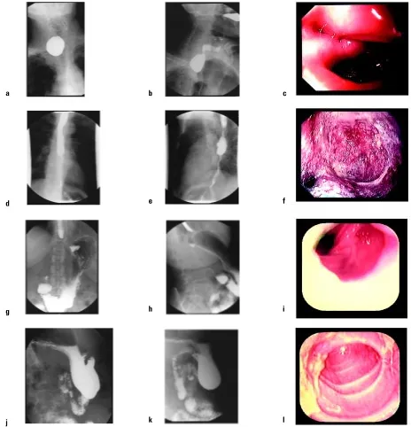
There were 22 cases of pharyngeal diverticula. Most of them morphologically and topographically corresponded to Zenker’s diverticula (Fig. 1a–c). In two cases diverticula bigger then 6 cm in longitudi-nal diameter were observed. Small right side Killian-Jamieson’s diverticulum was seen only in one case.
Radiological examination supplied a better view of Zenker’s diverticula in short and stocky patients due to difficulty with their endoscopic visualisation.
Oesophageal diverticula were observed in mid-dle and lower third in 2 and 4 cases, respectively (Fig. 1d–f). All of them were located on the level of or lower than tracheal bifurcation. There was one case of giant diverticulum arising from lower third of anterior oesophageal wall.
Figure 1. AP — anterior-posterior (a), lateral (b) radiological view of Zenker’s diverticula and its endoscopic picture (c); AP (d), lateral (e) radiological view of oesophageal diverticula and its endoscopic picture (f); AP (g), lateral (h) radiological view of stomach diverticula and its endoscopic picture (i); AP (j), lateral (k) radiological view of multiple duodenal diverticula. Endoscopic picture of upper duodenal diver-ticula (l) presented in the previous two pictures.
a
d
b c
e f
g h i
Four gastric diverticula were seen in superior half of the medial stomach curvature or close to it (Fig. 1g–i). Their size was similar to each other.
There were 9 cases of single and 4 multiple duode-nal diverticula in the examined group. The single ones were seen in bulb and descending part of duode-num (Fig. 1j–l). Four out of 6 bulbal diverticula were located on anterior wall. The remaining 2 arose from the posterior one. Diverticula of descending part of duodenum were located close to great duodenal papilla. Endoscopic retrograde cholangiopancreatog-raphy (ERCP) performed during same examination showed obstruction on the distal end of the com-mon bile duct. Multiple diverticula were observed in the descending part or distally, and were absent in duodenal bulb.
Statistical analysis showed that there were no sig-nificant differences between diverticula diameters measured during roentgenologic and endoscopic examination. All endoscopically observed changes in diverticula of thoracic oesophagus and infradia-phragmatic part of the alimentary tract were fully confirmed during radiological examinations.
DISCUSSION
Our data suggest that both medical procedures are complementary. However, the radiological examina-tion was better tolerated by patients, and gave a bet-ter view of pharyngeal diverticula. It is also the first, sometimes the only, choice for examination of the upper part of the alimentary tract in patients with strictures of oesophagus or at different levels [10, 15]. However, sampling and better visualisation of the diverticula opening testify to the unquestion-able superiority of endoscopy, but precise radiolo-gical evaluation completes the diagnostic protocol [5, 10, 11, 14, 15].
Most of the investigators choose contrast fluo-roscopy as the prime diagnostic tool, due to the fact that endoscopy adds little to the evaluation of diver-ticulum but may be indicated in the assessment of other oesophageal, gastric and duodenal abnorma-lities [5, 23]. Endoscopy seems to be necessary in patients with duodenal diverticula, especially those located in descending part of duodenum, in the vi-cinity of the major duodenal papilla or encompass-ing it, which may not be evident in X-ray study [5, 20, 22, 27, 33]. Such localisation can cause forma-tion of gallbladder stones, primary choledocholithi-asis and onset of chronic pancreatitis secondary to increasing ductal pressure by chronic obstruction of the distal opening of the hepatopancreatic duct [3,
8, 19, 20, 26, 27, 30, 33, 36]. Endoscopy should also be the initial investigation in cases of upper gas-trointestinal bleeding and perforations that compli-cate diverticula [16, 18].
The authors were unable to find any paper re-garding comparison of roentgenologic and endo-scopic methods in examination of the upper part alimentary tract diverticula. Upper gastrointestinal endoscopy as a gold standard in oesophageal, gas-tric and duodenal examinations showed its supe-riority in gastric and duodenal ulcer diagnosis. The radiological sensitivity is lower and depended on size of lesion and used method. Ott et al. [28] showed that only 56% of ulcers under 5 mm of diameter, and 88% of larger ones, could be detected with sin-gle-contrast method. Using double-contrast method they detected 45% of small ulcers and 78% of those larger ones. However, statistical analysis showed equal effectiveness of single- and double-contrast radiography. The superiority of endoscopy in ulcer detection was proved in other studies [4–7, 9, 11– –14, 17, 21, 23–25, 32, 34] The same results were also seen by investigators evaluating the effective-ness of endoscopic and radiological procedures in cases of gastritis, benign and malignant neoplasms [5, 11, 14, 18, 24]. However, large lesions, especially the ones of diameter smaller than the internal diam-eter of alimentary tract, are clearly visible in endo-scopic as well as radiological examination. The high, equal effectiveness of radiological examinations ob-served in our study seems to be secondary to the large size of the diagnosed diverticula. Other stud-ies, in which the effectiveness of both methods in diagnosis of smaller diverticula will be evaluated, are needed to complete our observations.
At present patient preferences are also impor-tant factors during the entire diagnostic process [7, 35]. Previous study showed that in the group of pa-tients who underwent both investigations, more patients, especially older ones, had preference for endoscopy preceded by pharmaceutical sedation [35]. When sedation was not routinely used, a smaller number of patients expressed a preference for up-per gastrointestinal endoscopy, which was also seen in our study.
REFERENCES
1. Afridi SA, Fichtenbaum CJ, Taubin H (1991) Review of duodenal diverticula. Am J Gastroenterol, 86: 935–938. 2. Baker ME, Zuccaro G, Achkar E, Rice TW (1999) Esopha-geal diverticula: patient assessment. Semin Thorac Cardiovasc Surg, 11: 326–336.
3. Chady G, Hart WJ, Roberts-Thomson IC (1997) An ana-lysis of the relationship between bile duct stones and periampullary duodenal diverticula. J Gastroenterol Hepatol, 12: 29–33.
4. Colin-Janes DG (1986) Endoscopy or radiology of up-per gastrointestinal symptoms? Lancet, 1: 1022.
5. Davenport PM, Morgan AG, Darnborgouh A, de
Dom-bal FT (1985) Can preliminary screening of dyspeptic patients allow more effective investigational tech-niques? BMJ, 290: 217–220.
6. Dooley CP, Larson AW, Stace NH, Renner IG, Valenzue-la JE, Eliasoph J, Colletti PM, Halls JM, Weiner JM (1984) Double-contrast barium meal and upper gastrointes-tinal endoscopy, a comparative study. Ann Intern Med, 101, 538–545.
7. Dooley CP, Weiner JM, Larson AW (1986) Endoscopy or ra-diography? The patient’s choice. Am J Med, 80: 203–207. 8. Egawa N, Kamisawa T, Tu Y, Sakaki N, Tsuruta K, Oka-moto A (1998) The role of juxtapopillary duodenal di-verticulum in the formation of gallbladder stones. Hepatogastroenterology, 45: 917–920.
9. Forrest JAH, Logan RAF (1977) Comparative diagnostic accuracy of barium meal and endoscopy. BMJ, 1: 50. 10. Freeny PC, Stevenson GW (eds.). (1994) Margulis and
Burhenne’s alimentary tract radiology — 5th edition.
Mos-by, St. Louis.
11. Gelfand DW, Chen YM, Ott DJ (1987) Multiphasic exa-minations of the stomach: efficacy of individual tech-niques and combinations of techtech-niques in detecting 1533 lesions. Radiology, 162: 829–834.
12. Gelfand DW, Dale WJ, Ott DJ (1984) The location and size of gastric ulcers: radiologic and endoscopic eva-luation. AJR, 143: 755–758.
13. Gelfand DW, Ott DJ (1981) Single- vs double-contrast gastrointestinal studies: critical analysis of reported statistics. AJR 137, 523–528.
14. Gelfand DW, Ott DJ, Munitz HA, Chen YM (1984) Ra-diology and endoscopy — a radiologic view-point. Ann Intern Med, 101: 550–552.
15. Halpert R.D., Feczko P.J.: Gastrointestinal Radiology. The Requisites. Second edition. 1999, Mosby, St. Louis. (Wydanie polskie: Różyło T.K. red. (Wydawnictwo Czelej, Lublin 2000.)
16. Hamada N, Ishizaki N, Shirahama K, Nakamura N, Murata R, Kadono J, Shimazaki T, Sameshima T, Mi-sono T, Taira A (2000) Multiple duodeno-jejunal diver-ticula causing massive intestinal bleeding. J Gastroen-terol, 35: 163–167.
17. Herlinger H, Glanville JN, Kree L (1977) An evaluation of the double contrast barium meal (DCBM) against endoscopy. Clin Radiol, 28: 307–314.
18. Hoare AM (1975) Comparative study between endos-copy and radiology in acute upper gastrointestinal haemorrhage. BMJ, 1: 27–30.
19. Huang FC, Chuang JH, Ko SF (1998) Intraluminal duodenal diverticulum in the formation of gallblad-der stones. J Pediatr Gastroenterol Nutr, 27: 593–595.
20. Kim MH, Myung SJ, Seo DW, Lee SK, Kim YS, Lee MH, Yoo BM, Min MI (1998) Association of periampullary diverticula with primary choledocholithiasis but not with secondary choledocholithiasis. Endoscopy, 30: 601–604. 21. Kreel L, Herlinger H, Glanville J (1973) Technique of the double contrast barium meal with examples of correlation with endoscopy. Clin Radiol, 24: 307–314. 22. Leivonen MK, Halttunen JA, Kivilaakso EO (1996) Duodenal diverticulum at endoscopic retrograde cho-langiopancreatography, analysis of 123 patients. Hepatogastroenterol, 43: 961–966.
23. Martin DF (1991) Useful collaboration between endosco-py and barium radiology. Br J Hosp Med, 45: 338–341. 24. Martin TR, Vennes JA, Silvis SE, Ansel HJ (1980) A
com-parison of upper gastrointestinal endoscopy and radio-graphy. J Clin Gastroenterol, 2: 21–25.
25. Montagne JP, Moss AA, Margulis AR (1978) Double--blind study of single and double contrast upper gas-trointestinal examinations using endoscopy as a con-trol. AJR, 130: 1041–1045.
26. Myung SJ, Kim MH, Seo DW, Lee SK (1998) Recurrent com-mon bile duct stones after endoscopic retrograde cholan-gioscopic stone removal: is there a role for associated pe-riampullary diverticulum? Endoscopy, 30: 871–872. 27. Novacek G, Walgram M, Bauer P, Schofl R, Gangl A,
Potzi R (1997) The relationship between juxtapapillary duodenal diverticula and biliary stone disease. Eur J Gastroenterol Hepatol, 9: 375–379.
28. Ott DJ, Gelfand DW, Wu WC (1982) Detection of gastric ulcer: comparison of single– and double-contrast exa-minations using endoscopy as a control. AJR, 139: 93–97. 29. Rice TW, Baker ME (1999) Midthoracic esophageal di-verticula. Semin Thorac Cardiovasc Surg, 11: 352–357. 30. Sandstad O, Osnes T, Skar V, Urdal P, Osnes M (2000) Structure and composition of common bile duct stones in relation to duodenal diverticula, gastric resection, cholecystectomy and infection. Digestion, 61: 181–188. 31. Schima W, Chober E, Stacher G, Franz P, Uranitsch K, Pokieser P, Wenzl E, Resch A, Herold CJ (1997) Asso-ciation of midoesophageal diverticula with oeso-phageal motor disorders. Videofluoroscopy and ma-nometry. Acta Radiol, 38: 108–114.
32. Stanley TV, Cocking JB (1978) Upper gastro-intestinal endoscopy and radiology in the elderly. Postgrad Med J, 54: 257–260.
33. Starkov IG, Strekalovskii VP, Vishnevskii VA, Grigorian RS (2000) Diverticuli of duodenal papilla region and their role in development of choledocholithiasis and strictures of bile and pancreatic ducts. Khirurgiia (Moscow), 3: 10–13. 34. Stevenson GW, Cox RR, Roberts CJC (1976) Prospec-tive comparison of double-contrast barium meal exa-mination and fibre-optic endoscopy in acute upper gastrointestinal haemorrhage. BMJ, 2: 723–724. 35. Stevenson GW, Norman G, Frost R, Somers S (1991)
Barium meal or endoscopy? A prospective randomized study of patient preference and physician decision making. Clin Radiol, 44: 317–321.