R E S E A R C H
Open Access
Age-related transcriptome changes in
Sox2+ supporting cells in the mouse
cochlea
Cheng Cheng
1,2†, Yunfeng Wang
3,4†, Luo Guo
4†, Xiaoling Lu
4†, Weijie Zhu
5, Waqas Muhammad
5,6, Liyan Zhang
5,
Ling Lu
1, Junyan Gao
7, Mingliang Tang
5, Fangyi Chen
8, Xia Gao
1*, Huawei Li
4*and Renjie Chai
4,5,9,10,11*Abstract
Background:Inner ear supporting cells (SCs) in the neonatal mouse cochlea are a potential source for hair cell (HC) regeneration, but several studies have shown that the regeneration ability of SCs decreases dramatically as mice age and that lost HCs cannot be regenerated in adult mice. To better understand how SCs might be better used to regenerate HCs, it is important to understand how the gene expression profile changes in SCs at different ages. Methods:Here, we usedSox2GFP/+mice to isolate the Sox2+ SCs at postnatal day (P)3, P7, P14, and P30 via flow cytometry. Next, we used RNA-seq to determine the transcriptome expression profiles of P3, P7, P14, and P30 SCs. To further analyze the relationships between these age-related and differentially expressed genes in Sox2+ SCs, we performed gene ontology (GO) analysis.
Results:Consistent with previous reports, we also found that the proliferation and HC regeneration ability of isolated Sox2+ SCs significantly decreased as mice aged. We identified numerous genes that are enriched and differentially expressed in Sox2+ SCs at four different postnatal ages, including cell cycle genes, signaling pathway genes, and transcription factors that might be involved in regulating the proliferation and HC differentiation ability of SCs. We thus present a set of genes that might regulate the proliferation and HC regeneration ability of SCs, and these might serve as potential new therapeutic targets for HC regeneration.
Conclusions:In our research, we found several genes that might play an important role in regulating the proliferation and HC regeneration ability of SCs. These datasets are expected to serve as a resource to provide potential new therapeutic targets for regulating the ability of SCs to regenerate HCs in postnatal mammals.
Keywords:RNA-seq, Proliferation, Differentiation, Sphere formation, Gene expression
© The Author(s). 2019Open AccessThis article is distributed under the terms of the Creative Commons Attribution 4.0 International License (http://creativecommons.org/licenses/by/4.0/), which permits unrestricted use, distribution, and reproduction in any medium, provided you give appropriate credit to the original author(s) and the source, provide a link to the Creative Commons license, and indicate if changes were made. The Creative Commons Public Domain Dedication waiver (http://creativecommons.org/publicdomain/zero/1.0/) applies to the data made available in this article, unless otherwise stated. * Correspondence:[email protected];[email protected];
†Cheng Cheng, Yunfeng Wang, Luo Guo and Xiaoling Lu contributed equally
to this work.
1Jiangsu Provincial Key Medical Discipline (Laboratory), Department of
Otolaryngology Head and Neck Surgery, Affiliated Drum Tower Hospital of Nanjing University Medical School, No. 321 Zhongshan Road, Nanjing 210008, China
4ENT Institute and Otorhinolaryngology Department of Affiliated Eye and
ENT Hospital, Key Laboratory of Hearing Medicine of NHFPC, Shanghai Engineering Research Centre of Cochlear Implant, State Key Laboratory of Medical Neurobiology, Fudan University, Room 611, Building 9, No. 83, Fenyang Road, Xuhui District, Shanghai 200031, China
Full list of author information is available at the end of the article
Introduction
Hair cells (HCs) in the inner ear play a critical role in converting mechanical sound waves into neural signals for hearing and play a critical role in maintaining
bal-ance [1, 2]. Multiple studies have reported that HCs in
non-mammalian vertebrates can be regenerated in both the auditory and vestibular systems after HC loss and thus lead to the complete recovery of hearing and
bal-ance function [3,4]. Conversely, HCs in the mammalian
cochlea can be spontaneously regenerated after damage only to a very limited extent and only in the neonatal cochlea and cannot be regenerated at all in adult ani-mals, and thus in adult mamani-mals, HC damage causes
permanent hearing loss [1, 4–10]. Finding a way to
re-generate HCs in mammals could possibly represent a cure for sensorineural hearing loss, which still has no treatment options other than prosthetic devices.
In the mouse organ of Corti, HCs and supporting cells (SCs) emerge from the same inner ear prosensory cells. These inner ear prosensory cells start to exit the cell cycle from the apical turn to the basal turn of the coch-lea. The apical prosensory cells exit the cell cycle at around embryonic day 12.5 (E12.5), and the basal pro-sensory cells exit the cell cycle at around E14.5. The inner ear prosensory cells start to differentiate into HCs and SCs beginning at the mid-base of the cochlea at around E13.5 and reaching the rest of the base and up
to the apex of the cochlea over the next few days [11].
SCs in the mouse inner ear have also been shown to be a reliable source for regenerating HCs after damage in vitro either through mitotic or direct differentiation
[10, 12–15]. Recent studies have demonstrated that the
SCs isolated from the neonatal mouse cochlea are
com-petent to form new HCs in culture [10, 16–18], but the
ability of SCs to form spheres in suspension cultures de-creases about 100-fold during the second and third
post-natal weeks [19]. In contrast, the adult mammalian
cochlea has almost no HC regeneration capacity, and at-tempts to stimulate the dormant regenerative capacity
have met with very limited success [15,20]. Multiple
fac-tors have been reported to be involved in regulating the process by which SCs regenerate HCs, including factors in the Wnt, Notch, Hedgehog, and STAT3 signaling
pathways [10, 21–24]. HC regeneration strategies have
only worked at all in the neonatal mouse cochlea, and none has been able to overcome the age barrier in the adult cochlea. An obvious limitation to these previous strategies has been a lack of understanding of the age-related changes in gene expression profiles, and possible age-related genes regulating the proliferation and HC re-generation ability of SCs have not been identified.
Sox2 is a universal stem cell marker, and it is also expressed in neural progenitor cells at different stages of
central nervous system development [25]. In the neonatal
mouse inner ear, Sox2 labels the SCs that have been shown to be a reliable source for regenerating HCs after damage. In this study, we performed RNA-seq profiling of
the Sox2+ SCs isolated fromSox2GFP/+transgenic mice at
four different postnatal time points and determined the age-related differential expression of genes that might be involved in regulating the proliferation and HC differenti-ation ability of Sox2+ SCs. The Sox2+ SCs we sorted
in-cluded Hensen’s cells, Deiters’ cells, pillar cells, inner
phalangeal cells, and the cells in the greater epithelium ridge. To further analyze the role of these age-related
dif-ferentially expressed genes, we constructed a protein–
pro-tein interaction network using STRING (Search Tool for the Retrieval of Interacting Genes/Proteins). These data-sets are expected to serve as a resource to provide poten-tial new therapeutic targets for regulating the ability of SCs to regenerate HCs in postnatal mammals.
Materials and methods Mice and genotyping
Sox2GFP/+mice were obtained from the Jackson Labora-tory (stock no. 17592). Transgenic mice were genotyped
using genomic DNA from tail tips by adding 180μl 50
mM NaOH, incubating at 98 °C for 1 h, and then adding
20μl 1 M Tris-HCl to neutralize the base. The
genotyp-ing primers were as follows: GFP forward: 5′-CAC ATG
AAG CAG CAC GAC TT-3′; GFP reverse: 5′-TGC
TCA GGT AGT GGT TGT CG-3′.
The cochleae were harvested at P3, P7, P14, and P30. All applicable international, national, and/or institutional guidelines for the care and use of animals were followed. All animal procedures were performed according to pro-tocols approved by the Animal Care and Use Committee of Southeast University and were consistent with the Na-tional Institutes of Health Guide for the Care and Use of Laboratory Animals. All efforts were made to minimize the number of animals used and to prevent their suffering.
Immunofluorescence
samples were washed again three times with 1× PBST and then mounted on slides in a mounting medium (DAKO, S3023). Cells were imaged with an LSM700 confocal microscope. The antibodies used in this study were anti-myosin7a (Proteus Bioscience, #25-6790, 1: 1000 dilution), anti-sox2 (Santa Cruz, #sc-17320, 1:500 dilution), Alexa Fluor 647 donkey anti-goat IgG (Invitro-gen, A-21447, 1:500 dilution), and Alexa Fluor 555 don-key anti-rabbit IgG (Invitrogen, A-31572, 1:500 dilution).
Flow cytometry
The cochleae were dissected in cold 1× HBSS (Gibco)
and transferred to 50μl 1× PBS in 1.5-ml Eppendorf
tubes. A total of 50μl 0.25% trypsin-EDTA (Invitrogen;
#25200-056) was added to the tubes, and these were
in-cubated for 8–12 min at 37 °C. The digestion was
stopped by the addition of 50μl trypsin inhibitor
(Worthington Biochem, #LS003570), and 200-μl
(Eppen-dorf, #22491245) and 1000-μl (Eppendorf, #22491253)
blunt pipette tips were used to triturate the tissues into single cell suspensions. The cells were filtered through a
40-μm strainer (BD Biosciences, 21008-949) to eliminate
clumps, and the GFP+ cells were sorted on a BD FACS Aria III flow cytometer (BD Biosciences).
Sphere-forming assay and differentiation assay
For the sphere-forming assay, the flow-sorted Sox2+ SCs
were cultured at a density of 2 cells/μl in Costar
ultra-low attachment dishes (Costar, 3473) for 5 days in DMEM/F12 (Gibco, 11330-032), 2% B27 (Invitrogen, 17504-044), 1% N2 (Invitrogen, 17502-048), IGF (50 ng/ ml, Sigma, I8779), EGF (20 ng/ml; Sigma, E9644), b-FGF (10 ng/ml, Sigma, F0291), heparan sulfate (20 ng/ml, Sigma, H4777), and 0.1% ampicillin (Sigma, A9518-5G). For the differentiation assay, we used both flow-sorted GFP+ SCs and spheres from the sphere-forming assay. In the cell-differentiation assay, the flow-sorted Sox2+
SCs were cultured at a density of 50 cells/μl on
laminin-coated four-well dishes for 10 days in DMEM/F12, 1% N2, 2% B27, EGF (20 ng/ml; Sigma, E9644), IGF (50 ng/ ml, Sigma, I8779), heparan sulfate (20 ng/ml, Sigma, H4777), b-FGF (10 ng/ml, Sigma, F0291), and 0.1% ampicillin. In the sphere-differentiation assay, the first-generation spheres were seeded on laminin-coated four-well dishes and cultured for 10 days in DMEM/F12 medium with 1% N2, 2% B27, and 0.1% ampicillin.
RNA extraction for RNA-seq analysis
Approximately 5000 GFP+ SCs were isolated by FACS and split into three fractions for separate replicates. RNA-seq libraries of FACS-purified cells were generated using the SMART-Seq v4 Ultra Low Input RNA Kit for Sequen-cing and the Illumina mRNA-Seq Sample Prep Kit. FACS-purified cells were suspended in 10× lysis buffer.
First-strand and second-First-strand cDNA synthesis, adaptor ligation, and PCR amplification were performed using the Illumina mRNA-Seq Sample Prep Kit. SPRI beads (Ampure XP, Beckman) were used in each purification step after RNA fragmentation for size selection. All librar-ies were analyzed for quality and concentration using an Agilent Bioanalyzer. Sequencing was performed using the Illumina HiSeq2500 150-bp Paired-End Platform, and FASTQ files of paired-end read files were generated.
Quantitative real-time PCR
We used the RNeasy Micro Kit (QIAGEN, 74004) to ex-tract the total RNA from ~ 20,000 FACS-sorted GFP+ SCs, and the RevertAid First Strand cDNA Synthesis Kit (Thermo, K1622) was used to synthesize cDNA. Real-time PCR was carried out by using the FastStart Universal SYBR Green Master (Rox) (Roche, 04913914001) on a Bio-Rad C1000 Touch thermal cycler. The expression
levels of the target genes were normalized toGapdh and
the q-PCR primers are listed in Additional file1.
Data analysis
The trimmomatic software was used to trim the RNA-seq reads in the FASTQ files. Clean reads were mapped to the mouse reference genome (mm9) using TopHat followed by transcript assembly and differential gene
ex-pression analysis using Cufflinks [26]. Genes and
tran-scripts were annotated using the RefGene database
(NCBI). Genes with a p value of less than or equal to
0.05 were considered significant. Gene ontology (GO) analysis with the functional annotation tool DAVID 6.7 was performed to assess the extent of functional
enrich-ment [27], which determines whether biological
pro-cesses are enriched within a list of genes. Protein functional association analysis was performed using STRING on genes in top enriched GO categories.
Statistical analysis
All of the data presented in the text are means ± stand-ard deviations, and we used GraphPad Prism 6 for
statis-tical analysis. For all experiments, n represents the
number of replicates, and at least three individual
exper-iments were conducted. Two-tailed, unpaired Student’st
tests were used to determine statistical significance when comparing two groups, and one-way ANOVA followed
by a Dunnett’s multiple comparisons test was used when
comparing more than two groups. Apvalue < 0.05 was
considered to be statistically significant.
Results
Neonatal SCs have higher sphere-forming ability compared with older SCs in vitro
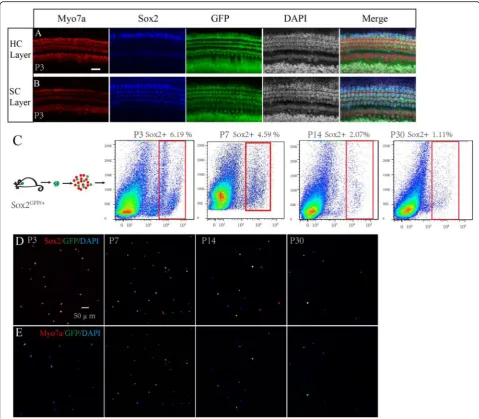
First, we performed an immunofluorescence assay to
ob-serve the GFP expression pattern inSox2GFP/+mice, and
we found that GFP was mainly expressed in Hensen’s
cells, Deiters’ cells, pillar cells, inner phalangeal cells,
and the greater epithelium ridge in the P3 mouse
coch-lea (Fig. 1a, b). We then used flow cytometry to sort the
Sox2+ SCs from cochleae dissected from mice at P3, P7, P14, and P30, and these made up 6.19% of the viable cells in the P3 mice, 4.59% of the viable cells in P7 mice, 2.07% of the viable cells in the P14 mice, and 1.11% of
the viable cells in the P30 mice (Fig. 1c). We observed
that the proportion of Sox2+ cells gradually decreased with age, and this might be because the increasing ossifi-cation with age made the dissection and dissociation of the organ of Corti more difficult at older ages. We then
performed immunofluorescence to double confirm the sorted cells and found that at P3 94.9 ± 2.3% and 94.5% ±
2.31% of the sorted cells were Sox2+and GFP+,
respect-ively, while none of the sorted cells was Myo7a+(Fig.1d,
e), suggesting that the flow-sorted cells were almost all Sox2+ SCs and that the sorted cells were highly pure.
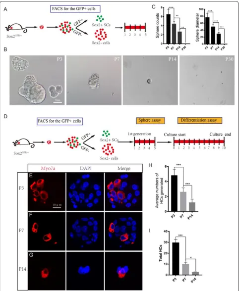
We next performed a sphere-forming assay using P3, P7, P14, and P30 SCs. A total of 200 isolated cells were plated onto a 96-well ultra-low attachment plate at a
density of 2 cells/μl for 5 days (Fig. 2a). We evaluated
the proliferation capacity of the SCs by quantifying the numbers and diameters of the spheres. Consistent with
previous reports [19], we found that 200 P3 Sox2+ SCs
Fig. 2(See legend on next page.)
could form around 7 spheres/well and that the diameter
of each sphere was more than 70μm (Fig.2b). However,
the spheres were fewer and smaller from P7 Sox2+ SCs and were even fewer and smaller from P14 Sox2+ SCs
(Fig. 2b, c). No spheres were observed from the P30
Sox2+ SCs (Fig. 2b, c). The greater sphere-forming
abil-ity of P3 SCs suggests that the neonatal (P3) SCs possess greater proliferation ability than aged (P7, P14, P30) SCs. In order to further evaluate the HC regeneration abil-ity of these spheres, we isolated the spheres derived from P3, P7, and P14 SCs and differentiated those spheres for 10 days and then immunostained them with the HC
marker Myo7a (Fig. 2d). We counted the Myo7a+ HCs
in each differentiated sphere and calculated the total Myo7a+ HCs that were generated from the original 200 flow cytometry-isolated Sox2+ SCs. We found that the P3 Sox2+ SC spheres generated significantly more Myo7a+ HCs than the P7 and P14 Sox2+ SC spheres
(Fig.2e–i). In summary, these results support prior
find-ings that neonatal (P3) SCs have a greater capacity to form spheres than aged (P7, P14, P30) SCs and that the spheres formed from neonatal SCs can generate more HCs than spheres formed from aged SCs.
Neonatal SCs have a greater capacity to regenerate HCs compared with aged SCs in vitro
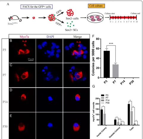
Most inner ear cell differentiation occurs during embry-onic development, but the neonatal mouse retains a lim-ited ability to regenerate HCs through the differentiation of SCs. This ability is quickly lost, however, and by the first week after birth, there is a notable decline in this re-generative activity. We cultured 5000 isolated Sox2+ P3, P7, P14, and P30 SCs in laminin-coated four-well dishes
at a density of 50 cells/μl for 10 days and then
immuno-stained them with the HC marker Myo7a (Fig. 3a). We
found that the P3 SCs generated significantly more Myo7a+ colonies than the P7 SCs, while no colonies were seen to develop from P14 and P30 SCs (5000 P3 SCs and P7 SCs generated 146.75 ± 12.71 and 76.5 ± 5.22
HCs inside of the colonies, respectively, p< 0.001,n= 3)
(Fig.3b–e). At P14 and P30, we only found the HCs
out-side of the colonies, suggesting that they were directly trans-differentiated from SCs. The total number of Myo7a+ HCs inside and outside of the colonies de-creased with age, suggesting that the ability of SCs to
regenerate HCs was significantly decreased with age
(Fig.3f).
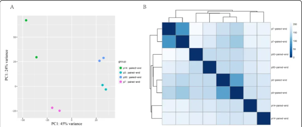
RNA-seq analysis of SCs isolated at different ages
To determine the gene expression profiles of SCs at differ-ent ages, RNA-seq analysis was performed on flow cytometry-isolated Sox2+ SCs from P3, P7, P14, and P30 basilar membranes. Three biological replicates were pre-pared for each time point. After alignment to the refer-ence genome (Mouse mm10, UCSC), the gene expression abundance was normalized to FPKM (fragments per kilo-base of transcript per million fragments mapped). We next explored the data set with principal component ana-lysis and sample clustering anaana-lysis. Replicates from the same group were well clustered and no outliers were
found (Fig. 4). We next carried out pairwise comparison
among all time points, and the genes that were differen-tially expressed within any two groups were marked. In total, we found 1296 differentially expressed genes.
Cell cycle analysis
The neonatal Sox2+ SCs had significantly greater prolif-eration and mitotic HC regenprolif-eration ability than the aged SCs; however, the detailed mechanism behind this difference remains unknown. To identify the possible genes regulating the age-dependent cell cycling of SCs, we used RNA-seq analysis to compare the expression of genes regulating the cell cycle and cell proliferation in P3, P7, P14, and P30 SCs. A prior study suggested that over 1000 cell cycle genes might exist in the average
mammalian cell [28], some of which had significant
ex-pression differences between SCs at different ages. We
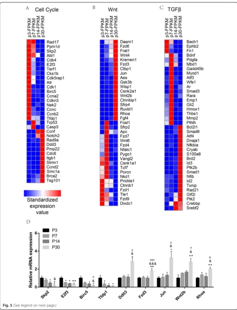
found thatRad17,Ppm1d,Skp2,Abl1,Cdk4,E2f3,Terf1,
Cks1b,Cdk5rap1,Atr, Cdk1, Birc5, Ccna2,Cdkn3,Nek2,
Ccnc, Ccnb2, and Tfdp1 were highly expressed in the
neonatal SCs compared with adult SCs and that Ccnf,
Rad9a, Ddit3, Pmp22, Cdc6, Itgb1, Stmn1, Ccnd2,
Smc1a, Brca2, andTsg101 were highly expressed in the
adult SCs compared with neonatal SCs (Fig.5a). Among
them, Skp2 [29–31], E2f3 [32, 33], Cdk1 [34, 35], Birc5
[36],Ddit3[37,38], andItgb1 [39] have already been re-ported in the inner ear. The results of the qPCR were consistent with RNA-seq results, thus confirming the
ex-pression difference in the cell cycle genes (Fig.5d).
How-ever, most of the differentially expressed cell cycle genes
(See figure on previous page.)
that we identified at different ages of SCs have not been characterized before in the inner ear and need to be fur-ther studied in the future.
Wnt signaling analysis
The Wnt signaling pathway is a highly conserved path-way and has been reported to be involved in multiple processes including proliferation, cell fate determination,
differentiation, and cell protection [40, 41]. In the inner
ear, activation of the Wnt signaling pathway is important
for HC regeneration and survival [8, 10, 12, 23, 42–47].
To determine which Wnt pathway factors are involved in regulating the age-dependent proliferation and HC re-generation ability of SCs, we measured the expression of over 147 genes, some of which had significant expression differences between SCs at different ages. We found that
Daam1, Fzd6, Frat1, Wnt4, Kremen1, Fzd3, Ctbp1, Jun,
Aes, Wisp1, Csnk2a1, Wnt2b, Ctnnbip1, Strp4, Ruvbl1, Fig. 3P3 Sox2+ SCs generated more HCs compared with the other three ages of SCs in vitro.aWe used the FITC channel to sort P3, P7, P14, and P30 Sox2+ SCs, and we cultured the sorted GFP+ cells at 50 cells/μl for 10 days.bP3 Sox2+ SCs generated a large number of Myo7a+ cells. cP7 Sox2+ SCs also could form colonies and generate Myo7a+ cells.d,eBoth P14 and P30 Sox2+ SCs could not form colonies, but the single cells could generate Myo7a+ cells.fP3 Sox2+ SCs formed more Myo7a+ cells compared with P7, P14, and P30 Sox2+ SCs.gBoth inside and outside of the colony, P3 SCs formed more Myo7a+ cells compared with P7, P14, and P30 Sox2+ SCs. ***p< 0.001. Scale bars are 10μm inb–h
Rhoa, and Fgf4 were significantly upregulated in adult
mice compared with neonatal mice, while Prickle1,
Ctnnb1, Fzd1, Tle1, Fzd9, and Dixdc1 were highly expressed in neonatal mice compared with adult mice
(Fig. 5b). Among them, Jun[48], Wnt2b [49, 50], Strp4
[51], Fgf4 [52, 53], Fzd1 [54], and Fzd3 [55, 56] have
already been reported in the inner ear. We performed qPCR to confirm the RNA-seq data, and the results were
consistent with the RNA-seq analysis (Fig.5d).
TGFβsignaling analysis
TGFβsignaling plays an important role in inner ear
de-velopment and HC regeneration [57,58], but studies of
TGFβ signaling in HC regeneration are still limited. To
determine which TGFβ pathway factors might be
in-volved in regulating HC regeneration, we examined the
expression of TGFβ pathway genes in the mouse
gen-ome in P3, P7, P14, and P30 SCs. We found thatSrebf2,
Crebbp, Ptk2, Gtf2i, Rad21, Id2, Txnip, Nfib, Nfkbia,
Ptk2b, Brd2, Id3, Smad1, S100a8, Atf4, Dnaja1, Cryab,
Bcl2l1, and Smad6 were significantly upregulated in
adult mice compared with neonatal mice, while Fn1,
Ephb2, and Bach1 were highly expressed in neonatal
mice compared with adult mice (Fig.5c). Among them,
Ephb2[59], Bdnf[60], andPdgfa[61] have already been reported in the inner ear.
Notch signaling analysis
Notch signaling plays an important role during the de-velopment and patterning of sensory HCs. The activa-tion of Notch signaling promotes the development of progenitor cells but prevents the differentiation of SCs into HCs. Inhibition of Notch signaling or Notch ligands
such as Dll1 and Jagged2 results in the generation of
supernumerary HCs in the mouse inner ear [62–64]. To
determine which Notch pathway genes are involved in regulating the age-dependent proliferation and HC re-generation ability of SCs, we measured over 1000 genes, some of which had significant expression differences among SCs at different ages. We found that the
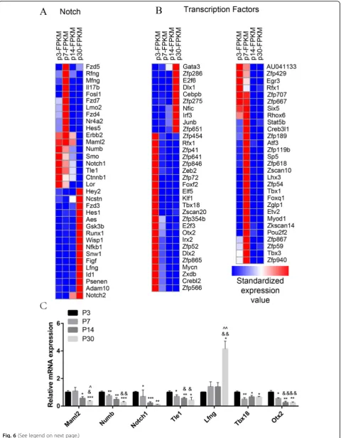
expres-sion of Maml2, Numb, Smo, Notch1, Tle1, and Lor
decreased with increasing age and that Hey2, Ncstn,
Hes1,Runx1,Wisp1, Nfkb1,Snw1,Figf,Lfng,Id1,Psenes,
Adam10, and Notch2were highly expressed in adult SCs
(Fig. 6a). Among these, Numb [65], Smo [21], Notch1
[43, 66, 67], Hey2 [68, 69], Hes1 [70, 71], Gsk3b [72],
Lfng [73, 74], Id1 [75, 76], and Adam10 [77–79] have
already been reported in the inner ear. We also performed qPCR to confirm the RNA-seq data, and the results were consistent with the RNA-seq analysis data
(Fig.6c).
Transcription factor analysis
Transcription factors (TFs) are regulatory proteins that control the expression of targeted genes by binding ei-ther to enhancer or promoter regions. TFs are involved in various processes, including inner ear development and HC regeneration. To determine which TFs might be involved in regulating HC regeneration, we examined the expression of 1324 TFs in the mouse genome in P3, P7, P14, and P30 SCs. We found that 9 TF genes (Zfp286,E2f6, Dlx1, Cebpb, Zfp275,Nfic,Irf3, Junb, and
Zfp651) were highly expressed in adult mice compared
to neonatal mice, while there were 28 TF genes (Zfp454,
Zfp41, Zfp641, Zfp846, Zeb2, Zfp72, Foxf2, Elf5, Klf1,
Tbx18, Zscan20, Zfp354b, Otx2, Irx2, Zfp52, Dlx2,
Fig. 5(See legend on next page.)
expressed in neonatal mice compared with adult mice
(Fig.6b). Some of the TF genes that are highly expressed
in neonatal SCs have been reported to play roles in pro-moting HC fate and patterning regulation during inner
ear development, including Rfx1 [80], Tbx18[81], Otx2
[82, 83], Dlx2 [84], andMycn [85]. We also performed
qPCR to confirm the RNA-seq data, and the results were
consistent with the RNA-seq analysis data (Fig. 6c). We
have identified many TFs that have not been character-ized before, and their involvement in the differential re-generation capacity in mouse cochlear SCs at different ages should be investigated in the future.
Gene ontology analysis of the genes that are differentially expressed in SCs of different ages
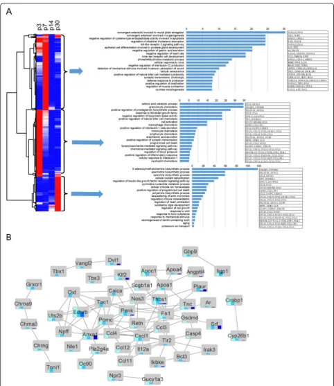
After clustering the expression of all 1296 differentially expressed genes in P3, P7, P14, and P30 Sox2+ SCs in a
heatmap (fold change > 2.0,q< 0.05), we applied GO
ana-lysis to the gene clusters. GO terms with the greatest
en-richment fold are shown on the right of Fig.7a, which also
shows the protein interaction network of these GO
enriched genes (Fig. 7b). GO analysis was applied to the
genes that were upregulated in SCs at different ages (fold
change > 2.0,p< 0.01). The genes with altered expression
in P3 Sox2+ SCs were highly enriched in functional cat-egories such as auditory receptor cell fate determination, neuron fate determination, signaling, and extracellular matrix formation and maintenance. Genes upregulated in P30 SCs were highly enriched in functional categories such as biosyntheic processes and positive regulation of programmed cell death.
Discussion
Several previous studies have shown that the ability of SCs to regenerate lost or damaged HCs decreases dra-matically with age; however, the detailed transcriptome profiles of SCs at different ages have not been studied. Here, we isolated SCs from P3, P7, P14, and P30 mice and compared their transcriptome expression profiles. We identified a set of differentially expressed genes, in-cluding cell cycle genes, signaling pathway genes, and TFs, that might be involved in regulating the prolifera-tion and HC differentiaprolifera-tion ability of SCs. Most of the differentially expressed genes identified in this study have not been investigated in the inner ear before and need to be further studied in the future.
In order to find the key genes regulating inner ear HC regeneration, our previous studies have reported the transcriptome profiles of SCs or Lgr5+ inner ear progen-itors, which are a sub-population of SCs, at different
lo-cations and under different treatment conditions [13,14,
86, 87]. We characterized the transcriptomes of Lgr5+
progenitor cells in the apical and basal turns of the
mouse cochlea [14]. Compared with our current results,
we found that the cell cycle genesCcnc,Cdk4,Nek2, and
Skp2were highly expressed both in the Lgr5+ progenitor
cells in the apical turn of the cochlea and in neonatal
mouse inner ear SCs. Also, the TF genes Irx2 and
Zfp667 were highly expressed both in the Lgr5+ pro-genitor cells in the apical turn of the cochlea and in
neo-natal mouse inner ear SCs, while Junb was highly
expressed both in the Lgr5+ progenitor cells in the basal turn of cochlea and in adult mouse inner ear SCs.
We also characterized the transcriptomes of Lgr5+
progenitor cells and other Lgr5− SCs [13]. Compared
with our current results, we found that the cell cycle
genesSkp2 and Terf1were highly expressed both in the
Lgr5+ progenitor cell and in neonatal mouse inner ear
SCs, while Ccnf, Notch2, Ppm22, Ccnd2, and Tsg101
were highly expressed both in the Lgr5− SCs and in
adult mouse inner ear SCs. The TF gene Zfp667 was
highly expressed both in the Lgr5+ progenitor cells and
neonatal mouse inner ear SCs, while Junb was highly
expressed both in the Lgr5−SCs and adult mouse inner
ear SCs. Among Wnt signaling pathway genes, Wisp1
and Rhoa were highly expressed both in the Lgr5− SCs
and the adult mouse inner ear SCs.
Next, we characterized the transcriptomes of Lgr5+ progenitor cells with or without neomycin injury to show the damage-induced transcriptome changes in the
Lgr5+ progenitors [87]. Compared with our current
re-sults, we found that the cell cycle geneTfdp1was highly
expressed both in the neomycin-treated Lgr5+
progeni-tors and neonatal mouse inner ear SCs, whileStmn1was
highly expressed both in the untreated Lgr5+ progeni-tors and in adult mouse inner ear SCs. The TF gene
Zfp52 was highly expressed both in the neomycin-treated Lgr5+ progenitors and in neonatal mouse inner
ear SCs, whileJunbwas highly expressed both in the
un-treated Lgr5+ progenitors and in adult mouse inner ear
SCs. Among Notch, Wnt, TGFβ signaling pathway
genes, Hes1, Ctnnbip1, Id2, and Id3 were highly
(See figure on previous page.)
Fig. 6(See legend on next page.)
expressed both in the untreated Lgr5+ progenitors and in adult mouse inner ear SCs.
Lastly, we characterized the transcriptomes of Lgr5+
progenitor cells and Lgr6+ progenitor cells [86].
Com-pared with our current results, we found that the TF
genesIlx2and AU041133were highly expressed both in
the Lgr6+ progenitors and in neonatal mouse inner ear
SCs; while the cell cycling genes Rad17 and Skp2 were
highly expressed both in the Lgr5+ progenitors and in neonatal mouse inner ear SCs. Among Notch signaling
pathway genes,Maml2was highly expressed both in the
Lgr6+ progenitors and in neonatal mouse inner ear SCs,
whileHey2,Hes1, andId1were highly expressed both in
the Lgr5+ progenitors and in adult mouse inner ear SCs. These candidate genes might play important roles in regulating HC regeneration in the inner ear.
Cell cycle analysis
Among the differentially expressed cell cycle-related
genes, Skp2, E2f3, Cdk1, Birc5, Ddit3, and Itgb1 have
been reported in the inner ear before. Skp2 is an F-box
protein that regulates the G1 to S transition by control-ling the stability of several G1 regulators, including p27, and it is expressed in the auditory epithelia and neurons at early stages of development. In the mature auditory
epithelium, overexpression ofSkp2alone can induce SC
proliferation but cannot induce new HC formation,
while overexpression ofSkp2combined with
overexpres-sion ofAtoh1generates new HCs [29–31]. This suggests
that regulation of HC regeneration requires multi-gene
coordination. Skp2 is also highly expressed in tumor
cells and promotes cell proliferation [88–90]. E2f3 is a
member of the E2F transcription factor family and is in-volved in regulating cell proliferation. In isolated human
islets, it can induce proliferation of βcells [91]. E2f3 is
barely expressed in the inner ear, but its expression in-creases in outer HC nuclei upon excessive noise
expos-ure [32, 33].Cdk1 is ubiquitously expressed throughout
the organ of Corti and spiral ganglion cells, and
inhib-ition ofCdk1and other cycldependent kinases can
in-duce differentiation of supernumerary HCs and Deiters’
cells in the developing organ of Corti in cultured rat
cochleae [34, 35]. Birc5 is expressed during embryonic
development and cannot be detected in most terminally differentiated tissues, and it is also highly expressed in many tumors such as pancreatic ductal adenocarcinoma
[92].Birc5is widely expressed in the organ of Corti, and it provides protection against ototoxin-induced
cytotox-icity [36]. Ddit3 is an endoplasmic reticulum stress
marker gene. In the animal model of acute hearing loss,
the expression ofDdit3is upregulated in the lateral wall
of the cochlea, and this high expression ofDdit3 might
lead to hearing loss because of endoplasmic reticulum
stress [37,38]. Itgb1is involved in the regulation of cell
migration and invasion of hepatoma carcinoma, breast
cancer, and gallbladder cancer [93–95]. It is expressed
throughout the otic area, including the fusion plate
epi-thelium and the periotic mesenchyme [39]. Rad17,
Ppm1d,Abl1,Cdk4,Terf1,Cks1b,Cdk5rap1,Atr,Ccna2,
Cdkn3, Nek2, Ccnc, Ccnb2, Tfdp1, Ccnf,Rad9a, Pmp22,
Cdc6,Stmn1,Ccnd2,Smc1a,Brca2, andTsg101have not been reported previously in the inner ear and need to be further studied in the future.
Wnt signaling analysis
Among the differentially expressed Wnt
signaling-related genes, Jun, Wnt2b, Strp4, Fgf4, Fzd1, Fzd3, and
Fzd6have been previously reported in the inner ear.Jun
has been implicated in the regulation of cell prolifera-tion, differentiaprolifera-tion, and apoptosis. It plays a critical role during inner ear development by mediating apoptosis
through the JNK pathway [48]. Wnt2b is expressed in
the endolymphatic duct; however, the role of Wnt2b in
inner ear development has not been reported [49, 50].
Sfrp4 is a Wnt pathway inhibitor that is involved in many diseases including obesity, type 2 diabetes, cancer,
and psoriasis [96]. In the inner ear,Sfrp4can be directly
targeted by miR-124 to regulate HC differentiation and
polarization in the organ of Corti [97].Fgf4 is present in
many cancerous and noncancerous tissues, indicating
that Fgf4 plays an important role in cell differentiation
and proliferation [98]. In zebrafish,Fgf4can be mediated
by miR-194 to regulate the development and
differenti-ation of sensory patches [52,53]. Frizzled signaling is
in-volved in diverse tissue closure processes, and defects in frizzled signaling result in some of the most common congenital anomalies in humans. In the organ of Corti at
E18,Fzd1is weakly expressed in the three outer rows of
sensory HCs and is strongly expressed in the flanking non-sensory epithelial cells and the underlying
phalan-geal and pillar cells, and Fzd1 mutations cause
mis-orientation of inner ear sensory HCs [54]. Fzd3 and
(See figure on previous page.)
Fig. 7Global comparisons of differentially expressed genes among four time points by hierarchical clustering and gene ontology analysis.aHierarchical clustering of FPKM of all differentially expressed genes. Red denotes above-average expression levels, and blue denotes below-average levels. Each row represents one gene, and each column represents one time point. Gene ontology analysis was performed on the highly expressed gene clusters in the P3, P7, and P30 groups.bSTRING network analysis of genes present in GO categories
Fzd6 are key regulators of planar cell polarity in the
mammalian cochlea. In the inner ear, both Fzd3 and
Fzd6 are localized on the lateral faces of sensory and
SCs in all sensory epithelia, and this localization overlaps withVangl2and suggests thatFzd3andFzd6might play an important role in the planar polarity of HCs because
Vangl2plays an important role in regulating hair bundle
orientation [55,56,99]. This suggests that different
Friz-zled genes in the inner ear have different functions.
Al-though Jun, Wnt2b, Strp4, Fgf4, Fzd1, Fzd3, and Fzd6
have been previously reported in the inner ear, the func-tion of these genes in HC regenerafunc-tion still need to be
studied further. Daam1, Frat1, Wnt4, Kremen1, Ctbp1,
Wisp1, Csnk2a1, Ctnnbip1, Ruvbl1, Rhoa, Prickle1,
Ctnnb1, Tle1, Fzd9, and Dixdc1 have not been reported previously in the inner ear and need to be further stud-ied in the future.
TGFβsignaling analysis
Among the differentially expressed TGFβ
signaling-related genes, Ephb2, Bdnf, and Pdgfa have previously
been reported in the inner ear. Ephb2 is a member of
the largest group of transmembrane receptor tyrosine
ki-nases, and deletion ofEphb2leads to vestibular
dysfunc-tion because of the reduced producdysfunc-tion of endolymph
[59].Bdnfacts as a nerve growth factor, and it promotes
the growth and survival of neurons in the central and
peripheral nervous systems [100]. In the inner ear, it
supports spiral ganglion neuron survival [60]. Pdgfais a
growth factor with restricted otic expression, and it
overlaps withFgf16 in the anterior and posterior cristae
in the chick inner ear [61]. Srebf2, Crebbp, Ptk2, Gtf2i,
Rad21, Id2, Txnip, Nfib, Nfkbia, Ptk2b, Brd2, Id3,
Smad1, S100a8, Atf4, Dnaja1, Cryab, Bcl2l1, Smad6,
Fn1, andBach1have not been reported previously in the
inner ear and need to be further studied in the future.
Notch signaling analysis
Among the differentially expressed Notch
signaling-related genes, Numb, Smo, Notch1, Hey2, Hes1, Gsk3b,
Lfng,Id1, andAdam10have previously been reported in
the inner ear.Numb is a cell fate determinant gene that
regulates cardiac progenitor cell differentiation and
car-diac morphogenesis [101]. In the auditory epithelium,
Numb expression has different patterns, which suggests
thatNumb plays an important role in cochlear
develop-ment [65].Smoencodes a membrane protein that is
es-sential for the transduction of Hedgehog signals into the
cytoplasm. Activation of Smo inhibits prosensory cell
differentiation into HCs or SCs and maintains their properties as prosensory cells, and conditional knockout
of theSmogene in the cochlea delays HC and SC
differ-entiation in the apical region [21].Notch1is the primary
Notch receptor expressed in the mouse inner ear, and
activation of Notch1 in developing auditory HCs causes
profound deafness, while deletion of Notch1 leads to
limited mitotic HC generation [43, 66]. Hey2is a
puta-tive Notch target gene and functions in cell fate
specifi-cation. Hey2 is expressed in the cochlear epithelium
prior to terminal differentiation, and its overexpression
overlaps with that of Hes1 in the developing cochlea.
The genetic inactivation ofHey2leads to increased
num-bers of mis-patterned inner HCs and outer HCs [70,71],
and activation of Hey2by FGF signaling blocks HC
dif-ferentiation [68,69].Gsk3 plays an important role in the
regulation of apoptosis and proliferation in the inner
ear, and activation of Gsk3 causes the release of
inflam-matory factors that can eventually lead to hearing loss,
while inactivation ofGsk3 increases the total number of
HCs [72, 102]. The Lfng gene is expressed in
non-sensory SCs in the mouse cochlea, but there is no
no-ticeable effect on HC differentiation in Lfng mutant
mice. However, mutation of Lfng suppresses the effects
of the Jag2mutations on inner HCs [73,74].Id1is able
to prevent differentiation of pluripotent cells, and in
bone marrow transplantation assays, reducing Id1
en-hanced hematopoietic stem cells’ self-renewal potential
[103].Id1is expressed within the cochlear duct in a
pat-tern that is consistent with a role in the regulation of HC development. However, there is no hearing deficit in
the absence of the Id1 gene, and the reason for this
might be compensatory effects by other Ids like Id3,
which has a similar expression pattern as Id1 in the
cochlea [75,76].Adam10is abundantly expressed in the
brain and is linked to epilepsy, Alzheimer’s disease,
Hunting’s disease, and developmental disorder Fragile X
syndrome because of its role in regulating the activity of
excitatory synapses [104,105].Adam10is also expressed
in the cochlea and vestibule, and inhibition of Adam10
after HC loss increases the proliferation of SCs in the
vestibular system [77–79]. Although Numb, Smo,
Notch1,Hey2,Hes1,Gsk3b,Lfng,Id1, andAdam10 have been reported in the inner ear, the functions of some of these genes in HC regeneration still need to be further
studied.Maml2, Tle1, Lor,Ncstn, Runx1, Wisp1,Nfkb1,
Snw1, Figf, Psenes, and Notch2 have not been reported previously in the inner ear and need to be further stud-ied in the future.
Transcription factor analysis
Among the differentially expressed TFs, Rfx1, Tbx18,
Otx2, Dlx2, and Mycnhave previously been reported in
the inner ear.Rfx1has an important function in brain
tu-mors and sensorineural hearing loss. Together withAtho1,
Rfx1/3 can induce HC-like cell differentiation, and the
conditional knockout of Rfx1/3 leads to severe hearing
loss and OHC damage [80, 106,107].Tbx18 is a critical
is also a target gene of the Hippo pathway [108]. The
ex-pression of Tbx18 during inner ear development is
re-stricted to the sub-region of the otic mesenchyme that is
fated to differentiate into fibrocytes, and Tbx18-deficient
mice show profound deafness and a complete disruption of the endocochlear potential that is essential for the
transduction of sound by sensory HCs [81]. Otx2 is a
regulator of embryonic development and embryonic neurogenesis. And it plays a role in brain, craniofacial, and
sensory organ development [109, 110]. In the inner ear,
Otx2 can induce Hes5 and lead to the differentiation of
both cochlear and macular neuroepithelium [82, 83]. In
the chick inner ear, the expression ofDlx1andDlx2
dur-ing the later stages of inner ear morphogenesis is limited to cochlear and vestibular nerves, and expression levels
are lower than in the early stages of morphogenesis [84].
Mycnis a critical factor in the development of the central
and peripheral nervous systems [111]. In humans, the
mu-tation ofMycncan cause structural and functional
abnor-malities of the inner ear [85].Zfp286,E2f6,Dlx1, Cebpb,
Zfp275, Nfic, Irf3, Junb, Zfp651, Zfp454, Zfp41, Zfp641,
Zfp846, Zeb2,Zfp72, Foxf2, Elf5, Klf1,Zscan20, Zfp354b,
Irx2, Zfp52, Zfp865, Zxdb, Crebl2, Zfp566, AU041133,
Zfp429,Egr3,Zfp707,Zfp667, andSix5have not been re-ported previously in the inner ear and need to be further studied in the future.
Conclusion
Consistent with previous reports, in this study we also found that neonatal SCs have significantly greater prolifer-ation and HC regenerprolifer-ation ability than adult SCs. We sys-tematically investigated the transcriptome differences between P3, P7, P14, and P30 SCs and identified several significantly differentially expressed genes that might regulate the proliferation and HC regeneration capacity of SCs of different ages. The transcriptomes of different ages of SCs reported here establish a framework for future characterization of the genes that regulate the prolifera-tion and HC regeneraprolifera-tion ability of SCs, and these genes
might represent new therapeutic targets for HC
regeneration.
Supplementary information
Supplementary informationaccompanies this paper athttps://doi.org/10. 1186/s13287-019-1437-0.
Additional file 1.q-PCR primers
Abbreviations
E:Embryonic day; GO: Gene ontology; HC: Hair cell; P: Postnatal day; SC: Supporting cell; STRING: Search Tool for the Retrieval of Interacting Genes/Proteins; TFs: Transcription factors
Acknowledgements Not applicable
Authors’contributions
CC, YW, LG, RC, XG, and HL designed the study. CC, XL, WZ, WM, LZ, and LL performed the laboratory experiments. CC, YW, LG, XL, JG, ML, LL, FC, and MT contributed to critical discussion and data analysis. CC, XG, MT, HL, and RC wrote the paper. All authors read and approved the final manuscript.
Authors’information Not applicable
Funding
This work was supported by grants from the National Key R&D Program of China (2017YFA0103903), the Strategic Priority Research Program of the Chinese Academy of Science (XDA16010303), the National Natural Science Foundation of China (81622013, 81970882, 81900941, 81570919, 81870721, 81771019, 81700913, 81670928, 81570921), Jiangsu Province Natural Science Foundation (BK20190121), Boehringer Ingelheim Pharma GmbH, the Fundamental Research Funds for the Central Universities (2242018k1G011), the Open Research Fund of the State Key Laboratory of Genetic Engineering, Fudan University (SKLGE1809), and the Project of Invigorating Health Care through Science, Technology and Education.
Availability of data and materials
The datasets during and/or analyzed during the current study are available from the corresponding author on reasonable request.
Ethics approval and consent to participate
All animal procedures were performed according to protocols approved by the Animal Care Committee of Southeast University and were consistent with the National Institutes of Health Guide for the Care and Use of Laboratory Animals. All efforts were made to minimize the number of animals used and to prevent their suffering.
Consent for publication Not applicable
Competing interests
The authors declare that they have no competing interests.
Author details
1Jiangsu Provincial Key Medical Discipline (Laboratory), Department of
Otolaryngology Head and Neck Surgery, Affiliated Drum Tower Hospital of Nanjing University Medical School, No. 321 Zhongshan Road, Nanjing 210008, China.2Research Institute of Otolaryngology, No. 321 Zhongshan
Road, Nanjing 210008, China.3Shanghai Fenyang Vision & Audition Center,
Shanghai, China.4ENT Institute and Otorhinolaryngology Department of
Affiliated Eye and ENT Hospital, Key Laboratory of Hearing Medicine of NHFPC, Shanghai Engineering Research Centre of Cochlear Implant, State Key Laboratory of Medical Neurobiology, Fudan University, Room 611, Building 9, No. 83, Fenyang Road, Xuhui District, Shanghai 200031, China.
5MOE Key Laboratory for Developmental Genes and Human Disease, State
Key Laboratory of Bioelectronics, Co-Innovation Center of Neuroregeneration, Institute of Life Sciences, Jiangsu Province High-Tech Key Laboratory for Bio-Medical Research, Southeast University, Nanjing 210096, China.
6Department of Biotechnology, Federal Urdu University of Arts, Science and
Technology, Gulshan-e-Iqbal Campus, Karachi, Pakistan.7Jiangsu
Rehabilitation Research Center for Hearing and Speech Impairment, Nanjing 210004, Jiangsu, China.8Department of Biomedical Engineering, Southern
University of Science and Technology, Shenzhen, China.9Co-Innovation
Center of Neuroregeneration, Nantong University, Nantong 226001, China.
10Institute for Stem Cell and Regeneration, Chinese Academy of Science,
Beijing, China.11Beijing Key Laboratory of Neural Regeneration and Repair,
Capital Medical University, Beijing 100069, China.
Received: 4 June 2019 Revised: 29 July 2019 Accepted: 1 October 2019
References
1. Ryals BM, Rubel EW. Hair cell regeneration after acoustic trauma in adult Coturnix quail. Science. 1988;240(4860):1774–6.
2. Lombarte A, Yan HY, Popper AN, Chang JS, Platt C. Damage and regeneration of hair cell ciliary bundles in a fish ear following treatment with gentamicin. Hear Res. 1993;64(2):166–74.
3. Cruz RM, Lambert PR, Rubel EW. Light microscopic evidence of hair cell regeneration after gentamicin toxicity in chick cochlea. Arch Otolaryngol Head Neck Surg. 1987;113(10):1058–62.
4. Corwin JT, Cotanche DA. Regeneration of sensory hair cells after acoustic trauma. Science. 1988;240(4860):1772–4.
5. Balak KJ, Corwin JT, Jones JE. Regenerated hair-cells can originate from supporting cell progeny - evidence from phototoxicity and laser ablation experiments in the lateral line system. J Neurosci. 1990;10(8):2502–12. 6. Brigande JV, Heller S. Quo vadis, hair cell regeneration? Nat Neurosci. 2009;
12(6):679–85.
7. Bermingham-McDonogh O, Rubel EW. Hair cell regeneration: winging our way towards a sound future. Curr Opin Neurobiol. 2003;13(1):119–26. 8. Cox BC, Chai R, Lenoir A, Liu Z, Zhang L, Nguyen DH, et al. Spontaneous
hair cell regeneration in the neonatal mouse cochlea in vivo. Development. 2014;141(4):816–29.
9. Warchol ME. Sensory regeneration in the vertebrate inner ear: differences at the levels of cells and species. Hearing Res. 2011;273(1–2):72–9.
10. Chai R, Kuo B, Wang T, Liaw EJ, Xia A, Jan TA, et al. Wnt signaling induces proliferation of sensory precursors in the postnatal mouse cochlea. Proc Natl Acad Sci U S A. 2012;109(21):8167–72.
11. Basch ML, Brown RM 2nd, Jen HI, Groves AK. Where hearing starts: the development of the mammalian cochlea. J Anat. 2016;228(2):233–54. 12. Shi F, Kempfle JS, Edge AS. Wnt-responsive Lgr5-expressing stem cells are
hair cell progenitors in the cochlea. J Neurosci. 2012;32(28):9639–48. 13. Cheng C, Guo L, Lu L, Xu X, Zhang S, Gao J, et al. Characterization of the
transcriptomes of Lgr5+ hair cell progenitors and Lgr5- supporting cells in the mouse cochlea. Front Mol Neurosci. 2017;10:122.
14. Waqas M, Guo L, Zhang S, Chen Y, Zhang X, Wang L, et al. Characterization of Lgr5+ progenitor cell transcriptomes in the apical and basal turns of the mouse cochlea. Oncotarget. 2016;7(27):41123–41.
15. Feghali JG, Lefebvre PP, Staecker H, Kopke R, Frenz DA, Malgrange B, et al. Mammalian auditory hair cell regeneration/repair and protection: a review and future directions. Ear Nose Throat J. 1998;77(4):276 80, 82–5. 16. Doetzlhofer A, White P, Lee YS, Groves A, Segil N. Prospective identification
and purification of hair cell and supporting cell progenitors from the embryonic cochlea. Brain Res. 2006;1091(1):282–8.
17. Golub JS, Tong L, Ngyuen TB, Hume CR, Palmiter RD, Rubel EW, et al. Hair cell replacement in adult mouse utricles after targeted ablation of hair cells with diphtheria toxin. J Neurosci. 2012;32(43):15093–105.
18. Sinkkonen ST, Chai RJ, Jan TA, Hartman BH, Laske RD, Gahlen F, et al. Intrinsic regenerative potential of murine cochlear supporting cells. Sci Rep. 2011;1:26.
19. Oshima K, Grimm CM, Corrales CE, Senn P, Martinez Monedero R, Geleoc GS, et al. Differential distribution of stem cells in the auditory and vestibular organs of the inner ear. J Assoc Res Otolaryngol. 2007;8(1):18–31. 20. Corwin JT, Oberholtzer JC. Fish n' chicks: model recipes for hair-cell
regeneration? Neuron. 1997;19(5):951–4.
21. Chen Y, Lu X, Guo L, Ni W, Zhang Y, Zhao L, et al. Hedgehog signaling promotes the proliferation and subsequent hair cell formation of progenitor cells in the neonatal mouse cochlea. Front Mol Neurosci. 2017;10:426. 22. Chen Q, Quan Y, Wang N, Xie C, Ji Z, He H, et al. Inactivation of STAT3
signaling impairs hair cell differentiation in the developing mouse cochlea. Stem Cell Rep. 2017;9(1):231–46.
23. Wu J, Li W, Lin C, Chen Y, Cheng C, Sun S, et al. Co-regulation of the Notch and Wnt signaling pathways promotes supporting cell proliferation and hair cell regeneration in mouse utricles. Sci Rep. 2016;6:29418.
24. Li W, Wu J, Yang J, Sun S, Chai R, Chen ZY, et al. Notch inhibition induces mitotically generated hair cells in mammalian cochleae via activating the Wnt pathway. Proc Natl Acad Sci U S A. 2015;112(1):166–71.
25. Dabdoub A, Puligilla C, Jones JM, Fritzsch B, Cheah KS, Pevny LH, et al. Sox2 signaling in prosensory domain specification and subsequent hair cell differentiation in the developing cochlea. Proc Natl Acad Sci U S A. 2008; 105(47):18396–401.
26. Trapnell C, Schatz MC. Optimizing data intensive GPGPU computations for DNA sequence alignment. Parallel Comput. 2009;35(8):429–40.
27. Huang DW, Sherman BT, Lempicki RA. Systematic and integrative analysis of large gene lists using DAVID bioinformatics resources. Nat Protoc. 2009;4(1): 44–57.
28. Forrest ARR, Taylor D, Grimmond S, Grp RG, Members G. Exploration of the cell-cycle genes found within the RIKEN FANTOM2 data set. Genome Res. 2003;13(6B):1366–75.
29. Minoda R, Izumikawa M, Kawamoto K, Zhang H, Raphael Y. Manipulating cell cycle regulation in the mature cochlea. Hear Res. 2007;232(1–2):44–51. 30. Dong Y, Nakagawa T, Endo T, Kim TS, Iguchi F, Yamamoto N, et al. Role of
the F-box protein Skp2 in cell proliferation in the developing auditory system in mice. Neuroreport. 2003;14(5):759–61.
31. Nakayama KI, Hatakeyama S, Nakayama K. Regulation of the cell cycle at the G1-S transition by proteolysis of cyclin E and p27Kip1. Biochem Biophys Res Commun. 2001;282(4):853–60.
32. Jamesdaniel S, Hu B, Kermany MH, Jiang H, Ding D, Coling D, et al. Noise induced changes in the expression of p38/MAPK signaling proteins in the sensory epithelium of the inner ear. J Proteome. 2011;75(2):410–24. 33. Pang J, Xiong H, Yang H, Ou Y, Xu Y, Huang Q, et al. Circulating miR-34a
levels correlate with age-related hearing loss in mice and humans. Exp Gerontol. 2016;76:58–67.
34. Liu YY, Wang GP, Peng Z, Guo JY, Wu Q, Xie J, et al. E2F1-CDK1 pathway activation in kanamycin-induced spiral ganglion cell apoptosis and the protective effect of CR8. Neurosci Lett. 2016;617:247–53.
35. Malgrange B, Knockaert M, Belachew S, Nguyen L, Moonen G, Meijer L, et al. The inhibition of cyclin-dependent kinases induces differentiation of supernumerary hair cells and Deiters' cells in the developing organ of Corti. FASEB J. 2003;17(14):2136–8.
36. Habtemichael N, Heinrich UR, Knauer SK, Schmidtmann I, Bier C, Docter D, et al. Expression analysis suggests a potential cytoprotective role of Birc5 in the inner ear. Mol Cell Neurosci. 2010;45(3):297–305.
37. Fujinami Y, Mutai H, Mizutari K, Nakagawa S, Matsunaga T. A novel animal model of hearing loss caused by acute endoplasmic reticulum stress in the cochlea. J Pharmacol Sci. 2012;118(3):363–72.
38. Fujinami Y, Mutai H, Kamiya K, Mizutari K, Fujii M, Matsunaga T. Enhanced expression of C/EBP homologous protein (CHOP) precedes degeneration of fibrocytes in the lateral wall after acute cochlear mitochondrial dysfunction induced by 3-nitropropionic acid. Neurochem Int. 2010;56(3): 487–94.
39. Matilainen T, Haugas M, Kreidberg JA, Salminen M. Analysis of Netrin 1 receptors during inner ear development. Int J Dev Biol. 2007;51(5):409–13. 40. Nusse R. Wnt signaling in disease and in development. Cell Res. 2005;15(1):28–32. 41. Pinto D, Clevers H. Wnt, stem cells and cancer in the intestine. Biol Cell.
2005;97(3):185–96.
42. Lu X, Sun S, Qi J, Li W, Liu L, Zhang Y, et al. Bmi1 regulates the proliferation of cochlear supporting cells via the canonical Wnt signaling pathway. Mol Neurobiol. 2017;54(2):1326–39.
43. Ni W, Lin C, Guo L, Wu J, Chen Y, Chai R, et al. Extensive supporting cell proliferation and mitotic hair cell generation by in vivo genetic reprogramming in the neonatal mouse cochlea. J Neurosci. 2016;36(33):8734–45.
44. Yu X, Liu W, Fan Z, Qian F, Zhang D, Han Y, et al. c-Myb knockdown increases the neomycin-induced damage to hair-cell-like HEI-OC1 cells in vitro. Sci Rep. 2017;7:41094.
45. Liu L, Chen Y, Qi J, Zhang Y, He Y, Ni W, et al. Wnt activation protects against neomycin-induced hair cell damage in the mouse cochlea. Cell Death Dis. 2016;7:e2136.
46. Chai R, Xia A, Wang T, Jan TA, Hayashi T, Bermingham-McDonogh O, et al. Dynamic expression of Lgr5, a Wnt target gene, in the developing and mature mouse cochlea. J Association Res Otolaryngol. 2011;12(4):455–69. 47. Shi F, Hu L, Edge AS. Generation of hair cells in neonatal mice by
beta-catenin overexpression in Lgr5-positive cochlear progenitors. Proc Natl Acad Sci U S A. 2013;110(34):13851–6.
48. Sanz C, Leon Y, Canon S, Alvarez L, Giraldez F, Varela-Nieto I. Pattern of expression of the Jun family of transcription factors during the early development of the inner ear: implications in apoptosis. J Cell Sci. 1999; 112(Pt 22):3967–74.
49. Lin ZS, Cantos R, Patente M, Wu DK. Gbx2 is required for the morphogenesis of the mouse inner ear: a downstream candidate of hindbrain signaling. Development. 2005;132(10):2309–18.
50. Choo D, Ward J, Reece A, Dou H, Lin Z, Greinwald J. Molecular mechanisms underlying inner ear patterning defects in kreisler mutants. Dev Biol. 2006; 289(2):308–17.
52. Wright TJ, Hatch EP, Karabagli H, Karabagli P, Schoenwolf GC, Mansour SL. Expression of mouse fibroblast growth factor and fibroblast growth factor receptor genes during early inner ear development. Dev Dyn. 2003;228(2): 267–72.
53. Cao H, Shi J, Du J, Chen K, Dong C, Jiang D, et al. MicroRNA-194 regulates the development and differentiation of sensory patches and statoacoustic ganglion of inner ear by Fgf4. Med Sci Monit. 2018;24:1712–23.
54. Yu H, Smallwood PM, Wang Y, Vidaltamayo R, Reed R, Nathans J. Frizzled 1 and frizzled 2 genes function in palate, ventricular septum and neural tube closure: general implications for tissue fusion processes. Development. 2010; 137(21):3707–17.
55. Wang Y, Guo N, Nathans J. The role of Frizzled3 and Frizzled6 in neural tube closure and in the planar polarity of inner-ear sensory hair cells. J Neurosci. 2006;26(8):2147–56.
56. Montcouquiol M, Sans N, Huss D, Kach J, Dickman JD, Forge A, et al. Asymmetric localization of Vangl2 and Fz3 indicate novel mechanisms for planar cell polarity in mammals. J Neurosci. 2006;26(19):5265–75. 57. Du XP, Li W, Gao XS, West MB, Saltzman WM, Cheng CJ, et al. Regeneration
of mammalian cochlear and vestibular hair cells through Hes1/Hes5 modulation with siRNA. Hearing Res. 2013;304:91–110.
58. Kawamoto K, Yagi M, Stover T, Kanzaki S, Raphael Y. Hearing and hair cells are protected by adenoviral gene therapy with TGF-beta1 and GDNF. Mol Ther. 2003;7(4):484–92.
59. Raft S, Andrade LR, Shao D, Akiyama H, Henkemeyer M, Wu DK. Ephrin-B2 governs morphogenesis of endolymphatic sac and duct epithelia in the mouse inner ear. Dev Biol. 2014;390(1):51–67.
60. McGuinness SL, Shepherd TK. Exogenous BDNF rescues rat spiral ganglion neurons in vivo. Otol Neurotol. 2005;26(5):1064–72.
61. Chapman SC, Cai Q, Bleyl SB, Schoenwolf GC. Restricted expression of Fgf16 within the developing chick inner ear. Dev Dyn. 2006;235(8):2276–81. 62. Kiernan AE, Cordes R, Kopan R, Gossler A, Gridley T. The Notch ligands DLL1
and JAG2 act synergistically to regulate hair cell development in the mammalian inner ear. Development. 2005;132(19):4353–62.
63. Bray SJ. Notch signalling: a simple pathway becomes complex. Nat Rev Mol Cell Biol. 2006;7(9):678–89.
64. Mizutari K, Fujioka M, Hosoya M, Bramhall N, Okano HJ, Okano H, et al. Notch inhibition induces cochlear hair cell regeneration and recovery of hearing after acoustic trauma. Neuron. 2013;77(1):58–69.
65. Gao Z, Chi FL, Huang YB, Yang JM, Cong N, Li W. Expression of Numb and Numb-like in the development of mammalian auditory sensory epithelium. Neuroreport. 2011;22(2):49–54.
66. Liu Z, Owen T, Fang J, Zuo J. Overactivation of Notch1 signaling induces ectopic hair cells in the mouse inner ear in an age-dependent manner. PLoS One. 2012;7(3):e34123.
67. Savoy-Burke G, Gilels FA, Pan W, Pratt D, Que J, Gan L, et al. Activated Notch causes deafness by promoting a supporting cell phenotype in developing auditory hair cells. PLoS One. 2014;9(9):e108160.
68. Doetzlhofer A, Basch ML, Ohyama T, Gessler M, Groves AK, Segil N. Hey2 regulation by FGF provides a Notch-independent mechanism for maintaining pillar cell fate in the organ of Corti. Dev Cell. 2009;16(1):58–69. 69. Benito-Gonzalez A, Doetzlhofer A. Hey1 and Hey2 control the spatial and
temporal pattern of mammalian auditory hair cell differentiation downstream of Hedgehog signaling. J Neurosci. 2014;34(38):12865–76. 70. Li S, Mark S, Radde-Gallwitz K, Schlisner R, Chin MT, Chen P. Hey2 functions
in parallel with Hes1 and Hes5 for mammalian auditory sensory organ development. BMC Dev Biol. 2008;8:20.
71. Korrapati S, Roux I, Glowatzki E, Doetzlhofer A. Notch signaling limits supporting cell plasticity in the hair cell-damaged early postnatal murine cochlea. PLoS One. 2013;8(8):e73276.
72. Park HJ, Kim HJ, Bae GS, Seo SW, Kim DY, Jung WS, et al. Selective GSK-3beta inhibitors attenuate the cisplatin-induced cytotoxicity of auditory cells. Hear Res. 2009;257(1–2):53–62.
73. Zhang N, Martin GV, Kelley MW, Gridley T. A mutation in the Lunatic fringe gene suppresses the effects of a Jagged2 mutation on inner hair cell development in the cochlea. Curr Biol. 2000;10(11):659–62.
74. Basch ML, Brown RM, Jen HI, Semerci F, Depreux F, Edlund RK, et al. Fine-tuning of Notch signaling sets the boundary of the organ of Corti and establishes sensory cell fates. eLife. 2016;5.
75. Jones JM, Montcouquiol M, Dabdoub A, Woods C, Kelley MW. Inhibitors of differentiation and DNA binding (Ids) regulate Math1 and hair cell formation during the development of the organ of Corti. J Neurosci. 2006;26(2):550–8.
76. Yan W, Young AZ, Soares VC, Kelley R, Benezra R, Zhuang Y. High incidence of T-cell tumors in E2A-null mice and E2A/Id1 double-knockout mice. Mol Cell Biol. 1997;17(12):7317–27.
77. Yan X, Lin JT, Wang H, Markus A, Wree A, Rolfs A, et al. Regional expression of the ADAMs in developing chicken cochlea. Dev Dyn. 2010;239(8):2256–65. 78. Lin J, Yan X, Wang C, Talabattula VA, Guo Z, Rolfs A, et al. Expression
patterns of the ADAMs in early developing chicken cochlea. Develop Growth Differ. 2013;55(3):368–76.
79. Warchol ME, Stone J, Barton M, Ku J, Veile R, Daudet N, et al. ADAM10 and gamma-secretase regulate sensory regeneration in the avian vestibular organs. Dev Biol. 2017;428(1):39–51.
80. Elkon R, Milon B, Morrison L, Shah M, Vijayakumar S, Racherla M, et al. RFX transcription factors are essential for hearing in mice. Nat Commun. 2015;6:8549. 81. Trowe MO, Maier H, Schweizer M, Kispert A. Deafness in mice lacking the
T-box transcription factor Tbx18 in otic fibrocytes. Development. 2008;135(9): 1725–34.
82. Palombo R, Porta G, Bruno E, Provero P, Serra V, Neduri K, et al. OTX2 regulates the expression of TAp63 leading to macular and cochlear neuroepithelium development. Aging. 2015;7(11):928–36.
83. Vendrell V, Lopez-Hernandez I, Alonso MBD, Feijoo-Redondo A, Abello G, Galvez H, et al. Otx2 is a target of N-myc and acts as a suppressor of sensory development in the mammalian cochlea. Development. 2015;142(16):2792. 84. Brown ST, Wang JM, Groves AK. Dlx gene expression during chick inner ear
development. J Comp Neurol. 2005;483(1):48–65.
85. Chen CP, Lin SP, Chern SR, Wu PS, Chang SD, Ng SH, et al. A de novo 4.4-Mb microdeletion in 2p24.3 --> p24.2 in a girl with bilateral hearing impairment, microcephaly, digit abnormalities and Feingold syndrome. Eur J Med Genet. 2012;55(11):666–9.
86. Zhang YP, Guo L, Lu XL, Cheng C, Sun S, Li W, et al. Characterization of Lgr6+ cells as an enriched population of hair cell progenitors compared to Lgr5+ cells for hair cell generation in the neonatal mouse cochlea. Front Mol Neurosci. 2018;11:147.
87. Zhang S, Zhang Y, Yu P, Hu Y, Zhou H, Guo L, et al. Characterization of Lgr5+ progenitor cell transcriptomes after neomycin injury in the neonatal mouse cochlea. Front Mol Neurosci. 2017;10:213.
88. Du S, Wang S, Zhang F, Lv Y. SKP2, positively regulated by circ_ODC1/miR-422a axis, promotes the proliferation of retinoblastoma. J Cell Biochem. 2019; 121:322–31.
89. He Z, Chen L, Wang Q, Yin C, Hu J, Hu X, et al. MicroRNA-186 targets SKP2 to induce p27(Kip1)-mediated pituitary tumor cell cycle deregulation and modulate cell proliferation. Korean J Physiol Pharmacol. 2019;23(3):171–9. 90. Zhao H, Pan H, Wang H, Chai P, Ge S, Jia R, et al. SKP2 targeted inhibition
suppresses human uveal melanoma progression by blocking ubiquitylation of p27. Onco Targets Ther. 2019;12:4297–308.
91. Rady B, Chen Y, Vaca P, Wang Q, Wang Y, Salmon P, et al. Overexpression of E2F3 promotes proliferation of functional human beta cells without induction of apoptosis. Cell Cycle. 2013;12(16):2691–702.
92. Liu SH, Hong Y, Markowiak S, Sanchez R, Creeden J, Nemunaitis J, et al. BIRC5 is a target for molecular imaging and detection of human pancreatic cancer. Cancer Lett. 2019;457:10–9.
93. Huang L, Li X, Gao W. Long non-coding RNA linc-ITGB1 promotes cell proliferation, migration, and invasion in human hepatoma carcinoma by up-regulating ROCK1. Biosci Rep. 2018;38(5).
94. Wang L, Zhang YJ, Lv WJ, Lu JH, Mu JS, Liu YB, et al. Long non-coding RNA Linc-ITGB1 knockdown inhibits cell migration and invasion in GBC-SD/M and GBC-SD gallbladder cancer cell lines (retracted article. See vol. 92, pg. 1815, 2018). Chem Biol Drug Des. 2015;86(5):1064–71.
95. Yan MD, Zhang LN, Li GQ, Xiao SW, Dai J, Cen XY. Long noncoding RNA linc-ITGB1 promotes cell migration and invasion in human breast cancer. Biotechnol Appl Bioc. 2017;64(1):5–13.
96. Guo HY, Xing YZ, Deng F, Yang K, Li YH. Secreted Frizzled-related protein 4 inhibits the regeneration of hair follicles. Peerj. 2019;6.
97. Huyghe A, Van den Ackerveken P, Sacheli R, Prevot PP, Thelen N, Renauld J, et al. MicroRNA-124 regulates cell specification in the cochlea through modulation of Sfrp4/5. Cell Rep. 2015;13(1):31–42.
98. Terada M, Yoshida T, Sakamoto H, Miyagawa K, Katoh O, Hattori Y, et al. Biological significance of the hst-1 gene. Princess Takamatsu Symp. 1989;20:71–80.
99. Fukuda T, Kominami K, Wang S, Togashi H, Hirata K, Mizoguchi A, et al. Aberrant cochlear hair cell attachments caused by Nectin-3 deficiency result in hair bundle abnormalities. Development. 2014;141(2):399–409.
100. Acheson A, Conover JC, Fandl JP, DeChiara TM, Russell M, Thadani A, et al. A BDNF autocrine loop in adult sensory neurons prevents cell death. Nature. 1995;374(6521):450–3.
101. Wu M, Li J. Numb family proteins: novel players in cardiac morphogenesis and cardiac progenitor cell differentiation. Biomol Concepts. 2015;6(2):137–48. 102. Ellis K, Driver EC, Okano T, Lemons A, Kelley MW. GSK3 regulates hair cell
fate in the developing mammalian cochlea. Dev Biol. 2019;453:191–205. 103. Singh SK, Singh S, Gadomski S, Sun L, Pfannenstein A, Magidson V, et al. Id1
ablation protects hematopoietic stem cells from stress-induced exhaustion and aging. Cell Stem Cell. 2018;23(2):252–65 e8.
104. Vezzoli E, Caron I, Talpo F, Besusso D, Conforti P, Battaglia E, et al. Inhibiting pathologically active ADAM10 rescues synaptic and cognitive decline in Huntington's disease. J Clin Investig. 2019;129(6):2390–403.
105. Prox J, Bernreuther C, Altmeppen H, Grendel J, Glatzel M, D'Hooge R, et al. Postnatal disruption of the disintegrin/metalloproteinase ADAM10 in brain causes epileptic seizures, learning deficits, altered spine morphology, and defective synaptic functions. J Neurosci. 2013;33(32):12915–28 28a. 106. Chen YC, Tsai CL, Wei YH, Wu YT, Hsu WT, Lin HC, et al. ATOH1/RFX1/RFX3
transcription factors facilitate the differentiation and characterisation of inner ear hair cell-like cells from patient-specific induced pluripotent stem cells harbouring A8344G mutation of mitochondrial DNA. Cell Death Dis. 2018;9(4):437.
107. Feng CZ, Zhang Y, Yin JB, Li J, Abounader R, Zuo ZY. Regulatory factor X1 is a new tumor suppressive transcription factor that acts via direct downregulation of CD44 in glioblastoma. Neuro-Oncology. 2014;16(8):1078–85.
108. Singh A, Ramesh S, Cibi DM, Yun LS, Li J, Li L, et al. Hippo signaling mediators Yap and Taz are required in the epicardium for coronary vasculature development. Cell Rep. 2016;15(7):1384–93.
109. Frantz GD, Weimann JM, Levin ME, McConnell SK. Otx1 and Otx2 define layers and regions in developing cerebral cortex and cerebellum. J Neurosci. 1994;14(10):5725–40.
110. Puelles E, Annino A, Tuorto F, Usiello A, Acampora D, Czerny T, et al. Otx2 regulates the extent, identity and fate of neuronal progenitor domains in the ventral midbrain. Development. 2004;131(9):2037–48.
111. Pession A, Tonelli R. The MYCN oncogene as a specific and selective drug target for peripheral and central nervous system tumors. Curr Cancer Drug Tar. 2005;5(4):273–83.
Publisher’s Note