The impact of gelatin and bovine serum albumin on the activities of
antioxidants in a food model.
Jumoke B Adeloye*
1, Michael H Gordon
21Department of Food and Nutritional Sciences, The School of Chemistry, Food and Pharmacy, University of Reading, Whiteknights, Reading RG6 6AP, UK
2Department of Food Science and Technology, Federal University of Technology, Akure, Nigeria
Abstract
Sunflower is very rich in polyunsaturated fatty acid (PUFA) having a health advantage but very susceptible to oxidation. The storage stability of sunflower oil-in-water emulsion was observed by measuring at 60°C the peroxide values, conjugated diene values and p-Anisidine values. Emulsion stability of the first set was in the following order BSA and caffeic acid>BSA and olive oil phenol ~ caffeic acid>phenol>BSA. The antioxidant activity of both caffeic acid and olive oil phenol extract was increased in the presence of BSA even though the protein showed a very low antioxidant effect itself in the emulsion. The second set ’ s order of stability was BSA and caffeic acid>gelatin and caffeic acid>Caffeic acid>BSA>gelatin. Both BSA and gelatin increased the antioxidant activity of caffeic acid in the emulsion but the BSA did better than the gelatin. FRAP assay to determine antioxidant activities and confocal microscopy to determine the distribution of protein in the emulsion was done.
Keywords: Antioxidants, Bovine serum albumin, Gelatin, Caffeic acid, Protein, Oxidative stability.
Accepted on 19 May, 2020
Introduction
Among the significant causes of deterioration in the quality of production of both natural and processed foods is the vulnerability of lipids in food to oxidation. This usually results in the production of undesirable flavors, off odors, unpleasant appearances and taste as well. Oxidation of lipids involves the reaction between oxygen and unsaturated lipids especially polyunsaturated fatty acids (PUFA). Saturated lipids have been shown to be more stable to lipid oxidation than the unsaturated lipids [1-3]. However unsaturated lipids are of great health benefits contrary to the saturated lipids that have been implicated in a range of health problems. The wide study of lipid oxidation in bulk oil has provided an appreciable knowledge of the factors that support oxidation [4]. Studies have shown that a significant difference exists in lipid oxidation between bulk oil and that in an emulsion or emulsified fat. Most food systems exist as an emulsion in the oil-in-water form such as cream, milk, mayonnaise or in the water-in-oil form such as butter and margarine. This type of food system is characterized by three distinct regions: the interior of the droplets, the continuous phase and the interfacial region. Various factors affect the lipid ’ s susceptibility to oxidation in an emulsion such as the oil droplet size, pH, partitioning, interfacial characteristics, antioxidants and other molecules in the system that interacts with the lipid molecules. The polarity and surface activity of the molecules present in an emulsion dictates their partitioning between the three distinct regions. It’s a great interest of the food manufacturers to find measures of preventing or retarding lipid oxidation in foods. Removal of oxygen is not perfectly practicable as it is one of the most abundant elements on the surface of the earth and virtually consumed by all living organisms [1,5]. According to
lower interfacial tension thereby improving the stability of oil-water emulsion has encouraged widely their use as emulsifiers in food products. More so the antioxidant activity of amino acids in proteins has been shown to scavenge free radicals and to chelate prooxidative metals [9]. Most of the phenolic compounds possess one or more hydroxyl-group occupying different positions on the molecular structure. Therefore, phenolic can interact with proteins in non-covalently manner by hydrogen bonding, and also by hydrophobic bonding in covalently manner depending on the polarity of the phenolic compound [1]. The binding of phenolic compounds with BSA, β-lactoglobulin, α-lactoglobulin, α-casein, β-casein and gelatin and their influence on the antioxidant activity of phenolic have been shown by studies. Several studies are been done on the antioxidant activity of phenols in an oil-in-water emulsion individually and in combination with other compounds present in the system. Almajano and Gordon have described the increase in the antioxidant activity of olive oil phenol in the presence of the protein, BSA [10-12]. BSA and gelatin are surface active, used as an emulsifier in stabilising emulsions. BSA is a globular protein found in the plasma. Gelatin comes from skins and skeletons of bovine and porcine. Health safety issue concerning bovine spongiform encephalopathy (BSE) instigated the generation of gelatin from other sources such as fish skin and bones from seafood ’ s. Aewsiri, Benjakul, Visessanguan and Tanaka, described the emulsifying features of marine sourced gelatin to be poorer compared to the gelatin sourced from mammals. The aim of this study was to investigate the antioxidant properties of caffeic acid in the presence of BSA and gelatin in an oil-in-water emulsion prepared with sunflower oil stripped of tocopherols [13].
Materials and Methods
ChemicalsCaffeic acid, bovine serum albumin (BSA), gelatin, ferrous sulphate, barium chloride, ammonium thiocyanate, Folin-ciocalteu’s phenol reagent, isooctane, polyoxyethylene sorbitan monolaurate (Tween 20), p-anisidine, glacial acetic acid, cumene hydroperoxide, 2,4,6-tripyridyl-S-triazine (TPTZ), sodium sulfate trihydrate, ferric chloride hexahydrate, Nile blue, aluminium oxide were purchased from Sigma-Aldrich Company Ltd. (United Kingdom). Organic solvents used were analytical and HPLC grade purchased from Fisher Scientific Ltd. The virgin olive oil and sunflower oil were purchased in a local store in Reading (UK).
Extraction of olive oil phenolic fraction
In accordance with Montedoro et al and Pirisi et al, Olive oil (100 g) in hexane (100 ml) was extracted with methanol/water (35 mL 80:20 v/v) five times. The collected fractions were combined and evaporated under vacuum at 40-60 OC to dryness. Dry extracts were re-dissolved in methanol and stored at -20°C [14,15].
Total phenolic content
The Colorimetric quantification of the total phenolic content of the olive oil methanolic extracts was done with the
Folin-Ciocalteu’s Assay. Briefly, extract (100 µl) was pipetted into a 10 mL volumetric flask, mixed with distilled water (5 mL), Folin-Ciocalteu’s reagent (500 µl), and after 3 minutes of brief incubation, 10% saturated sodium carbonate (4 mL) was added and the volume was completed with distilled water. Incubation for 90 minutes at room temperature in the dark took place after the solution was mixed. The absorbance of the blue color produced following the reduction of the Folin-Ciocalteu ’ s reagent was measured at 765 nm with the CECIL 1000 series Spectrophotometer. Quantification was based on the standard curve generated with caffeic acid (0-10 µg/ml). The analyses were made in triplicate and values were reported as mg caffeic acid equivalent (CAE) per kg oil [16].
Removal of tocopherols from sunflower oil
In accordance with Yoshida et al, sunflower oil was stripped of tocopherols by column chromatography using alumina. Oil was passed over the aluminum oxide in a column (24/29). Exposure to strong light as well as oxygen was prevented by wrapping the column and the collection vessel with aluminum foil while the oil was sucked through the column by suction [17]
Preparation of emulsion
1.) Oil-in-water emulsions (50 mL) were prepared as described by Bonoli-Carbogin, Cerretani, Bendini, Almajano and Gordon by dissolving Tween 20 (1%) in acetate buffer (0.1M, pH 5.4) containing antioxidant either with or without BSA (0.2% w/w). Emulsions were prepared by dropwise dispensation of the stripped sunflower oil to the aqueous phase, cooling in an ice bath with continuous sonication by a Vibracell Sonicator (Sonics and Materials, Danbury, CT, USA) for 15minutes. The emulsion samples were coded SC (Control), SB (with BSA but without phenol), SP (with phenol but without BSA), SCa (with Caffeic acid but without BSA), SBP (with phenol and BSA), SBCa (with Caffeic Acid and BSA) [10].
2.) Another set of oil-in water emulsion either with or without BSA (0.2%) and gelatin (0.2%) in acetate buffer containing antioxidant was prepared. These samples were coded SF (without BSA and Gelatin), SFB (with BSA), SFG (with Gelatin), SFC (with Caffeic acid), SFBC (with BSA and Caffeic acid) and SFGC (with Gelatin and Caffeic acid).
Emulsion incubation/storage
All emulsions were stored in duplicate in glass tubes with a stopper in the dark (inside the oven) at 60 C. Periodically, aliquots of each emulsion were taken for measurement of peroxide value (PV), Conjugated diene content (CD) and p-anisidine value (P-AV).
p-Anisidine value (P-AV)
The spectrophotometric determination of the PA was done according to the Official AOCS method no. Ti 1a-64. The emulsion was dissolved in Isooctane and absorbance was read at 350 nm before and after reaction with p-anisidine reagent.
The determination was done by the AOCS Official methods no cd 18-90 with the absorbance measured with CECIL 1000series spectrophotometer at 233 nm.
Lipid hydroperoxide determination
Spectrophotometric determination of lipid hydroperoxide was done using the ferrous thiocyanate method adapted from Nuchi, McClements and Decker and Shantha and Decker. Briefly, isooctane/2-propanol (1.5 mL 3:2v/v) was added to emulsion (0.3 g), vortexed, centrifuged (2000 rpm for 2 minutes). The upper layer (0.1 mL) was removed and diluted one hundred times with analytical ethanol. The ethanolic dilution (0.2 mL) was pipetted into a tube containing methanol/ butanol (2.8 mL 1:2 v/v), added freshly prepared ferrous thiocyanate solution, mixed and incubated at room temperature for 20 minutes after which absorbance was measured at 510 nm (CECIL 1000, Cecil instrument Ltd, Cambridge UK). Peroxide value was estimated based on the standard curve generated with cumene hydroperoxide (10 µM-200 µM). All analysis was done in duplicate [18,19].
Ferric reducing/antioxidant power (FRAP) assay
The FRAP assay was based on the reduction of the complex ferric 2,4,6-tripyridyl-S-triazine (Fe3+-TPTZ) by antioxidants to a blue color ferrous complex (Fe2+-TPTZ) and adapted from Benzie and Straine. The antioxidant potential of the samples was determined against a standard curve of ascorbic acid (250 µM-1250 µM). FeCl3.6H2O was weighed in a suitable amount such that the final concentration would be 20 mM, 1mL of 1M HCl solution was added, dissolved in some distilled water and diluted to 50 mL with H2O. A suitable mass of TPTZ was weighed such that its final concentration would be 10 mM, dissolved in absolute EtOH and diluted to 50 mL. To prepare 300 mM CH3COOH/CH3COONa buffer solution at pH 3.6, 3.1g of CH3COONa.3H2O was weighed and 16 mL glacial acetic acid was added, diluted with water to 1 L. FRAP reagent was freshly prepared daily as follows; pH. 3.6 acetic buffer, 10 mM TPTZ, and 20 mM FeCl3.6H20 solution were mixed in this order at a volume ratio of 10:1:1. To the FRAP reagent (3 mL) in test tubes, samples (100 µL) were added, vortexed and allowed incubation for 6 minutes at room temperature in the dark. The absorbance was then measured at 593 nm using the spectrophotometer (CECIL 1000, Cecil instrument Ltd, Cambridge UK). The analysis was conducted in duplicate [20].
Partitioning coefficient determination
This was done using Folin-Ciocalteu’ s method. A suitable aliquot of the methanolic olive oil extracts was mixed with sunflower oil in a separating funnel. Sodium acetate buffer solution (0.1M, pH. 5.4) with and without BSA (0.2%) was added and shaken together. The portions obtained were subjected to Folin-Ciocalteu ’ s assay. The estimations were based on the standard curve generated with caffeic acid (0-10 µg/mL). All analysis was done in duplicate.
Confocal microscopy
The study of the microstructure of the emulsion was done using the Leica TCS SP2 AOBS confocal laser scanning microscope mounted on a Leica inverted DM IRE2 microscope. Nile Red / Nile Blue mixture was used to stain the emulsion containing protein. A drop of dye was mixed with the sample. The stained sample was placed on a microscope slide and a coverslip was placed on the slide while preventing air or bubbles from being trapped between the sample and the coverslip. The microscope examination was done using x10 objectives and the excitation of the sample was done with the 633 nm Helium-Neon laser with an emission band of 630-750 nm.
Statistical analysis
All the experimental data were subjected to One-Way Analysis of Variance (ANOVA) and the differences between means at 5% level were evaluated by Tukey’s HSD posthoc test. The Prism 5 package was used for the data analysis.
Results and Discussion
Estimation of the total phenol content
The extracted virgin olive oil phenol fraction was subjected to Folin-Ciocalteu’s assay as described above for the estimation of the total phenol content. The total phenol content was 154.5 mg/kg oil ± 14.19 mg/kg oil as caffeic acid equivalents. The value falls into the range of 50-500 mg/kg of total phenolics in virgin olive oil expressed as caffeic acid equivalents by Tsimidou, Papadopoulos, and Boskou [21].
Antioxidant activity of olive oil phenols and caffeic acid in an oil-in water emulsion in the presence of protein
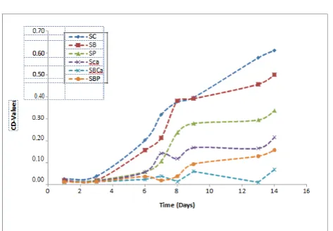
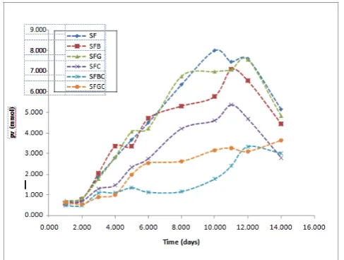
caffeic acid in the presence of BSA. For emulsions without BSA, the caffeic acid emulsion was more stable than the phenol emulsion although their antioxidant activity showed no significant difference (p>0.05). The determination of the primary oxidation products by means of the conjugated diene values also followed the same trend with the PV analysis as shown in Figure 2. Caffeic acid antioxidant activity was strongly increased in the presence of BSA and there was a significant difference (p<0.05) between the individual caffeic acid antioxidant activity from the combination of caffeic acid and BSA antioxidant activity. Similarly, the antioxidant activity of phenol and caffeic acid in the absence of BSA was not significantly different (p>0.05) but caffeic acid was still considered more active than the phenol because the emulsion was more stable with the caffeic acid. Figure 3 shows the trend in the increase of the p-Anisidine value for monitoring the secondary oxidation products. The antioxidant activity order was SBCa>SCa>SBP>SP>SB. The combination of caffeic acid and BSA was more active than the individual caffeic acid or BSA. However, in this case the caffeic acid in the absence of BSA was more active than individual phenol and the combination of phenol and BSA although there was no significant difference (p>0.05) among them. The other set of emulsions prepared with caffeic acid with or without BSA or gelatin and stored at 60°C was monitored for both primary and secondary oxidation by determining the peroxide values and the p-anisidine values, thereby investigating the antioxidant activity of caffeic acid in the presence of the proteins. At the initial stage of the oxidation, the PV of the emulsions was not significantly different (p>0.05) from one another although the PV of the SFBC was low compared with the others. Figure 4 showed the increase in the PV of the emulsions as observed during the storage period. By day 5 there was a significant difference (p<0.05) in the PV of SFG and SFB from SFBC and SFGC and by day 8 there was a significant difference (p<0.05) in the PV between the SFC from SFBC and SFGC. SFBC and SFGC showed no significant difference (p>0.05) in their antioxidant activity but SFBC was still considered to be more active as the emulsions were more stable. SFB and SFG were not significantly different (p>0.05) from each other and from the control as well. At the later stage of the oxidation, SFC was not significantly different (p>0.05) from the SFGC, however, SFGC oxidative stability was high. Table 1 shows the significant in difference of the oxidative stability of the additives in the emulsions by the end of the study. The increase in the PA value as monitored during storage is shown in Figure 5. The highest PA value 11.185 was reached at day 12 in SFC and the lowest at this point was 1.960 in SFBC. There was no significant difference (p>0.05) between SFC, SFB and SFG. SFBC, and SFGC were significantly different (p<0.05) from SFB and SFG. Though SFGC emulsions were more stable, its antioxidant activity was not strongly significantly different from SFC. Similarly, in line with the PV result, the antioxidant activity of the SFBC was significantly different (p<0.05) from SFC. The total oxidative value (TOTOX) of the SBCa, SFBC, and SFGC from the two sets of emulsions was low compared to SCa and SFC. The BSA itself has no antioxidant activity but usually used as additives in food and functions as an
emulsifier, an important surface-active agent that stabilizes emulsions. The two sets of emulsions investigated for lipid oxidation in the presence of BSA with the phenol extract and caffeic acid as antioxidants showed retardation in lipid oxidation thereby resulting in a more stable emulsion during storage compared to the emulsions with the individual antioxidants or the individual protein. These findings agree with the study of the synergistic effect of BSA on the antioxidant activity of Caffeic acid and Epigallocatechin gallate (EGCG) and to a lesser extent in Trolox shown by Almajano et al. The lower stability of the emulsion containing gelatin with caffeic acid compared with the emulsion containing BSA with caffeic acid might be due to the type of interactions between gelatin and caffeic acid and the source of the gelatin which determines its emulsifying and surface-active capacity [12,22].
Figure 1. The trend of monitored peroxide values of sunflower oil-in-water emulsion containing antioxidants and proteins. SC, control sample; SB, emulsion with BSA only; SP, emulsion with phenol only; SCa, emulsion with caffeic acid only; SBCa, emulsion with BSA and caffeic acid; SBP, emulsion with BSA and phenol.
Figure 3. The trend of monitored p-anisidine values of sunflower oil-in-water emulsion containing antioxidants and proteins. SC, control Sample; SB, emulsion with BSA only; SP, emulsion with phenol only; SCa, emulsion with caffeic acid only; SBCa, emulsion with BSA and caffeic acid; SBP, emulsion with BSA and phenol.
Figure 4. The trend of monitored peroxide values of sunflower oil-in-water emulsion containing antioxidants and proteins. SF, control Sample; SFB, emulsion with BSA only; SFG, emulsion with gelatin only; SFC, emulsion with caffeic acid only; SFBC, emulsion with BSA and caffeic acid; SFGC, emulsion with gelatin and caffeic acid.
Figure 5. The trend of change in the monitored p-anisidine values of sunflower oil-in-water emulsion containing antioxidants and proteins. SF, control sample; SFB, emulsion with BSA only; SFG, emulsion with gelatin only; SFC, emulsion with caffeic acid only; SFBC, emulsion with BSA and caffeic acid; SFGC, emulsion with gelatin and caffeic acid.
Table 1. The antioxidant potential of caffeic acid in the presence and absence BSA and gelatin by FRAP assay.
Samples FRAP (µM)
BSA 0.08 ± 0.02
Gelatin 0.08 ± 0.02
Caffeic acid 1.20 ± 0.08
BSA-Caffeic acid 1.10 ± 0.05
Gelatin-Caffeic acid 0.63 ± 0.01
Antioxidant partitioning behaviour
extract resulting in loss of hydroxyl group from the phenol, reducing its affinity to the aqueous phase, as a result reducing its solubility and then it’s partitioning into the aqueous phase. Schwartz, Frankel and German described the decrease in the proportion of hydrophilic antioxidants in the aqueous phase in an emulsified mixture at a lowered pH. The phenol extract bound to BSA might be more dispersed at the oil-water interface where the BSA acts as a surface-active agent. Partitioning behavior of the antioxidant in a heterogeneous system is an important parameter to be considered when choosing an antioxidant. The distribution of prooxidants and antioxidants in a heterogeneous system and their contact with the lipid component of fatty foods determine the oxidative stability of the fatty acids. Phenolics are water-soluble and polar antioxidants that are effective in retarding lipid oxidation in bulk oil and less effective in an emulsion because they go into the aqueous phase, unlike the lipophilic antioxidants that are effective in an emulsion because they are located at the oil-water interface where oxidation is initiated [1,25].
Ferrous reducing antioxidant power (FRAP) assay
The potential of the antioxidant, caffeic acid to inhibit the oxidative effect of reactive oxidative species (ROS) in the absence and presence of BSA and gelatin was determined by Ferric reducing/antioxidant power (FRAP) assay as previously described. The principle behind the FRAP assay is the reduction of ferric tripyridyl triazine (Fe III-TPTZ) to a blue-colored ferrous form. This is a redox reaction that occurs at a low pH because the antioxidant functioned as a reductant in reducing the ferric ion to ferrous and this is measured at 510nm. The higher the absorbance reading the better is the total reducing potential of the antioxidant. The FRAP value of caffeic acid in the presence of BSA was higher than the FRAP value of the Individual BSA. Caffeic acid in the presence of gelatin FRAP value was as well higher than the FRAP value for the individual gelatin. The FRAP value of caffeic acid in the presence of BSA was higher than the FRAP value of caffeic acid in the presence of gelatin. However, the FRAP value of caffeic acid was higher than that of BSA-caffeic acid, gelatin-caffeic acid, BSA, and gelatin (Table 1). This result was in line with the study by Aewsiri et al that described the FRAP value of free tannic acid to be higher than the FRAP values of the gelatin modified with tannic acid and oxidized tannic acid. The reduced antioxidant potential of caffeic acid in the presence of protein showed by FRAP assay might be attributed to the loss of free hydroxyl groups in binding to the protein and the unavailability of the phenol hydroxyl groups of an antioxidant might reduce the antioxidant potential. Though the FRAP value showed that caffeic acid had the highest antioxidant potential but this does not correlate with its ability to retard lipid oxidation in an emulsion since the oxidative stability of the emulsion containing caffeic acid was reduced compared to the BSA and caffeic acid-containing emulsion as well as the gelatin and caffeic acid-containing emulsion. This less antioxidant activity of caffeic acid in the emulsion might be due to its partitioning behavior in the multiphasic system. Caffeic acid is a water-soluble, hydrophilic antioxidant having two hydroxyl groups on its molecular structure. Being
hydrophilic, its affinity for aqueous phase is more than for the lipid phase or the oil-water phase and since oxidation is at the oil-water phase and not in the aqueous phase its ability to retard oxidation is hence reduced. BSA and gelatin, on the other hand, are surface-active that have got no antioxidant potential but are emulsifiers that stabilize an emulsion. The increased antioxidant activity of the caffeic acid in the presence of BSA and gelatin resulting in high oxidative stability in an emulsion is attributed to its transportation to the oil-water phase by its binding with BSA and gelatin forming a BSA-antioxidant and gelatin-BSA-antioxidant adduct [12,26-29].
Confocal microscopy
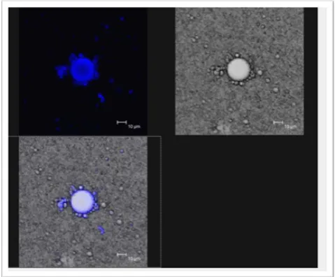
The microstructure of the emulsion containing caffeic acid and BSA was examined with the Confocal Laser Scanning microscope. The microstructure examination result showed that the BSA was homogeneously distributed on the surface of the oil droplets. The best images were obtained with a Nile Red / Nile blue mixture as shown in Figure 6. This further explained why and how in the presence of BSA, caffeic acid performed well in retarding lipid oxidation in an emulsion. Binding itself to BSA, it is being transported to the oil-water phase where oxidation takes place and becomes effective in inhibiting the formation of lipid hydroperoxides [1,12].
Figure 6. Confocal microscopy images of the distribution of protein molecules on the oil droplets in an oil-in water emulsion stained with Nile Red/Nile Blue dye.
Conclusion
antioxidants combine with protein molecules present in the food.
References
1. McClements DJ, Decker EA. Lipid oxidation in oil-in-water emulsions: Impact of molecular environment on chemical reactions in Heterogeneous food systems. J Food Sci 2000;65:1270-82.
2. Nawar WW. Food Chemistry. In Food Science and Technology, 3rd ed.; Fennema, OR, Ed.; Marcel Dekker: New York 1996;225-319.
3. Iqbai S, Bhanger MI. Stabilization of Sunflower oil by garlic extract during accelerated storage. J Food chem 2007;100:246-54.
4. Halliwell B, Murcia NA, Chirico S, et al. Free Radicals and Antioxidants in food and in vivo: What they do and how they work in Crit Rev Food Sci Nutri 1995;35:17-20. 5. Coupland JN, McClements DJ. Lipid Oxidation in Food
Emulsions. Trends of Food Sci Tech 1996;7:83-91.
6. Seo HS, Kwak SY, Lee YS. Antioxidant activities of histidine containing caffeic acid-dipeptides. Bioorganic and medicinal chemistry letters 2010;20:4266-72.
7. Jung UK, Lee SK, Hun S, et al. Antioxidant effects of Natural lecithin on borage oil. Food Science and Biotechnology 2001;3:1-6.
8. Porter WL. Paradoxical behaviour of antioxidants in food and biological systems. Toxicology and industrial Health 1993;9:93-122.
9. Djordjenic D, Cercai L, Alamed J, et al. Stability of Citral in protein-gum Arabic-stabilised oil-in water emulsions. J Food Chem 2008;106:698-705.
10. Almajano MP, Delgado ME, Gordon MH. Changes in the antioxidant properties of protein solutions in the presence of epigallocatechin gallate. Journal Food Chemistry 2007;101:126-30.
11. Aewsiri T, Benjakul S, Visessanguan W, et al. Antioxidative activity and emulsifying properties of cuttle fish skin Gelatin modified by oxidised phenolic compounds. J Food Chem 2009;117:160-8.
12. Almajano MP, Gordon MH. Synergistic Effect of BSA on Antioxidant Activities In Model Food Emulsions. J Agr Food Chem 2004;81:275-80.
13. Aewsiri T, Benjakul S, Visessanguan W, et al. Chemical compositions and functional properties of Gelatin from pre-cooked tuna fish. Int J Food Sci Technol 2008;43:685-93. 14. Montedoro GF, Servili G, Baldioli M, et al. Simple and
Hydrolyzable Phenolic compounds in virgin olive oils. Their extraction, separation, and Quantitative and semi quantitative evaluation by HPLC. J Agri Food Chem 1992;40:1571-6.
15. Pirisi FM, Cabras P, Falqui Cao C, et al. Phenolic Compounds in virgin olive oil. Reappraisal of the extraction, HPLC separation and quantification procedures. J Agri Food Chem 2000;48:1191-6.
16. Papoti VT, Tsimidou MZ. Looking through the qualities of a fluorimetric assay for the total phenol content estimation
in virgin olive oil, olive fruit or leaf polar extract. J Food Chem 2009;112:246-52.
17. Yoshida H. Influence of Fatty Acids of different unsaturation in the oxidation of purified vegetable oils during microwave irradiation. J Sci Food Agric 1993;62:41-7.
18. Nuchi CD, McClements DJ, Decker EA. Impact of Tween 20, hydroperoxides and iron on the oxidation of methyl linoleate and salmon oil dispersions. J Agri Food Chem 2001;49: 4912-19.
19. Shantha NC, Decker EA. Rapid sensitive, iron-based spectrophotometric methods for determination of peroxide values of food lipids. Journal of AOAC International 1994;177: 421-4.
20. Benzie IFF, Strain JJ. he ferric reducing ability of plasma (FRAP) as a measure of “antioxidant power”: The FRAP assay. Analytical Biochemistry 1996;239:70-6.
21. Tsimidou M, Papadopoulos G, Boskou D. Determination of Phenolic compound in virgin olive oil by Reversed-phase HPLC with Emphasis on UV detection. J Food Chem 1992;44:53-60.
22. Aewsiri, SoottawatBenjakul, WonnopVisessanguan, et al. Antioxidative activity and emulsifying properties of cuttle fish skin gelatin-tannic complex as influenced by types of interaction. Innovative Food Science and Emerging Technologies 2010;11:712-20.
23. Huang SW, Frankel EN, Aeschbach R, et al. Partitioning of Selected Antioxidants in Corn oil-water Model Systems. J Agr Food Chem 1997;45:1991-4.
24. Di Mattia CD, Sacchetti G, Mastrocola D, Pittia P. Effect of phenolic antioxidants on the dispersion state and chemical stability of olive oil O/W emulsions. Food Research International 2009;42:1163-70.
25. Schwarz K, Frankel EN, German JB. Partitioning Behaviour of Antioxidative Phenolic Compounds in Heterophasic System. Fett / Lipid 1996;98:115-21.
26. Aewsiri, SoottawatBenjakul, WonnopVisessanguan, et al. Antioxidative activity and emulsifying properties of cuttle fish skin gelatin-tannic complex as influenced by types of interaction. Innovative Food Science and Emerging Technologies 2010;11:712-20.
27. Bonoli-Carbognin M, Cerretani L, Bendini A, et al. Bovine serum albumin produces a synergistic increase in the antioxidant activity of virgin olive oil phenolic compounds in oil-in water emulsions. Journal of Agricultural and Food Chemistry 2008;56:7076-81.
28. AOCS Official method cd 18-90 In official Methods and Recommended Practices of the American oil Chemists’ Society, 4th ed.; Firestone, D. Ed.; American oil Chemists’ Society: Champaign, IL I 1989;.
29. AOCS official method Ti La-64, In official Methods and Recommended Practices of the American Oil Chemists’ Society: Champaign, IL 1989;.
*Correspondence to:
Department of Food and Nutritional Sciences The School of Chemistry, Food and Pharmacy University of Reading
Whiteknights, Reading RG6 6AP
UK
Tel: 2348144074461