R E V I E W
Open Access
Engineering in-vitro stem cell-based
vascularized bone models for drug
screening and predictive toxicology
Alessandro Pirosa
1, Riccardo Gottardi
1,2, Peter G. Alexander
1and Rocky S. Tuan
1*Abstract
The production of veritable in-vitro models of bone tissue is essential to understand the biology of bone and its surrounding environment, to analyze the pathogenesis of bone diseases (e.g., osteoporosis, osteoarthritis, osteomyelitis, etc.), to develop effective therapeutic drug screening, and to test potential therapeutic strategies. Dysregulated interactions between vasculature and bone cells are often related to the aforementioned pathologies, underscoring the need for a bone model that contains engineered vasculature. Due to ethical restraints and limited prediction power of animal models, human stem cell-based tissue engineering has gained increasing relevance as a candidate approach to overcome the limitations of animals and to serve as preclinical models for drug testing. Since bone is a highly vascularized tissue, the concomitant development of vasculature and mineralized matrix requires a synergistic interaction between osteogenic and endothelial precursors. A number of experimental approaches have been used to achieve this goal, such as the combination of angiogenic factors and three-dimensional scaffolds, prevascularization strategies, and coculture systems. In this review, we present an overview of the current models and approaches to generate in-vitro stem cell-based vascularized bone, with emphasis on the main challenges of vasculature engineering. These challenges are related to the choice of biomaterials, scaffold fabrication techniques, and cells, as well as the type of culturing conditions required, and specifically the application of dynamic culture systems using bioreactors.
Keywords:tissue engineering, vascularized bone, three-dimensional microphysiological systems, biomaterial, scaffold, preclinical model, bioreactors
Background
Bone architecture and development
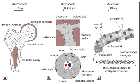
Bones are hierarchically organized over multiple length scales. Macroscopically, bone can be divided into com-pact and trabecular tissues, each with very different mechanical strength and stiffness (Fig. 1a). The micro-scopic architecture of the former is characterized by osteons and Haversian channels containing nerves and blood supply, whereas the latter is comprised of inter-connected trabeculae and the presence of the bone mar-row [1] (Fig.1b). At a molecular level, bone extracellular matrix (ECM) is composed largely of the fibrous macro-molecule collagen type I and mineralized inorganic
hydroxyapatite crystals [2] (Fig. 1c). Bone is primarily vascularized by an arterial network, but within the bone marrow cavity and the Haversian channels the vascula-ture branches into thin-walled capillaries, whose funda-mental role is the exchange of nutrients and signals between blood and bone cells [3].
Bone fulfills a wide range of physiological functions. As essential structural and load-bearing elements, bones rep-resent the foundation of physical locomotion and protect our internal organs. Due to the presence of the bone mar-row, long bones serve as the tissue origin of the biological components required for hematopoiesis. Moreover, osse-ous tissues can trap potentially harmful metals (e.g., lead), as well as maintain the homeostasis of key electrolytes via calcium and phosphate ion storage [4]. Bone formation can occur through two distinct pathways, intramembra-nous and endochondral ossification. In both cases, the first step is the condensation of mesenchymal cells to * Correspondence:[email protected];[email protected]
1Center for Cellular and Molecular Engineering, Department of Orthopaedic Surgery, University of Pittsburgh School of Medicine, 450 Technology Drive, Pittsburgh, PA 15219, USA
Full list of author information is available at the end of the article
© The Author(s). 2018Open AccessThis article is distributed under the terms of the Creative Commons Attribution 4.0
produce a template for subsequent bone formation [5]. Intramembranous bone formation (typical of flat bones) involves the direct differentiation of mesenchymal pro-genitor cells into osteoblasts, whereas endochondral ossifi-cation (typical of long bones) involves the initial differentiation of the mesenchymal progenitor cells into chondrocytes, followed by their hypertrophy, matrix mineralization, and replacement by bone tissue [6]. Both intramembranous and endochondral ossification occur in close proximity to vascular ingrowth. During intramem-branous ossification, capillaries invade the differentiating mesenchymal zone, whereas in endochondral ossification, hypertrophic chondrocytes recruit the infiltrating vascula-ture. Initial vascularization is followed by invasion of oste-oclasts and osteoblasts, with coordinated resorption of hypertrophic cartilage and subsequent mineralization of the ECM and bone formation [7]. Hence, vascularization occurs from the ossification centers toward the growth plate and determines the rate of bone ossification [8]. It is interesting to note that many of the processes that occur during long bone formation are recapitulated during frac-ture healing [9], underscoring the importance of know-ledge of bone development for the design of more effective strategies for bone repairing [6]. Vascular endo-thelial growth factor (VEGF) is a key regulator of angio-genesis and thus bone development [10]. For instance, VEGF-A gene depletion was shown to attenuate the re-sorption of hypertrophic chondrocytes and bone
formation, highlighting the role of VEGF-dependent angiogenesis during bone formation [11]. VEGF-A levels were also found to be dependent on the oxygen-sensing hypoxia-inducible factor (HIF)-1αpathway in the modula-tion of bone mass, thus confirming the role of neoangio-genesis for the homing of osteoblast progenitor cells and for providing bone formation/promoting factors [12]. In fact, besides bone development, vasculature is also essen-tial for bone remodeling, and fracture healing is also highly dependent on the vasculature. Dysregulated inter-actions between the vasculature and bone cells are the basis of a number of different pathologies [13]. Some of these, such as avascular necrosis and osteoporosis, are as-sociated with a diminished vascular supply [14, 15], whereas others such as Gorham–Stout disease, a form of idiopathic osteolysis caused by abnormal proliferation of vascular structures originating in the bone [16], and Klippel-Trénaunay syndrome, characterized by vessel mal-formations and overgrowth of bones and soft tissues [17], are caused by excessive vascularization.
Bone and related pathologies
Bone diseases can cause loss of bone strength and density, and they may arise from nutrient deficiencies, abnormal development, genetic disorders, impaired vasculature, and other causes. Table1summarizes and compares the main pathologies affecting bone.
Osteoporosis refers to the loss of bone density result-ing from an altered balance of the bone remodelresult-ing process, and affects approximately 10 million US adults 50 years of age and older [18]. The most widely used osteoporosis treatment is the administration of bispho-sphonates, which shorten the osteoclast life span and in-hibit bone resorption [19]. Although general risk factors of osteoporosis are well documented, little is known about the role of vasculature [20]. Some studies have re-vealed a connection between low bone mineral density and increased cardiovascular morbidity/mortality [21, 22]. Endothelial cells (ECs) are known regulators of vascular tone by releasing vasodilator molecules, such as nitric oxide (NO), and they have been addressed as a po-tential link between cardiovascular diseases and osteo-porosis. Studies in rats showed that the inhibition of NO production or NO synthase (NOS) activity was followed by marked bone loss [23, 24], while human studies revealed lower NOS expression resulting from estrogen deficiency [25–27]. Since the presence of estrogen receptors has been found in human ECs [28, 29], it is possible that estrogen deficiency seen in postmenopausal women could alter the endothelial function of bone microcirculation. Although these studies suggest that endothelial dysfunction may play a role in the develop-ment of osteoporosis, the exact causal relationship has yet to be determined.
Osteoarthritis is the main cause of disability in the USA [30], and its hallmark is the progressive degener-ation of cartilage. However, OA affects the whole joint
and all tissues play a role in the disease [31]. In particu-lar, the subchondral bone has been reported to be crit-ical in the pathogenesis of OA [32]. During movement, there is continuous functional interaction across the osteochondral junction. Under the diseased state, altered mechanical loading in cartilage induces changes in bone and vice versa [33,34]. The communication between the two tissues, however, is not limited to mechanical coup-ling and the associated mechanotransduction. Recent evidence indicates that the calcified cartilage and sub-chondral bone are not an impermeable barrier, and some molecules are capable of diffusing across the osteochon-dral junction [35–38]. Blood vessels and microchannels have been found to reach from the subchondral bone all the way to the uncalcified cartilage, and there is evidence of contact between uncalcified cartilage and subchondral bone and the marrow spaces [33, 39–41]. During OA, the osteochondral junction is significantly altered, allow-ing greater transport and cellular crosstalk between cartilage and bone [32,38,42]. Another hallmark change of the osteochondral junction occurring during OA is in-creased vascularization and neoangiogenesis [38, 43], which may further contribute to the molecular crosstalk between cartilage and bone. Part of this signaling in-volves an increase in the VEGF level in osteoarthritic chondrocytes compared to those in healthy cartilage [43], possibly contributing to the induction of vascular invasion as part of a proregenerative mechanism. In turn, ECs have recently been reported to enhance chon-drogenic differentiation of mesenchymal stem cells
Table 1Etiology, current treatments, and role of vasculature in the main pathologies affecting bone
Etiology/risk factors Current treatments Role of vasculature
Osteoporosis, loss of bone density
Altered balance of bone remodeling: greater bone removal by osteoclasts and then production by osteoblasts [18]
Administration of bisphosphonates, which shorten osteoclast life span and inhibit bone resorption [19]
Possible link between decreased production of vasodilator molecules by endothelial cells and increased bone loss [20]
Osteoarthritis, progressive degeneration of cartilage and bone
Traumatic, congenital, postoperative, metabolic, endocrine; age, joint overuse, obesity are common risk factors [274]
Symptomatic treatments through physiotherapy, orthopedic aids and orthoses, pharmacotherapy, total joint replacement [275]
Increased vascularization and neoangiogenesis in the joint; increase in VEGF level in osteoarthritic chondrocytes [43]
Osteomyelitis, infection within bone
Infection byStaphylococcus aureus, but also by other Gram-negative cocci and Gram-positive bacilli [45]
Parenteral course of broad-spectrum antibiotics and surgical debridement [46]
Poor vascularity can cause both development of the infection and resistance to antibiotics [46]
Osteonecrosis, death of bone cells, arthritis, and destruction of bone
Inadequate vascular supply to the bone; long-term steroid treatment, alcohol abuse, joint injury, arthritis, cancer are common risk factors [52]
Nonsteroidal anti-inflammatory drugs to reduce pain and inflammation; bone surgery, grafting, and joint replacement [276]
Compromised subchondral
microcirculation, vascular interruption, intravascular occlusion, and extravascular compression [277]
Fractures, loss of bone contiguity
Mainly trauma; osteoporosis, low mineral density, age, tumors are common risk factors [278–280]
Fracture reduction and immobilization; bone autograft, allograft, or synthetic materials [59]
Vascular supply is critical for fracture healing; VEGF treatment can enhance fracture repair [57]
Osteosarcoma, bone malignancy
Occurring mostly in the medullary cavity of long bones: environmental factors, chromosomal abnormalities, p53 mutation are common risk factors [72]
Depending on the stage, chemotherapy, radiation therapy, surgery (amputation, grafting, local excision) [281]
Vasculature is critical for tumor survival, osteosarcoma generally involves downregulation of anti-angiogenic factors [73,74]
(MSCs) [44], suggesting the potential of significant mo-lecular interplay between subchondral bone vasculature and cartilage, an aspect that has not been much investi-gated. Overall, increased vascularity in the subchondral bone is associated with OA severity in cartilage and with clinical disease activity [33].
Another pathogenic bone condition with devastating consequences is osteomyelitis (OM). OM can be broadly defined as an infection within the bone and is classified by duration (acute or chronic), pathogenesis (trauma, contiguous spread, hematogeneous, surgical), site, ex-tent, or type of patient [45]. Poor vascularity is a prime cause for both the development of an infection and re-sistance to antibiotics [46]. Acute OM can be eradicated before osteonecrosis occurs if the infection is treated promptly and aggressively with antibiotics and surgical debridement [46]. However, in an established/chronic in-fection, fibrous tissue and chronic inflammatory cells en-capsulate the infected site, reducing vascular supply, inhibiting an effective inflammatory response, and limit-ing the action of antibiotics [46]. In addition, the bac-teria become encapsulated within a biofilm, that both protects the bacteria from the body’s defenses and anti-biotics and serves as a chronic source of new bacterial infection [45–47]. Several therapies intended to enhance blood flow to vascular or chronically infected areas have been tested in experimental models or clinical trials, in-cluding hyperbaric oxygen therapy, PRP, and VEGF de-livery [47–49]. Recently, a VEGF gene-transfected muscle flap was shown to be effective as a treatment to supplement systemic antibiotic treatment in the manage-ment of experimanage-mental OM in a rodent model [50, 51]. These strategies seek to enhance the body’s own defense and the effectiveness of antibiotics by enhancing blood flow to avascular tissue.
Osteonecrosis, or avascular necrosis (AVN), occurs when the blood supply to the bone is disrupted, pre-cipitating death of the bone cells, arthritis, and destruc-tion of the hip joint. Common risk factors include long-term steroid treatment, alcohol, abuse, joint in-jury, arthritis, and cancer—all associated with altered blood supply [52]. The risk greatly increases with cor-ticosteroid use (to treat pain and inflammation), bisphosphonate (to prevent bone loss), and anti-angiogenic therapy (in the treatment of some cancers or leprosy) [53,54]. Areas susceptible to AVN have sev-eral common characteristics: they have limited routes of vascular supply, they undergo relatively high rates of bone turnover, and they may have higher than normal exposure to bacterial infections [55]. Evidence that re-duced vascular supply causes AVN is indicated by the rise in AVN incidence following anti-angiogenic treat-ments used in cancer therapy, such as the VEGF-speci-fic antibody bevacizumab, the tyrosine kinase inhibitor
sunitinib (Sutent), and the mTOR inhibitor rapamycin, and new treatments for leprosy, such as lenalidomide. Osteoclasts and blood vessels are closely associated during bone remodeling, and recent studies indicate that osteoclasts promote angiogenesis through secre-tion of factors such as matrix metalloproteinase 9 (MMP9). Poor vascularization occurs in certain inflam-matory and immunosuppressive states and in the pres-ence of infection as well. While the pathogenesis of AVN is largely agreed upon, the cellular and molecular mechanisms are less understood, and thus counter-treatments to prevent the condition during anti-resorptive and anti-cancer treatments are not available.
Trauma-related injuries can lead to bone fractures, whose healing is critically dependent upon an adequate vascular supply. Breakage of the bone initiates the frac-ture healing process, which begins with an inflammatory reaction, followed by the processes typical of endochon-dral ossification for a period of up to 28 days. Neovascu-larization is critical for successful bone formation, since vascular endothelium interruption is the first event fol-lowing trauma, which could lead to the formation of necrotic tissue. Experimental evidence showed impaired bone formation with administration of anti-angiogenic factors [56], whereas VEGF treatment enhanced fracture repair [57]. Clinical evidence showed that in the pres-ence of decreased vascular perfusion to the fracture, the incidence of impaired healing (delayed union or non-union) increases from 10–15% to 46% [58]. Standard treatment to augment bone healing and prevent delayed union or nonunion is represented by the use of auto-graft, alloauto-graft, or synthetic materials [59]. Bone marrow-derived endothelial progenitor cells (EPCs) par-ticipate in the generation of new blood vessels (vasculo-genesis) at the site of injury [60,61], and they have been shown, in combination with bone marrow-derived MSCs, to augment bone healing [62,63]. Significant pro-gress in improving fracture healing is hindered by the limited knowledge of the molecular mechanisms leading to nonunions; thus, a better understating of the bio-logical pathways involved in this process would certainly benefit from the development of specific clinical therapies.
reservoir of lead, contributing to systemic toxicity [68], and it is the target tissue for the actions of a number of teratogens (e.g., valproic acid, thalidomide, etc.) [69,70]. Studying the mechanism of how these toxins act at the cellular and molecular levels with bone is indeed crucial to developing new treatments.
Osteosarcoma is the most common bone malignancy af-fecting predominantly adolescents and young adults, with a 5-year survival rate of about 50–60% [71]. Osteosarcoma occurs mostly in the medullary cavity within the metaphy-sis of long bones, an active bone growing region, and then can propagate to the bone cortex and the surrounding soft tissues. The molecular pathogenesis of this tumor is quite complex and involves several elements, such as environ-mental factors (e.g., UV and ionizing radiation, methylcho-lanthrene and chromium salts, etc.), chromosomal abnormalities, p53 mutations, and so forth [72]. As for many other tumors, vasculature is a critical factor for the survival and proliferation of cancer cells; in fact, osteosar-coma generally involves downregulation of anti-angiogenic factors, such as thrombospondin-1 [73] and pigment epithelium-derived factor (PEDF) [74]. Although osteosarcoma is known as a vascular tumor, there are contradictory data about the correlation between the microvascular density/VEGF expression and the forma-tion of metastasis [75, 76]. Emerging evidence suggests that the blood vessels of bone may play a role in the inter-actions between other tumors and the bone microenviron-ment in the pathogenesis of bone metastasis [77]. The extravasation of tumor cells can be related to the molecu-lar receptors typical of a tissue/organ-specific blood vessel [78]. Interestingly, blood vessels located in the metaphysics of long bones express specific adhesion pro-teins, such as P-selectin and E-selectin, which have been shown to promote interaction and subsequent adhesion of tumor cells [79]. One widely studied example of this kind of interaction is the preferential spreading of breast cancer metastasis to bone. In fact, breast cancer cells express chemokine receptors, integrins, cadherins, and bone-remodeling factors that contribute to the successful and preferential spread of tumor to bone [79].
Due to the multifactorial nature of bone diseases, the aging population, and increased occurrence of trauma-related injuries, bone disorders represent a big concern and the current treatments do not provide optimal out-comes. Furthermore, any alteration in the vascular supply may lead to an increased susceptibility to osteo-porosis, osteonecrosis, and osteomyelitis [8]. Animal models have been traditionally utilized in research to mimic these human pathologies. Nevertheless, on mul-tiple occasions, animal models have been unsuccessful and unreliable in predicting the processes and develop-ment of human pathologies and the response to drug candidates, because their physiology is fundamentally
different from that of humans. To overcome this defi-ciency, tissue engineering may represent an alternative platform by establishing in-vitro pathophysiological models based on human cells to study the causes and progression of specific diseases, and to develop and test candidate therapeutic strategies.
In-vitro modeling of 3D vascularized bone models through tissue engineering approaches
The successful development of in-vitro engineered bone is critically dependent on the ability to introduce a 3D vascular network that guarantees adequate oxygenation, mass transfer, nutrient delivery, and by-product removal [80]. Here, we summarize the challenges in generating in-vitro vascularized bone constructs and the main ma-terials, scaffolds, and cells required to achieve this goal, with a focus on stem cell employment.
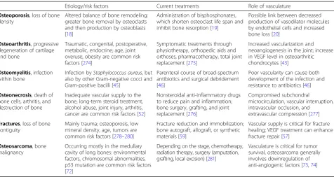
Challenges of in-vitro vascularized bone engineering Bone is a highly vascularized organ and the development of vasculature and mineralized matrix requires a syner-gistic interaction between osteogenic and endothelial precursors [81]. These mechanisms have been studied in animal models, but they are still not understood due to the complexity of the in-vivo environment. In-vitro scale-up of bioengineered tissues is known to be limited by diffusion issues; therefore, the establishment of a functional vasculature within the construct could be es-sential to generate an accurate and large enough model capable of mimicking the native tissue. Current vascularization strategies comprise the use of angiogenic factors combined with 3D scaffolds (Fig. 2a), prevascu-larization strategies (Fig. 2b), and the use of coculture systems (Fig.2c) [82,83].
namely cell type (osteogenic and vasculogenic), media, seeding methodology, culture (static or dynamic), scaf-folds, and microenvironment (e.g., oxygen tension) [83]. In the past decade, different attempts to generate vascu-larized bone constructs using osteogenic precursor cells and vascular progenitor cells have been made, but the majority of these studies were conducted through in-vivo models [86–88]. It has been difficult to create a ver-itable in-vitro model containing bone tissue and vasculature-like structures within the same construct. A recent attempt to introduce simultaneously osteogenic differentiation and vasculature development in-vitro was made by Tsigkou et al. [89], which combined human bone marrow MSCs and human umbilical vein endothe-lial cells (HUVECs) seeded on a polymeric scaffold and
in a hydrogel, respectively. They observed the formation of capillary-like structures 4–7 days after implantation in a mouse model, and MSCs were found to be necessary for the development of a stable vasculature. The main outstanding issue is still the generation of accurate in-vitro models, due to the difficulties of obtaining quality mineralized bone matrix, further complicated by the introduction of the vasculature.
Bone mineralized matrix formation is provided by os-teoblasts during embryonic development and postnatal growth, yet about 95% of the cellular population residing in the adult skeleton is represented by osteocytes. Osteocytes are the terminally differentiated osteoblasts embedded in the lacunae within the bone matrix. They are characterized by cellular processes radially spread
Fig. 2Vascularization strategies for engineered bone constructs.aScaffold loading with proangiogenic factors in-vivo.bScaffold preseeding with
toward the mineralized matrix inside tiny tunnels called canaliculi, filled by canalicular fluid [90]. The presence of osteocytes in an engineered bone construct is an indi-cation of a mature developed osseous tissue. Although osteocytes play a secondary role in bone tissue forma-tion, they are functionally important in its homeostasis, mechanosensation, and mechanotransduction [91, 92]. Osteocytes sense canalicular fluid flow-derived shear stress through their primary cilium [92] and specific ion channels and mechanotransduction proteins, releasing in response paracrine signaling molecules such as NO, ATP, and prostaglandins, which regulate osteoblast and osteoclast activity [93–95]. The lacuna–canalicular net-work connects the osteocytes to the vasculature and there is evidence linking osteocyte apoptosis and re-duced VEGF production with consequent impaired vas-cularity and bone strength [96]. Taken together, these findings highlight the importance of the presence of mature osteocytes in an engineered bone. For this rea-son, many investigations are focused on the derivation of mature osteocytes from stem cells to better under-stand their physiology and assess their potential in bone engineering [97–99].
Bone is also innervated by sensory and sympathetic nerve fibers that, in addition to skeletal pain transmis-sion, play a role in bone metabolism. Bone cells have been recently shown to express adrenoceptors, receptors for norepinephrine (NE), calcitonin gene-related peptide (CGRP), substance P, and vasoactive intestinal peptide (VIP) [100, 101]. NE reuptake influences bone remodel-ing by osteoclasts [102], CGRP deficiency was linked to decreased bone formation in mice [102], while SP has been shown to have both dose-dependent bone-resorbing and formation activity [102]. These are just a handful of examples on how innervation has been linked to bone production and turnover; thus, an in-vitro engi-neered bone construct would certainly benefit from the presence of nerve endings and related neurotransmitters. However, the generation of a vascularized and inner-vated bone construct clearly presents an additional level of complexity in terms of integrating and regulating such different tissue types [103].
Another parameter to consider when developing verit-able in-vitro vascularized bone is the incorporation of osteoclast activity inside the engineered constructs. Osteoclasts are essential for bone remodeling by resorb-ing bone matrix and generatresorb-ing physical space for osteo-blasts and ECs, thus allowing the formation of new bone tissue and vasculature [104]. The dissolution of the inorganic phase of bone matrix takes place by the secre-tion of hydrochloric acid, while the organic matrix is resorbed by secreted enzymes, like cathepsin K and me-talloproteinase 9 [105]. The resorption activity as well as the area and depth of the resorption pits can be analyzed
in-vitro using osteoclast-like cells derived from mono-cytes, for instance the RAW 264.7 cell line [106]; how-ever, to date, there are considerable variations in the examined parameters. There is no specific, single func-tional osteoclast cell marker; thus, different parameters should be analyzed together, such as tartrate-resistant acid phosphatase 5b (TRAP 5b) [107], morphological changes from monocytes [104], and 3D volume characterization of pits [108]. Furthermore, as different materials can affect the response of osteoclasts, the choice of the scaffold is critical for an optimized bone engineering strategy. Differ-ent authors have studied the resorption ability of osteo-clasts using different scaffolds/materials, from dentin to bone substitutes, as well as calcium phosphate and bio-active glasses [109–112], and have observed specific re-sorption rates depending on the material used. For example, Keller et al. [112] found that natural bioma-terials were resorbed more rapidly than synthetic ones, while Badran et al. [111] showed an inhibition of osteoclast activity by increasing mineral density.
As highlighted by Alexander et al. in a recent minire-view [31], a TE approach can be used for the develop-ment of in-vitro preclinical models of normal/ pathological tissue function, but an effective high-throughput assay should consist of a minimal system with well-defined performance parameters. These systems should model the structure and function of hu-man tissue, as well as the physiological response to dif-ferent stimuli, such as the interaction with adjacent tissues [113–115]. Bone actively interacts with cartilage, muscles, ligaments, tendons, and many other tissues in its physiological function within the musculoskeletal system [116–120]. An ideal in-vitro model of bone should consider the possibility of studying its interaction with other tissues, thus posing significant challenges in accom-modating different, tissue-specific microenvironments.
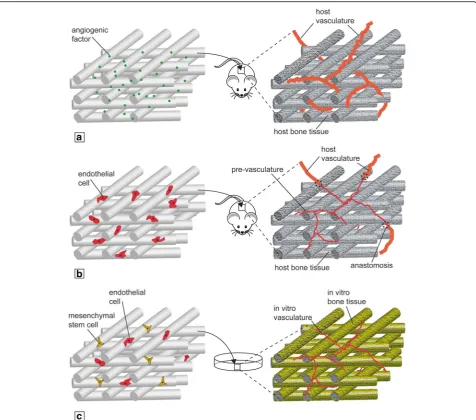
Although different models have been created to recap-itulate the interaction between bone and other adjacent tissues, to the authors’knowledge, an in-vitro engineered model coupling vascularized bone to another tissue has not yet been developed. Such capability would be par-ticularly relevant when engineering the osteochondral junction, as only the simultaneous presence of bone matrix, vasculature, and cartilage can enhance or permit veritable behavior of the complex. In fact, the interac-tions between vasculature and cartilage are well known during in-vivo skeletogenesis [123, 124], and numerous in-vitro studies have documented the molecular ex-changes driving the inhibition of chondrocyte differenti-ation, including the pivotal role exerted by VEGF secreted by ECs [125–127]. However, a model also con-taining subchondral bone would better mimic the com-plex interaction among the three tissues. For these reasons, a triphasic scaffold was produced by the authors using a recently developed 3D-printed microphysiologi-cal tissue system (MPS) bioreactor that allows the separ-ate flow of specific media to the chondral and osseous components, while maintaining them in contact and allowing tissue–tissue communication [128] (Fig.3). The cartilage construct was engineered incorporating human bone marrow-derived MSCs in a photocrosslinkable gelMA hydrogel, while the vascularized osseous con-struct was obtained by seeding MSCs and HUVECs in a poly(ε-caprolactone) (PCL) scaffold, produced through additive manufacturing as described by Puppi et al. [129]. Results from our preliminary assessments suggest
a better differentiation of MSCs in the presence of the HUVECs, which form interconnected tubular-like struc-tures in the osseous compartment.
Overall, the development of a successful in-vitro model of vascularized bone is heavily dependent on optimal se-lection of scaffold/matrix able to guide the formation of the different tissues, and on fine control of cell signaling.
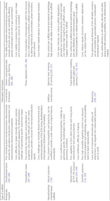
Materials
Mineralized bone and associated vasculature are charac-terized by different morphological and structural proper-ties, thus different biomaterials are needed for the engineering of both tissues to match their characteris-tics. Different materials have been investigated for the development of bone constructs, and evaluated both in-vitro and in-vivo. Calcium phosphates (CaPs) have been of great interest due to their osteoconductive properties. The majority of research has been focused on two main forms of CaPs, hydroxyapatite (HAp) and beta-tricalcium phosphate (β-TCP), or the combination of both [130]. Other inorganic materials that have been exploited in bone engineering are bioactive glasses [131], which have been shown to improve the formation of a hydroxycarbonate apatite layer by osteoblasts in-vitro and to support bone formation in-vivo [132]. The most commonly used polymeric materials for bone engineer-ing include many natural polymers, such as collagen, fibrin, alginate, silk, hyaluronic acid, chitosan, and poly-hydroxyalkanoates [133]. These biopolymers offer eco-nomic and environmental advantages, such as low
Fig. 3aCross-sectional bioreactor schematic.bMacroscopic and histological analysis of engineered osteochondral interface.cLive/dead staining of capillary-like network formed by HUVECs in bone compartment (GFP, green = HUVEC; Calcein Blue AM, blue = live cells; EthD-1, red = dead cells).
manufacture/disposal costs and renewability, as well as biological advantages, such as supporting cell signaling, adhesion, and cell-mediated degradation and remodeling [134]. Synthetic polymers have also been thoroughly in-vestigated as engineered bone scaffolds, by virtue of their controllable and reproducible chemical–physical and degradation properties, which can be tailored to the re-quirements of the target applications [135]. The different classes of polymers that have received significant atten-tion include saturated poly(α-hydroxesters), such as poly(glycolic acid) (PGA), poly(lactic acid) (PLA), and PCL, and biodegradable polyurethanes, covering a wide range of properties, including mechanical strength, elasticity, biodegradability, hydrophilicity, and so forth [135]. When considering the development of a capillary-like network within a mineralized bone construct, the most current models include the embedding of endothelial-derived cells in hydrogels, such as Matrigel®, collagen, and fibronectin [136–138]. However, these materials have limitations in mechanical stability, durability, and immunogenicity when implanted [139, 140]. One recent example to overcome these limitations is represented by the use of methacrylated gelatin (gelMA), produced by conjugating methacrylate groups to the amine-containing side groups of gelatin, which becomes a photocrosslinkable hydrogel [141]. This ma-terial is characterized by the advantages of both natural and synthetic polymers, allowing the fine-tuning of mechanical properties, while preserving biological cues. Different studies in the past have reported the capability of creating vascular-like structures using gelMA and ECs [80,89,142].
There have been a number of investigations that ex-plored a variety of biomaterials for the engineering of vascularized bone; however, identifying the most suitable biomaterials remains a difficult task, since each material has its inherent drawbacks. Ceramics are characterized by brittleness and the biodegradation rate not matching the formation of new bone tissue; natural polymers pos-sess low mechanical, thermal, and chemical stability; and synthetic polymers lack biological cues. A logical path to overcome these limitations would be to combine differ-ent materials to obtain constructs with improved proper-ties. An example of this paradigm is the development of nanocomposite materials based on biopolymers and cer-amic nanofillers, in an attempt to exploit the biological activity of natural polymers as well as the osteoconduc-tivity of ceramics [143].
Scaffolds
All of the biomaterials already described require multi-step processing to develop 3D scaffolds/matrices capable of inducing and supporting osteoblast proliferation and differentiation, as well as vasculature development.
Accordingly, in addition to osteoinductivity and osteo-conductivity, an ideal scaffold should have functional physical properties such as interconnected macroporos-ity with pore size larger than 100μm for optimal osteo-blast differentiation and formation of new blood vessels, and biocompatible stiffness to match the mechanical properties of native bone [144]. Substrate mechanical properties greatly influence stem cell osteogenic differ-entiation, as well as EC behavior [145]; for instance, a scaffold Young’s modulus in the range 25–40 kPa directs MSCs toward osteoblastic differentiation [146, 147]. Current technologies do not allow the production of scaffolds with the exact mechanical properties of bone;
however, a number of groups have explored
development-inspired precursor templates to instruct stem cells to form a mature tissue [148–151]. Although such constructs do not possess the same mechanical properties as native bone initially, they are engineered in-vitro with sufficient stiffness to be implanted in a load-bearing region and are able to guide stem cells to develop mature bone. For instance, Daly et al. [152] pro-duced an MSC-laden 3D-printed hypertrophic cartilage template, made of a reinforced alginate bioink, which developed into functional vascularized bone when im-planted in mice.
Biologically inspired scaffolds should also harbor sig-nals that act to induce the simultaneous development of bone and vasculature. Different biological scaffolds have been used for the development of in-vitro vascularized bone models, including decellularized bone and specific ECM preparations. Decellularized bone has been histor-ically considered and applied as a biologhistor-ically derived matrix, able to induce mineralized bone production by osteoprogenitor cells. A recent study by Correia et al. [81] showed the employment of decellularized bone plugs as scaffold for HUVECs and MSCs seeded in a fibrin carrier. Vasculature was able to grow inside the porosity of the scaffold both in-vitro and then in- vivo after subcutaneous implantation in mice. The use of decellularized ECM has been reported recently by Gao et al., who developed a vascular patch using MSCs in a decellularized human aortic matrix [153]. Another strat-egy involves the functionalization of “smart” scaffolds with angiogenic and osteogenic factors such as VEGF, providing a highly localized signal to control stem cell fate [154].
(TIPS) technique, Mannella et al. [155] developed a por-ous scaffold with a pore size gradient able to mimic the porosity of the cancellous bone. Although several studies about the production of bone scaffolds using the afore-mentioned techniques have been published, these meth-odologies are lacking in control over the fine architecture of the structure, specifically in relation to pore morphology and interconnection, fundamental re-quirements for the introduction of vasculature. There are no precise values for specific bone and/or vascular ingrowth; pore sizes greater than 100 μm have been shown to favor bone formation [156], while capillary density is promoted when pore sizes are greater than 300 μm [130]. For these reasons, solid freeform fabrica-tion (SFF) or additive manufacturing (AM) techniques are attracting great interest, owing to their capabilities of producing predefined interconnected porous structures, using natural and synthetic polymers, as well as ceramics as starting materials [157]. AM techniques comprise the layer-by-layer building of 3D structures, based on computer-aided design (CAD) and computer-aided manufacturing (CAM) processes. Depending on the specific working principles, AM can be classified into laser-based systems (e.g., selective laser sintering, stereo-lithography), printing-based systems (e.g., 3D printing), and nozzle/extrusion-based systems (e.g., fused depos-ition modeling, computer-aided wet-spinning), each allowing for specific scaffold macroarchitectural and microarchitectural features [158]. AM has significantly improved the technical ability to control key factors in bone scaffolds, such as composition, pore geometry, size, and interconnectivity, as well as scaffold mechanical per-formance. The fine-tuning of morphological parameters provided by AM allowed researchers to produce scaf-folds for the concomitant development of osseous and vascular tissue. 3D printing has been thoroughly exploited for the generation of bone scaffolds with vas-cular integration, mostly using ceramics [159], but also synthetic polymers [160], natural polymers [160], and composites [160]. 3D-printed scaffolds have also been loaded with bioactive molecules, such as BMP and VEGF, to promote bone formation and vascularization respectively. Novel studies are exploring the incorpor-ation of other molecules, such as KR-34893 indene com-pound that stimulates MSC differentiation and mineral deposition [160], oxygen-releasing agents like calcium peroxide to solve O2diffusion issues [160], and platelet-rich fibrin to stimulate bone marrow-derived MSC dif-ferentiation [160]. AM and 3D-printing technologies have also produced great advancements in the field of microfluidics systems and organ-tissue chips that em-ploy stem cells for in-vitro disease modeling and drug screening [161–163]. In-vitro vascularized bone models have steadily benefited from this technology. For
instance, Jusoh et al. [164] recently reported a microflui-dics platform based on HAp-loaded ECM to study angiogenesis and osteogenesis in a vascularized bone model. Furthermore, AM can be integrated with other scaffold fabrication methods to fabricate hybrid architec-tures with unique structural feaarchitec-tures, as described in detail by Giannitelli et al. [165]. Although AM tech-niques were widely studied for the development of bone scaffolds, to date, there are few studies employing them to develop in-vitro models of vascularized bone. A sum-mary of the main scaffolds and fabrication techniques employed in bone engineering is presented in Table2.
MSCs. MSC osteogenesis has been also stimulated by combining low-intensity pulsed ultrasound (LIPUS), as mechanical stimulation, and a 3D-bioprinted PEG–RGD construct [177]. Other materials have been employed, in various formulations, for the development of bioprinted bone constructs, ranging from agarose and gelatin to PEG [178–180]. The integration of bioprinted vascula-ture and mechanically stiffer substrates would greatly improve the production of functional in-vitro vascular-ized bone. One example of this paradigm featured a dual 3D bioprinting system based on fused deposition model-ing (FDM) and selective laser ablation (SLA) [181]. The authors used alternate deposition of stiff polylactide (PLA) fibers and MSC/HUVEC-laden gelMA hydrogel to achieve proper mechanical strength and biological cues of complex vascularized bone constructs. Another approach comprised the production of a mandible frag-ment using an integrated tissue-organ printer (ITOP), which was able to bioprint Pluronic-F127 hydrogel laden with human amniotic fluid-derived stem cells (hAFSCs), in combination with a PCL-based mechanical backbone, and featuring incorporated microchannels for nutrient diffusion [182]. Kolesky et al. bioprinted a thick vascular-ized tissue using different polymeric templates and fugi-tive inks. The construct was laden with MSCs in combination with HUVECs, as well as other parenchy-mal and stroparenchy-mal cells, showing robust osteogenesis and functional perfusion of the vasculature [183].
The great landscape of current scaffold fabrication technologies has provided a wealth of choices for the generation of in-vitro vascularized bone constructs. Future attention should be focused on high-throughput and high-resolution AM techniques that are able to pro-duce functional vasculature within a mature bone con-struct. The key aspect is the biocompatibility of these methods with the chosen cells of interest in order to ob-tain their optimal functionality.
Osteogenic and vascular precursors
When considering the development of an in-vitro model of vascularized bone, the selection of suitable cell sources is crucial. A desired cell source must have little to no limitation in terms of availability and be easy to maintain and manipulate in-vitro. Adult MSCs are widely employed for somatic TE by virtue of their ability to differentiate into multiple lineages, such as cartilage, fat, muscle, and bone [184]. MSCs can be readily iso-lated from different tissue sources, such as adipose, muscle, bone, and in particular, bone marrow, which is the most widely used source [185]. In fact, bone marrow-derived MSCs have been benchmarked as one of the most appropriate cell sources for bone TE due to their well-defined osteogenic differentiation [186–188]. By using an appropriate culture medium, MSCs can be
expanded without differentiation, and induced to undergo specific differentiation to a stable phenotype in culture [189]. In addition to bone marrow-derived MSCs, adipose-derived MSCs (ASCs) are now a widely accepted source for bone tissue engineering applications and have been employed also in numerous preclinical and clinical studies [190]. Induced pluripotent stem cells (iPSCs) and embryonic stem cells (ESCs) have also been studied as potential cell sources for bone regeneration [191–194]. Although pluripotent ESCs are a promising cell candidate for the development of fully functional vascularized bone in-vitro as they can form all special-ized cell types constituting the human bone, including its vasculature [195], ESCs research also raises ethical and political controversies due to their derivation from early human embryos [196]. For these reasons, the use of iPSCs obtained by reprogramming of adult somatic cells, thus avoiding the ethical problems inherent in ESC research, has gained significant attention.
Current models of biomimetic, engineered vascular-ized bone, fabricated by culturing human MSCs on 3D scaffolds resembling the matrix of native bone [197], re-quire a vascular compartment created using other cell sources. Various sources of stem cells, such as ESCs, MSCs, and iPSCs, have been identified as potential can-didates for vascular engineering [198–202]. Endothelial cells have also been successfully differentiated from am-niotic fluid stem cells [203,204]. The choice of vascular precursor cells is crucial for functional production of a proper vasculature within the in-vitro bone model. Mature ECs have been traditionally used to stimulate angiogenesis [205], among them HUVECs, which repre-sent one of the most commonly employed cells in vascu-larized bone engineering. These cells naturally form vessel-like structures when cultured in hydrogels and, most importantly, they were demonstrated to enhance MSC osteogenesis in-vitro [86, 206–208]. Another cell type that has gained attention are the EPCs, which were found to be 10 times more proliferative than HUVECs [209] and have been recently used, also in combination with MSCs, to improve vasculogenesis in-vivo [210–213] and in tissue engineering applications [214–216]. Blood vessel development was also achieved by employing iPSC-derived ECs [217].
frequently used to study osteoclastogenesis in-vitro [221]. Primary monocytes can also be differentiated into osteoclast-like cells and used to study osteoclastogenesis in-vitro [222]. Furthermore, osteoclasts have been ob-tained by differentiating macrophages derived from human iPSCs [99].
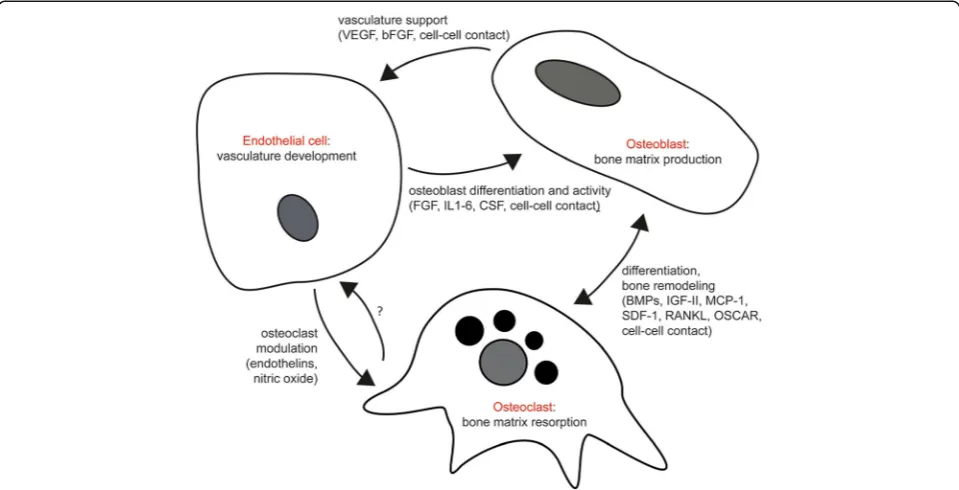
These well-established, strong relationships among the constituent cell types, taken together, underscore the im-portant and fundamental requirement of these cells in the success for development of vascularized bone in-vitro and the production of a veritable bone model (Fig.4).
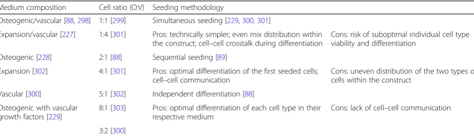
Culture media
Based on the cells selected for the development of a vas-cularized bone construct, the choice of culture media and seeding methodology has to be carefully considered to avoid negative, undesired effects. Different medium formulations and supplements have been identified for optimal osteogenic differentiation of stem cells. Dexamethasone, β-glycerophosphate, ascorbic acid, 1,25-dihydroxyvitamin D3, and BMP-2 have significant osteogenic inductive activity on MSCs by influencing different aspects of their biology, such as activation of specific genes (osteopontin, core binding factorα1, alka-line phosphatase, osteocalcin) as well as signaling path-ways like Wnt [184]. Regarding the culture of endothelial progenitors, VEGF, basic fibroblast growth factor (bFGF), and epidermal growth factor (EGF) are
usually employed to stimulate endothelial progenitor ex-pansion and differentiation/angiogenesis [223]. The use of individual differentiation medium, endothelial or osse-ous, generally yields satisfactory vasculature and miner-alized matrix formation, albeit separately [224]. However, given the need for simultaneous development of bone and vasculature, a coculture system must be set up, and investigators have used either a combination of the two differentiation media [88, 206,225–227], a sin-gle type of medium [88, 228], or osteogenic medium with some vascular growth factors [229]. In the case of a coculture system, while different cell types can be seeded simultaneously to avoid uneven distribution inside the construct and to allow communication between them, the choice of culture media is very important to assure cosurvival and differentiation. In another approach, dif-ferent cell types can be expanded and difdif-ferentiated separately and then seeded together, thus avoiding the media problem; however, some critical cell–cell interac-tions will be missing and, as discussed earlier, the gener-ation of a veritable in-vitro model, which requires recapitulation of the interaction between bone cells and vasculature, may thus be compromised. Another param-eter that needs to be considered is the cell ratio. Signal-ing between osteogenic and vascular precursors, as well as their adult counterparts in the mature organ, directs their functional integration (see Fig.4). Altering the bal-ance of this crosstalk compromises the engineering of
Fig. 4Primary cellular interactions between ECs, osteoblasts, and osteoclasts in production of vascularized bone. VEGF vascular endothelial growth factor, bFGF basic fibroblast growth factor, IL interleukin, CSF colony-stimulating factor, BMP bone morphogenetic protein, IGF insulin-like growth factor, MCP-1
monocyte chemoattractant protein-1, SDF stromal cell-derived factor-1, RANKL receptor activator of nuclear factor kappa-Βligand, OSCAR
the vascularized bone tissue. Investigators have studied varying combinations between osteogenic and vascular precursors, ranging from a 1:1 mix to unbalanced ratios in favor of osteogenic or endothelial precursors. Although a positive trend can be identified in the use of a 1:1 ratio, the results are also heterogeneous and an ideal ratio cannot be defined a priori [230]. Table3 sum-marizes the main media and cell ratios that have been used in the engineering of vascularized bone and their major benefits and drawbacks.
A recently published review by Liu et al. [83] covered different aspects of the medium combinations and seed-ing methodologies used in coculture systems for vascu-larized bone tissue engineering. However, due to the heterogeneity of the experimental parameters used in coculture studies, a clear trend is difficult to establish.
Considering the tight interactions between osteogenic and vascular stem cells during development, and the functional integration between bone and blood vessels in the mature organ, the development of veritable in-vitro models of this organ should comprise both types of cells cultured in a mixed medium in order to not miss the key interplay between osseous and vascular precursors/ mature cells.
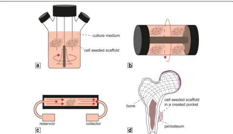
Bioreactors
Bioreactors are generally defined as any device that is able to dynamically sustain biological and/or biochem-ical processes under precisely monitored and controlled experimental and operating conditions (e.g., pH, temperature, mechanical stimuli, time, nutrient supply, and waste removal) [231]. Different types of experimen-tal setups have been developed to optimize cell seeding and mass transport, such as spinner flasks (Fig. 5a), ro-tating wall vessels (RWVs) (Fig.5b), and perfusion biore-actors (Fig. 5c). Spinner flasks and RWVs minimize the nutrient gradient and metabolite concentration around the construct, while perfusion bioreactors directly perfuse media inside the scaffold, thus assuring mass
transport within the porosity [232]. Using optimal ma-terial–scaffold–cell systems, coupled with the higher control over experimental parameters, has made biore-actors ideal means for the development of 3D tissues in-vitro. This is particularly true for the engineering of bio-logical interfaces or complex tissues, like the osteochon-dral junction and vascularized bone. Bioreactors, in combination with MSCs, have in fact been used for the recreation of the osteochondral tissue interface, as re-ported in a number of studies [121, 233, 234]. In particular, culturing in flow perfusion bioreactors has been shown to upregulate expression of osteoblastic markers [235–237], and these bioreactors have been employed for the production of engineered bone [238– 240]. To improve the amount and quality of bone be-yond what has been produced by an in-vitro process, various studies have reported the use of“in-vivo bioreac-tor” systems to produce a clinically relevant amount of vascularized bone (Fig. 5d). These approaches were based on the body’s healing mechanism that supports the formation of neotissue in different kinds of scaffolds, ranging from calcium-alginate gel, to β-TCP, to natural bovine bone mineral-coated titanium meshes implanted subperiosteally in-vivo [241–243]. The engineered bone produced with this approach could be harvested from the “living bioreactor” host and subsequently trans-planted successfully at a bone defect site. While in-vivo engineering surely ensures production of enhanced vas-cularized bone, high-throughput assays require a large number of identical samples that cannot be performed by in-vivo systems, thus necessitating the development of in-vitro platforms of vascularized bone engineering. In addition to improving in-vitro osteogenic differenti-ation of MSCs in the presence of ECs, several studies have reported better matrix mineralization by osteogenic MSCs cultured under dynamic conditions rather than static ones [237,244–246]. However, the combination of these two strategies using a flow perfusion bioreactor did not significantly enhance osteogenic differentiation
Table 3Main strategies of coculturing osteogenic and vascular precursors for vascularized bone engineering
Medium composition Cell ratio (O:V) Seeding methodology
Osteogenic/vascular [88,298] 1:1 [299] Simultaneous seeding [229,300,301]
Expansion/vascular [227] 1:4 [301] Pros: technically simpler; even mix distribution within the construct; cell–cell crosstalk during differentiation
Cons: risk of suboptimal individual cell type viability and differentiation
Osteogenic [228] 2:1 [88] Sequential seeding [89]
Expansion [302] 4:1 [301] Pros: optimal differentiation of the first seeded cells; cell–cell communication
Cons: uneven distribution of the two types of cells within the construct
Vascular [300] 5:1 [302] Independent differentiation [88]
Osteogenic with vascular growth factors [229]
8:1 [303] Pros: optimal differentiation of each cell type in their respective medium
Cons: lack of cell–cell communication
3:2 [300]
of MSCs in the presence of ECs [247, 248], likely be-cause shear stress affects the function of ECs [249–251]. However, another approach was used by Nishi et al. [252], who cultured MSCs and ECs on a porous poly(-lactic acid) scaffold in a rotating wall vessel bioreactor. The flow environment created by the RWV bioreactor, coupled with the interaction between the cell types, en-hanced the distribution and differentiation of cells in the scaffold. To date, there are only a limited number of studies using a bioreactor in combination with bone and vascular progenitor cells, due to the technical and bio-logical challenges of codifferentiating the two cell types together. The successful construction of a physiologically compatible bioreactor for engineered vascularized bone, to achieve fast and easy visualization of the target cells and their interactions, must take into account a number of design features, including noninvasive optical access, automation, uniform cell seeding, overall chamber di-mensions, checkpoint markers, and mechanical stimuli [253,254].
In utilizing engineered vascularized bone models in-vitro, instead of harvesting tissue samples and analyzing them at designated time points, the nondestructive tracking of cells and new matrix formation should result in a reduction of resources and permit real-time under-standing of the processes. One example is represented by the work reported by Bersini et al. [255], who devel-oped a biological 3D in-vitro microfluidic model to study
breast cancer metastasis to bone. This microfluidics plat-form is based on hydrogel-containing chambers placed between surface-accessible microchannels, allowing high-resolution real-time imaging of single-cell behavior, cell–cell communication, cell–matrix interactions, and cell population dynamics [256]. Another useful high-throughput platform was developed by Moya et al. [257] to study vasculature in real time. They produced an in-vitro 3D metabolically active stroma (~ 1 mm3volume) containing a living and dynamically perfused human capillary network, by combining human endothelial colony forming cell-derived ECs (ECFC-ECs) and a poly-dimethylsiloxane (PDMS) micromold. Although diffu-sion of oxygen was demonstrated to not be a limiting step in-vitro, thus allowing the development of tissue constructs in the order of centimeters in thickness [258], the major technical problem is nutrient diffusion. The density of the ECM plays an important role in the diffu-sion of nutrients and the morphogenesis of capillaries [259], and this is particularly true for mineralized bone matrix. In addition, real-time visualization of the process occurring inside the bone matrix is also limited by the opacity of the bone matrix itself, thus requiring the de-velopment of specific bioreactor or in-vitro models to overcome this limitation. X-ray microcomputed tomog-raphy (μCT) has been extensively used in the field of bone tissue engineering due to its ability to provide rapid, nondestructive 3D images of bone and bone
Fig. 5Schematic of main bioreactors used for production of 3D constructs for TE applications:aspinner flask,brotating wall vessel,cperfusion,
scaffold microstructure at 1–50 μm resolution [260, 261]. Various groups have developed μCT-compatible bioreactors [262, 263], utilizing low radio-opacity mate-rials, such as polysulfone, with dimensions matched to those of standard μCT chambers. Another requirement is that such systems should allow the study of the vascu-lature within in-vitro vascularized bone, which, however, is complicated by the 3D nature and limited access to the internal microvasculature due to the loss of optical access. While the analysis of thin sections of engineered vascularized bone could address this issue, it would obviate the purpose of recapitulating the 3D in-vivo structure. A more viable solution is the use of contrast-enhanced μCT for the investigation of microvasculature [264], which was previously studied in soft tissues, such as the kidney, heart, and liver [265–267]. This method is based on the injection of a radio-opaque contrast agent within the vasculature prior to imaging. However, again, the opacity and density of the bone matrix put some limitations on visualization of the vessel microstructure when this is performed in a bone model. It is thus not surprising that few studies have been published on the analysis of bone microvasculature usingμCT [268, 269], illustrating the future potential of contrast-enhanced μCT for the evaluation of bone neovascularization in tissue-engineered constructs.
Perspectives and future directions
The definition of the parameters that must be accommo-dated in the design of in-vitro models of vascularized bone represents a challenging task for researchers. The complex interactions between blood vessels and bone have limited our ability to develop veritable in-vitro con-structs of physiological systems representing human bone. First, from a biological point of view, the codiffer-entiation of endothelial precursors and osteogenic stem cells is hindered by the different mechanisms and signals to which these cells respond when recapitulating in-vivo development. As shown in numerous studies, a com-promise between endothelial and osteogenic signals/cues must be considered, in order to minimize undesired dif-ferentiation of the cell types. On the other hand, predif-ferentiation of vasculature or bone cells does not permit the critical communications between the two physio-logical systems during development or repair, thus com-promising the morphogenesis of the actual structure of the tissue complex and in a manner similar to native bone. This aspect is of high relevance when studying the effects of drugs and/or toxicants on the structure and development of bone, as well as screening for potential treatments. In other words, the engineered vascularized bone model must present advantages over the majority of studies that have been performed in-vitro on 2D cul-tures of separate endothelial and osteogenic cells or
cocultures, and through in-vivo animal models [270, 271]. From an engineering point of view, the design of a system which could maintain the vitality and differenti-ation of different types of cells for long-term culture is also a significant challenge. Furthermore, experimental parameters must be controlled (i.e., system variables must be clearly defined) and, as described earlier, the system must be accessible for real-time imaging and evaluation. Bioreactors have partially solved the need to have in-vitro systems which could mimic the full physio-logical structure of bone and, at the same time, have allowed analysis of the constructs.
High-throughput screening of drugs or toxicant relies on a minimal system defined by precise parameters that can be stimulated by specific stimuli, such as mechan-ical, biochemmechan-ical, genetic, and so forth. To address this, our group has developed a triphasic model composed of blood vessels, bone, and cartilage to study the inter-action between these three different tissues [128]. Our 3D-printed microphysiological system, in combination with stem cells, allows for the codifferentiation of the three different tissues, and is also responsive to the introduction of different stimuli, including stress-inducing factors, such as IL-1β for the study of osteo-arthritis, female hormones like estrogens for the study of osteoporosis [272], or teratogens to study their effect on skeletal development. The capabilities of additive manu-facturing have enabled us to produce customized scaf-folds and bioreactors to meet the requirements of vascularized bone engineering, coupled with a high-throughput platform for drug screening and toxicology assessment. In the future, bioprinting could represent a suitable on-demand platform for the versatile fabrication of specific cellular patterns at the micrometer scale for the production of vascularized bone, but also a broad range of other engineered tissues [166]. While still in its early stage, bioprinting can also benefit from the wide range of additive manufacturing techniques and bioreac-tor technologies to generate veritable in-vitro models of vascularized bone that could help understand the physiopathology of bone, and generate valuable treat-ment strategies. To become an economically viable resource, the production of in-vitro 3D models of vascu-larized bone should also consider the introduction of a certain level of automation, to reduce human interven-tion as well as product variability. An interesting and comprehensive review about this topic was recently pub-lished by Costa et al. [273], who reviewed the current automated tools and strategies for the production of bone substitutes.
Conclusions
preclinical models of normal or pathological tissues for applications in drug screening and toxicity assessment. For effective high-throughput assays, minimal systems and accurate modeling of the structure/function of the studied human tissue or organ are required. Bone path-ologies present significant disease burden, underscoring the need to develop more effective treatment strategies. The development of in-vitro high-throughput microphy-siological models that faithfully recapitulate the physio-pathology of bone is thus highly relevant, particularly given the cost and intrinsic genetic difference of animal models. Different biological and engineering challenges must be overcome, including the right choice of stem cells, regulation of codifferentiation of vasculature and bone, control of system parameters and stimuli, access for real-time imaging, and functional evaluation. In the pursuit of this initiative, additive manufacturing tech-niques and bioreactor technologies have increased our ability to produce systems that integrate cells, scaffolds, and biological/environmental stimuli to create in-vitro models of native osseous tissue, and to study the pro-cesses regulating its biology.
Abbreviations
EC:Endothelial cell; EPC: Endothelial progenitor cell; ESC: Embryonic stem
cell; HUVEC: Human umbilical vein endothelial cell; iPSC: Induced pluripotent
stem cell; MSC: Mesenchymal stem cell;μCT: Microcomputed tomography
Acknowledgements
The authors acknowledge funding support from the sources indicated.
Funding
This work was supported by the Commonwealth of Pennsylvania, Department of Health, the Environmental Protection Agency (R835736), the US Department of Defense (W81XWH-14-1-0217 and W81XWH-14-2-0003), CASIS (GA-2016-236), the Ri.MED Foundation, and the National Institutes of Health (1UG3 TR002136).
Availability of data and materials
Not applicable to this article, as no data were generated or analyzed.
Authors’contributions
All authors participated in the writing and review of the manuscript. All authors read and approved the final manuscript.
Ethics approval and consent to participate Not applicable.
Competing interests
The authors declare that they have no competing interests.
Publisher’s Note
Springer Nature remains neutral with regard to jurisdictional claims in published maps and institutional affiliations.
Author details 1
Center for Cellular and Molecular Engineering, Department of Orthopaedic Surgery, University of Pittsburgh School of Medicine, 450 Technology Drive, Pittsburgh, PA 15219, USA.2Ri.MED Foundation, Via Bandiera 11, Palermo 90133, Italy.
References
1. Thompson WR, Gottardi R, Stearns KM, Rubin J, Ambrosio F, Tuan RS.
Biologics in cartilage, bone repair, and regeneration. In: Hughes CC, Childers M, editors. Appl Regen Med to Orthop Phys Ther. La Crosse: Orthopaedic
Section: APTA; 2014. p. 1–24.
2. Laurencin CT, Ambrosio AMA, Borden MD, Cooper JA. Tissue engineering:
Orthopedic applications. Annu Rev Biomed Eng. 1999;1:19–46.
3. Cowin SC, Cardoso L. Blood and interstitial flow in the hierarchical pore
space architecture of bone tissue. J Biomech. 2015;48:842–54.
4. Amini AR, Laurencin CT, Nukavarapu SP. Bone tissue engineering: recent
advances and challenges. Crit Rev Biomed Eng. 2012;40:363–408.
5. Yang Y. Skeletal morphogenesis during embryonic development. Crit Rev
Eukaryot Gene Expr. 2009;19:197–218.
6. Fröhlich M, Grayson WL, Wan LQ, Marolt D, Drobnic M, Vunjak-Novakovic G.
Tissue engineered bone grafts: biological requirements, tissue culture and
clinical relevance. Curr Stem Cell Res Ther. 2008;3:254–64.
7. Chung AS, Ferrara N. Developmental and pathological angiogenesis. Annu
Rev Cell Dev Biol. 2011;27:563–84.
8. Kanczler JM, Oreffo ROC. Osteogenesis and angiogenesis: the potential for
engineering bone. Eur Cell Mater. 2008;15:100–14.
9. Hu DP, Ferro F, Yang F, Taylor AJ, Chang W, Miclau T, et al. Cartilage to
bone transformation during fracture healing is coordinated by the invading vasculature and induction of the core pluripotency genes. Development.
2017;144:221–34.
10. Ferrara N, Gerber H-P, LeCouter J. The biology of VEGF and its receptors.
Nat Med. 2003;9:669–76.
11. Haigh JJ, Gerber HP, Ferrara N, Wagner EF. Conditional inactivation of
VEGF-A in areas of collagen2a1 expression results in embryonic lethality in the
heterozygous state. Development. 2000;127:1445–53.
12. Schipani E, Maes C, Carmeliet G, Semenza GL. Regulation of
osteogenesis-angiogenesis coupling by HIFs and VEGF. J Bone Miner Res. 2009;24:1347–53.
13. Carulli C, Innocenti M, Brandi ML. Bone vascularization in normal and
disease conditions. Front Endocrinol. 2013;4:106.
14. Burkhardt R, Kettner G, Böhm W, Schmidmeier M, Schlag R, Frisch B, et al.
Changes in trabecular bone, hematopoiesis and bone marrow vessels in aplastic anemia, primary osteoporosis, and old age: a comparative
histomorphometric study. Bone. 1987;8:157–64.
15. Smith DW. Is avascular necrosis of the femoral head the result of inhibition
of angiogenesis? Med Hypotheses. 1997;49:497–500.
16. Gorham LW, Stout AP. Massive osteolysis (acute spontaneous absorption of
bone, phantom bone, disappearing bone); its relation to hemangiomatosis.
J Bone Joint Surg Am. 1955;37–A:985–1004.
17. Funayama E, Sasaki S, Oyama A, Furukawa H, Hayashi T, Yamamoto Y. How
do the type and location of a vascular malformation influence growth in
Klippel-Trénaunay syndrome? Plast Reconstr Surg. 2011;127:340–6.
18. Wright NC, Looker AC, Saag KG, Curtis JR, Delzell ES, Randall S, et al. The
recent prevalence of osteoporosis and low bone mass in the United States based on bone mineral density at the femoral neck or lumbar spine. J Bone
Miner Res. 2014;29:2520–6.
19. Cosman F, de Beur SJ, LeBoff MS, Lewiecki EM, Tanner B, Randall S, et al.
Clinician’s guide to prevention and treatment of osteoporosis. Osteoporos
Int. 2014;25:2359–81.
20. Alagiakrishnan K, Juby A, Hanley D, Tymchak W, Sclater A. Role of vascular
factors in osteoporosis. J Gerontol A Biol Sci Med Sci. 2003;58:M362–6.
21. Browner WS, Pressman AR, Nevitt MC, Cauley JA, Cummings SR. Association
between low bone density and stroke in elderly women. The study of
osteoporotic fractures. Stroke. 1993;24:940–6.
22. Samelson EJ, Kiel DP, Broe KE, Zhang Y, Cupples LA, Hannan MT, et al.
Metacarpal cortical area and risk of coronary heart disease: the Framingham
Study. Am J Epidemiol. 2004;159:589–95.
23. Wimalawansa SJ, De Marco G, Gangula P, Yallampalli C. Nitric oxide donor
alleviates ovariectomy-induced bone loss. Bone. 1996;18:301–4.
24. Kasten TP, Collin-Osdoby P, Patel N, Osdoby P, Krukowski M, Misko TP, et al.
Potentiation of osteoclast bone-resorption activity by inhibition of nitric
oxide synthase. Proc Natl Acad Sci U S A. 1994;91:3569–73.
25. MacRitchie AN, Jun SS, Chen Z, German Z, Yuhanna IS, Sherman TS, et al.
Estrogen upregulates endothelial nitric oxide synthase gene expression in