Introduction
Aastrom Biosciences, founded in 1989, has developed a proprietary cell-processing technology that enables the manufacture of ixmyelocel-T, a patient-specific multi-cellular therapy expanded from a patient’s own bone marrow. Aastrom has developed a bioreactor speci fically
designed for the ex vivo expansion of autologous marrow-derived stem and progenitor cells that mimics the in vivo
environment of bone marrow tissue. Two key cell types are expanded in this 12 ± 1-day culture process: stromal cells, including mesenchymal stem cells (MSCs), and alternatively activated macrophages. Aastrom has advanced ixmyelocel-T into late-stage clinical develop-ment in critical limb ischemia (CLI), including the completion of a phase 2 trial and concurrence with the US Food and Drug Administration (FDA) on a phase 3 study through the Special Protocol Assessment (SPA) process. In addition, the phase 2b clinical program in patients with dilated cardiomyopathy (DCM) will initiate in 2012. Other areas of research are ongoing.
Adult stems cells can be found in the central nervous system, skeletal muscles, pancreas, liver, adipose tissue, and the bone marrow and blood. Stem cells found in the bone marrow have been studied for almost 50 years [1]. Two primary types of stem cells are found in the bone marrow: hemato poietic stem cells (HSCs), and stromal cells (including MSCs, multipotent stromal cells, and endothelial progenitors).
HSCs are responsible for forming all the types of blood cells in the body. Approved treatment uses for stem cells are primarily for HSCs collected from the bone marrow or the peripheral blood for the treatment of specific types of cancers (leukemia, lymphoma, and myeloma). Stromal cells are a mixed population of support cells that generate the regulatory niches that support blood cell formation from HSCs [2]. Recently, progenitor cells with the capacity to differentiate into vascular endothelial cells have been identified in bone marrow and peripheral blood, but this population appears to be restricted to the endothelial lineage and would therefore be a committed precursor. While a committed endothelial progenitor is not a ‘stem cell’ per se, it is a cell type that can be thera-peutically effective without the need to strictly maintain multipotentiality, in theory providing a source of new blood vessels [3].
Aastrom scientists have focused their research on the expansion of adult cells taken from the bone marrow of an individual patient. The expanded cell product, ixmyelocel-T, is then injected into ischemic tissue in the same patient.
Abstract
Aastrom Biosciences has developed a proprietary cell-processing technology that enables the manufacture of ixmyelocel-T, a patient-specific multicellular therapy expanded from a small sample of a patient’s own bone marrow. Ixmyelocel-T is produced under current good manufacturing practices (cGMP) in a fully closed, automated system that expands mesenchymal stem cells (MSCs) and macrophages. While the cell types in ixmyelocel-T are the same as those found in the bone marrow, the numbers of MSCs and alternative macrophages are greater in ixmyelocel-T. We propose that the mixture of expanded MSCs and alternatively activated macrophages promote long-term tissue repair of ischemic tissue. The multiple cell types in ixmyelocel-T have a range of biological activities that are likely to contribute to a complex mechanism of action. Clinical trial data collected to date support the potential for ixmyelocel-T as an efficacious and safe treatment for ischemic cardiovascular indications, including critical limb ischemia (CLI) and a severe form of heart failure, dilated cardiomyopathy (DCM). The CLI clinical program has completed phase 2 and has reached concurrence with the Food and Drug Administration (FDA) on a phase 3 study (REVIVE) through the Special Protocol Assessment (SPA) process. The phase 3 study began screening patients in February 2012. The DCM clinical program will initiate phase 2b in 2012.
© 2010 BioMed Central Ltd
The Aastrom experience
Ronnda L Bartel*, Caryn Cramer, Kelly Ledford, Amy Longcore, Christopher Parrish, Theresa Stern, Sharon Watling
and Frank Zeigler
RE VIE W
*Correspondence: [email protected]
Aastrom Biosciences, Inc., Domino’s Farms, Lobby K, 24 Frank Lloyd Wright Drive, Ann Arbor, MI 48105, USA
fore, taking a small sample of bone marrow from a patient and expanding the cell populations ex vivo is an obvious benefit to the patient. Ixmyelocel-T is manufactured from a small sample (approximately 60 ml) of autologous bone marrow aseptically withdrawn from the posterior iliac crest of a patient under local anesthesia and conscious sedation during a 20-minute outpatient procedure. The bone marrow aspirate sample is shipped overnight to Aastrom’s manufacturing facility in an insulated shipping container supplied by Aastrom that is qualified to maintain the aspirate at ambient temperature. Aastrom is in a unique position in the field of cell therapy, having developed a product under current good manufacturing practices (cGMP) in a fully closed, automated system for the expansion of stem and progenitor cells [7].
Ixmyelocel-T is produced by placing collected bone marrow aspirate cells into a bioreactor system under controlled conditions, and harvesting the cells after a specified amount of time. The ixmyelocel-T manufactur-ing process is initiated by usmanufactur-ing an automated, closed system (SEPAX Cell Separation System manufactured by Biosafe, Houston, TX, USA) to perform a Ficoll-based density gradient centrifugation process to deplete red blood cells and purify bone marrow mononuclear cells (BMMNCs). The purified BMMNCs are collected and trans ferred into a single-use, sterile, disposable cell cassette that is a component of Aastrom’s proprietary, automated, closed cell processing system. The system uniformly distributes the cells over the culture surface and then controls the culture conditions, including tem-pera ture, culture medium exchange, and gas exchange.
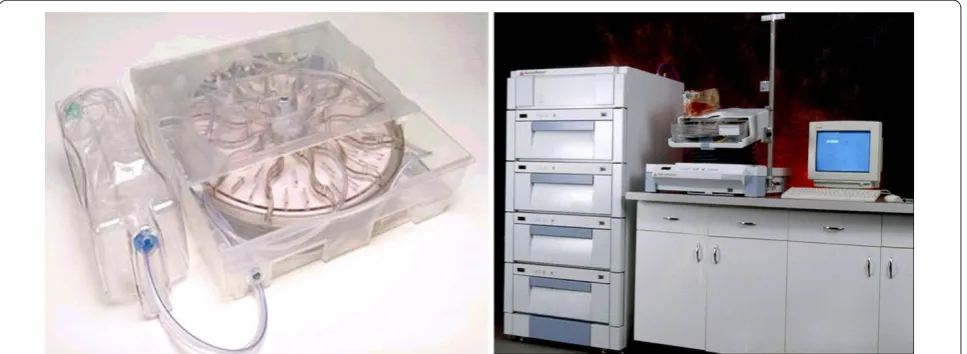
After 12 ± 1 days of culture, cells are washed and har-vested from the cassette by a multistep, automated process. The cells are then concentrated by centrifugation to a final volume suitable for patient administration. Ixmyelocel-T is shipped overnight to the treatment site using a qualified gel-packed shipping container that maintains the product at hypothermic temperature; in this container the product has a 72-hour shelf life. Components of the manufacturing system are pictured in Figure 1 [8].
evidence of an effect: characterization of ixmyelocel-T and preclinical research
Because of the mixed cell composition of ixmyelocel-T, a wide range of biological activities relevant to the repair
ixmyelocel-T may impact the repair of damaged ischemic tissue.
Characterization of the cell populations
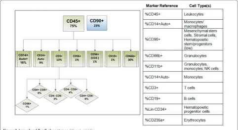
Ixmyelocel-T is composed of a mixture of cell types that include those expected to be found in the BMMNC population. These include myeloid cells (that is, granulo-cytes, monogranulo-cytes, and mixed myeloid progenitors) and lymphoid cells (that is, T cells, B cells, and mixed lymphoid progenitors) that express CD45 on the cell surface and CD90+ MSCs/stromal cells, and CD45+CD14+ autofluorescent+ (CD14+Auto+) macrophages. While the cell types are similar to those found in the BMMNC population, the numbers of CD90+ and CD14+Auto+ cells are significantly greater in ixmyelocel-T due to expansion during the manufacturing process.
Figure 2a-d shows a graphical representation of the changes in the cell types during the culture process, including the fold-change in cell phenotypes. A summary of the cell phenotypes present in ixmyelocel-T is pre-sented in Figure 3.
The prevailing scientific view is that a mixture of re-generative cell types like MSCs and alternatively activated macrophages (CD90+ and CD14+Auto+, respect ively), rather than a single cell type, are required to promote long-term tissue regeneration and repair [9,10]. Though not within the scope of this review, a significant body of scientific research supports the role of bone marrow-derived MSCs and myeloid cells in the restoration of blood flow to chronic ischemic tissues. Bone marrow maintains a small reservoir of these two cell types that the Aastrom process significantly expands while maintaining many of the mononuclear cells from the original bone marrow sample.
Not unexpectedly, these diverse cell types display a wide range of biological activities relevant to the repair and regeneration of ischemic tissue, including tissue remodeling and immunomodulation and the promotion of angiogenesis. We propose that these activities are the most likely responsible for the complex and multiple mechanisms of action in this multicellular therapy.
Tissue remodeling
Figure 1. Aastrom manufacturing platform. Left: single-use cell cassette. Right: highly automated instrument platform.
types that may promote remodeling of the extracellular matrix (ECM) in ischemic tissue. The ECM regulates many aspects of cell behavior and within a single tissue, it is constantly being remodeled as cells build and reshape their environment. Remodeling rates are particularly high during periods of infection and wound repair [12]. Using proteomic techniques, ixmyelocel-T has been shown to secrete a variety of ECM proteins and matrix metalloproteins (MMPs) that remodel the ECM [13]. Additionally, the MSCs in ixmyelocel-T have been shown to maintain a significant osteoblastic potential and, when combined with a bone inductive matrix, efficiently regenerate bone in a mouse calvarial defect model. The MSCs from ixmyelocel-T have also been shown to engraft and participate in new bone formation based on localization and expression of a bone-specific ECM gene linked to a fluorescent reporter gene [14].
The CD14+ macrophages in ixmyelocel-T are actively phagocytic, and this property could provide for clearance of necrotic tissue and apoptotic cells in diseased or damaged tissues. Figure 4 shows that the CD14 cells actively internalize acetylated low-density lipoprotein as measured by flow cytometry.
Immunomodulation
Successful repair and regeneration of ischemic tissue requires the removal of damaged or necrotic cells and tissue, as well as the resolution of pathological inflam ma-tion. Ixmyelocel-T may promote vascular re covery by
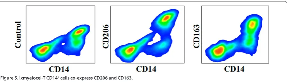
exerting protective effects on the endothelium through a population of alternatively activated macro phages that regulate inflammation and tissue remodeling. Activated macrophages can be proinflammatory (classi cal or M1 type macrophages, associated with T helper 1 lympho-cytes (Th1)) or can be alternatively activated and anti-inflam matory (alternative or M2 type macrophages, associated with Th2). Macro phages that are M2 have been associated with the resolution of inflammation and progression of tissue repair and healing after ischemic damage or infection. Alternatively activated macrophages in ixmyelocel-T express scavenger receptors CD163 and CD206 (Figure 5), consistent with an alternative activa-tion pheno type (M2).
Figure 3. Ixmyelocel-T cell phenotypes. NK, natural killer.
Analysis of the cytokine secretion profile of ixmyelocel-T has been shown to be consistent with the ixmyelocel-Th2/M2 phenotype. The pro-inflammatory cytokines IL-1α and IL-1β are barely detected (approximately <10 pg/ml), while anti-inflammatory cytokines IL-10 and IL-1 recep-tor antagonist (IL-1ra) are actively secreted by ixmyelocel-T, primarily by the CD14+Auto+ cells (approximately 5,000 to 10,000 pg/ml). The lack of Th1/M1 pro-inflammatory cytokines (IL-1α and IL-1β), and a high-level secretion of Th2/M2 anti-inflammatory cytokines (IL-10 and IL-1ra) demonstrate that ixmyelocel-T is likely promoting resolution of inflammation rather than further contributing to pathological inflammation.
Multiplex measurement of secreted cytokines from ixmyelocel-T product has been shown in sorted cell popu-la tions. Ixmyelocel-T secretes a distinct and characteristic array of cytokines, chemokines and growth factors, including biologically significant levels of angiogenic (vascular endothelial growth factor, angiopoietin 1, angio poietin 2, hepatocyte growth factor, IL-8) and anti-inflammatory or immune regulatory factors (adiponectin, IL-1ra, IL-6, IL-10, IP-10, monocyte chemotactic protein-1 and transforming growth factor-β) together with low or undetectable levels of pivotal pro-inflammatory cyto-kines (such as IL-1α, IL-1β, IL-15, IL-17, tumor necrosis factor-α and most notably lacking interferon and IL-12 as quantified by ELISA and Luminex-based assays). A summary of the multiplex cytokine analysis of BMMNCs compared to CD90+ and CD14+Auto+ from ixmyelocel-T is shown in Figure 6.
Aastrom is continuing to evaluate the properties of ixmyelocel-T through in vitro cell biology as well as testing in relevant animal models. A summary of clinical trial data collected for the two ongoing cardiovascular programs (DCM and CLI) are described below.
evidence of an effect: clinical research Dilated cardiomyopathy program
DCM is a form of heart failure; it is a progressive disease, the third most common cause of heart failure and the
most frequent cause of heart transplantation [15]. DCMs are associated with both systolic abnormalities (difficulty of the left ventricle to empty or eject blood from its chamber) and diastolic abnormalities (increased resis-tance to filling of one or both ventricles). Heart enlarge-ment and poor function generally lead to progressive heart failure with further decline in the ability of the heart to contract and pump blood around the body efficiently.
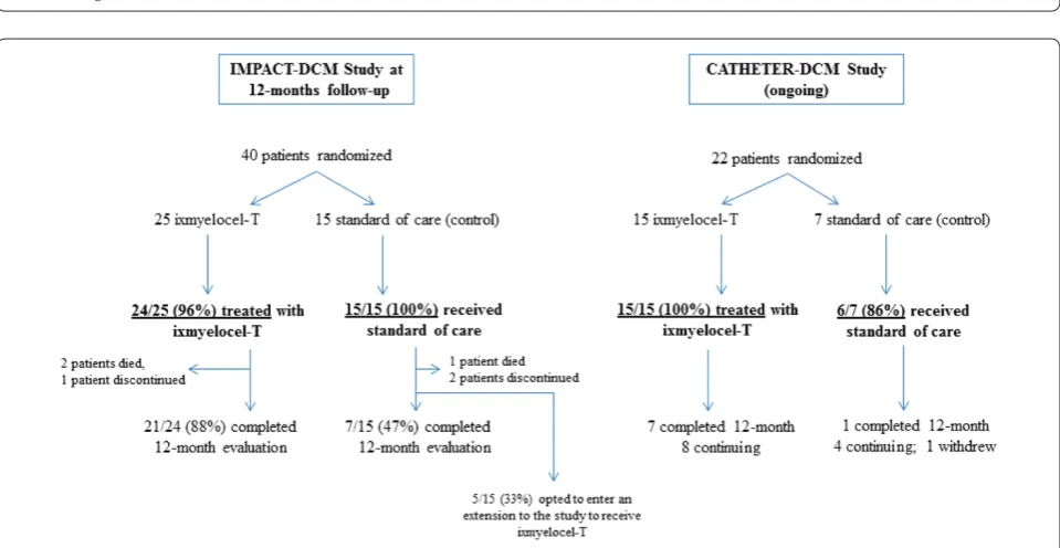
Ixmyelocel-T received orphan product designation for the treatment of DCM in 2007. Aastrom has conducted an exploratory phase 2a program in patients with heart failure due to DCM using intramyocardial surgical and transendocardial catheter delivery of ixmyelocel-T. To date, two phase 2a open-label studies using the two different delivery methods (Study ABI-55-0712-1 (IMPACT-DCM) surgical delivery, and Study ABI-55-0811-1 (CATHETER-DCM) catheter delivery) have been conducted. Both studies were open-label, multi center, randomized (ixmyelocel-T or standard of care) studies in patients with a diagnosis of ischemic DCM (IDCM) or nonischemic DCM (NIDCM). The numbers of patients in each study are presented in Figure 7.
ixmyelocel-T-treated patients (both IDCM and NIDCM) had improvement in New York Heart Association class over the 12 months following treatment. Eight patients (five IDCM and three NIDCM) receiving ixmyelocel-T had an improvement of two classes (from class III to class I) from screening to 12 months. There was also a trend toward improved function, with a higher percentage of ixmyelocel-T-treated IDCM patients show-ing an increased 6-minute walk performance compared
to the IDCM control patients. Left ventricular structural indices showed a trend toward improvement in septal thickness in the IDCM patients.
Critical limb ischemia program
CLI is the most severe form of peripheral arterial disease that results from markedly reduced blood flow to the legs, feet, and hands and is usually caused by athero-sclerosis. An estimated 3 to 5% of adults aged 40 years Figure 6. Summary of the multiplex cytokine analysis of bone marrow mononuclear cells (BMMNCs) compared to ixmyelocel-T CD90+
and CD14+Auto populations.Cells were cultured for approximately 24 hours in multiwell plates and cytokine levels were measured in comparison to blank medium negative controls. CD90+ and CD14+Auto+ populations were sorted using fluorescence-activated cell sorting to high purity prior to culture. Non-paired data from two or more independent donors assayed in replicate are expressed as the mean cytokine concentration ± standard error in pg/ml. IL-1ra, IL-1 receptor antagonist; HGF, hepatocyte growth factor; MIP, macrophage inflammatory protein; VEGF, vascular endothelial growth factor.
and older have peripheral arterial disease in the United States [17]. It is estimated that 5% to 10% of peripheral arterial disease patients over 50 years of age will develop CLI within 5 years [18]. Many patients with CLI have multiple co-morbidities, which may often prevent them from having open bypass or endovascular surgical
procedures. It is estimated that up to 40% of CLI patients are not candidates for surgery [19]. Major amputation is necessary when there is overwhelming infection that threatens the patient’s life, when rest pain cannot be controlled, or when there is extensive skin and tissue loss.
Table 1. Summary of efficacy - all treated patients (IMPACT-DCM)
IDCM NIDCM
Ixmyelocel-T Control Ixmyelocel-T Control
Percent of patients with MACE adverse events
Enrolled patients 12 7 12 8
All MACE events (n (%)) 6 (50.0) 5 (71.4) 5 (41.7) 2 (25.0)
Total MACE events 8 8 8 3
Event on day 0/injection (n (%)) 2 (16.7) 0 1 (8.3) 0
Total MACE events day 0/injection 2 0 1 0
Event on days 1 to 365 (n (%)) 3 (25.0) 4 (57.1) 4 (33.3) 1 (12.5)
Total MACE events days 1 to 365 5 7 7 1
Event on day 366+ (n (%)) 1 (8.3) 1 (14.3) 0 1 (12.5)
Total MACE events day 366+ 1 1 0 2
Number of patients with improvements from baseline in efficacy/total patients in group
12 months 9/10 1/3 7/9 2/3
Increase in six minute walk test 12 months 6/10 0/3 8/9 3/3
Increase in echocardiogram ejection fraction 12 months 3/7 0/2 4/7 1/2
IDCM, ischemic dilated cardiomyopathy; MACE, major adverse cardiac event; NIDCM, non-ischemic dilated cardiomyopathy; NYHA, New York Heart Association.
A phase 2b clinical study has been successfully com-pleted in the CLI program with positive results presented at the American Heart Association Scientific Sessions 2011 in November 2011 [20,21]. A pivotal phase 3 clinical study under a SPA approved by the FDA began screening patients in early 2012. The phase 2 study (RESTORE-CLI) was a randomized, double-blind, placebo-controlled study in patients with no options for revascularization. Safety data were evaluated from 77 aspirated patients (53 ixmyelocel-T and 24 control); efficacy data were evalu-ated in 72 treevalu-ated patients (48 ixmyelocel-T and 24 control) (Figure 8).
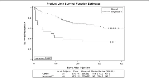
As expected with an autologous cell product, the safety profile showed that the ixmyelocel-T was well tolerated, with a similar adverse event and serious adverse event profile in the ixmyelocel-T group relative to the control group. RESTORE-CLI was not powered to show statistical significance for efficacy endpoints; despite that limitation, however, there was a statistically significant difference in the time to first occurrence of treatment failure. The treatment failure composite, which consisted of major amputation of the index leg, all-cause mortality, doubling of wound total surface area from baseline and de novo gangrene, is a phase 2 surrogate for the phase 3 amputation-free survival (major amputation of the index leg, all-cause mortality) endpoint. Time to first occur-rence of treatment failure is the earliest day at which any of the treatment failure events occurred. There was a 62%
risk reduction in treatment failure over the 12-month follow-up in the ixmyelocel-T group compared to the control group (hazard ratio 0.38, 95% confidence interval = 0.20 to 0.74; Figure 9). As shown in Table 2, the individual components of the treatment failure composite endpoint all trended in the same direction, favoring ixmyelocel-T treatment, with the exception of all-cause mortality that was the same in both treatment groups.
Conclusions
Ixmyelocel-T is a patient-specific multicellular therapy expanded from a patient’s own bone marrow. The product is manufactured using a fully closed, highly automated bioreactor system specifically designed for the ex vivo expansion of autologous marrow-derived stem Figure 9. Kaplan-Meier curve: time to treatment failure (RESTORE-CLI).Kaplan-Meier survival plot of time to treatment failure (major
amputation of injected leg, all-cause mortality, doubling of total wound surface area from baseline, de novo gangrene) for all patients injected. Censored observations are indicated by plus symbols. CL, confidence limit; NA, not available. Reprinted with permission of the author [21].
Table 2. Contribution to treatment failure composite endpoint in treated patients (Ixmyelocel-T or control)
Ixmyelocel-T Control
Endpoint: n (%) N = 48 N = 24
Major amputation 6 4
All-cause mortality 2 1
Doubling in total wound surface areaa 5 7
De novo gangrene 6 4
Total 19 (39.6%)b 16 (66.7%)b
aPatient must have come into the study with a wound to be eligible to
and progenitor cells that mimics the in vivo environment of bone marrow tissue. Characterization of ixmyelocel-T has shown a mixture of MSCs and alternatively activated macrophages that have a wide range of biological activities relevant to the repair and regeneration of ischemic tissue. This mixture of cell types has multiple mechanisms of action, including tissue remodeling and immunomodulatory functions that target the many underlying causes of severe, chronic cardiovascular diseases. Clinical trial data collected to date support the potential for ixmyelocel-T as an efficacious and safe treatment for CLI and DCM; however, data from phase 3 clinical programs are needed for confirmation.
Abbreviations
BMMNC, bone marrow mononuclear cells; CLI, critical limb ischemia; DCM, dilated cardiomyopathy; ECM, extracellular matrix; ELISA, enzyme-linked immunosorbent assay; FDA, Food and Drug Administration; HSC, hematopoietic stem cell; IDCM, ischemic dilated cardiomyopathy; IL, interleukin; IL-1ra, IL-1 receptor antagonist; MSC, mesenchymal stem cell; NIDCM, non-ischemic dilated cardiomyopathy; SPA, Special Protocol Assessment; Th, T helper.
Competing interests
All authors are employees of Aastrom Biosciences. Ixmyelocel-T is protected by patents US 7,871,605 and EP246551 issued in 2011 “Mixed cell populations for tissue repair and separation technique for cell processing”.
Published: 9 July 2012
References
1. Friedenstein AJ, Chailakhjan RK, Lalykina KS: The development of fibroblast
colonies in monolayer cultures of guinea-pig bone marrow and spleen cells.Cell Tissue Kinetics 1970, 3:393-403.
2. Williams A, Hare JM: Mesenchymal stem cells: biology, pathophysiology,
translational findings, and therapeutic implications for cardiac disease. Circ Res 2011, 109:923-940.
3. National Institutes of Health: The Adult Stem Cell (Stem Cell Information)
[http://stemcells.nih.gov/info/scireport/chapter4]
4. Weissman LL: Stem cells: units of development, units of regeneration, and
units in evolution. Cell 2000, 100:157-168.
5. Shi Y, Hu G, Su J, Li W, Chen Q, Shou P, Xu C, Chen X, Huang Y, Zhu Z, Huang X,
Han X, Xie N, Ren G: Mesenchymal stem cells: a new strategy for
immunosuppression and tissue repair. Cell Res 2010, 20:510-518. 6. Lannert H, Able T, Becker S, Sommer M, Braun M, Stadtherr P, Ho AD:
Optimizing BM harvesting from normal adult donors. Bone Marrow Transplant 2008, 42:443-447.
7. Mandalam RK, Koller MR, Smith AK: Ex vivo hematopoietic expansion for
bone marrow transplantation. In Ex Vivo Cell Therapy. Edited by Schindhelm K, Nordon R. San Diego: Academic Press; 1999:273-291.
8. Goltry K, Hampson B, Venturi N, Bartel R: Large-scale production of adult
stem cells for clinical use. In Emerging Technology Platforms for Stem Cells. Edited by Lakshmipathy U, Chesnut JD, Thyagarajan B. John Wiley and Sons, Inc.; 2009:153-168.
9. van Weel V, van Tongeren RB, van Hinsbergh VW, van Bockel JH, Quax PH:
Vascular growth in ischemic limbs: a review of mechanisms and possible therapeutic stimulation.Ann Vasc Surg 2008, 22:582-597.
10. Shireman PK: The chemokine system in arteriogenesis and hind limb
ischemia.J Vasc Surg 2007, 45(Suppl A):A48-A56.
11. Caplan AI, Dennis JE: Mesenchymal stem cells as trophic mediators.J Cell
Biochem 2006, 98:1076-1084.
12. Daley WP, Peters SB, Larsen M: Extracellular matrix dynamics in
development and regenerative medicine.J Cell Sci 2007, 121:255-264.
13. Prahalad AK, Hock JM: Proteomic characteristics of ex vivo-enriched adult
human bone marrow mononuclear cells in continuous perfusion cultures. J Proteome Res 2009, 8:2079-2089.
14. Yin D, Wang Z, Gao Q, Sundaresan R, Parrish C, Yang Q, Krebsbach PH, Lichtier
AC, Rowe DW, Hock J, Liu P: Determination of the fate and contribution of
ex vivo expanded human bone marrow stem and progenitor cells for bone formation by 2.3ColGFP.Mol Ther 2009, 17:1967-1978.
15. Goswami V: Dilated Cardiomyopathy (Medscape Reference: Drugs, Diseases and Procedures) [http://emedicine.medscape.com/article/152696-overview] 16. Patel A, Hamman B, Bruckner B, Lattouf O, Smedira N, Bartel R, Watling S:
Safety and efficacy of ixmyelocel-T, an expanded patient-specific mixed cell product, in dilated cardiomyopathy (IMPACT-DCM). J Cardiac Failure
2011, 17(Suppl):S58.
17. Selvin E, Erlinger TP: Prevalence of and risk factors for peripheral arterial disease in the United States: results from the National Health and Nutrition Examination Survey, 1999-2000.Circulation 2004, 110:738-743. 18. Norgren L, Hiatt WR, Dormandy JA, Nehler MR, Harris KA, Fowkes FG; TASC II
Working Group: Inter-society consensus for the management of peripheral
arterial disease (TASC II).Eur J Vasc Endovasc Surg 2007, 33 Suppl:S1-S75. 19. Powell RJ, Simons M, Mendelsohn FO, Daniel G, Henry TD, Koga M, Morishita
R, Annex BH: Results of a double-blind, placebo-controlled study to assess
the safety of intramuscular injection of hepatocyte growth factor plasmid to improve limb perfusion in patients with critical limb ischemia. Circulation 2008, 118:58-65.
20. Marston W, Powell R, Dall’Olmo C, Guzman R, Moore CJ, Martinez J, Halloran B, Henke P, Stiff P, Comerota A, Velazquez O, Tzeng E, Mendelsohn F, Henry T,
Bohannon WT, Saucedo J, Bartel R, Longcore A, Stern T, Watling S:
Patient-specific cellular therapy (Ixmyelocel-T) is safe and improves time to treatment failure in patients with critical limb ischemia and no revascularization options.Circulation 2011, 124:A8547.
21. Powell RJ, Marston W, Berceli SA, Guzman R, Henry T, Longcore A, Stern T, Watling S, Bartel R: Cellular therapy with ixmyelocel-T to treat critical limb ischemia: the randomized, double-blind, placebo-controlled RESTORE-CLI trial.Mol Ther 20:1280-1286.
doi:10.1186/scrt117
Cite this article as: Bartel RL, et al.: The Aastrom experience.Stem Cell Research & Therapy 2012, 3:26.