R E V I E W
Open Access
Mesenchymal stem cells: potential
application for the treatment of hepatic
cirrhosis
Yongting Zhang
1†, Yuwen Li
2†, Lili Zhang
1, Jun Li
1and Chuanlong Zhu
1*Abstract
Nowadays, orthotopic liver transplantation is considered
the most efficient approach to the end stage of chronic
hepatic cirrhosis. Because of the limitations of orthotopic
liver transplantation, stem cells are an attractive
therapeutic option. Mesenchymal stem cells (MSCs)
especially show promise as an alternative treatment
for hepatic cirrhosis in animal models and during
clinical trials. Nevertheless, the homing of transplanted
MSCs to the liver occurs in limited numbers. Therefore,
we review the strategies for enhancing the homing of
MSCs, mainly via the delivery routes, optimizing cell
culture conditions, stimulating the target sites, and
genetic modification.
Keywords:
Mesenchymal stem cells, Cirrhosis, Homing
Background
Cirrhosis is the end stage of progressive fibrosis that is
caused by various reasons and that responds poorly to
medical conservative treatment. Chronic damage to the
liver leads to the extensive accumulation of extracellular
matrix (ECM) among the hepatocytes. Epidemiological
data state that 1.03 million cirrhotic patients worldwide
die each year from severe associated complications [
1
].
Currently, liver transplantation is the most effective
therapy for advanced hepatic diseases. Among those
fortu-nate enough to receive liver transplantation, the survival
rates at 3, 12, and 36 months are 94%, 88%, and 79%,
respectively [
2
]. However, we should be take into account
the lack of donor organs, the high costs, and the long-term
use of immunosuppressants after transplantation. Thus,
* Correspondence:chuanlong@yahoo.com
†Equal contributors
1Department of Infectious Disease, the First Affiliated Hospital with Nanjing
Medical University, Nanjing, China
Full list of author information is available at the end of the article
there is an urgent need to find alternative therapeutic
strat-egies. Recent studies have shown that hepatocytes in the
cirrhotic liver still have the potential to regenerate, but
there is an imbalance between regeneration and necrosis
[
3
]. A potential hypothesis states that a fully functioning
part of the liver could be created through the proliferation
of the infused cells that will remodel the injured liver. It is
doubtful whether increasing the number of hepatocytes
alone would be an effective treatment for the patients.
Based on the proof-of-concept, hepatocytes were
transplanted to treat liver-related diseases [
4
]. Because of
the limited number of hepatocytes and the lack of their
proliferation and stability in vitro, the efficacy of grafted
hepatocytes decreased progressively. Hence, it is crucial
to find another readily available cell source.
This review aims to highlight all currently available
evidence regarding the use of stem cells for treatment of
liver cirrhosis and to determine whether there is any
factual basis for their potential.
Stem cells in regenerative medicine
Stem cells, termed as clonogenic undifferentiated cells,
cannot just self-renew indefinitely but can differentiate into
a variety of cell lineages, including pluripotent embryonic
stem cells (ESCs), induced pluripotent stem cells (iPSCs),
hematopoietic stem cells (HSCs), hepatic stem cells,
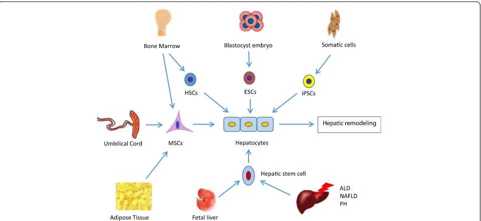
mesen-chymal stem cells (MSCs), and so forth (Fig.
1
).
Splenic teratomas could be formed after infusion of
ESCs [
5
]. The application of ESCs is therefore limited
because of their potential for malignancy. iPSCs are
artificially derived from a nonpluripotent cell and thus
ethical issues remain the major obstacle to their clinical
administration. Furthermore, the only available source of
HSCs is the hematopoietic system, and this restricts
their clinical application. Hepatic stem cells have been
identified in fetal as well as mature liver. During
embry-onic development, the cells within the liver bud are
recognized as hepatoblasts which are bipotent, giving
© The Author(s). 2018Open AccessThis article is distributed under the terms of the Creative Commons Attribution 4.0 International License (http://creativecommons.org/licenses/by/4.0/), which permits unrestricted use, distribution, and reproduction in any medium, provided you give appropriate credit to the original author(s) and the source, provide a link to the Creative Commons license, and indicate if changes were made. The Creative Commons Public Domain Dedication waiver (http://creativecommons.org/publicdomain/zero/1.0/) applies to the data made available in this article, unless otherwise stated.
rise to both hepatocytes and bile-duct epithelial cells.
Moreover, cells in the ductal plates in fetal and neonatal
livers are also hepatic stem cells. Their capacity to
repopulate the liver upon transplantation is also well
stud-ied in animal models [
6
,
7
]. Hepatic progenitor cells
(HPCs), also defined as hepatic stem cells, are rare in
nor-mal adult livers (0.01%), located in the Canals of Hering,
and all regenerative responses are mainly granted by
ma-ture hepatocytes except in certain disease states [
7
].
HPCs are activated after liver injury, such as alcoholic
liver disease (ALD) and nonalcoholic fatty liver disease
(NAFLD) [
8
]. Oxidative stress, which plays the main role
in the pathogenesis of ALD and NAFLD, promotes the
accumulation and differentiation of HPCs into
hepato-cytes [
9
,
10
]; furthermore, HPCs can differentiate into
hepatocytes in vivo and promote liver regeneration after
partial hepatectomy or acute toxic liver injury [
11
]. This
suggests that infusion of the progenitor cells may
allevi-ate the damage of hepatocytes which is caused by
long-lasting oxidative stress or partial hepatectomy.
The proliferation of HPCs as a response to chronic
liver damage is minimal [
11
] and is correlated with the
severity and localization of the inflammatory infiltrate
[
12
]. Manipulation of the HPC microenvironment may
be used as a therapeutic approach for the alleviation of
liver insufficiency [
11
,
12
].
In addition, evidence has suggested that mesenchymal
cells through the processes of mesenchymal-epithelial or
epithelial-mesenchymal transition (MET/EMT) may
con-tribute to adult liver regeneration during chronic liver
injury [
13
]. Mesenchymal cells in the liver may be
derived not only from their own progenitor cells but
also from the bone marrow (BM) by migrating to the
injured liver [
14
,
15
], although this statement is
contro-versial. This suggests that not only HPCs but also
mesenchymal cells simultaneously contribute to the
initiation and development of liver diseases, although
the mechanisms remain unclear [
16
]. This indicates
that the interaction between HPCs and mesenchymal
cells is important for remodeling of injured liver. The
accumulating evidence suggests that HPCs could be the
best alternative treatment for hepatic damage; however,
HPCs may cause carcinogenesis and fibrogenesis, as
has been shown in vitro [
6
]. Before thorough viewing of
their therapeutic potential, a better knowledge of the
factors that determine HPC differentiation and their
possible malignant transformation is necessary.
The therapeutic potential of MSCs has been
exten-sively investigated as well as their differentiation,
immu-noregulatory properties, and secretion of trophic factors.
In contrast to ESCs, iPSCs, and HPCs, MSCs do not
have any ethical problems and have become the ideal
alternative.
During the past few years, MSCs have been mainly
isolated from the bone marrow (BM-MSCs). Alternative
sources of MSCs have been proposed, such as from
adipose tissue (AD-MSCs), umbilical cord blood
(CB-MSCs), umbilical cord (UC-(CB-MSCs), and amniotic fluid.
The application of MSCs
BM-MSCs are capable of undergoing differentiation into
hepatic cells and recovering liver function, indicated by
the apoptosis of hepatic stellate cells, decreased
trans-forming growth factor (TGF)-
β
1, and alpha-smooth
muscle actin (
α
-SMA) gene expression [
17
]. AD-MSCs,
which are more immunocompatible and easier to isolate
than BM-MSCs, have a protective role against liver
fibrosis [
18
]. UC-MSCs show a more beneficial
immuno-genic profile and stronger overall immunosuppressive
potential than BM-MSCs [
19
].
Although MSC differentiation into hepatocytes has been
demonstrated in vivo, evidence suggests that various
trophic and immunomodulatory factors play a key
thera-peutic role in the treatment of liver fibrosis. The trophic
factors, which are secreted by MSCs, prevent apoptosis of
hepatocytes with the help of antiapoptotic factors
(hepato-cyte growth factor (HGF) and insulin-like growth factor
(IGF-1)), angiogenetic factors (vascular endothelial growth
factor (VEGF)), mitogenetic factors (epidermal growth
factor (EGF), HGF, and nerve growth factor (NGF)), and
TGF-
α
[
20
,
21
]. Because of the smaller and less complex
immunogenic potency, MSC-free therapy might constitute
a better alternative treatment.
Further clinical trials have evaluated the efficiency of
transplanted MSCs for treating patients with liver
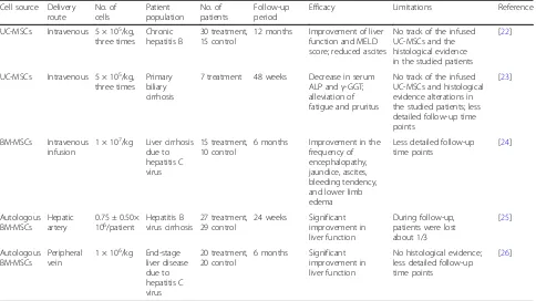
fibro-sis. Several clinical trials have been designed to evaluate
their therapeutic potential in hepatic cirrhosis treatment
[
22
–
26
] (Table
1
). The results of the studies seem to be
promising, with improvements in model for end-stage
liver disease (MELD) score and metabolic parameters,
but data on histological improvement are weak.
Long-term outcomes after UC-MSC treatment would be
preferable for patients with liver cirrhosis [
22
,
23
],
although the short-term efficacy of infused BM-MSCs
is favorable [
24
–
26
]. It should be noted that the
number of infused cells, the delivery route, and the
frequency of injection per patient vary in the studies.
Different sources of MSCs and various populations of
patients may be more convincing for any therapeutic
effect. Moreover, AD-MSCs and UC-MSCs have better
immunocompatibility, and they are more vitalized and
much easier to isolate than BM-MSCs from older
patients [
18
,
19
]. The efficacy of autologous BM-MSCs
may suffer from aging differentiation and deficiency in
vitality [
18
,
22
]. In contrast, allogeneic UC-MSCs are
free from these limitations [
19
,
22
]. Furthermore, for
prognosis and better analysis on the difference
be-tween stem cells, the follow-up time of patients should
be prolonged with the creation of time points. The
results are also limited because of small sample sizes
and absence of control groups [
22
–
26
]. Currently,
there are no standardized protocols for clinical trials
and it is not possible to monitor whether the infused
MSCs home to the targeted organs or not.
Gholamrezanezhad et al. [
27
] have shown that there
was no significant improvement in liver function after a
Table 1
MSCs in clinical trials treating liver fibrosis
Cell source Delivery route
No. of cells
Patient population
No. of patients
Follow-up period
Efficacy Limitations Reference
UC-MSCs Intravenous 5 × 10⁵/kg,
three times
Chronic hepatitis B
30 treatment, 15 control
12 months Improvement of liver function and MELD score; reduced ascites
No track of the infused UC-MSCs and the histological evidence in the studied patients
[22]
UC-MSCs Intravenous 5 × 10⁵/kg,
three times
Primary biliary cirrhosis
7 treatment 48 weeks Decrease in serum ALP andγ-GGT; alleviation of fatigue and pruritus
No track of the infused UC-MSCs and histological evidence alterations in the studied patients; less detailed follow-up time points
[23]
BM-MSCs Intravenous infusion
1 × 10⁷/kg Liver cirrhosis due to hepatitis C virus
15 treatment, 10 control
6 months Improvement in the frequency of encephalopathy, jaundice, ascites, bleeding tendency, and lower limb edema
Less detailed follow-up time points
[24]
Autologous BM-MSCs
Hepatic artery
0.75 ± 0.50×
106/patient Hepatitis Bvirus cirrhosis 27 treatment,29 control 24 weeks Significantimprovement in
liver function
During follow-up, patients were lost about 1/3
[25]
Autologous BM-MSCs
Peripheral vein
1 × 10⁶/kg End-stage liver disease due to hepatitis C virus
20 treatment, 20 control
6 months Significant improvement in liver function
No histological evidence; less detailed follow-up time points
[26]
ALPalkaline phosphatase,BM-MSCbone marrow-derived mesenchymal stem cell,γ-GGTglutamyl transpeptidase,MELDmodel for end-stage liver disease,MSC mesenchymal stem cell,UC-MSCumbilical cord-derived mesenchymal stem cell
1-month period of follow-up because the homing ability
of BM-MSCs into the liver occurred in just a limited
number of infused cells. Peng et al. [
28
] also mentioned
that the homing ability of MSCs is the main cause why
autologous MSC transplantation did not achieve
accept-able long-term effects on the prognosis of a patient. The
lingering problem of cell-based therapies is whether the
delivered cells home within the injured sites or not and
how to increase their homing ability.
Homing
Migration or homing within the injured tissues is
influ-enced by multiple factors including the delivery route,
the number of infused cells, culture conditions, and
others. We review various factors that are related to the
migration of MSCs.
Administration routes of MSCs
The delivery route for MSCs seems to be crucial for
thera-peutic efficiency. Traditional administration of MSCs is
mainly via intrahepatic injection, intrasplenic injection, and
by intravenous infusion. Systemic delivery of cells may
cause a large number of rapid losses of cells within the
capillaries, especially in the lungs, which creates a short
lifespan for remaining MSCs [
29
]. Furthermore, infusion of
cells with heparin significantly decreases the number of
entrapped AD-MSCs within the lungs and increases the
number of cells which are accumulated in the liver [
30
].
The vascular patency may be an essential factor for MSCs
flowing into the targeted tissue. Intrahepatic injection
appeared to be the ideal way to administer stem cells, with
less entrapment of cells in the circulation, and more MSCs
differentiating into hepatocytes [
31
]. Furthermore,
admin-istration of the MSCs via the portal vein or hepatic artery
shows homing efficacy less than 5% and 20
–
30%,
respect-ively [
32
,
33
]. The hepatic artery thus seems to be the best
delivery route and shows better homing efficacy; however,
the vascular patency should be checked before infusion.
Optimizing cultivation conditions
During expansion, freshly isolated MSCs lose ligands or
receptors which respond to migratory signals [
34
].
Migration is a passage-dependent process; with a higher
number of passage there is a decrease in efficacy of
homing. Also, high culture confluence impairs the
mi-gration of MSCs due to upregulation of tissue inhibitor
of metalloproteinase (TIMP)-3 [
35
]. Moreover, hypoxia
induces the expression of leptin which is associated
with activation of both the STAT3/hypoxia-inducible
factor-1
α
(HIF-1
α
)/VEGF and stromal cell-derived
factor (SDF)-1/CXCR4 signaling pathways [
36
]. It is
suggested that hypoxic preconditioning augments the
recruitment of MSCs.
Stimulating the target site to recruit MSC mobilization
In the acute phase of injury, inflammatory cytokines
which were released from the damaged tissues recruit
monocytes for tissues repair. Compared with
unirradi-ated mice, more MSCs homed in mice that received
total body irradiation [
37
], suggesting that infused MSCs
are moved first to injured sites. However, in patients
with a subchronic or chronic phase of the disease, some
indispensable chemokines for homing may be minimal
or absent; therefore external stimuli may provide a
sim-ple and available novel approach for homing.
Perry et al. [
38
] used degenerate electrical waveforms
for patients with skin scars and showed that electrical
stimulation significantly reduced scar scores and may
guide cell migration. Furthermore, the physiological
electrical field induced MSCs to graft to the anode in
vitro, which had no influence on cell senescence and
phenotype [
39
]. Meanwhile, pulsed focused ultrasound
noninvasive local pressure waves deposit energy within
the targeted tissues that change the level of local
chemoattractants and enhance the efficacy of homing
[
40
]. Mechanical stretching could also enhance engrafted
MSC homing within injured tissues via hypoxia,
vascularization, and proliferation [
41
]. In summary,
external stimuli may be used to control or induce direct
migration of MSCs.
Genetically modified MSCs
Because of the presence of specific integration between
ligand and receptor, one hypothesis is that changing the
level of the receptor/ligand on MSCs may improve the
efficiency of homing within the targeted tissues.
In the acute phase of injury, the damaged tissue
releases numerous stromal cell-derived factors (SDF-1
α
),
but their receptor (CXCR4) is at a low level on the
cultured MSCs. MSCs with overexpressed CXCR4 have
better migration potential toward SDF-1
α
and secrete
more trophic factors, including HGF and VEGF which
stimulate hepatocyte regeneration [
42
]. Ryu et al. [
43
]
further explained that Akt, ERK, and p38 signal
path-ways are also related to the SDF-1/CXCR4 axis.
MicroRNAs or noncoding RNAs target mRNA for
deg-radation or inhibition and may determine the migration of
MSCs. More than 60 different microRNAs in MSCs have
been recently described and some of them are involved in
migration, including let7, microRNA-10b, microRNA-27b,
microRNA-335, and microRNA-886-3b [
47
].
Overex-pressed microRNA-211 through the
STAT3/micro-RNA-211/STAT5A signal pathway enhanced migration
[
48
]. Upregulation of 221 and
microRNA-26b enhanced MSC migration via the chemotactic
response towards HGF through activation of PI3K/Akt
signaling [
49
]. In addition, some other microRNAs
suppressed migration of MSCs
—
microRNA-27b
sup-pressed the directional migration of MSCs by targeting
SDF-1
α
, and overexpression of microRNA-124
signifi-cantly inhibited the chemotactic migration towards
HGF by downregulation of Wnt/
β
-catenin signaling
[
50
]. It is suggested that microRNAs are involved in MSC
potential, including their differentiation, paracrine
func-tion, proliferafunc-tion, survival, and migration. Upregulation
or downregulation of microRNAs in MSCs could regulate
the migration.
Conclusion
The present review demonstrates that stem cell therapy
has a favorable therapeutic effect. Currently, the crucial
factor that determines the benefit of MSCs is the
homing efficacy. The disadvantages of MSC therapy in
clinical trials include the risks of iatrogenic
tumorigen-esis, cellular embolism, and the optimum time for the
infusion of cells. Moreover, its safety in clinical trials
should be approved by institutional ethics committees.
In conclusion, the results on MSCs which were used for
the treatment of liver fibrosis are promising, but we
need to know the underlying mechanism of their
thera-peutic effects.
Abbreviations
AD-MSC:Adipose-derived mesenchymal stem cell; ALD: Alcoholic liver disease; BM-MSC: Bone marrow-derived mesenchymal stem cell; CB-MSC: Cord blood-derived mesenchymal stem cell; c-met: Cellular mesenchymal to epithelial transition factor; CXCR4: Chemokine receptor type 4; ECM: Extracellular matrix; EGF: Epidermal growth factor; ESC: Embryonic stem cell; HGF: Hepatocyte growth factor; HIF-1α: Hypoxia-inducible factor-1α; HPC: Hepatic progenitor cell; HSC: Hematopoietic stem cell; IGF-1: Insulin-like growth factor-1; iPSC: Induced pluripotent stem cell; MELD: Model for end-stage liver disease; MSC: Mesenchymal stem cell; NAFLD: Nonalcoholic fatty liver disease; NGF: Nerve growth factor; SDF: Stromal cell-derived factor; TGF: Transforming growth factor; TIMP: Tissue inhibitor of metalloproteinase; UC-MSC: Umbilical cord-derived mesenchymal stem cell; VEGF: Vascular endothelial growth factor
Acknowledgements
We thank Dr. Jiaying Liu for supporting us with human UCB-MSCs.
Funding
This work was supported by the National Natural Science Foundation of China (No. 81770591), the Gilead Sciences Research Scholars Program in Liver Disease—Asia, the Key Medical Talents Fund of Jiangsu Province (ZDRCA2016007) and the Medical Innovation Team Project of Jiangsu Province (CXTDA2017023).
Availability of data and materials
Not applicable.
Authors’contributions
YZ, YL, LZ, JL, and CZ designed the manuscript and analyzed the literature. YZ, YL, and CZ wrote the manuscript and prepared the table. All authors reviewed and approved the final manuscript.
Ethics approval and consent to participate
Not applicable.
Consent for publication
All authors consent to the publication of this manuscript.
Competing interests
The authors declare that they have no competing interests.
Publisher
’
s Note
Springer Nature remains neutral with regard to jurisdictional claims in published maps and institutional affiliations.
Author details
1Department of Infectious Disease, the First Affiliated Hospital with Nanjing
Medical University, Nanjing, China.2Department of Pediatrics, the First Affiliated Hospital with Nanjing Medical University, Nanjing, China.
References
1. Lozano R, Naghavi M, Foreman K, Lim S, Shibuya K, Aboyans V, Abraham J, Adair T, Aggarwal R, Ahn SY, Alvarado M, Anderson HR, Anderson LM, Andrews KG, Atkinson C, Baddour LM, Barker-Collo S, Bartels DH, Bell ML, Benjamin EJ, Bennett D, Bhalla K, Bikbov B, Bin Abdulhak A, Birbeck G, Blyth F, Bolliger I, Boufous S, Bucello C, Burch M, Burney P, Carapetis J, Chen H, Chou D, Chugh SS, Coffeng LE, Colan SD, Colquhoun S, Colson KE, Condon J, Connor MD, Cooper LT, Corriere M, Cortinovis M, de Vaccaro KC, Couser W, Cowie BC, Criqui MH, Cross M, Dabhadkar KC, Dahodwala N, De Leo D, Degenhardt L, Delossantos A, Denenberg J, Des Jarlais DC, Dharmaratne SD, Dorsey ER, Driscoll T, Duber H, Ebel B, Erwin PJ, Espindola P, Ezzati M, Feigin V, Flaxman AD, Forouzanfar MH, Fowkes FG, Franklin R, Fransen M, Freeman MK, Gabriel SE, Gakidou E, Gaspari F, Gillum RF, Gonzalez-Medina D, Halasa YA, Haring D, Harrison JE, Havmoeller R, Hay RJ, Hoen B, Hotez PJ, Hoy D, Jacobsen KH, James SL, Jasrasaria R, Jayaraman S, Johns N, Karthikeyan G, Kassebaum N, Keren A, Khoo JP, Knowlton LM, Kobusingye O, Koranteng A, Krishnamurthi R, Lipnick M, Lipshultz SE, Ohno SL, Mabweijano J, MI MF, Mallinger L, March L, Marks GB, Marks R, Matsumori A, Matzopoulos R, Mayosi BM, McAnulty JH, McDermott MM, McGrath J, Mensah GA, Merriman TR, Michaud C, Miller M, Miller TR, Mock C, Mocumbi AO, Mokdad AA, Moran A, Mulholland K, Nair MN, Naldi L, Narayan KM, Nasseri K, Norman P, O'Donnell M, Omer SB, Ortblad K, Osborne R, Ozgediz D, Pahari B, Pandian JD, Rivero AP, Padilla RP, Perez-Ruiz F, Perico N, Phillips D, Pierce K, Pope CA 3rd, Porrini E, Pourmalek F, Raju M, Ranganathan D, Rehm JT, Rein DB, Remuzzi G, Rivara FP, Roberts T, De León FR, Rosenfeld LC, Rushton L, Sacco RL, Salomon JA, Sampson U, Sanman E, Schwebel DC, Segui-Gomez M, Shepard DS, Singh D, Singleton J, Sliwa K, Smith E, Steer A, Taylor JA, Thomas B, Tleyjeh IM, Towbin JA, Truelsen T, Undurraga EA,
Venketasubramanian N, Vijayakumar L, Vos T, Wagner GR, Wang M, Wang W, Watt K, Weinstock MA, Weintraub R, Wilkinson JD, Woolf AD, Wulf S, Yeh PH, Yip P, Zabetian A, Zheng ZJ, Lopez AD, Murray CJ, AlMazroa MA, Memish ZA. Global and regional mortality from 235 causes of death for 20 age groups in 1990 and 2010: a systematic analysis for the Global Burden of Disease Study 2010. Lancet. 2012;380(9589):2095–128.
2. Freeman RB Jr, Steffick DE, Guidinger MK, Farmer DG, Berg CL, Merion RM. Liver and intestine transplantation in the United States, 1997–2006. Am J Transplant. 2008;8(4Pt 2):958–76.
3. Issa R, Zhou X, Constandinou CM, Fallowfield J, Millward-Sadler H, Gaca MD, Sands E, Suliman I, Trim N, Knorr A, Arthur MJ, Benyon RC, Iredale JP. Spontaneous recovery from micronodular cirrhosis: evidence for incomplete resolution associated with matrix cross-linking. Gastroenterology. 2004; 126(7):1795–808.
4. Sokal EM, Smets F, Bourgois A, Van Maldergem L, Buts JP, Reding R, Bernard Otte J, Evrard V, Latinne D, Vincent MF, Moser A, Soriano HE. Hepatocyte transplantation in a 4-year-old girl with peroxisomal biogenesis disease: technique, safety, and metabolic follow-up. Transplantation. 2003;76(4):735–8. 5. Ishii T, Yasuchika K, Machimoto T, Kamo N, Komori J, Konishi S, Suemori H,
Nakatsuji N, Saito M, Kohno K, Uemoto S, Ikai I. Transplantation of embryonic stem cell-derived endodermal cells into mice with induced lethal liver damage. Stem Cells. 2007;25(12):3252–60.
6. Miyajima A, Tanaka M, Itoh T. Stem/progenitor cells in liver development, homeostasis, regeneration, and reprogramming. Cell Stem Cell. 2014;14(5):561–74. 7. Schmelzer E, Zhang L, Bruce A, Wauthier E, Ludlow J, Yao HL, Moss N,
Melhem A, McClelland R, Turner W, Kulik M, Sherwood S, Tallheden T, Cheng N, Furth ME, Reid LM. Human hepatic stem cells from fetal and postnatal donors. J Exp Med. 2007;204(8):1973–87.
8. Li D, Cen J, Chen X, Conway EM, Ji Y, Hui L. Hepatic loss of survivin impairs postnatal liver development and promotes expansion of hepatic progenitor cells in mice. Hepatology. 2013;58(6):2109–21.
9. Dubuquoy L, Louvet A, Lassailly G, Truant S, Boleslawski E, Artru F, Maggiotto F, Gantier E, Buob D, Leteurtre E, Cannesson A, Dharancy S, Moreno C, Pruvot FR, Bataller R, Mathurin P. Progenitor cell expansion and impaired hepatocyte regeneration in explanted livers from alcoholic hepatitis. Gut. 2015;64(12):1949–60.
10. Nobili V, Carpino G, Alisi A, Franchitto A, Alpini G, De Vito R, Onori P, Alvaro D, Gaudio E. Hepatic progenitor cells activation, fibrosis, and adipokines production in pediatric nonalcoholic fatty liver disease. Hepatology. 2012; 56(6):2142–53.
11. Español-Suñer R, Carpentier R, Van Hul N, Legry V, Achouri Y, Cordi S, Jacquemin P, Lemaigre F, Leclercq IA. Liver progenitor cells yield functional hepatocytes in response to chronic liver injury in mice. Gastroenterology. 2012;143(6):1564–75.
12. Libbrecht L, Desmet V, Van Damme B, Roskams T. Deep intralobular extension of human hepatic“progenitor cells”correlates with parenchymal inflammation in chronic viral hepatitis: can“progenitor cells”migrate? J Pathol. 2000;192:373–8.
13. Xie G, Diehl AM. Evidence for and against epithelial-to-mesenchymal transition in the liver. Am J Physiol Gastrointest Liver Physiol. 2013;305(12): G881–90.
14. Asahina K, Tsai SY, Li P, Ishii M, Maxson RE Jr, Sucov HM, Tsukamoto H. Mesenchymal origin of hepatic stellate cells, submesothelial cells, and perivascular mesenchymal cells during mouse liver development. Hepatology. 2009;49(3):998–1011.
15. Si-Tayeb K, Lemaigre FP, Duncan SA. Organogenesis and development of the liver. Dev Cell. 2010;18(2):175–89.
16. Lua I, James D, Wang J, Wang KS, Asahina K. Mesodermal mesenchymal cells give rise to myofibroblasts, but not epithelial cells, in mouse liver injury. Hepatology. 2014;60(1):311–22.
17. Jang YO, Kim MY, Cho MY, Baik SK, Cho YZ, Kwon SO. Effect of bone marrow-derived mesenchymal stem cells on hepatic fibrosis in a thioacetamide-induced cirrhotic rat model. BMC Gastroenterol. 2014;14:198. 18. Schubert T, Xhema D, Vériter S, Schubert M, Behets C, Delloye C, Gianello P, Dufrane D. The enhanced performance of bone allografts using osteogenic-differentiated adipose-derived mesenchymal stem cells. Biomaterials. 2011; 32(34):8880–91.
19. Baksh D, Yao R, Tuan RS. Comparison of proliferative and multilineage differentiation potential of human mesenchymal stem cells derived from umbilical cord and bone marrow. Stem Cells. 2007;25(6):1384–92. 20. Mohammadi Gorji S, Karimpor Malekshah AA, Hashemi-Soteh MB, Rafiei A,
Parivar K, Aghdami N. Effect of mesenchymal stem cells on doxorubicin-induced fbrosis. Cell J. 2012;14:142–51.
21. Eom YW, Shim KY, Baik SK. Mesenchymal stem cell therapy for liver fibrosis. Korean J Intern Med. 2015;30(5):580–9.
22. Zhang Z, Lin H, Shi M, Xu R, Fu J, Lv J, Chen L, Lv S, Li Y, Yu S, Geng H, Jin L, Lau GK, Wang FS. Human umbilical cord mesenchymal stem cells improve liver function and ascites in decompensated liver cirrhosis patients. J Gastroenterol Hepatol. 2012;27(Suppl 2):112–20.
23. Wang L, Li J, Liu H, Li Y, Fu J, Sun Y, Xu R, Lin H, Wang S, Lv S, Chen L, Zou Z, Li B, Shi M, Zhang Z, Wang FS. Pilot study of umbilical cord-derived mesenchymal stem cell transfusion in patients with primary biliary cirrhosis. J Gastroenterol Hepatol. 2013;28(Suppl 1):85–92.
24. El-Ansary M, Abdel-Aziz I, Mogawer S, Abdel-Hamid S, Hammam O, Teaema S, Wahdan M. Phase II trial: undifferentiated versus differentiated autologous
mesenchymal stem cells transplantation in Egyptian patients with HCV induced liver cirrhosis. Stem Cell Rev. 2012;8(3):972–81.
25. Xu L, Gong Y, Wang B, Shi K, Hou Y, Wang L, Lin Z, Han Y, Lu L, Chen D, Lin X, Zeng Q, Feng W, Chen Y. Randomized trial of autologous bone marrow mesenchymal stem cells transplantation for hepatitis B virus cirrhosis: regulation of Treg/Th17 cells. J Gastroenterol Hepatol. 2014;29(8):1620–8. 26. Salama H, Zekri AR, Medhat E, Al Alim SA, Ahmed OS, Bahnassy AA, Lotfy
MM, Ahmed R, Musa S. Peripheral vein infusion of autologous mesenchymal stem cells in Egyptian HCV positive patients with end stage liver disease. Stem Cell Res Ther. 2014;5(3):70.
27. Gholamrezanezhad A, Mirpour S, Bagheri M, Mohamadnejad M, Alimoghaddam K, Abdolahzadeh L, Saghari M, Malekzadeh R. In vivo tracking of 111In-oxine labeled mesenchymal stem cells following infusion in patients with advanced cirrhosis. Nucl Med Biol. 2011;38(7):961–7. 28. Peng L, Xie DY, Lin BL, Liu J, Zhu HP, Xie C, Zheng YB, Gao ZL. Autologous
bone marrow mesenchymal stem cell transplantation in liver failure patients caused by hepatitis B: short-term and long-term outcomes. Hepatology. 2011;54(3):820–8.
29. Zhang L, Li K, Liu X, Li D, Luo C, Fu B, Cui S, Zhu F, Zhao RC, Chen X. Repeated systemic administration of human adipose-derived stem cells attenuates overt diabetic nephropathy in rats. Stem Cells Dev. 2013;22(23):3074–86. 30. Yukawa H, Watanabe M, Kaji N, Okamoto Y, Tokeshi M, Miyamoto Y,
Noguchi H, Baba Y, Hayashi S. Monitoring transplanted adipose tissue-derived stem cells combined with heparin in the liver by fluorescence imaging using quantum dots. Biomaterials. 2012;33(7):2177–86. 31. Chamberlain J, Yamagami T, Colletti E, Theise ND, Desai J, Frias A, Pixley J,
Zanjani ED, Porada CD, Almeida-Porada G. Efficient generation of human hepatocytes by the intrahepatic delivery of clonal human mesenchymal stem cells in fetal sheep. Hepatology. 2007;46(6):1935–45.
32. Puppi J, Strom SC, Hughes RD, Bansal S, Castell JV, Dagher I, Ellis EC, Nowak G, Ericzon BG, Fox IJ, Gómez-Lechón MJ, Guha C, Gupta S, Mitry RR, Ohashi K, Ott M, Reid LM, Roy-Chowdhury J, Sokal E, Weber A, Dhawan A. Improving the techniques for human hepatocyte transplantation: report from a consensus meeting in London. Cell Transplant. 2012;21(1):1–10. 33. Khan AA, Shaik MV, Parveen N, Rajendraprasad A, Aleem MA, Habeeb MA,
Srinivas G, Raj TA, Tiwari SK, Kumaresan K, Venkateswarlu J, Pande G, Habibullah CM. Human fetal liver-derived stem cell transplantation as supportive modality in the management of end-stage decompensated liver cirrhosis. Cell Transplant. 2010;19(4):409–18.
34. Jin J. Compared treatment of primary and repeated bone marrow mesenchymal stem cell transplantation on the acute myocardial infarction. NanJing: Nanjing Medical University; 2009.
35. De Becker A, Van Hummelen P, Bakkus M, Vande Broek I, De Wever J, De Waele M, Van Riet I. Migration of culture-expanded human mesenchymal stem cells through bone marrow endothelium is regulated by matrix metalloproteinase-2 and tissue inhibitor of metalloproteinase-3. Haematologica. 2007;92(4):440–9.
36. Hu X, Wu R, Jiang Z, Wang L, Chen P, Zhang L, Yang L, Wu Y, Chen H, Chen H, Xu Y, Zhou Y, Huang X, Webster KA, Yu H, Wang J. Leptin signaling is required for augmented therapeutic properties of mesenchymal stem cells conferred by hypoxia preconditioning. Stem Cells. 2014;32(10):2702–13. 37. François S, Bensidhoum M, Mouiseddine M, Mazurier C, Allenet B, Semont A,
Frick J, Saché A, Bouchet S, Thierry D, Gourmelon P, Gorin NC, Chapel A. Local irradiation not only induces homing of human mesenchymal stem cells at exposed sites but promotes their widespread engraftment to multiple organs: a study of their quantitative distribution after irradiation damage. Stem Cells. 2006;24(4):1020–9.
38. Perry D, Colthurst J, Giddings P, McGrouther DA, Morris J, Bayat A. Treatment of symptomatic abnormal skin scars with electrical stimulation. J Wound Care. 2010;19(10):447–53.
39. Zhao Z, Watt C, Karystinou A, Roelofs AJ, McCaig CD, Gibson IR, De Bari C. Directed migration of human bone marrow mesenchymal stem cells in a physiological direct current electric field. Eur Cell Mater. 2011;22:344–58. 40. Burks SR, Ziadloo A, Kim SJ, Nguyen BA, Frank JA. Noninvasive pulsed
focused ultrasound allows spatiotemporal control of targeted homing for multiple stem cell types in murine skeletal muscle and the magnitude of cell homing can be increased through repeated applications. Stem Cells. 2013;31(11):2551–60.
42. Marquez-Curtis LA, Gul-Uludag H, Xu P, Chen J, Janowska-Wieczorek A. CXCR4 transfection of cord blood mesenchymal stromal cells with the use of cationic liposome enhances their migration toward stromal cell-derived factor-1. Cytotherapy. 2013;15(7):840–9.
43. Ryu CH, Park SA, Kim SM, Lim JY, Jeong CH, Jun JA, Oh JH, Park SH, Oh WI, Jeun SS. Migration of human umbilical cord blood mesenchymal stem cells mediated by stromal cell-derived factor-1/CXCR4 axis via Akt, ERK, and p38 signal transduction pathways. Biochem Biophys Res Commun. 2010;398(1):105–10. 44. Trusolino L, Bertotti A, Comoglio PM. MET signalling: principles and
functions in development, organ regeneration and cancer. Nat Rev Mol Cell Biol. 2010;11(12):834–48.
45. Ishikawa T, Factor VM, Marquardt JU, Raggi C, Seo D, Kitade M, Conner EA, Thorgeirsson SS. Hepatocyte growth factor (HGF)/c-met signaling is required for stem cell mediated liver regeneration. Hepatology. 2012;55(4): 1215–26.
46. Liu J, Pan G, Liang T, Huang P. HGF/c-Met signaling mediated mesenchymal stem cell-induced liver recovery in intestinal ischemia reperfusion model. Int J Med Sci. 2014;11(6):626–33.
47. Clark EA, Kalomoiris S, Nolta JA, Fierro FA. Concise review: microRNA function in multipotent mesenchymal stromal cells. Stem Cells. 2014; 32(5):1074–82.
48. Hu X, Chen P, Wu Y, Wang K, Xu Y, Chen H, Zhang L, Wu R, Webster KA, Yu H, Zhu W, Wang J. MiR-211/stat5a signaling modulates migration of mesenchymal stem cells to improve its therapeutic efficacy. Stem Cells. 2016;34(7):1846–58.
49. Zhu A, Kang N, He L, Li X, Xu X, Zhang H. MiR-221 and miR-26b Regulate chemotactic migration of MSCs toward HGF through activation of Akt and FAK. J Cell Biochem. 2016;117(6):1370–83.
50. Yue Q, Zhang Y, Li X, He L, Hu Y, Wang X, Xu X, Shen Y, Zhang H. MiR-124 suppresses the chemotactic migration of rat mesenchymal stem cells toward HGF by downregulating Wnt/β-catenin signaling. Eur J Cell Biol. 2016;95(9):342–53.

